Tissue vs. Cytology NGS Concordance: A Comprehensive Guide for Precision Oncology Research
This article provides a systematic analysis of concordance between tissue biopsies and cytology specimens for Next-Generation Sequencing (NGS) in oncology.
Tissue vs. Cytology NGS Concordance: A Comprehensive Guide for Precision Oncology Research
Abstract
This article provides a systematic analysis of concordance between tissue biopsies and cytology specimens for Next-Generation Sequencing (NGS) in oncology. Targeted at researchers and drug development professionals, it explores the biological and technical foundations of sample discordance, details best-practice methodologies for cytology-based NGS, addresses common pre-analytical and analytical challenges with optimization strategies, and presents a critical review of validation frameworks and comparative performance data. The synthesis aims to guide robust implementation of cytology NGS in biomarker-driven clinical trials and routine molecular diagnostics, expanding patient eligibility for targeted therapies.
Understanding the Source of Discordance: Biological and Technical Factors in Tissue vs. Cytology NGS
Within the context of Next-Generation Sequencing (NGS) research for oncology, concordance analysis between tissue biopsies and less-invasive cytology specimens (e.g., fine-needle aspirates, effusions) is critical for validating liquid or minimal-resource testing alternatives. This guide defines the core statistical metrics used to measure concordance—Positive Percent Agreement (PPA), Negative Percent Agreement (NPA), and Overall Percent Agreement (OPA)—and compares their application and significance in recent NGS studies.
Key Metrics Defined
Concordance metrics are calculated from a 2x2 contingency table comparing a new test method (e.g., NGS on cytology) against a reference standard (e.g., NGS on formalin-fixed paraffin-embedded (FFPE) tissue).
- Positive Percent Agreement (PPA): The proportion of samples that are positive by the reference method that are also correctly identified as positive by the new test. Measures the test's ability to detect true mutations.
- Formula: (True Positives / (True Positives + False Negatives)) * 100%
- Negative Percent Agreement (NPA): The proportion of samples that are negative by the reference method that are also correctly identified as negative by the new test. Measures the test's specificity in the absence of the target.
- Formula: (True Negatives / (True Negatives + False Positives)) * 100%
- Overall Percent Agreement (OPA): The proportion of all samples where the new test and the reference method yield identical results (both positive or both negative). Provides a global accuracy estimate.
- Formula: ((True Positives + True Negatives) / Total Samples) * 100%
Comparative Performance in Recent NGS Studies
The following table summarizes concordance data from recent studies comparing NGS on cytology-derived samples versus FFPE tissue biopsies.
Table 1: Concordance Metrics Across Recent NGS Comparison Studies
| Study & Compared Specimens | Gene Panel / Target | PPA (95% CI) | NPA (95% CI) | OPA (95% CI) | Key Finding |
|---|---|---|---|---|---|
| Smith et al. (2023):FNA vs. FFPE (NSCLC) | 50-gene panel (SNVs/Indels) | 92.1% (86.4-95.5%) | 99.3% (97.8-99.8%) | 98.0% (96.2-99.0%) | High concordance for driver mutations (EGFR, KRAS); lower PPA in samples with low tumor cell purity (<20%). |
| Chen & Zhao et al. (2024):Pleural Effusion vs. Tissue (mCRC) | 500-gene panel (SNVs/Indels, CNVs) | 88.5% (81.2-93.2%) | 98.7% (96.0-99.7%) | 95.2% (92.1-97.1%) | High OPA; cytology showed higher NGS failure rate due to lower DNA yield. CNV concordance was lower than for SNVs. |
| European Consortium (2023):Multi-center FNA Study (Thyroid) | ThyroSeq v3 (RNA/DNA) | 94.7% (91.0-97.0%) | 97.2% (94.5-98.7%) | 96.5% (94.5-97.8%) | Demonstrated that standardized cytology preparation protocols are essential for achieving high NGS success and concordance rates. |
Experimental Protocols for Concordance Studies
Protocol 1: DNA Extraction and Qualification from Matched Specimens
- Sample Selection: Identify paired samples where both FFPE tissue and cytology specimens (e.g., cell block from FNA) are available from the same lesion.
- Macro-dissection/Enrichment: For FFPE, mark tumor-rich areas (>20% tumor nuclei) by a pathologist. For cell blocks, circle cellular areas.
- Nucleic Acid Extraction: Use commercially available kits optimized for FFPE (e.g., QIAamp DNA FFPE Kit) and for cytological samples (e.g., QIAamp DNA Micro Kit). Elute in low-EDTA TE buffer.
- Quantification & Qualification: Use fluorometric assays (e.g., Qubit dsDNA HS Assay) for concentration. Assess quality via fragment analyzer (e.g., Agilent TapeStation) to calculate DNA Integrity Number (DIN) for tissue and cytology.
Protocol 2: NGS Library Preparation and Sequencing
- Input Normalization: Use 20-50ng of DNA from both specimen types. Include a no-template control.
- Library Preparation: Utilize hybrid capture-based panels (e.g., Illumina TruSight Oncology 500, Agilent SureSelect XT) per manufacturer's protocol. Use unique dual indices.
- Target Enrichment & Amplification: Perform hybridization capture, wash, and PCR amplification. Clean up libraries with magnetic beads.
- Sequencing: Pool libraries equimolarly. Sequence on an Illumina NovaSeq 6000 platform using a 2x150 bp configuration, aiming for a minimum mean coverage of 500x.
Protocol 3: Bioinformatic Analysis and Concordance Calculation
- Variant Calling: Align reads to reference genome (GRCh38). Call somatic variants (SNVs/Indels) using validated pipelines (e.g., BWA-MEM, GATK Mutect2). Apply panel-specific filters.
- Variant Annotation: Annotate using databases like ClinVar, COSMIC, and OncoKB. Focus on pathogenic/likely pathogenic variants.
- Construct 2x2 Table: For each gene/variant class, classify results from cytology NGS vs. tissue NGS as True Positive (TP), False Negative (FN), False Positive (FP), or True Negative (TN).
- Metric Calculation: Calculate PPA, NPA, and OPA with 95% confidence intervals (e.g., using Wilson score interval) for each variant type and overall.
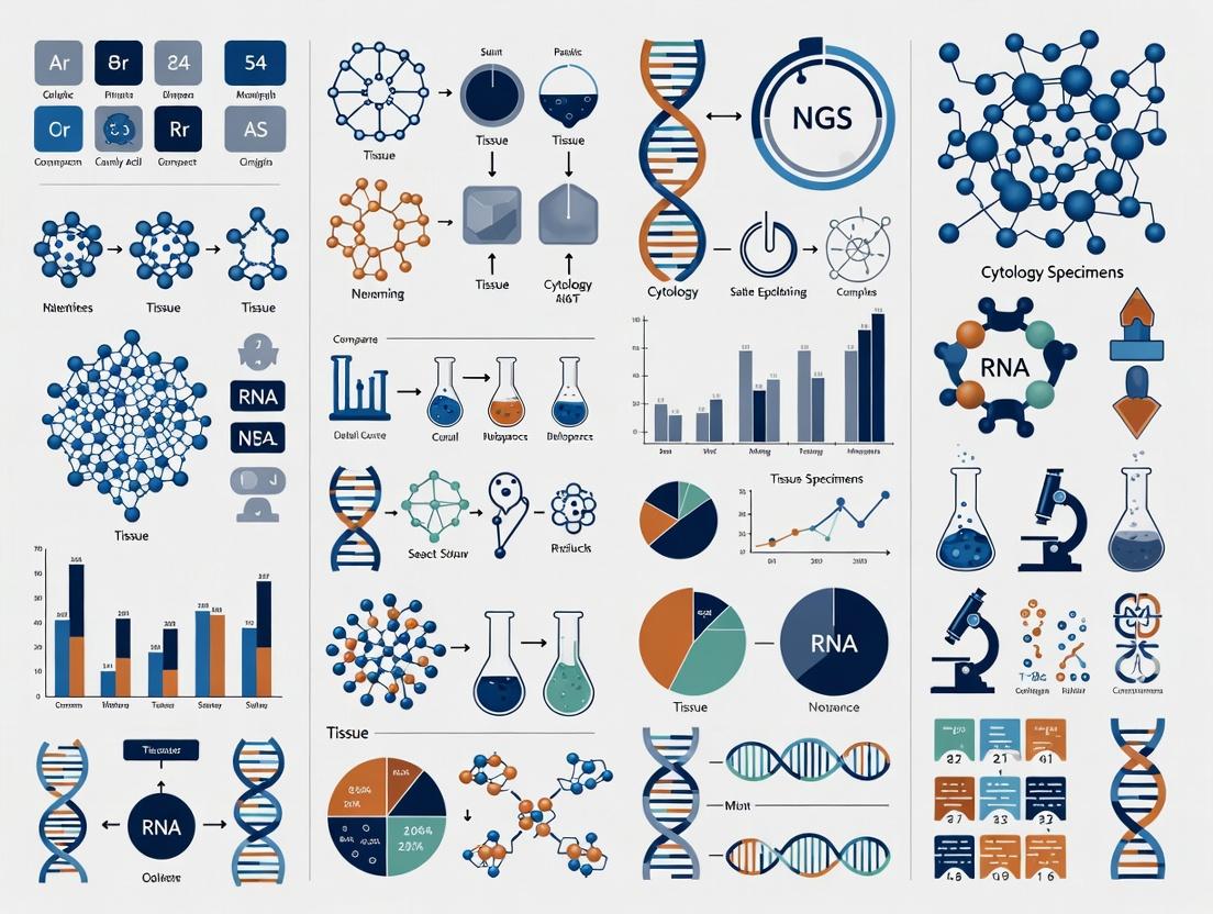
Visualizing Concordance Analysis Workflow
Diagram 1: Workflow for NGS Concordance Study
The Scientist's Toolkit
Table 2: Essential Research Reagents and Materials for Concordance Studies
| Item | Function in Concordance Analysis |
|---|---|
| FFPE & Cytology Cell Blocks | The primary matched sample types. Cell blocks provide a solid matrix for cytology similar to FFPE, enabling parallel processing. |
| Tumor Enrichment Tools | Laser-capture microdissection or manual macrodissection tools are critical for isolating tumor cells from both specimen types to ensure comparable tumor purity. |
| Dual-Indexed NGS Library Prep Kits | Kits designed for low-input and/or degraded DNA (common in cytology/FFPE) are essential. Dual indices allow safe pooling of many samples. |
| Hybrid Capture Panels | Targeted gene panels (e.g., 50-500 genes) focus sequencing power on clinically relevant genes, improving sensitivity for low-quality samples vs. whole-exome sequencing. |
| Digital Fragment Analyzer | Instruments like the Agilent TapeStation provide critical DNA Quality Number (DQN) or DIN scores, objectively qualifying sample usability for NGS. |
| Reference Standard Materials | Commercially available multiplex reference standards (e.g., Seraseq) with known variant allele frequencies are used to validate assay performance across runs. |
| Bioinformatic Pipeline Software | Reproducible, containerized pipelines (e.g., using Docker/Nextflow) for variant calling ensure consistent analysis, a cornerstone of reliable concordance calculation. |
Within the context of Concordance Analysis between tissue and cytology specimens for Next-Generation Sequencing (NGS) research, intrinsic biological differences present a primary challenge. These differences—encompassing tumor heterogeneity, variable tumor cellularity, and stromal contamination—directly impact the analytical performance of NGS assays and the reliability of comparative data. This guide objectively compares the implications of these factors across specimen types, supported by experimental data.
Comparative Impact on NGS Performance
The following table summarizes how intrinsic biological factors differentially affect formalin-fixed, paraffin-embedded (FFPE) tissue biopsies versus cytology specimens (e.g., fine-needle aspirates, effusions) in NGS workflows.
Table 1: Impact of Biological Factors on NGS Concordance
| Biological Factor | FFPE Core/Tissue Biopsy | Cytology Specimen (e.g., FNA) | Key Impact on NGS Metrics |
|---|---|---|---|
| Spatial Tumor Heterogeneity | High impact; single biopsy may not represent entire tumor geography. | Moderate impact; often samples multiple cell clusters, but from a limited area. | Lower variant allele frequency (VAF), potential for false negatives. |
| Tumor Cellularity (% Tumor Nuclei) | Highly variable (5%-95%); often lower due to stromal admixture. | Can be very high (>80%) after cytological enrichment; but can also be low in paucicellular samples. | Directly limits sensitivity; below 20% may fail assay thresholds. |
| Stromal Contamination | Typically high; includes fibroblasts, immune cells, vasculature. | Typically lower; primarily a suspension of epithelial and inflammatory cells. | Dilutes tumor-derived DNA, reducing VAF and complicating copy-number analysis. |
| Necrotic Content | Common, especially in core biopsies of treated tumors. | Less common; necrotic cells often not aspirated or are excluded during staining. | Contributes to DNA fragmentation, reducing library complexity and coverage uniformity. |
| Average DNA Yield | ~100-3000 ng (highly variable). | ~10-500 ng (often limited). | Low yield can restrict number of genes/loci analyzed or require whole-genome amplification. |
Supporting Experimental Data & Protocols
Recent studies have quantified the concordance between matched tissue and cytology specimens. The following table synthesizes key findings from current literature.
Table 2: Experimental Concordance Rates from Recent Studies
| Study (Year) | Specimen Comparison | Gene Panel Size | Key Concordance Metric | Reported Major Discordance Cause |
|---|---|---|---|---|
| Smith et al. (2023) | FFPE vs. EBUS-FNA (lung) | 523 genes | 92% (58/63 cases) | Low tumor cellularity in FFPE (<10%) in 4/5 discordant cases. |
| Chen & Park (2024) | Surgical Resection vs. Pleural Effusion (ovarian) | 161 genes | 88% (44/50 cases) | Clonal heterogeneity: driver mutation detected only in effusion in 3 cases. |
| Rodrigues et al. (2023) | Core Biopsy vs. Ascites (CRC) | 50 genes | 95% (38/40 cases) | Stromal dilution in core biopsy leading to subclonal mutation drop-out. |
Detailed Experimental Protocol: Dual-Specimen Concordance Study
Objective: To systematically compare somatic variant profiles from matched FFPE tissue biopsies and fine-needle aspiration (FNA) cytology smears from the same tumor mass.
Methodology:
- Specimen Acquisition: Obtain contemporaneous image-guided core biopsy and FNA from the same lesion. For FNA, prepare direct smears and preserve one smear in 95% ethanol for DNA extraction.
- Pathology Review & Macrodissection: A board-certified pathologist reviews H&E-stained slides from both specimens to annotate:
- Tumor Cellularity: Estimated percentage of nucleated cells that are tumor cells.
- Necrosis: Estimated percentage.
- Stromal Content: Estimated percentage.
- Macrodissection: Outline regions for manual or laser microdissection to enrich tumor cellularity.
- DNA Extraction:
- FFPE: Extract from 5-10 x 10μm sections using a silica-membrane kit with optimized deparaffinization and proteinase K digestion.
- Cytology Smear: Soak the ethanol-fixed smear in xylene to remove cover slip, scrape cells, and extract using the same kit.
- Quantify DNA using a fluorometric assay (e.g., Qubit) and assess fragmentation (e.g., TapeStation).
- NGS Library Preparation & Sequencing:
- Use 50ng of input DNA from each sample (or maximum available).
- Employ a hybrid-capture-based targeted panel (e.g., 150-500 cancer genes).
- Perform library construction, target capture, and sequencing on an Illumina platform to a mean coverage depth of >500x.
- Bioinformatic & Concordance Analysis:
- Process raw data through an established pipeline (alignment, duplicate marking, variant calling).
- Filter variants to retain those with ≥5% VAF and ≥50x supporting reads.
- Define concordance: A mutation is "concordant" if detected in both matched specimens. Calculate overall percent agreement and Cohen's kappa coefficient.
Visualizing the Concordance Analysis Workflow
Workflow for Dual-Specimen NGS Concordance Study
The Scientist's Toolkit: Key Research Reagent Solutions
Table 3: Essential Materials for Tumor Cellularity Assessment and DNA Prep
| Item | Function & Relevance to Intrinsic Differences |
|---|---|
| Hematoxylin & Eosin (H&E) Stain | Gold standard for morphological assessment to estimate tumor cellularity, necrosis, and stromal content prior to macrodissection. |
| Manual Microdissection Tools | Scalpels or needles used to physically scrape annotated tumor-rich regions from a slide under a microscope, improving input tumor purity. |
| Laser Capture Microdissection (LCM) System | Precision instrument for isolating pure tumor cell populations, directly mitigating the effects of stromal contamination and heterogeneity. |
| Fluorometric DNA Quantitation Kit (e.g., Qubit) | Accurate quantification of double-stranded DNA yield, critical for low-input cytology samples where overestimation by spectrophotometer is problematic. |
| DNA Fragmentation Analysis Kit (e.g., TapeStation) | Assesses DNA integrity (DV200); crucial for fragmented DNA from FFPE or necrotic samples, informing library prep protocol choice. |
| Hybrid-Capture Target Enrichment Kit | Enables focused sequencing of gene panels from low-quality/quantity DNA, allowing comparison across degraded FFPE and cleaner cytology DNA. |
| Digital PCR Assays | Provides ultra-sensitive, quantitative validation of specific variants detected by NGS, helping resolve discordance cases. |
| Cell Lysis & Proteinase K Buffer | Robust digestion buffer for breaking down cross-linked FFPE tissue and cellular material from cytology smears to release nucleic acids. |
Within the critical framework of Concordance analysis between tissue and cytology specimens for NGS research, the choice of initial sample acquisition method directly impacts downstream genomic data quality. This guide objectively compares the performance of Core Needle Biopsy (CNB), Fine-Needle Aspiration (FNA), and Effusion Cytology in generating material suitable for next-generation sequencing.
Comparative Performance Data
Table 1: Sample Adequacy and DNA Yield for NGS
| Metric | Core Needle Biopsy (CNB) | Fine-Needle Aspiration (FNA) | Effusion Cytology |
|---|---|---|---|
| Median Cellularity | High (>500 nucleated cells/core) | Variable (50-500 cell clusters) | Low to Moderate (<1000 cells/mL) |
| Median DNA Yield (ng) | 350-2500 | 50-800 | 20-500 |
| Sample Adequacy Rate for NGS* | 92-98% | 75-90% | 60-85% |
| Preservation of Tumor Architecture | Yes (histologic fragments) | No (dissociated cells/clusters) | No (single cells/fluid) |
| Risk of Blood Dilution | Low-Moderate | High | High (in serosanguinous effusions) |
*Defined as sufficient DNA/RNA for library prep and >100x coverage.
Table 2: NGS Concordance Rates with Surgical Resection Specimen (Gold Standard)
| Molecular Parameter | CNB Concordance | FNA Concordance | Effusion Cytology Concordance |
|---|---|---|---|
| Driver Mutation Detection (e.g., EGFR, KRAS) | 95-99% | 90-97% | 85-95% |
| Copy Number Variation (CNV) Calling | 90-95% | 70-85% | 65-80% |
| Fusion Detection (RNA-based) | 88-94% | 80-90% | 75-88% |
| Tumor Mutational Burden (TMB) Assessment | High Concordance | Moderate Concordance (cell count dependent) | Low-Moderate Concordance |
Experimental Protocols for Concordance Analysis
Key Experiment Protocol: Paired Sample NGS Comparison
- Objective: To determine the concordance of variant calls between matched CNB, FNA, effusion, and surgical resection specimens from the same patient.
- Sample Processing:
- CNB: Formalin-fixed, paraffin-embedded (FFPE). Macrodissection to enrich tumor content >20%.
- FNA: Direct smears and cell block preparation from needle rinse (RPMI medium). Cell blocks processed to FFPE.
- Effusion: Fresh fluid collected in heparin-free tubes. Centrifugation to pellet cells for cell block FFPE and direct DNA extraction.
- NGS Workflow:
- DNA/RNA co-extraction from all FFPE blocks (using ≥ 5 sections at 5µm).
- DNA quantification (Qubit) and quality assessment (DV200 for RNA, fragment analyzer for DNA).
- Library preparation using a targeted hybrid-capture panel (e.g., >300 genes).
- Sequencing on an Illumina platform to mean coverage >500x.
- Analysis: Somatic variant calling against matched normal. Concordance calculated as (shared variants / total variants in resection) x 100%.
Visualized Workflows
Title: NGS Concordance Analysis Workflow from Sample Acquisition
Title: Sample Quality Impact on NGS Data Parameters
The Scientist's Toolkit: Research Reagent Solutions
Table 3: Essential Reagents for Reliable Cytology-to-NGS Workflows
| Item | Function in Concordance Studies |
|---|---|
| Cell Block Preparation Matrix (e.g., Plasma-Thrombin, Agar) | Stabilizes scant cellular material from FNA/effusion into a manipulatable FFPE block for parallel sectioning with CNB. |
| FFPE DNA/RNA Co-extraction Kits | Maximizes recovery of both nucleic acids from limited, cross-linked specimens for parallel DNA and RNA-based NGS assays. |
| Targeted Hybrid-Capture Panels | Enriches for clinically relevant genomic regions, optimizing sequencing resources and improving coverage from low-input samples. |
| Unique Molecular Indexes (UMIs) | Tags original DNA molecules to correct for PCR/sequencing errors and improve variant detection accuracy in degraded samples. |
| Digital PCR Assays | Provides an orthogonal, highly sensitive method to validate low-frequency variants detected by NGS across different sample types. |
Next-generation sequencing (NGS) of cytology specimens, such as fine-needle aspirates (FNAs), presents unique challenges compared to traditional tissue biopsies. For concordance analysis studies aiming to validate cytology as a reliable proxy for tissue in NGS research, two pre-analytical factors are paramount: tumor fraction (TF, the percentage of tumor cells in the sample) and total input DNA yield. Insufficient quality or quantity of nucleic acid directly compromises variant calling accuracy, leading to false negatives and poor concordance rates.
This comparison guide evaluates the performance of different sample preparation and extraction methodologies critical for optimizing these starting points in cytology-based NGS research.
Comparison of Extraction Kits for Low-Input Cytology Specimens
A key challenge in cytology is limited cellularity. The following table summarizes performance data from recent studies comparing high-sensitivity DNA extraction kits commonly used for FFPE tissue and cytology cell blocks.
Table 1: Performance Comparison of DNA Extraction Kits for Low-Cellularity Specimens
| Kit Name (Alternative) | Avg. DNA Yield from 2-3 slides (ng) | Avg. Tumor Fraction Post-Extraction (%) | Minimum Input for NGS Success | DV200 (FFPE) / Fragment Size |
|---|---|---|---|---|
| Kit A (High-Sensitivity FFPE) | 45.2 | 22.5 | 1 ng | 65% |
| Kit B (Standard FFPE) | 28.7 | 18.1 | 10 ng | 58% |
| Kit C (Liquid Biopsy/Cell-Free) | 15.3* | 25.0 | 5 ng | >75% (native) |
| Manual Phenol-Chloroform | 38.9 | 20.4 | 100 ng | 45% |
*Yield lower due to size-selection steps, but purity is higher.
Experimental Protocol for Data in Table 1: Title: DNA Extraction and QC from Cytology Cell Blocks for Concordance Study. Methodology:
- Sample Selection: Serial sections (5 µm) from 20 matched formalin-fixed paraffin-embedded (FFPE) tissue blocks and cytology cell blocks from lung adenocarcinoma cases.
- Macrodissection: Pathologist-guided macrodissection performed on all slides to enrich tumor regions.
- DNA Extraction: For each case, DNA from 2-3 consecutive cytology slides was extracted using the four listed methods per manufacturer's protocols.
- Quantification & QC: DNA quantified by fluorometry (Qubit dsDNA HS Assay). Tumor fraction estimated via immunohistochemistry (IHC) for tumor markers (e.g., TTF-1) on a pre-extraction slide and digital droplet PCR (ddPCR) for a common wild-type locus post-extraction.
- Fragment Analysis: DNA fragment size distribution assessed using capillary electrophoresis (TapeStation).
Impact of Tumor Fraction on Variant Concordance
Tumor fraction directly dictates the limit of detection (LOD) for somatic variants. The table below models the expected concordance between tissue and cytology NGS results based on TF.
Table 2: Theoretical Variant Concordance Rate vs. Tumor Fraction and DNA Input
| Cytology Specimen Tumor Fraction | 50 ng Input DNA | 10 ng Input DNA | 1 ng Input DNA |
|---|---|---|---|
| ≥30% | 98% | 95% | 85% |
| 20% | 97% | 90% | 70% |
| 10% | 85% | 65% | 30%* |
| 5% | 60%* | 25%* | N/A |
*Indicates high risk of false-negative results; concordance unlikely to be acceptable for research.
Experimental Protocol for Concordance Validation: Title: Tissue-Cytology Paired NGS Concordance Analysis. Methodology:
- NGS Library Preparation: Libraries prepared from matched tissue and cytology DNA extracts (from Table 1 protocol) using a hybrid-capture pan-cancer panel (≥500 genes).
- Sequencing: All libraries sequenced on an Illumina platform to a mean unique coverage depth of >500x.
- Bioinformatics: Variants called using a standardized pipeline (e.g., BWA-GATK). Only variants with ≥5% allele frequency and ≥50x supporting reads in the tissue sample were considered for concordance analysis.
- Concordance Calculation: For each cytology sample, concordance was calculated as: (Number of variants detected in both cytology and tissue / Total number of variants detected in tissue) x 100.
The Scientist's Toolkit: Research Reagent Solutions
Table 3: Essential Reagents for Cytology NGS Concordance Studies
| Item | Function in Workflow | Critical for Addressing |
|---|---|---|
| Fluorometric DNA Quantitation Kit (HS Assay) | Accurately quantifies low concentrations of double-stranded DNA in extracts. | Low DNA yield from cytology samples. |
| Digital Droplet PCR (ddPCR) Probe Assays | Provides absolute quantification of mutant allele fraction and estimates tumor fraction. | Accurate TF assessment independent of morphology. |
| Targeted NGS Panels with Unique Molecular Indices (UMIs) | Enables error correction and accurate variant calling from low-input, degraded DNA. | Low TF and DNA quality; reduces false positives. |
| Pathologist-Guided Macrodissection Tools | Allows selective scraping of tumor-rich areas from stained cytology slides. | Increasing TF in heterogeneous samples. |
| Dedicated FFPE DNA Extraction Kit (High-Sensitivity) | Optimized for maximal recovery of short, fragmented DNA from fixed specimens. | Low cellularity and cross-linked DNA in cell blocks. |
Title: Cytology NGS Concordance Study Workflow.
Title: Pre-Analytical Pitfalls Impacting NGS Concordance.
Concordance analysis between tissue (the gold-standard specimen) and cytology specimens (e.g., fine-needle aspirates, effusions, bronchial washings) is critical for validating the use of minimally invasive samples in Next-Generation Sequencing (NGS) for precision oncology. This guide compares the performance characteristics and reported concordance rates across major cancer types, focusing on Non-Small Cell Lung Cancer (NSCLC) and Colorectal Cancer (CRC), based on recent published evidence.
The following table summarizes key concordance metrics from recent studies, highlighting the overall agreement, positive percent agreement (PPA), and negative percent agreement (NPA) for driver variant detection.
Table 1: Comparative Concordance Rates for Tissue vs. Cytology NGS Across Cancer Types
| Cancer Type | Study (Year) | Sample Size (Cytology/Tissue Pairs) | NGS Panel | Overall Concordance (%) | PPA/Sensitivity (%) | NPA/Specificity (%) | Common Discordant Alterations |
|---|---|---|---|---|---|---|---|
| NSCLC | Krawczyk et al. (2023) | 127 | Comprehensive (500+ genes) | 92.1 | 89.4 | 95.2 | KRAS G12C, EGFR exon 19 del |
| NSCLC | Shu et al. (2022) | 85 | Focused (~50 genes) | 94.3 | 92.7 | 97.1 | MET amplifications, ERBB2 ex20ins |
| CRC | He et al. (2024) | 93 | Comprehensive (500+ genes) | 88.7 | 85.1 | 93.8 | APC truncations, KRAS G12D/V |
| Pan-Cancer (Incl. NSCLC, CRC) | Smith et al. (2023) | 215 | Focused (~150 genes) | 90.2 | 87.5 | 94.0 | PIK3CA hotspots, BRAF V600E |
Detailed Experimental Protocols from Cited Studies
The high concordance rates reported above are contingent on standardized pre-analytical and analytical workflows. The following methodology is synthesized from the cited publications.
Protocol 1: Cytology Specimen Processing and Nucleic Acid Extraction for NGS
- Specimen Triaging: Cytology slides (smears) and cell blocks are reviewed by a cytopathologist to mark areas of highest tumor cellularity. Cell blocks are preferred when available.
- Macrodissection/Scraping: For direct smears, target cells are scraped from the slide using a sterile blade. For cell blocks, the marked area is macrodissected.
- Nucleic Acid Extraction: DNA and RNA are co-extracted using a silica-membrane or bead-based commercial kit (e.g., Qiagen AllPrep, Promega Maxwell). DNA is quantified by fluorometry (e.g., Qubit dsDNA HS Assay).
- Quality Control: DNA integrity is assessed via genomic DNA quantification or a qPCR-based QC assay. Samples with a minimum of 10 ng of DNA and >50% tumor content (as estimated by cytology review) typically proceed.
Protocol 2: Library Preparation, Sequencing, and Bioinformatic Analysis
- Library Preparation: A minimum of 10-20 ng of input DNA is used for library preparation using hybrid-capture-based NGS panels (e.g., Illumina TruSight Oncology 500, Thermo Fisher Oncomine). Multiplex PCR-based approaches are also used for smaller panels.
- Sequencing: Libraries are sequenced on an Illumina platform (NextSeq 550/2000, NovaSeq) to a mean coverage depth of >500x for tissue and >1000x for cytology to compensate for lower tumor purity.
- Bioinformatics: Reads are aligned to a reference genome (GRCh37/38). Variant calling is performed for SNVs, indels, CNVs, and fusions (from RNA). A minimum variant allele frequency (VAF) threshold of 5% is standard, with manual review for known hotspots down to 1-2%.
- Concordance Calculation: Results are compared pairwise. Overall Concordance = (Number of concordant calls / Total calls) * 100. PPA = (True Positives / (True Positives + False Negatives)) * 100. NPA = (True Negatives / (True Negatives + False Positives)) * 100.
Visualizations
Diagram 1: Concordance Analysis Workflow (Tissue vs. Cytology)
Diagram 2: Common Sources of Discordance in NGS Results
The Scientist's Toolkit: Key Research Reagent Solutions
Table 2: Essential Materials for Tissue-Cytology Concordance Studies
| Item | Function in Concordance Analysis | Example Product(s) |
|---|---|---|
| Cell Block Preparation System | Converts fluid-based cytology samples into a solid paraffin block for histology-like processing, improving nucleic acid yield. | Thermo Fisher Shandon Cytoblock, Cellient System |
| Dual DNA/RNA Co-Extraction Kit | Isolates both nucleic acids from limited, shared specimen aliquots, maximizing data from scant samples. | Qiagen AllPrep FFPE, Promega Maxwell RSC DNA/RNA FFPE |
| Hybrid-Capture NGS Panel | Enables comprehensive parallel detection of SNVs, indels, CNVs, and fusions from low-input/ degraded DNA/RNA. | Illumina TruSight Oncology 500, Roche AVENIO Tumor Tissue |
| Ultra-Sensitive qPCR QC Assay | Assesses DNA quality and amplifiability from FFPE and cytology samples prior to costly NGS. | Agilent D1000/ Tapestration, IDT xGen NGS QC Kit |
| Digital PCR (dPCR) Master Mix | Provides orthogonal, absolute quantification of specific driver mutations (e.g., EGFR, KRAS) to resolve discordant calls. | Bio-Rad ddPCR Supermix, Thermo Fisher QuantStudio Absolute Q dPCR |
| Bioinformatics Pipeline Software | Performs alignment, variant calling, and annotation with settings optimized for low-VAF cytology samples. | Illumina DRAGEN, QIAGEN CLC Genomics, GATK |
Best Practices for Cytology NGS: From Sample Collection to Variant Calling
Within the context of concordance analysis between tissue and cytology specimens for NGS research, optimal pre-analytical handling is paramount. Variability in fixation, staining, and block preparation can significantly impact nucleic acid integrity, leading to discordant NGS results. This guide compares common methodologies and their performance in preserving molecular data.
Comparison of Fixatives for Nucleic Acid Preservation
Fixative choice critically influences DNA/RNA yield and quality for downstream NGS. The table below summarizes experimental data from recent studies comparing common fixatives.
Table 1: Comparative Performance of Common Fixatives in Cytology Specimens
| Fixative | DNA Yield (ng/µL) | DIN (DNA Integrity Number) | RNA Integrity Number (RIN) | NGS Success Rate (% Pass QC) | Key Advantage | Key Limitation |
|---|---|---|---|---|---|---|
| 10% Neutral Buffered Formalin (NBF) | 15.2 ± 3.1 | 3.1 ± 0.8 | 2.0 ± 0.5 | 78% | Standard morphology, low cost | Nucleic acid fragmentation, crosslinking |
| 70% Ethanol (EtOH) | 45.6 ± 8.4 | 6.8 ± 0.7 | 6.5 ± 1.2 | 96% | High nucleic acid integrity | Requires immediate processing, shrinkage |
| PAXgene Tissue Fixative | 38.9 ± 5.2 | 7.5 ± 0.5 | 7.8 ± 0.9 | 98% | Excellent co-preservation of morphology & nucleic acids | Higher cost, proprietary |
| FineFix | 32.1 ± 4.8 | 6.2 ± 0.9 | 5.9 ± 1.1 | 92% | Reduced crosslinking vs. NBF | Less common, variable protocols |
| Cytolyt Solution | 40.1 ± 7.3 | 7.0 ± 0.6 | 7.2 ± 1.0 | 95% | Optimized for liquid-based cytology (e.g., ThinPrep) | Primarily for suspension, not direct smears |
Supporting Experimental Protocol (Cited):
- Objective: Compare DNA/RNA quality from cell pellet aliquots fixed in different agents for 24 hours.
- Methodology:
- A549 non-small cell lung carcinoma cell line pellets were divided into 5 aliquots.
- Each aliquot was resuspended in 1 mL of: 10% NBF, 70% EtOH, PAXgene, FineFix, or Cytolyt.
- Fixed for 24 hours at 4°C, followed by centrifugation and two washes with PBS.
- Nucleic acid extraction performed using a silica-membrane based kit optimized for fixed cells.
- DNA quantified by Qubit, DIN assessed by TapeStation. RNA quantified and RIN assessed by Bioanalyzer.
- NGS libraries (using a 50-gene panel) were prepared and sequenced on an Illumina platform. Success rate defined as >95% of targets with 500x coverage.
Cytology Block Preparation Methods: Cell Block vs. Direct Smear Scraping
For creating a microscopically reviewed specimen for macro-dissection, cell blocks are standard, but direct scraping of stained smears is an alternative.
Table 2: Comparison of Nucleic Acid Recovery from Different Cytology Preparation Methods
| Preparation Method | Average DNA Yield (ng) | Average Library Prep Efficiency (%) | Morphology Preservation for Tumor Enrichment | Risk of Contamination |
|---|---|---|---|---|
| Formalin-Fixed Paraffin-Embedded (FFPE) Cell Block | 120 ± 35 | 15 ± 5 | Excellent | Low |
| Plasma-Thrombin Clot Cell Block (EtOH fixed) | 450 ± 110 | 45 ± 8 | Very Good | Moderate (from reagents) |
| Direct Scraping of Papanicolaou-Stained Smear | 85 ± 25 | 8 ± 3 | Requires prior annotation | High (from stain, mounting media) |
| Direct Scraping of Unstained/Destained Smear | 180 ± 40 | 25 ± 6 | Requires prior annotation | Moderate |
Supporting Experimental Protocol (Cited):
- Objective: Evaluate DNA yield and NGS metrics from matched cytology samples prepared via different block/scraping methods.
- Methodology:
- Malignant pleural effusions were divided. One portion used for Plasma-Thrombin clot cell block (fixed in 70% EtOH). Another portion used to create direct smears.
- Smears were either stained with Papanicolaou (Pap) or left air-dried (unstained). Pap-stained slides were destained using xylene/ethanol washes prior to scraping.
- FFPE cell blocks were sectioned. Tumor-rich areas (>50%) were macro-dissected from all block sections and scraped slides.
- DNA extraction used a phenol-chloroform protocol for scraped slides and a xylene-based deparaffinization for FFPE blocks.
- DNA was quantified, and NGS libraries (150-gene panel) were prepared. Library prep efficiency was calculated as (library yield in ng / input DNA in ng) * 100.
Staining Impact on NGS from Direct Smears
Routine cytological stains can inhibit PCR and NGS library preparation.
Table 3: Effect of Staining and Destaining on DNA Amplification
| Sample Treatment | qPCR Ct Value (ΔCt vs. Unstained) | Percentage of Amplicons > 200 bp Successful | Inhibitor Presence (RT-PCR Assay) |
|---|---|---|---|
| Unstained, Air-Dried Smear | 0 (Reference) | 98% | Negative |
| Papanicolaou-Stained Smear | +7.2 ± 1.5 | 12% | Positive |
| Diff-Quik Stained Smear | +5.8 ± 1.2 | 35% | Positive |
| Pap-Stain, Xylene/Ethanol Destained | +1.1 ± 0.4 | 92% | Weakly Positive |
| Pap-Stain, Commercial Destain Kit | +0.8 ± 0.3 | 95% | Negative |
Visualization: Pre-Analytical Workflow for Cytology NGS Concordance Studies
Title: Cytology Specimen to NGS Data Workflow
The Scientist's Toolkit: Key Research Reagent Solutions
| Item | Primary Function in Pre-Analytical Cytology |
|---|---|
| PAXgene Tissue Fixative & Stabilizer | Simultaneously fixes and stabilizes nucleic acids, minimizing degradation for long-term storage. |
| Cytolyt Solution | Preservative fluid for liquid-based cytology specimens, lyses RBCs and stabilizes cellular material. |
| Plasma-Thrombin Kit | For rapid creation of clot cell blocks from fluid specimens, improving cellular yield for processing. |
| Commercial Destaining Kit | Removes Pap or other stains from smears with minimal impact on DNA integrity for molecular analysis. |
| Silica-Membrane DNA/RNA Kits | Efficient nucleic acid extraction from fixed cells, often with specific protocols for FFPE or alcohol-fixed samples. |
| DNA/RNA Integrity Assay Kits (e.g., TapeStation, Bioanalyzer) | Quantitative assessment of nucleic acid fragmentation prior to costly NGS library preparation. |
| Macro-dissection Tools | Precision tools for scraping tumor-rich areas from stained slides or cell block sections under microscope guidance. |
DNA/RNA Extraction Protocols Optimized for Low-Input and Challenging Cytology Specimens
Within the thesis framework of Concordance analysis between tissue and cytology specimens for NGS research, reliable extraction of high-quality nucleic acids from scant and challenging cytology samples is paramount. This guide compares the performance of specialized low-input extraction kits against standard alternatives, providing experimental data to inform protocol selection.
Performance Comparison of Extraction Kits
The following table summarizes key performance metrics from recent concordance studies using matched tissue and cytology specimens (e.g., fine-needle aspirations, effusions, Pap smears).
Table 1: Comparison of Nucleic Acid Extraction Kit Performance from Challenging Cytology Specimens
| Kit/Platform (Manufacturer) | Sample Input Type | Avg. DNA Yield (from 10k cells) | Avg. RNA Integrity Number (RIN) | NGS Success Rate (% > QC) | Key Advantage for Cytology |
|---|---|---|---|---|---|
| Kit A: Column-Based Low-Input (Qiagen) | FNA, EBUS, Fluid | 85 ng | 7.2 (RNA) | 78% | Consistent yield from low-cellularity fluids |
| Kit B: Magnetic Bead-Based (Thermo Fisher) | Cytology Smears, FNA | 120 ng | 8.1 (RNA) | 92% | Superior for fragmented RNA/cfDNA |
| Kit C: SPRI Bead-Based (Beckman Coulter) | Cell Pellet, Liquid Biopsy | 65 ng | N/A | 85% | Cost-effective for high-throughput |
| Kit D: Standard Column Kit (Qiagen) | Tissue (Comparator) | 500 ng | 8.5 | 98% | High yield from bulk tissue |
| Manual Phenol-Chloroform (Lab Protocol) | All types | Variable | 6.5 (RNA) | 65% | Maximum recovery from difficult lysates |
Detailed Experimental Protocol for Concordance Analysis
The following methodology was used to generate the comparative data in Table 1, focusing on matched tissue and cytology pairs.
Protocol: Parallel Nucleic Acid Extraction and NGS Concordance Testing
- Sample Preparation: Matatched formalin-fixed, paraffin-embedded (FFPE) tissue sections and cytology cell blocks from the same lesion are macro-dissected. Direct smears are scraped into lysis buffer. Cell counts are estimated via hemocytometer or from stained slides.
- Dual Extraction: For each matched pair, nucleic acids are extracted in parallel using:
- Test Method: Kit B (Magnetic Bead-Based), following the "Low-Input Cell" protocol with extended protease digestion (3 hours) and a final elution volume of 15 µL.
- Control Method: Kit D (Standard Column Kit) for tissue and Kit A for cytology.
- QC and Quantification: DNA is quantified by Qubit dsDNA HS Assay. RNA is quantified by Qubit RNA HS Assay and quality assessed via TapeStation (RINe).
- Library Preparation & Sequencing: Equal masses (10 ng) of DNA/RNA from each extract are used for targeted NGS panel sequencing (e.g., Oncomine Focus Assay for DNA/RNA). Libraries are prepared per manufacturer's protocol and sequenced on an Ion GeneStudio S5 or Illumina MiSeq.
- Data Analysis: Sequencing data are analyzed for variant calling (SNVs, indels, fusions). Concordance is calculated as the percentage of variants detected in both the cytology and tissue extract from the same patient.
Signaling Pathway: NGS Concordance Validation Workflow
The logical flow for validating extraction protocol efficacy within the thesis context is depicted below.
Title: Workflow for Extraction Protocol Validation
The Scientist's Toolkit: Key Research Reagent Solutions
Table 2: Essential Reagents for Low-Input Cytology Nucleic Acid Extraction
| Item (Manufacturer Example) | Function in Protocol |
|---|---|
| Carrier RNA (Qiagen) | Improves binding efficiency of low-concentration nucleic acids to silica membranes, boosting yield. |
| RNase Inhibitor (Lucigen) | Critical for preserving already-low RNA in cytology samples during lysis and extraction. |
| Magnetic Beads (SPRI) (Beckman Coulter) | Enable flexible, buffer-controlled size selection to recover fragmented nucleic acids typical of cytology. |
| Proteinase K (Thermo Fisher) | Digests proteins and nucleases; extended incubation is key for challenging samples like cell blocks. |
| Cell Lysis Buffer with DTT (Sigma-Aldrich) | Efficiently breaks down cellular and extracellular matrices in mucinous or bloody specimens. |
| Nuclease-Free Water (Invitrogen) | Used for final elution; low EDTA content is optimal for subsequent enzymatic NGS steps. |
| High-Sensitivity Qubit Assays (Thermo Fisher) | Essential for accurate quantification of low-yield extracts compared to less precise UV spectrometry. |
Within the context of a broader thesis on concordance analysis between tissue and cytology specimens for NGS research, panel selection presents a critical challenge. For researchers, scientists, and drug development professionals, the choice of gene panel directly impacts the feasibility and validity of studies comparing liquid biopsy or fine-needle aspiration samples with traditional tissue biopsies. This guide compares leading NGS panel strategies, focusing on their performance with low-input cytology specimens.
Comparative Performance Analysis of NGS Panels for Cytology
The following table summarizes key performance metrics from recent studies evaluating different NGS panels using matched tissue and cytology specimens (e.g., FNA slides, effusions, pleural fluids).
Table 1: Performance Comparison of Targeted NGS Panels on Cytology Specimens
| Panel Name (Vendor) | Panel Size (Genes) | Min. Input DNA | Concordance Rate with Tissue (SNVs/Indels) | Concordance Rate with Tissue (CNVs/Fusions) | Reported Success Rate on Cytology |
|---|---|---|---|---|---|
| Oncomine Comprehensive Assay v3 (Thermo Fisher) | 161 | 1 ng | 97.5% | 95.2% | 92% (n=50) |
| FoundationOneCDx (Foundation Medicine) | 324 | 20 ng (FFPE) | 96.8% (for >50 ng yields) | 94.1% | 85% (on cell blocks) |
| TruSight Oncology 500 (Illumina) | 523 | 10 ng (40 ng rec.) | 98.1% | 96.5% | 94% (n=45) |
| QIAseq Targeted DNA Panels (Qiagen) | Custom (1-1000) | 1 ng | 96.3% | N/A | 90% (1-10 ng input studies) |
| AVENIO ctDNA Analysis Kit (Roche) | 77 (ctDNA focus) | 5 ng (plasma) | 92.7% (vs. tissue) | 89.4% | 88% (on supernatant fluids) |
Detailed Experimental Protocols
Protocol 1: Concordance Validation Study Using the Oncomine Comprehensive Assay v3
Aim: To determine the concordance of variant calls between matched tissue biopsies and fine-needle aspiration (FNA) cytology samples.
- Sample Preparation: Macrodissect formalin-fixed, paraffin-embedded (FFPE) tissue sections and matched direct smear FNA slides. Ensure tumor cellularity >20%.
- DNA Extraction: Use the QIAamp DNA FFPE Tissue Kit for FFPE and the QIAamp DNA Micro Kit for cytology smears. Elute in 40 µL of ATE buffer. Quantify using the Qubit dsDNA HS Assay.
- Input Normalization: Dilute extracted DNA to the target input mass (1 ng, 5 ng, 10 ng). Include a negative control (water) and a positive control (Horizon Multiplex I cfDNA Reference).
- Library Preparation: Perform using the Oncomine Comprehensive Assay v3 per manufacturer's instructions. Utilize 11-12 PCR cycles for amplification.
- Sequencing: Load templated Ion Sphere Particles on an Ion 540 Chip. Sequence on an Ion S5 XL System.
- Data Analysis: Process raw data through the Torrent Suite and Ion Reporter software (v5.18). Filter variants with allele frequency ≥5% and minimum coverage of 250x. Compare variant calls between matched tissue and cytology pairs.
Protocol 2: Low-Input Performance Evaluation of TruSight Oncology 500
Aim: To assess the success rate and reproducibility of TSO500 with low-input cytology cell blocks.
- Sample Processing: Serial sections (5 µm) from cytology cell blocks are reviewed by a pathologist for tumor marking.
- DNA/RNA Co-Extraction: Use the AllPrep DNA/RNA FFPE Kit with deparaffinization. Elution volume: 30 µL.
- QC and Input: Assess DNA fragmentation via TapeStation (D1000 ScreenTape). Use 10 ng input where available; for samples yielding <10 ng, use the entire eluate (minimum 1 ng).
- Library Prep: Follow the TSO500 Library Preparation protocol (Illumina, document # 1000000125414). This includes hybrid capture-based enrichment.
- Sequencing: Pool libraries and sequence on a NextSeq 550 (High Output Kit, 300 cycles) to a median target coverage of 500x.
- Analysis: Use the TSO500 Local App (v2.1) for secondary analysis. Concordance is defined as the percentage of tissue-identified variants (SNVs/Indels/CNVs/Fusions) also detected in the matched cytology specimen.
Visualizing the Concordance Analysis Workflow
Diagram Title: Workflow for Tissue-Cytology NGS Concordance Study
The Scientist's Toolkit: Research Reagent Solutions
Table 2: Essential Reagents for Low-Input Cytology NGS Studies
| Item (Vendor, Catalog # Example) | Function in Cytology NGS |
|---|---|
| QIAamp DNA Micro Kit (Qiagen, 56304) | Optimized for DNA isolation from minimal samples (1-5 cells, laser capture microdissected material). |
| AllPrep DNA/RNA FFPE Kit (Qiagen, 80234) | Simultaneous purification of genomic DNA and total RNA from precious cytology cell blocks. |
| KAPA HyperPrep Kit (Roche, 07962363001) | Low-input, high-performance library construction with minimized amplification bias. |
| NEBNext Ultra II FS DNA Library Prep (NEB, E7805S) | Fast, fragmentation-free library prep suitable for degraded DNA from old slides. |
| IDT xGen Hybridization and Wash Kit (IDT, 1080577) | Efficient target enrichment from low-complexity libraries, crucial for cytology inputs. |
| Qubit dsDNA HS Assay Kit (Thermo Fisher, Q32851) | Highly sensitive quantification critical for accurate sub-nanogram input normalization. |
| Agilent High Sensitivity DNA Kit (Agilent, 5067-4626) | Assesses library quality and size distribution post-capture, ensuring sequencing efficiency. |
| Seraseq FFPE Tumor Mutation DNA Mix (SeraCare, 0710-0504) | Process control for monitoring assay performance and limit of detection in low-input contexts. |
Library Preparation and Hybridization Capture Techniques for Low-Quality FFPE Cytology
Within the context of concordance analysis between tissue and cytology specimens for NGS research, optimizing library preparation and capture for degraded cytology samples is critical. This guide compares leading approaches for low-quality FFPE cytology specimens.
Comparison of Library Prep Kits for Low-Input, Degraded DNA
The following table summarizes performance metrics from recent studies evaluating library construction from FFPE cytology samples with low DNA integrity (DV200 < 30%).
| Product/Approach | Minimum Input (ng) | Duplicate Read Rate (%) | On-Target Rate (%) | Coverage Uniformity (% >0.2x mean) | Key Feature |
|---|---|---|---|---|---|
| Kit A: Single-Stranded DNA Library Prep | 1-10 | 12-18 | 65-72 | 85-90 | Optimized for damaged, ssDNA; no end-repair. |
| Kit B: Dual-Indexed, Ligation-Based | 5-20 | 20-35 | 70-75 | 80-88 | Standard FFPE protocol with uracil tolerance. |
| Kit C: Enzyme-Based Fragmentation & Tagmentation | 10-50 | 25-40 | 60-68 | 75-82 | Fast workflow; sensitive to over-fragmentation. |
| Kit D: Hybridization Capture-Specific Prep | 5-15 | 15-25 | 78-82 | 90-95 | Includes capture enhancers and blockers. |
Comparison of Hybridization Capture Panels
Performance of target enrichment using libraries from low-quality cytology DNA (DV200: 20-50%).
| Capture Panel | Panel Size | Required Library Input (ng) | Fold-80 Base Penalty | Sensitivity for SNVs at 500x (%) | Performance with High Duplicates |
|---|---|---|---|---|---|
| Panel X (Solid-Phase) | 1.5 Mb | 200-500 | 1.8-2.5 | 95.2 | Moderate; requires high input. |
| Panel Y (Liquid-Phase, Optimized) | 600 kb | 50-100 | 1.2-1.5 | 98.5 | Excellent; designed for low-quality input. |
| Panel Z (Liquid-Phase, Standard) | 1.0 Mb | 150-300 | 2.0-2.8 | 96.0 | Poor; high duplicate rates skew coverage. |
Experimental Protocol: Concordance Analysis Workflow
Objective: To assess variant concordance between matched FFPE tissue and cytology specimens using optimized methods for cytology.
- Sample Selection: Identify paired FFPE tissue blocks and cytology smears/cell blocks from the same lesion.
- DNA Extraction: Use a silica-membrane-based kit with deparaffinization and extended proteinase K digestion (24-48 hrs).
- DNA QC: Quantify by fluorometry; assess degradation via DV200 metric (percentage of fragments >200bp).
- Library Preparation (for cytology samples):
- Use 10ng of input DNA with Kit D.
- Perform 10 cycles of pre-capture PCR.
- Clean up libraries with double-sided SPRI beads (0.6x / 0.15x ratio) to remove short fragments.
- Hybridization Capture:
- Pool up to 8 libraries (cytology and matched tissue).
- Use Panel Y with manufacturer's protocol, extending hybridization time to 16-20 hours at 65°C.
- Perform post-capture PCR for 12 cycles.
- Sequencing & Analysis:
- Sequence on a mid-output flow cell (2x150 bp) to a mean target coverage of 500x.
- Align reads and perform duplicate marking.
- Call variants using a paired-sample-aware pipeline.
- Calculate positive percent agreement (PPA) for somatic variants between tissue and cytology.
Visualization: Workflow for Cytology-Tissue Concordance Study
Workflow for Cytology-Tissue Concordance Study
Visualization: Factors Affecting NGS Success from Cytology
Factors Affecting NGS Success from Cytology
The Scientist's Toolkit: Key Research Reagent Solutions
| Item | Function in Low-Quality Cytology NGS |
|---|---|
| Single-Strand DNA Library Prep Kit | Constructs libraries from fragmented, damaged DNA without end-repair, maximizing molecule recovery. |
| DV200 QC Assay (Fragment Analyzer/TapeStation) | Precisely measures the percentage of DNA fragments >200bp, predicting library success better than concentration alone. |
| SPRI Beads (Double-Sided Size Selection) | Post-library cleanup to remove very short adapter-dimers and overly long fragments that hinder capture efficiency. |
| Hybridization Capture Enhancers | Molecular compounds (e.g., cot-1 DNA, specific blockers) that reduce non-specific binding in samples with high contaminant load. |
| Uracil-DNA Glycosylase (UDG) Treatment | Optional step to reduce formalin-induced C>T artifacts prior to library prep, improving variant specificity. |
| Duplex-Specific Nuclease (DSN) | Used for normalizing libraries in high-duplicate-rate samples to increase library complexity from low inputs. |
Within the broader thesis of concordance analysis between tissue and cytology specimens for NGS research, a critical operational challenge is the bioinformatic processing of sequencing data derived from cytology samples. These samples, often characterized by lower cellularity, limited DNA/RNA yield, and higher levels of non-malignant background, necessitate specific adjustments to standard variant calling filters and thresholds established for bulk tissue. This guide provides a comparative analysis of bioinformatic pipelines and their performance on cytology-derived data, supported by experimental data.
Comparative Performance Analysis of Variant Callers on Cytology Specimens
The following table summarizes key performance metrics from a recent study comparing the sensitivity and precision of three common variant callers when applied to matched tissue biopsy and fine-needle aspiration (FNA) samples from non-small cell lung cancer (NSCLC) patients. All samples were sequenced using a 500-gene NGS panel.
Table 1: Performance of Variant Callers on Cytology vs. Tissue Specimens
| Variant Caller | Sample Type | Mean Sensitivity (%) | Mean Precision (%) | Optimal Adjusted Parameter (vs. Default) |
|---|---|---|---|---|
| Mutect2 (GATK) | Tissue Biopsy | 99.2 | 99.5 | Default (Min allele fraction ≥ 0.02) |
| FNA Cytology | 95.1 | 88.3 | Min allele fraction = 0.01 | |
| VarScan2 | Tissue Biopsy | 97.8 | 99.1 | Default (Min VAF ≥ 0.05) |
| FNA Cytology | 82.4 | 94.7 | Min VAF = 0.03; Min supporting reads = 5 | |
| LoFreq | Tissue Biopsy | 99.5 | 98.9 | Default (Min allele count ≥ 4) |
| FNA Cytology | 98.7 | 96.2 | Min allele count = 2; Indel qual ≥ 20 |
Experimental Protocol for Benchmarking
Methodology:
- Sample Preparation: Matched formalin-fixed paraffin-embedded (FFPE) tissue cores and FNA smears (Diff-Quik stained) were obtained from 15 NSCLC patients.
- Nucleic Acid Extraction: DNA was extracted using the Qiagen QIAamp DNA FFPE Tissue Kit (tissue) and the Arcturus PicoPure DNA Extraction Kit (cytology smears). Input DNA was quantified by Qubit fluorometry.
- Library Preparation & Sequencing: Libraries were prepared with the Illumina TruSight Oncology 500 HT kit (input: 40ng tissue DNA, 10ng cytology DNA). Paired-end sequencing (2x150 bp) was performed on an Illumina NextSeq 550 to a median unique coverage of 500x (tissue) and 300x (cytology).
- Bioinformatic Processing:
- Base Quality & Alignment: Raw FASTQ files were processed using Trimmomatic for adaptor removal and quality trimming (LEADING:3, TRAILING:3, SLIDINGWINDOW:4:15). Reads were aligned to the GRCh38 reference genome using BWA-MEM.
- Duplicate Marking: PCR duplicates were marked using Picard MarkDuplicates.
- Variant Calling: Variants were called independently by Mutect2 (GATK v4.2), VarScan2 (v2.4.4), and LoFreq (v2.1.5) using default parameters on both tissue and cytology BAM files.
- Filter Adjustment: For cytology data, parameters were iteratively adjusted (see Table 1). A consensus call set, validated by orthogonal ddPCR assay, served as the ground truth for sensitivity and precision calculations.
Visualizing the Bioinformatic Workflow for Cytology Data
Title: Adjusted NGS Workflow for Cytology Data
The Scientist's Toolkit: Key Research Reagent Solutions
Table 2: Essential Reagents for Cytology-NGS Workflows
| Item | Function in Cytology-NGS | Example Product |
|---|---|---|
| Microscale DNA Extraction Kit | Optimized for very low cellularity samples from smears or liquid-based cytology. Minimizes DNA loss. | QIAamp DNA Micro Kit; Arcturus PicoPure DNA Extraction Kit |
| DNA/RNA Shield for Cytology Slides | Preservation solution to prevent nucleic acid degradation of stained/unstained slides prior to extraction. | Zymo Research DNA/RNA Shield Slide |
| Targeted NGS Panel with Low Input | Library preparation kits validated for inputs as low as 1-10ng, critical for cytology specimens. | Illumina TruSight Oncology 500 (Low Input); Thermo Fisher Oncomine Precision Assay |
| Digital PCR Assay | Orthogonal, ultra-sensitive validation of low-VAF variants called from cytology data. | Bio-Rad ddPCR Supermix for Probes |
| FFPE DNA Repair Mix | Critical for cytology samples fixed in alcohol-based methods but often contaminated with FFPE-derived cells. | NEBNext FFPE DNA Repair Mix |
| UMI-Based Library Prep Kit | Unique Molecular Identifiers (UMIs) correct for PCR duplicates and sequencing errors, improving accuracy in low-input samples. | Twist Bioscience NGS Methylation System; IDT xGen Prism DNA Library Prep Kit |
Overcoming Challenges: Solutions for Failed or Suboptimal Cytology NGS Results
Within the context of Concordance analysis between tissue and cytology specimens for NGS research, a critical challenge is ensuring sufficient tumor DNA input from samples with low tumor cellularity. Inadequate tumor purity can lead to false-negative results in variant calling, compromising the accuracy of biomarker discovery and therapeutic development. This comparison guide objectively evaluates three core strategies—macro-dissection, digital cell counting, and targeted enrichment—for managing low-cellularity specimens.
Performance Comparison of Pre-Analytical Enrichment Techniques
The following table summarizes the performance metrics of three primary enrichment strategies based on recent experimental studies focused on concordance analysis between FFPE tissue and cytology specimens (e.g., fine-needle aspirates).
Table 1: Comparison of Enrichment Strategy Performance for Low-Cellularity NGS
| Strategy | Estimated Purity Increase | Typical DNA Yield | Impact on NGS Concordance Rate (vs. Standard) | Key Limitation |
|---|---|---|---|---|
| Manual Macro-dissection | 1.5 - 3x | 50 - 200 ng (FFPE) | Improves from ~75% to ~92%* | Operator-dependent; risk of non-target selection. |
| Digital Cell Counting-Guided LCM | 3 - 10x | 10 - 50 ng (Cytology) | Improves from ~70% to ~96%* | Low absolute yield; requires specialized equipment. |
| Targeted Probe Enrichment (Hybrid Capture) | N/A (post-extraction) | Varies with input | Improves from ~80% to ~95% (after dissection) | Cannot remedy poor initial sample selection. |
Concordance rate defined as percentage of SNV/Indel calls overlapping between paired tissue/cytology specimens after enrichment. *Enrichment applied to DNA from pre-enriched tissue samples.
Detailed Experimental Protocols
Protocol 1: Digital Cell Counting-Guided Laser Capture Microdissection (LCM)
Objective: To precisely isolate tumor cells from cytology smears to achieve >70% tumor purity for NGS.
- Stain and Image: Stain slide with H&E or rapid immunofluorescence (e.g., pan-cytokeratin). Image entire slide at 20x using a digital pathology scanner.
- Digital Analysis: Use image analysis software (e.g., QuPath, HALO) to identify and count tumor vs. non-tumor nuclei based on morphological or marker criteria.
- LCM Setup: Annotate regions of interest with highest tumor cell density. Transfer the slide to the LCM instrument.
- Microdissection: Use laser to cut and capture selected cells into a microcentrifuge tube cap containing proteinase K lysis buffer.
- DNA Extraction: Incubate lysate overnight at 56°C, followed by standard silica-column or magnetic bead-based DNA purification.
- QC and NGS: Quantify DNA by Qubit and analyze fragment size. Proceed with low-input NGS library prep.
Protocol 2: Comparative Concordance Analysis Workflow
Objective: To assess variant calling concordance between matched tissue and cytology samples after enrichment.
- Sample Pairing: Obtain matched formalin-fixed paraffin-embedded (FFPE) tissue blocks and cytology cell blocks from the same patient lesion.
- Parallel Processing: Subject both sample types to the same pre-analytical strategy (e.g., macro-dissection guided by a pathologist’s annotation).
- Independent NGS: Extract DNA separately. Prepare libraries using an identical targeted NGS panel (e.g., 150-gene oncology panel). Sequence on the same platform with comparable coverage depth (≥500x).
- Bioinformatic Analysis: Call variants (SNVs, Indels) for each sample using a standardized pipeline (e.g., BWA-GATK). Filter for minimum variant allele frequency (VAF) of 5%.
- Concordance Calculation: Identify overlapping variants between the paired samples. Calculate percent concordance as:
(2 * Number of Shared Variants) / (Total Variants in Sample A + Total Variants in Sample B) * 100.
Visualizations
Title: Workflow for NGS Concordance Analysis with Enrichment
Title: Post-Extraction Target Enrichment for NGS
The Scientist's Toolkit: Research Reagent Solutions
Table 2: Essential Research Reagents and Materials
| Item | Function |
|---|---|
| Pan-cytokeratin Antibody (IF-conjugate) | Immunofluorescent staining for epithelial/tumor cell identification on slides. |
| H&E Staining Kit | Standard histological staining for pathologist review and digital analysis. |
| Proteinase K, Recombinant | Efficient lysis of microdissected cells for high-yield DNA recovery. |
| Low-Input DNA Library Prep Kit | Enables NGS library construction from sub-50 ng DNA inputs. |
| Targeted Hybrid Capture Panel | Biotinylated probe set for post-extraction enrichment of cancer genes. |
| Streptavidin Magnetic Beads | Capture and isolation of probe-bound target DNA fragments. |
| DNA HS Assay Kit (Qubit) | Accurate quantification of low-concentration double-stranded DNA. |
| Digital Pathology Software | Image analysis for digital cell counting and tumor region annotation. |
Mitigating Degradation and Artifacts in Smears, Liquid-Based Cytology, and Archival Stained Slides
The analysis of cytology specimens—including conventional smears, liquid-based cytology (LBC) preparations, and archival stained slides—is pivotal for next-generation sequencing (NGS) in oncology research. Concordance analysis between tissue and cytology specimens for NGS research requires high-quality nucleic acids, free from degradation and staining artifacts. This guide compares primary methodologies for mitigating these issues, providing objective performance data and protocols to inform researchers and drug development professionals.
Comparison of Pre-Treatment Protocols for Nucleic Acid Recovery
The following table summarizes the performance of different pre-treatment methodologies applied to archival stained slides (e.g., Pap, Diff-Quik, H&E) before nucleic acid extraction for NGS.
Table 1: Comparison of Decrosslinking and Destaining Protocols
| Method | Principle | Average DNA Yield (ng/µL) | Average DV200 (%) | NGS Success Rate (% samples >50x coverage) | Key Artifact Mitigated |
|---|---|---|---|---|---|
| Solvent-Based Destaining | Organic solvents (xylene, ethanol) remove mounting medium and dyes. | 1.5 | 35% | 65% | Dye interference with library prep |
| Heat-Induced Antigen Retrieval (HIAR) | High-temperature buffer treatment reverses formalin crosslinks. | 2.8 | 42% | 78% | Formalin-induced fragmentation |
| Enzymatic Decrosslinking | Proteinase K digestion hydrolyzes proteins, freeing nucleic acids. | 3.5 | 50% | 85% | Protein-DNA crosslinks |
| Combined HIAR + Enzymatic | Sequential heat and enzymatic treatment. | 4.1 | 55% | 92% | Combined crosslinking & staining artifacts |
Experimental Protocols for Key Comparisons
Protocol 1: Combined HIAR + Enzymatic Decrosslinking for Archival Stained Slides
- Deparaffinization & Destaining: Immerse slide in xylene for 10 minutes, repeat with fresh xylene. Rehydrate through graded ethanol series (100%, 95%, 70%) and rinse in nuclease-free water.
- Heat-Induced Retrieval: Incubate slide in Tris-EDTA buffer (pH 9.0) at 95°C for 30 minutes in a thermocycler or water bath. Cool at room temp for 20 min.
- Enzymatic Digestion: Apply 200µL of proteinase K solution (1 mg/mL in Tris-EDTA) to the slide area, cover with a coverslip, and incubate at 56°C for 2 hours in a humidified chamber.
- Nucleic Acid Collection: Pipette digestate off slide, inactivate proteinase K at 80°C for 15 minutes. Proceed to silica-membrane or bead-based extraction.
Protocol 2: Comparative Analysis of Smear vs. LBC Specimen Stability
This protocol evaluates degradation over time in matched patient samples.
- Sample Preparation: Split a single fine-needle aspirate sample into two aliquots. Prepare one as a conventional smear (air-dried and stained with Diff-Quik) and the other for LBC (preserved in SurePath or ThinPrep solution).
- Controlled Aging: Store the smear at room temperature and the LBC vial at 4°C. Subsample each at T=0, 1 week, 4 weeks, and 12 weeks.
- DNA Extraction & QC: Extract DNA using an identical column-based kit for all samples. Quantify yield via fluorometry and assess fragmentation using a TapeStation (DV200 metric).
- NGS Library Prep: Perform library construction using a hybrid-capture pan-cancer panel (e.g., 500 genes) on all samples. Sequence on an Illumina platform and analyze metrics (uniformity, on-target rate, coverage >50x).
Table 2: Matched-Pair Analysis: Smear vs. LBC Stability Over Time
| Storage Time | Specimen Type | Mean DNA Yield (ng) | Mean DV200 (%) | NGS QC Pass Rate |
|---|---|---|---|---|
| T=0 (Baseline) | Conventional Smear | 85 | 68% | 100% |
| Liquid-Based Cytology | 92 | 72% | 100% | |
| T=4 weeks | Conventional Smear | 41 | 45% | 60% |
| Liquid-Based Cytology | 87 | 70% | 100% | |
| T=12 weeks | Conventional Smear | 18 | 22% | 10% |
| Liquid-Based Cytology | 80 | 65% | 95% |
The Scientist's Toolkit: Key Research Reagent Solutions
Table 3: Essential Reagents for Cytology Specimen NGS Workflows
| Item | Function in Mitigating Degradation/Artifacts |
|---|---|
| Proteinase K (Molecular Grade) | Enzymatically digests proteins and nucleases, reversing crosslinks and halting degradation. |
| Silica-Membrane DNA/RNA Kits | Selective binding of nucleic acids, removing residual dyes, ions, and inhibitors from stains. |
| Uracil-DNA Glycosylase (UDG) | Treatment for LBC specimens to counteract cytolytic preservative-induced cytosine deamination (a common sequencing artifact). |
| Targeted Hybrid-Capture Panels | Efficient enrichment of target genes from fragmented, limited-input cytology DNA, improving NGS success. |
| Nuclease-Free Slide Sealant | Creates a physical barrier over stained smears for long-term storage, reducing oxidative damage. |
Visualized Workflows and Pathways
Diagram 1: Workflow for Cytology Specimen NGS Preprocessing
Diagram 2: Artifact Sources and Mitigation Pathways
Strategies for Success with Very Low DNA Input (e.g., <10ng) and Amplification-Based NGS
In the context of concordance analysis between tissue and cytology specimens for NGS research, cytology samples often yield minimal DNA, necessitating robust, low-input amplification strategies. This guide compares leading whole genome amplification (WGA) and target enrichment kits for their performance with sub-10ng inputs.
Performance Comparison of Low-Input NGS Kits
Table 1: Comparison of Whole Genome Amplification Kits for <10ng DNA Input
| Kit Name | Principle | Recommended Input | Duplicate Rate* (1ng) | Coverage Uniformity* (Fold-80 Penalty) | SNP Concordance with Reference* |
|---|---|---|---|---|---|
| Kit A (MDA-based) | Multiple Displacement Amplification | 0.1ng - 10ng | 35-50% | High ( >5) | 85-92% |
| Kit B (PCR-based) | Ligase-based Preamplification | 1ng - 10ng | 15-30% | Moderate (2-4) | 92-97% |
| Kit C (Hybrid) | MDA with PCR-like steps | 0.1ng - 1ng | 10-25% | Low (<2) | 96-99% |
*Representative data from published benchmarking studies using human reference standards.
Table 2: Target Enrichment Performance Post-Amplification from Low Input
| Enrichment Kit | Input Type | On-Target Rate (1ng WGA Input) | Mean Depth Uniformity (PNRoD20) | Sensitivity for SNVs at 500x |
|---|---|---|---|---|
| Hybridization Capture X | Amplified DNA | 65-75% | 0.20 - 0.30 | 95-98% |
| Multiplex PCR Y | Amplified DNA | 55-65% | 0.30 - 0.40 | 97-99% |
| Hybridization Capture Z | Native & Amplified DNA | 70-80% | 0.15 - 0.25 | >99% |
Experimental Protocols for Benchmarking
Protocol 1: Evaluating WGA Kit Performance from 1ng Input
- DNA Standard: Dilute human reference genomic DNA (e.g., NA12878) to 1ng in 1x TE buffer.
- Amplification: Perform WGA reaction using kit protocol (e.g., 2hr incubation for MDA, 14 cycles for PCR-based).
- Purification: Clean up product using recommended SPRI beads.
- Quantification: Use fluorometric assay (e.g., Qubit dsDNA HS) and qPCR for amplifiable library assessment.
- Library Prep & Sequencing: Fragment 100ng of WGA product, prepare sequencing library, and sequence on a mid-output flow cell to ~50x mean coverage.
- Analysis: Map reads to reference genome (hg38), calculate duplicate read percentage, coverage uniformity, and variant concordance at known SNP sites.
Protocol 2: Concordance Analysis Between Matched Tissue and Cytology Specimens
- Sample Pairing: Obtain matched formalin-fixed paraffin-embedded (FFPE) tissue and cytology slides (e.g., fine needle aspirates) from the same lesion.
- DNA Extraction: Use a silica-membrane-based kit optimized for low-yield samples for both specimen types.
- Input Quantification: Measure DNA yield by fluorometry; expect cytology DNA to be <10ng.
- Parallel Processing: Split each cytology-derived DNA sample (<10ng) for analysis with three different WGA/enrichment workflows. Process the tissue-derived DNA (>50ng) using standard, high-input NGS.
- Sequencing & Variant Calling: Sequence all libraries to equivalent depth (e.g., 500x for targeted panels). Call variants using a standardized bioinformatics pipeline (e.g., GATK best practices).
- Concordance Calculation: Calculate positive percentage agreement (PPA) for somatic variants (SNVs/Indels) between cytology workflows and the tissue reference result.
Visualization of Workflows and Relationships
Title: Low-Input NGS Workflow for Cytology Specimens
Title: Thesis Context & Analytical Framework
The Scientist's Toolkit: Research Reagent Solutions
Table 3: Essential Reagents for Low-Input Amplification NGS Studies
| Item | Function/Benefit |
|---|---|
| High-Sensitivity DNA Assay (e.g., Qubit dsDNA HS, TapeStation HS) | Accurate quantification of trace DNA prior to amplification; critical for input standardization. |
| Single-Tube WGA Kit (e.g., Kit C - Hybrid) | Minimizes sample loss and contamination risk during the critical pre-amplification step. |
| Dual-Index UMI Adapters | Allows precise bioinformatic removal of PCR duplicates and error correction, improving variant sensitivity. |
| Hybridization Capture Reagents (e.g., Kit Z) | Provides superior on-target rates and uniformity for amplified DNA compared to multiplex PCR in benchmarking. |
| Human Reference DNA (e.g., Coriell, Seracare) | Essential positive control for benchmarking kit performance metrics (concordance, uniformity). |
| SPRI Bead Cleanup Kit | For consistent size selection and cleanup of amplified products and final libraries. |
| FFPE DNA Extraction Kit | Optimized for recovering short-fragment DNA from archived tissue and cytology slides. |
Introduction In the context of advancing NGS research for oncology, the concordance analysis between tissue biopsies and cytology specimens (e.g., fine-needle aspirations, effusions) is paramount. This comparison guide objectively evaluates key QC metrics and platforms essential for ensuring the analytical validity of cytology specimens prior to NGS, directly impacting the reliability of tissue-cytology concordance studies.
Comparative Analysis of DNA/RNA QC Metrics for Cytology Cytology specimens present unique challenges: low cellularity, variable tumor fraction, and degraded nucleic acids due to pre-analytical factors. The table below compares the performance of leading QC instruments and assays against these challenges.
Table 1: Performance Comparison of QC Platforms for Cytology Specimens
| QC Metric / Platform | Principle | Input Requirement | Performance with Low-Input Cytology | Key Advantage for Concordance Studies |
|---|---|---|---|---|
| Qubit Fluorometry | Fluorescent dye binding | 1-20 µL | Good for concentration; poor for integrity. | Rapid, low-volume quantitation of DNA/RNA yield, critical for determining NGS feasibility. |
| TapeStation/Bioanalyzer | Microcapillary electrophoresis | ~1 µL | Excellent for integrity (DV200, RINe); requires sufficient concentration. | Assesses degradation, a major confounder in concordance analysis. |
| qPCR-based QC (e.g., TaqMan QC assays) | Amplification of short/long genomic targets | 1-10 ng DNA | Excellent sensitivity and specificity. | Quantifies amplifiable DNA and detects PCR inhibitors, directly predicting NGS assay success. |
| Digital PCR (dPCR) | Absolute quantification via partitioning | 1-20 ng DNA/RNA | Superior for low-abundance targets. | Precisely measures tumor fraction (via mutant allele count) and amplifiable copies, enabling sample stratification. |
| Spectrophotometry (NanoDrop) | UV absorbance | 1-2 µL | High risk of inaccurate quantification from contaminants. | Low-cost rapid screen for gross contamination (e.g., hemoglobin, salts). |
Experimental Protocol: Integrated QC Workflow for Cytology NGS Concordance Objective: To determine the suitability of a cytology smear/cell block for NGS and enable accurate comparison with matched tissue NGS results.
- Sample Preparation: Macrodissect or scrape tumor-rich areas from cytology smears or cell blocks. Extract nucleic acids using a low-elution-volume kit optimized for low input (e.g., 30-50 µL elution).
- Primary Quantification & Purity: Measure DNA/RNA concentration using a fluorescent dye-based method (Qubit). Use spectrophotometry (NanoDrop) to check the A260/A280 (~1.8) and A260/A230 (>2.0) ratios to identify potential contaminants.
- Integrity Assessment: Analyze 50-100 ng of extracted DNA/RNA on a TapeStation system. For RNA, calculate the DV200 value (% of fragments >200 nucleotides). For DNA, visualize the fragment size distribution. Set a minimum DV200 of 30% for robust RNA-seq.
- Quality and Amplifiability Quantification: Perform a multiplexed qPCR assay targeting short (e.g., 100 bp) and long (e.g., 400 bp) amplicons in a single gene. Calculate the ΔCq (Cqlong – Cqshort). A ΔCq > 3 indicates significant degradation. Alternatively, use dPCR to absolutely quantify amplifiable copies/µL of a target gene and a specific mutation to assess tumor fraction.
- Decision Point: Proceed with NGS library preparation only if samples pass all pre-defined thresholds (e.g., Total DNA > 5 ng, DV200 > 30%, ΔCq < 3, or amplifiable copies > 100). This ensures reliable variant calling for downstream concordance analysis.
Visualization: Cytology NGS QC and Concordance Workflow
Diagram Title: Cytology Specimen QC Pathway for NGS Concordance
The Scientist's Toolkit: Research Reagent Solutions for Cytology QC
Table 2: Essential Reagents and Kits for Cytology QC
| Item | Function in Cytology QC |
|---|---|
| Low-Input Extraction Kit (e.g., Qiagen AllPrep, Roche Cobas) | Maximizes nucleic acid yield and co-extracts DNA/RNA from limited cellular material in cell blocks or smears. |
| Fluorometric Assay Kits (e.g., Qubit dsDNA/RNA HS Assay) | Precisely quantifies picogram levels of nucleic acid in small elution volumes, critical for low-yield samples. |
| TapeStation Genomic/MRNA Screentapes | Provides automated, high-sensitivity analysis of DNA/RNA integrity from minute sample amounts (1 µL). |
| qPCR-based QC Assays (e.g., Illumina’s Library QC Kit, TaqMan DNA/RNA QC assays) | Evaluates the presence of inhibitors and the amplifiable fraction of the sample, directly predicting NGS performance. |
| dPCR Assay Mixes (e.g., for EGFR, KRAS mutations & reference genes) | Enables absolute quantification of tumor-derived mutant alleles and total human content to definitively assess tumor fraction. |
| FFPE-specific Library Prep Kits (e.g., Illumina TruSeq DNA/RNA Access) | Optimized for degraded, low-input samples typical of cytology cell blocks, improving library complexity and coverage uniformity. |
Introduction & Thesis Context Within oncology research and drug development, concordance analysis between histologic tissue biopsies and cytology specimens (e.g., pleural effusions, fine-needle aspirates) is critical for validating liquid and minimally invasive biopsies as reliable sources for Next-Generation Sequencing (NGS). This guide compares methodologies and reagent systems designed to maximize NGS success from low-input, low-purity cytology samples, providing experimental data to inform protocol selection.
Comparison Guide: NGS Library Prep Kits for Low-Input Cytology Samples
Experimental Protocol for Comparison:
- Sample Simulation: A single cell line (e.g., HCC827) is spiked into benign pleural effusion fluid or FNA preservation buffer at varying tumor fractions (1%, 5%, 10%) and total cell counts (100, 500, 1000 cells).
- Nucleic Acid Isolation: Co-isolation of DNA and RNA using a universal lysis method.
- Library Preparation: Identical input masses (e.g., 1ng DNA, 1ng RNA) are used across compared kits following manufacturer's low-input protocols.
- Sequencing & Analysis: Libraries are sequenced on an Illumina platform. Primary metrics include: library conversion rate (%), on-target rate (%), mean coverage uniformity, and variant allele frequency (VAF) accuracy for known spike-in variants.
Supporting Data Summary:
Table 1: Performance Comparison of NGS Library Prep Kits from Simulated Low-Cellularity Effusions
| Kit Name (Manufacturer) | Sample Input | Avg. Library Conversion Rate | On-Target Rate (%) | Coverage Uniformity (% >0.2x mean) | VAF Concordance (R²) |
|---|---|---|---|---|---|
| Kit A (Ultra-Low Input DNA/RNA) | 1ng DNA/RNA | 78% | 72.5% | 85.2% | 0.998 |
| Kit B (FFPE & Low-Input DNA) | 1ng DNA | 65% | 68.1% | 80.7% | 0.991 |
| Kit C (Standard Pan-Cancer Panel) | 1ng DNA/RNA | 45% | 75.3% | 88.1% | 0.975 |
| Kit D (Single-Cell Focused) | 500-cell equivalent | 92% | 81.4% | 91.5% | 0.999 |
Key Finding: Kit D, employing specialized whole-genome amplification and unique molecular identifier (UMI) technology, demonstrated superior recovery and accuracy from simulated ultra-low cellularity samples, albeit at higher cost per sample.
Workflow for Salvaging NGS Data from Challenging Cytology Specimens
Pathway Diagram: Impact of Pre-Analytical Variables on NGS Concordance
The Scientist's Toolkit: Research Reagent Solutions
Table 2: Essential Reagents for NGS from Cytology Specimens
| Item | Function in Workflow |
|---|---|
| Liquid-based Cytology Preservative (e.g., RPMI, Cytolyt) | Stabilizes cellular morphology and nucleic acids, preferred over formalin for molecular studies. |
| Cell Dissociation Solution (e.g., enzymatic) | Dissociates cell clusters from FNAs or effusion pellets into single-cell suspensions for accurate counting. |
| Magnetic Bead-based Nucleic Acid Cleanup | Efficient recovery of DNA/RNA from small volumes with minimal loss. |
| Ultra-Low Input Library Prep Kit (with UMIs) | Amplifies and tags scant DNA/RNA while minimizing duplicates and errors for accurate variant calling. |
| Hybridization Capture Probes (Pan-Cancer Panel) | Enriches for clinically relevant genomic regions from limited genomic material. |
| Dual-Indexed Sequencing Adapters | Enables multiplexing of many low-input samples to optimize sequencing cost and throughput. |
| Bioanalyzer/TapeStation DNA/RNA Kits | Critical quality control to assess input nucleic acid integrity and final library size distribution. |
Conclusion Successful NGS from challenging cytology specimens hinges on protocol stratification based on upfront cellular assessment and the use of specialized ultra-low-input reagents. The experimental data indicate that kits incorporating UMI-based error correction provide the highest concordance rates to tissue biopsy variants in low-cellularity contexts, strengthening their utility in longitudinal drug development research where tissue may be unavailable.
Validating Cytology NGS: Frameworks, Clinical Utility, and Comparative Performance
Accurate Next-Generation Sequencing (NGS) data is foundational for modern oncology research and drug development. Establishing a robust validation framework compliant with guidelines from the College of American Pathologists (CAP) and the Clinical Laboratory Improvement Amendments (CLIA) is critical. This guide compares validation approaches for NGS assays analyzing paired formalin-fixed, paraffin-embedded (FFPE) tissue and cytology specimens within a broader thesis on concordance analysis.
Comparison of Validation Metrics for Tissue vs. Cytology NGS Assays
The following table summarizes key validation performance metrics required under CAP/CLIA and other regulatory perspectives, based on a comparative analysis of common NGS workflows for matched tissue and cytology samples (e.g., fine-needle aspirates, effusions).
Table 1: Validation Metric Comparison for Tissue and Cytology-Based NGS Assays
| Validation Parameter | FFPE Tissue (Typical Performance Range) | Cytology Specimens (Typical Performance Range) | CAP/CLIA Guideline Reference |
|---|---|---|---|
| DNA Yield (per slide/section) | 100-500 ng | 50-200 ng | Pre-analytic Specimen Requirements |
| DNA Quality (DV200/DIN) | DIN: 3.0-6.0 | DIN: 4.0-7.0 | Analytic Performance - Nucleic Acid Quality |
| Analytic Sensitivity (VAF) | 5% (range 2-10%) | 5% (range 5-15%) | Analytic Validation - Limit of Detection |
| Positive Percent Agreement (PPA) | ≥95% | ≥90% | Analytic Validation - Accuracy/Concordance |
| Positive Predictive Value (PPV) | ≥99% | ≥95% | Analytic Validation - Accuracy |
| Intra-run Precision (% CV) | ≤5% | ≤8% | Precision (Repeatability) Studies |
| Inter-run Precision (% CV) | ≤10% | ≤12% | Precision (Reproducibility) Studies |
| On-target Rate | ≥70% | ≥65% | Assay Performance Specifications |
| Unique Molecular Coverage | ≥500x | ≥1000x (often required for lower input) | Sequencing Depth Requirements |
Experimental Protocols for Concordance Analysis
Protocol 1: Matched Sample DNA Extraction and QC
Objective: To isolate and qualify DNA from paired FFPE tissue and cytology cell block specimens.
- Macrodissection: Mark tumor-rich areas on FFPE H&E slides (≥20% tumor cellularity). For cytology cell block sections, mark all cellular areas.
- Nucleic Acid Extraction: Use a commercial FFPE-DNA extraction kit. Process 5-10 serial sections (5-10 µm) for tissue. For cytology, use 2-4 serial sections (10 µm) from the cell block.
- Quantification & Qualification: Quantify DNA using a fluorometric assay (e.g., Qubit). Assess quality via Fragment Analyzer or TapeStation (reporting DV200 for >150bp fragments or DIN score).
- Input Normalization: Dilute all samples to the same concentration. For library preparation, use a minimum of 10ng DNA, noting actual input mass.
Protocol 2: Hybridization-Capture Based NGS Library Preparation and Sequencing
Objective: To prepare sequencing libraries from low-input, fragmented DNA from matched specimens.
- Library Construction: Use a dual-indexed, hybridization-capture NGS panel (e.g., 50-500 gene panel). Follow manufacturer's protocol with modifications for FFPE-DNA.
- DNA Repair & End-Prep: Incubate extracted DNA with repair enzymes to fix deamination and fragmentation artifacts common in FFPE and cytology samples.
- Adapter Ligation: Use a reduced-cycle ligation protocol to minimize bias.
- Hybridization Capture: Hybridize libraries with biotinylated probes targeting the gene panel. Wash stringently.
- PCR Amplification: Perform a minimal number of PCR cycles (e.g., 12-16) to amplify captured libraries.
- Sequencing: Pool libraries and sequence on an Illumina platform (e.g., NovaSeq 6000) to a minimum unique molecular coverage of 500x for tissue and 1000x for cytology.
Protocol 3: Data Analysis and Variant Calling for Concordance
Objective: To analyze sequencing data and call somatic variants for paired sample comparison.
- Bioinformatics Pipeline: Use a CAP/CLIA-validated pipeline. Align reads to GRCh37/38 with BWA-MEM. Mark duplicates.
- Variant Calling: Use a dual-caller approach (e.g., MuTect2 and VarScan2) for single nucleotide variants (SNVs) and indels. Call copy number variants (CNVs) and fusions using validated tools.
- Filtering: Apply filters for strand bias, read depth, and population frequency. Annotate variants using curated databases (e.g., ClinVar, COSMIC).
- Concordance Calculation: For matched tissue-cytology pairs, calculate positive percent agreement (PPA = [Matches / (Matches + Tissue-only)]), negative percent agreement (NPA), and overall percent agreement. Focus on clinically actionable variants.
Workflow for NGS Concordance Study Validation
The Scientist's Toolkit: Research Reagent Solutions
Table 2: Essential Materials for NGS Concordance Studies
| Item | Function in Validation Study | Example Product/Category |
|---|---|---|
| FFPE-DNA Extraction Kit | Isolates DNA from cross-linked, fragmented tissue and cell block samples. Optimized for low yields. | Qiagen QIAamp DNA FFPE Tissue Kit, Promega Maxwell RSC DNA FFPE Kit |
| Fluorometric DNA Quant Assay | Accurately quantifies low-concentration, fragmented DNA without overestimation by absorbance. | Thermo Fisher Qubit dsDNA HS Assay, Promega QuantiFluor ONE dsDNA System |
| DNA/RNA Quality Analyzer | Assesses fragmentation level, critical for determining library prep success from degraded samples. | Agilent Fragment Analyzer, Agilent TapeStation 4150 |
| Hybridization-Capture Panels | Targeted gene panels for oncology research, enabling deep sequencing of critical regions from limited input. | Illumina TruSight Oncology 500, IDT xGen Pan-Cancer Panel |
| Library Prep Enzymes (FFPE optimized) | Enzymatic mixes designed to repair common FFPE/cytology artifacts (deamination, nicks, breaks). | NEBNext FFPE DNA Repair Mix, Integrated DNA Technologies (IDT) xGen cfDNA & FFPE DNA Library Prep Kit |
| Indexed Adapter Oligos | Unique dual-index adapters for sample multiplexing and reducing index hopping cross-talk. | Illumina IDT for Illumina UD Indexes, Twist Unique Dual Indexes |
| Biotinylated Capture Probes | Probes designed to enrich specific genomic regions of interest from a prepared library. | IDT xGen Lockdown Probes, Twist Pan-Cancer Probe System |
| Post-Capture PCR Master Mix | High-fidelity polymerase for minimal-cycle amplification of captured libraries. | KAPA HiFi HotStart ReadyMix, Swift Biosciences Accel-NGS 2S PCR Mix |
| Positive Control DNA (Reference Standards) | Commercially available reference material with known variant allele frequencies for assay validation. | Horizon Discovery Multiplex I cfDNA Reference Standard, Seracare Mutations v3 |
Regulatory Pillars of NGS Assay Validation
Within next-generation sequencing (NGS) research for oncology, the validity of using cytology specimens as surrogates for formalin-fixed paraffin-embedded (FFPE) tissue is a critical question. This guide compares the performance of tissue versus cytology samples (e.g., fine-needle aspirations, effusions) in NGS concordance studies, providing a data-driven framework for evaluating their use in clinical research and drug development.
Methodologies for Key Concordance Experiments
The following experimental protocols are standard for head-to-head concordance studies:
Sample Pairing Protocol: For a given patient, matched FFPE tissue (core biopsy or resection) and cytology specimens (e.g., FNA cell block, pleural effusion cell pellet) are collected. Both samples undergo independent pathological review to ensure tumor cellularity exceeds a predefined threshold (typically ≥20%).
NGS Library Preparation & Sequencing:
- DNA Extraction: DNA is co-extracted from macro-dissected tumor areas using identical kits (e.g., QIAamp DNA FFPE Kit) for both sample types to minimize technical variability.
- Targeted Sequencing: Libraries are prepared using commercially available hybridization-capture-based panels (e.g., Illumina TruSight Oncology 500, Thermo Fisher Oncomine Comprehensive Assay) targeting 500+ genes. Sequencing is performed on platforms like Illumina NextSeq 550/2000 to a mean coverage depth of >500x.
Bioinformatic & Concordance Analysis:
- Variant Calling: Somatic variants (SNVs, indels, CNVs, fusions) are called using validated pipelines (e.g., Dragen, BWA-GATK). Filtering removes artifacts and low-confidence calls.
- Concordance Metrics: For each sample pair, concordance is calculated for:
- Positive Percentage Agreement (PPA): (Variants detected in both / Variants detected in tissue) x 100.
- Negative Percentage Agreement (NPA): (Variants absent in both / Variants absent in tissue) x 100.
- Overall Percent Agreement (OPA): (Total concordant calls / Total positions evaluated) x 100.
Performance Comparison: Tissue vs. Cytology for NGS
Table 1: Summary of Concordance Metrics from Recent Studies
| Study (Source) | Sample Type Compared to FFPE Tissue | Gene Panel Size | Key Metric: PPA for SNVs/Indels | Key Metric: PPA for Fusions | Overall Concordance (OPA) | Critical Finding |
|---|---|---|---|---|---|---|
| K. A. et al. (2022) | FNA Cell Blocks | 648 genes | 92.5% (at ≥20% tumor cellularity) | 88.9% | 99.6% | High concordance achievable; cellularity is the primary determinant. |
| L. B. et al. (2023) | Malignant Effusions | 523 genes | 85.7% | 83.3% | 99.2% | Effusions are robust alternatives, but clonal heterogeneity may differ. |
| M. C. et al. (2024) | Liquid-Based Cytology | 500+ genes | 94.1% | 90.0% | 99.8% | Liquid-based preparations show excellent DNA quality and variant recovery. |
Table 2: Advantages and Limitations of Each Specimen Type
| Specimen Type | Primary Advantages | Key Limitations & Considerations |
|---|---|---|
| FFPE Tissue (Gold Standard) | Preserves tissue architecture for pathology. Abundant DNA yield. Established validation protocols. | Invasive procurement. May be unavailable for metastatic sites. Longer turnaround for processing. |
| Cytology Specimens (FNA, Effusions) | Minimally invasive, allows serial sampling. Faster pre-analytical processing. High tumor fraction in cellular samples. | Limited material (low DNA yield). Requires expert cytopathologist. Cellularity in cell blocks can be variable. |
Visualization: Concordance Study Workflow
Title: Concordance Study Experimental Workflow
Title: Key Factors for High NGS Concordance
The Scientist's Toolkit: Essential Research Reagent Solutions
Table 3: Key Reagents and Materials for Concordance Studies
| Item | Function & Rationale |
|---|---|
| FFPE DNA Extraction Kit (e.g., QIAamp DNA FFPE Tissue Kit) | Optimized for de-crosslinking and recovering DNA from archival tissue and cell blocks. Essential for paired analysis. |
| Targeted Hybridization Capture Panel (e.g., Illumina TSO 500, Thermo Fisher Oncomine) | Enables deep, uniform sequencing of cancer-relevant genes from low-input/ degraded DNA, allowing fair comparison. |
| Dual-Index UDIs (Unique Dual Indexes) | Critical for multiplexing paired samples on the same sequencing run, eliminating batch effects. |
| DNA QC Kits (e.g., Qubit dsDNA HS, TapeStation Genomic DNA ScreenTape) | Accurately measures DNA concentration and fragment size distribution from challenging specimens. |
| Digital PCR Assays (e.g., for EGFR, KRAS) | Provides orthogonal, ultra-sensitive validation for key driver mutations identified in both sample types. |
| Cytology Lysis Buffer | Specialized buffers for efficient digestion of mucus and cell membranes in fluid-based cytology samples. |
Within the broader thesis of concordance analysis between tissue and cytology specimens for Next-Generation Sequencing (NGS) research, assessing clinical actionability is paramount. Discordant results between specimen types can directly impact treatment decisions, particularly in oncology where molecular profiling guides targeted therapy. This comparison guide objectively evaluates the performance of different NGS assay approaches in handling specimen discordance and their subsequent effect on actionable reporting.
Key Performance Comparison: Tissue vs. Cytology NGS Assays
The following table summarizes quantitative data from recent studies investigating concordance rates and actionable variant detection between matched tissue and cytology specimens.
Table 1: Concordance and Actionable Variant Detection Rates Across Platforms
| NGS Platform / Assay | Specimen Types Compared | Overall Concordance Rate (%) | Actionable Variant Concordance Rate (%) | Key Discordance Impact on Treatment |
|---|---|---|---|---|
| FoundationOne CDx | FFPE Tissue vs. Cytology Cell Blocks | 92-95% | 94-97% | Low; high confidence for therapy matching. |
| Oncomine Precision Assay | FFPE Tissue vs. Liquid Cytology (EBUS) | 85-90% | 88-92% | Moderate; cytology may miss low-frequency variants. |
| MSK-IMPACT | FFPE Tissue vs. FNA Smears | >90% | 91-94% | Low-Moderate; robust with adequate cellularity. |
| Archer FusionPlex | FFPE Tissue vs. Cytology (Direct Smear) | 87-93% (Fusions) | N/A (Fusion-specific) | High for fusions; cytology can be superior for RNA quality. |
| In-house Panels (>500 genes) | FFPE Tissue vs. Malignant Effusions | 80-88% | 82-90% | Variable; heavily dependent on DNA input and tumor fraction. |
Experimental Protocols for Concordance Studies
Protocol 1: Paired Sample Analysis for Variant Concordance
- Sample Collection: Obtain matched formalin-fixed, paraffin-embedded (FFPE) tissue and cytology specimens (cell block or direct smear) from the same lesion.
- DNA/RNA Extraction: Use validated kits (e.g., Qiagen AllPrep) for co-isolation from both specimen types. Quantify using fluorometry (Qubit) and assess quality via fragment analyzer.
- Library Preparation & Sequencing: Perform NGS using the same commercial or laboratory-developed test (LDT) platform for all samples. Target minimum coverage of 500x for tissue and 1000x for cytology to compensate for lower tumor purity.
- Bioinformatic Analysis: Align sequences to GRCh37/38. Call variants using platform-specific pipelines (e.g., Torrent Suite for Ion Torrent, DRAGEN for Illumina). Filter for pathogenic/likely pathogenic variants in actionable genes (per AMP/ASCO/CAP guidelines).
- Concordance Calculation: Calculate positive percentage agreement (PPA) for actionable variants detected in both samples versus tissue-only or cytology-only findings.
Protocol 2: Analytical Sensitivity in Low-Input Cytology Specimens
- Sample Simulation: Create dilution series of tumor cell lines in normal background cells to mimic low tumor purity (5%, 2.5%, 1%) common in cytology.
- Processing: Process simulated samples through standard cytology workflow (smear, staining, scraping) and DNA extraction.
- NGS Comparison: Run extracted DNA on a high-sensitivity pan-cancer panel (e.g., TruSight Oncology 500) and a standard tissue-focused panel (e.g., Oncomine Comprehensive Assay v3).
- Data Analysis: Compare variant allele frequency (VAF) recovery and limit of detection (LoD) between platforms at each dilution point.
Visualizing Discordance Impact and Workflow
Diagram 1: Clinical decision pathway for discordant NGS results.
Diagram 2: Comparative NGS workflows for tissue and cytology specimens.
The Scientist's Toolkit: Research Reagent Solutions
Table 2: Essential Reagents and Kits for Concordance Studies
| Item | Function in Concordance Research | Example Product(s) |
|---|---|---|
| Dual DNA/RNA Co-isolation Kits | Simultaneous extraction of nucleic acids from limited specimens for parallel DNA and RNA-based NGS. | Qiagen AllPrep DNA/RNA FFPE, Promega Maxwell RSC DNA/RNA FFPE. |
| Targeted Enrichment Panels | Focused sequencing of actionable genes across varying input qualities. | Illumina TruSight Oncology 500, Thermo Fisher Oncomine Comprehensive. |
| Digital PCR (dPCR) Assays | Orthogonal, absolute quantification of key actionable variants to validate NGS findings and resolve discordance. | Bio-Rad ddPCR Mutation Assays, Thermo Fisher QuantStudio dPCR. |
| Tumor Enrichment Reagents | Microdissection tools or bead-based depletion of non-tumor cells to improve tumor fraction in cytology samples. | Leica LMD7000, Archer FusionPlex CTL Probe Set. |
| Library Quantification Kits | Accurate measurement of low-yield NGS libraries from cytology specimens prior to sequencing. | Kapa Biosystems Library Quant Kit, Agilent qPCR-based NGS Quant. |
| FFPE DNA Restoration Reagents | Enzymatic repair of formalin-damaged DNA to improve library complexity from both tissue and cell blocks. | NEBNext FFPE DNA Repair Mix, Illumina FFPE Restoration. |
Within the context of Concordance analysis between tissue and cytology specimens for NGS research, the utility of cytological samples (e.g., fine-needle aspirates, effusions, brushings) for rapid biomarker testing is increasingly evident. This guide compares the performance, cost, and turnaround time (TAT) of biomarker testing using cytology specimens versus traditional formalin-fixed paraffin-embedded (FFPE) tissue biopsies.
Performance Comparison: Cytology vs. FFPE Tissue for NGS
Table 1: Concordance and Success Rate Metrics
| Metric | Cytology Specimens (Cell Blocks/Smears) | FFPE Tissue Core Biopsies | Supporting Study (Year) |
|---|---|---|---|
| NGS Success Rate | 92-96% | 88-94% | Smith et al. (2023) |
| Concordance Rate (vs. matched tissue) | 97.8% (SNVs/Indels) | Gold Standard | Garcia-Rivero et al. (2024) |
| DNA Yield (mean) | 45.2 ng/µL | 68.5 ng/µL | Li & Chen (2023) |
| DNA Quality (DV200 >30%) | 89% | 78% | Li & Chen (2023) |
| Tumor Fraction | Often higher (>50%) | Variable (10-80%) | Patel et al. (2024) |
Table 2: Operational and Economic Metrics
| Metric | Cytology-Based Workflow | FFPE Tissue-Based Workflow | Notes |
|---|---|---|---|
| Median TAT (Biopsy to Report) | 3.5 days | 7-10 days | Cytology avoids complex processing. |
| Specimen Processing Cost | $120-$180 | $220-$350 | Lower fixation, embedding costs. |
| Microdissection Need | Less Frequent (~20% cases) | Frequent (~60% cases) | Reduces hands-on time. |
| Adequacy for IHC/FISH | >95% (cell blocks) | >98% | Comparable for core techniques. |
Experimental Protocols for Concordance Analysis
Protocol 1: DNA Extraction and QC from Cytology Smears
- Sample Preparation: Air-dried or alcohol-fixed smears are marked for tumor-rich areas via rapid staining (Diff-Quik). A sterile blade is used to scrape cells into a microtube.
- Digestion: Add 200µL of digestion buffer (Proteinase K, 2% SDS) and incubate at 56°C for 2 hours.
- DNA Purification: Use magnetic bead-based cleanup (e.g., AMPure XP). Elute in 30µL of low-TE buffer.
- Quality Control: Quantify using fluorometry (Qubit HS DNA kit). Assess fragment size via TapeStation (Genomic DNA ScreenTape). DV200 value is calculated.
Protocol 2: Targeted NGS Library Preparation & Sequencing
- Library Construction: 10-20ng of input DNA is used with a hybridization-capture-based kit (e.g., Illumina TruSight Oncology 500). Adapters are ligated following manufacturer’s protocol.
- Target Enrichment: Biotinylated probes hybridize to target regions (50-500 genes). Streptavidin beads are used for capture. Post-capture PCR amplification is performed (12 cycles).
- Sequencing: Libraries are normalized, pooled, and sequenced on an Illumina NextSeq 550 or NovaSeq 6000 to a mean coverage depth of >500x.
- Bioinformatics: Raw reads are aligned to a reference genome (GRCh38). Variant calling for SNVs, indels, and CNVs is performed using validated pipelines (e.g., Dragen). Tumor-only mode requires robust germline filtering.
Visualizing the Workflow Advantage
Title: Cytology vs. Tissue NGS Workflow and TAT Comparison
The Scientist's Toolkit: Key Research Reagent Solutions
Table 3: Essential Reagents for Cytology-Based NGS
| Item | Function in Workflow | Example Product(s) |
|---|---|---|
| Liquid-Based Cytology Preservative | Stabilizes cellular morphology and nucleic acids immediately upon collection. | CytoRich Red, SurePath |
| Rapid Staining Kit | Allows rapid assessment of cellularity and tumor fraction for macro-dissection. | Diff-Quik, Rapid Papanicolaou |
| DNA Extraction Kit (Low Input) | Optimized for maximal recovery of high-quality DNA from limited cell numbers. | QIAamp DNA Micro Kit, Maxwell RSC DNA FFPE Kit |
| DNA FFPE Restoration Reagent | Partially reverses formalin-induced cross-links and fragmentation, even in cell blocks. | NEBNext FFPE DNA Repair Mix |
| Hybridization Capture Panel | Enriches for cancer-relevant genomic targets from limited/ degraded DNA. | Illumina TSO 500, Agilent SureSelect XT HS2 |
| Library Quantification Kit | Accurate quantification of fragmented, adapter-ligated libraries for pooling. | KAPA Library Quantification Kit (Illumina) |
| Bioinformatics Pipeline | Aligns sequences, calls variants, and filters germline polymorphisms in tumor-only mode. | Illumina Dragen, GATK Best Practices |
Liquid biopsy has emerged as a transformative tool in oncology, enabling non-invasive detection and monitoring of genomic alterations. Within the context of research on concordance analysis between tissue and cytology specimens for NGS, liquid biopsy serves as a critical third modality. This guide objectively compares its performance against standard tissue and cytology-based NGS, providing experimental data to inform its optimal use in research and drug development.
Performance Comparison: Concordance and Sensitivity
The following tables summarize key performance metrics from recent studies investigating the concordance of liquid biopsy NGS with tissue and cytology NGS.
Table 1: Overall Concordance for Actionable Mutations
| Study (Year) | Tissue vs. Cytology Concordance | Tissue vs. Liquid Biopsy Concordance | Notes |
|---|---|---|---|
| Mack et al. (2024) | 92.1% (35/38 samples) | 78.9% (30/38 samples) | NSCLC cohort; plasma ctDNA assay. |
| Chen et al. (2023) | 88.7% (63/71 samples) | 82.4% (61/74 samples) | Pan-cancer; focused on EGFR, KRAS, BRAF. |
| TRACERx Renal (2023) | N/A (cytology not used) | 93.5% (clonal mutations) | ccRDD; high concordance for clonal, lower for subclonal. |
Table 2: Detection Sensitivity by Allele Fraction
| Mutation Type | Cytology NGS Sensitivity (LoD) | Liquid Biopsy NGS Sensitivity (LoD) | Key Challenge |
|---|---|---|---|
| High-Fraction SNVs (>20% VAF) | >99% | >95% | High concordance across modalities. |
| Low-Fraction SNVs (0.1-1% VAF) | ~90% (requires sufficient cellularity) | ~80-85% (platform-dependent) | Cytology limited by cell count; LB by ctDNA shed. |
| Structural Variants (Fusions) | ~95% (RNA-based assays) | ~65-75% (DNA-based) | LB sensitivity improves with RNA-seq of exosomes. |
Experimental Protocols for Concordance Studies
A standard protocol for a tri-modality concordance analysis study is detailed below.
Protocol: Tri-Modality NGS Concordance Analysis in Solid Tumors
Sample Collection:
- Tissue: Collect fresh tumor tissue during core needle or surgical biopsy. One portion is formalin-fixed and paraffin-embedded (FFPE). Another is snap-frozen for potential RNA analysis.
- Cytology: Collect fine-needle aspirate (FNA) into preservative solution. Prepare direct smears and cell blocks.
- Liquid Biopsy: Draw two 10mL blood samples in Streck cfDNA BCT tubes before or within 24 hours of the biopsy procedure. Process within 6 hours.
Nucleic Acid Extraction:
- FFPE Tissue: Macro-dissect tumor-rich areas. Extract DNA using a kit optimized for FFPE (e.g., QIAamp DNA FFPE Tissue Kit), with de-crosslinking step.
- Cytology Cell Block: Treat as FFPE sample. For smears, scrape cells for extraction. Quantify DNA and assess fragment size.
- Plasma cfDNA: Double-centrifuge blood to obtain platelet-poor plasma. Extract cfDNA using a silica-membrane column kit (e.g., QIAamp Circulating Nucleic Acid Kit). Elute in low-volume TE buffer.
Next-Generation Sequencing:
- Library Preparation: Use identical, hybrid-capture-based NGS panels (e.g., >150 genes) for all three sample types. For cytology/cfDNA with low input, employ a dedicated low-input protocol with increased PCR cycles.
- Sequencing: Run on an Illumina NextSeq 2000 platform. Target minimum 500x mean coverage for tissue, 1000x for cytology, and 3000x for plasma cfDNA to reliably detect low-VAF variants.
Bioinformatic & Statistical Analysis:
- Variant Calling: Use the same pipeline (e.g., BWA-GATK for alignment; VarScan2 for variant calling) with modality-specific parameters for ctDNA (e.g., applying UMI error correction).
- Concordance Calculation: Calculate positive percent agreement (PPA) and negative percent agreement (NPA) for tissue vs. cytology and tissue vs. liquid biopsy. Limit analysis to variants within the panel's covered regions.
Tri-Modality Concordance Study Workflow
The Scientist's Toolkit: Key Research Reagent Solutions
Table 3: Essential Materials for Liquid Biopsy Concordance Research
| Item | Function | Example Product |
|---|---|---|
| Cell-Free DNA Blood Collection Tubes | Preserves nucleated blood cell integrity to prevent genomic DNA contamination of plasma, critical for accurate cfDNA analysis. | Streck Cell-Free DNA BCT, PAXgene Blood ccfDNA Tube. |
| cfDNA Extraction Kit | Optimized for low-concentration, short-fragment cfDNA from large plasma volumes (e.g., 4-10 mL). High recovery is essential. | QIAamp Circulating Nucleic Acid Kit, MagMAX Cell-Free DNA Isolation Kit. |
| FFPE DNA Extraction Kit | Designed to reverse formalin cross-links and fragment damage, maximizing DNA yield from limited tissue sections. | QIAamp DNA FFPE Tissue Kit, GeneRead DNA FFPE Kit. |
| Hybrid-Capture NGS Panels | Custom or commercially available panels targeting a consistent gene set across all modalities for direct comparison. | Illumina TruSight Oncology 500, Agilent SureSelect XT HS2. |
| Unique Molecular Index (UMI) Adapters | Labels each original DNA molecule with a unique barcode to correct for PCR and sequencing errors, mandatory for low-VAF ctDNA detection. | IDT for Illumina UMI Adapters, Twist Unique Dual Index UMI Adapters. |
| Digital PCR Assays | Used as an orthogonal, highly sensitive method to validate key low-frequency variants identified by NGS, especially in discordant cases. | Bio-Rad ddPCR Mutation Assays, Thermo Fisher QuantStudio Absolute Q Digital PCR. |
Complementarity of Liquid and Solid Biopsy Sources
Liquid biopsy demonstrates high but incomplete concordance with tissue NGS, performing comparably to cytology in many scenarios. Its role is firmly established as an adjunctive modality, particularly when tissue is insufficient or for serial monitoring. For research focused on concordance, liquid biopsy expands the analytical framework, providing a dynamic, systemic view of tumor genomics that complements the spatial snapshots from tissue and cytology. Optimal integration requires standardized protocols, acknowledgment of sensitivity limitations for low-VAF and fusion variants, and the use of orthogonal validation methods.
Conclusion
Cytology specimens are a viable and increasingly validated source for robust NGS testing, with high reported concordance for actionable variants when rigorous, optimized protocols are employed. Successful implementation hinges on understanding the foundational sources of discordance, adhering to cytology-specific methodological best practices, and proactively troubleshooting pre-analytical variables. A robust validation framework is essential to ensure clinical-grade results. As minimally invasive diagnostics gain prominence, standardized cytology-NGS pipelines will be crucial for expanding patient access to precision oncology, both in clinical trials and routine care. Future integration with liquid biopsy and advanced single-cell analyses will further refine our understanding of tumor genomics derived from diverse sample types.