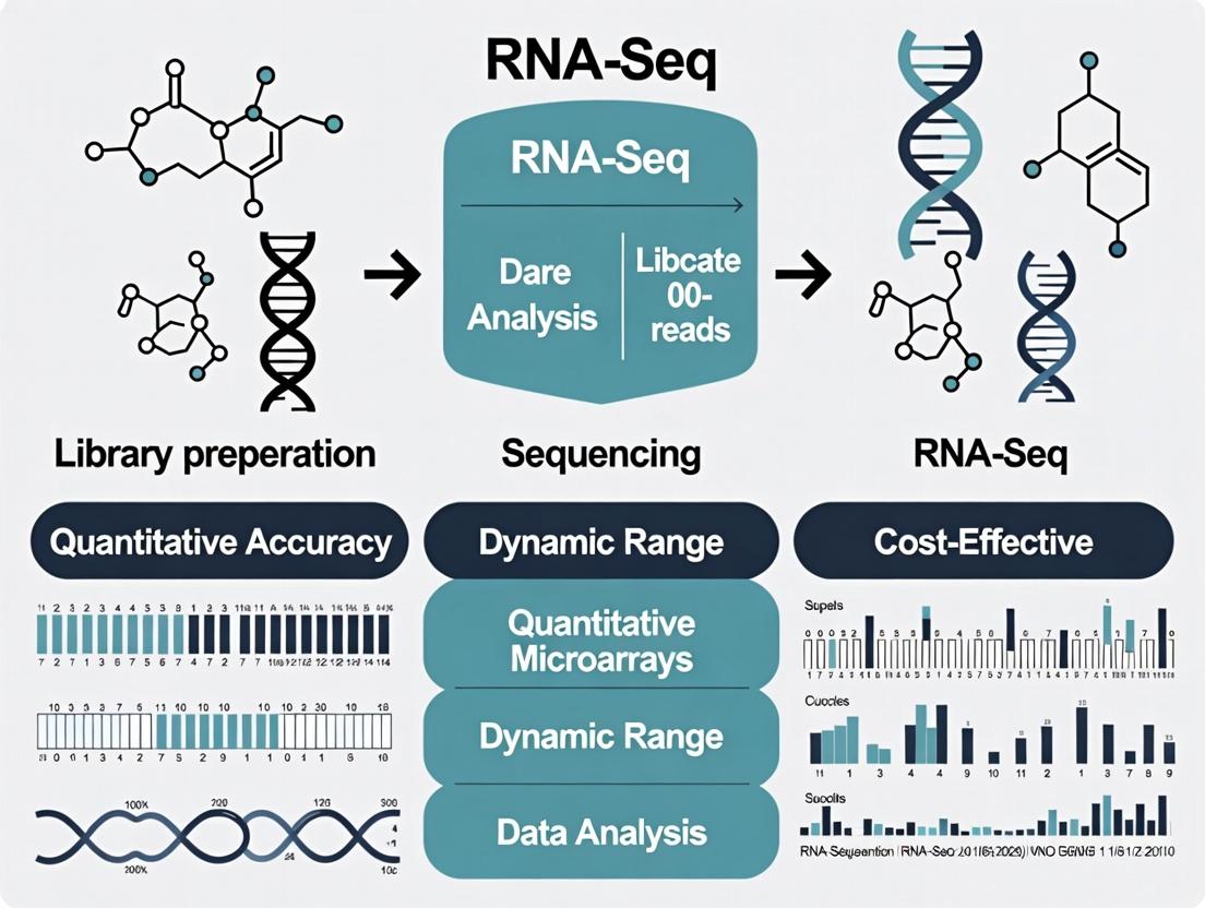
RNA-Seq vs. Microarrays: A Comprehensive Guide to Superior Gene Expression Analysis in Modern Research
This article provides researchers, scientists, and drug development professionals with a definitive comparison of RNA sequencing (RNA-Seq) and microarray technologies for gene expression analysis.
RNA-Seq vs. Microarrays: A Comprehensive Guide to Superior Gene Expression Analysis in Modern Research
Abstract
This article provides researchers, scientists, and drug development professionals with a definitive comparison of RNA sequencing (RNA-Seq) and microarray technologies for gene expression analysis. We explore the foundational principles of both methods before delving into the key technical and practical advantages of RNA-Seq, including its broader dynamic range, discovery of novel transcripts, and quantitative precision. The guide covers essential methodological considerations, common troubleshooting scenarios, and validation strategies to ensure robust data. By synthesizing current evidence, we demonstrate why RNA-Seq has become the dominant platform, enabling more accurate biomarkers, deeper biological insights, and accelerated therapeutic discovery.
From Probes to Reads: Understanding the Core Technologies of Microarrays and RNA-Seq
This article details the technical foundations of DNA microarrays, a transformative technology that enabled high-throughput gene expression analysis. While microarrays established a critical legacy in genomics, their limitations in the modern research context provide a clear rationale for the transition to RNA-Seq, which offers superior sensitivity, dynamic range, and discovery potential.
Core Technology and Workflow
A DNA microarray is a solid-surface platform (typically glass or silicon) onto which thousands to millions of nucleic acid probes are immobilized in a precise grid. Each probe is a short, sequence-specific DNA fragment that hybridizes to complementary target sequences from a sample.
Experimental Protocol: Two-Color Microarray Analysis
Objective: To compare gene expression levels between two biological samples (e.g., treated vs. untreated, diseased vs. healthy).
Key Steps:
- RNA Extraction & Quality Control: Total RNA is isolated from both sample types and assessed for integrity (e.g., RIN > 8.0 using Bioanalyzer).
- Reverse Transcription & Fluorescent Labeling: RNA from Sample A is converted to cDNA and labeled with Cy5 (red fluorescent dye). RNA from Sample B is labeled with Cy3 (green dye). Common method: Amino-allyl dUTP incorporation followed by dye coupling.
- Hybridization: The labeled cDNA pools are mixed and competitively hybridized to the microarray slide for 12-16 hours at 65°C in a specialized hybridization chamber.
- Washing & Scanning: Stringent washes remove non-specifically bound cDNA. The slide is scanned at wavelengths specific for Cy3 and Cy5.
- Image & Data Analysis: Software quantifies fluorescence intensity at each spot. The ratio of Cy5 to Cy3 fluorescence per spot represents the relative expression level of that gene in Sample A vs. B.
Diagram: Two-Color Microarray Experimental Workflow
Inherent Limitations of DNA Microarray Technology
The legacy of microarrays is defined by their specific constraints, which are fundamentally addressed by RNA-Seq.
Table 1: Quantitative Limitations of Microarray Technology
| Limitation | Description | Typical Impact/Value |
|---|---|---|
| Dependence on Prior Knowledge | Can only detect sequences complementary to pre-designed probes. | 0% discovery of novel transcripts/splice variants. |
| Limited Dynamic Range | Signal saturates at high fluorescence intensities; background limits low-end detection. | ~2-3 orders of magnitude (10²–10⁴). |
| Background Noise & Cross-Hybridization | Non-specific binding of similar sequences to a probe. | Can obscure low-abundance transcript signals. |
| Probe Design Issues | Performance varies based on probe sequence specificity and melting temperature (Tm). | Requires complex normalization algorithms. |
| Comparative Nature | Two-color arrays provide only relative expression (ratios), not absolute quantitation. | Requires a co-hybridized reference sample. |
Technical Limitation Pathways
The core constraints of the technology create a cascade of analytical challenges.
Diagram: Cascade of Microarray Limitations to Analytical Impact
The Scientist's Toolkit: Key Research Reagents & Materials
Table 2: Essential Microarray Experiment Reagents
| Item | Function in Protocol | Critical Specification |
|---|---|---|
| Microarray Slide | Solid support with spatially arrayed DNA probes. | Probe density, batch consistency, surface chemistry. |
| Fluorescent dNTPs (Cy3/Cy5) | Incorporation during cDNA synthesis for target labeling. | High specific activity, matched coupling efficiency. |
| Hybridization Buffer | Medium for target-probe interaction. | Contains blockers (Cot-1 DNA, poly-dA) to reduce non-specific binding. |
| SSC/SDS Wash Buffers | Post-hybridization stringency washes. | Precise saline concentration and temperature control. |
| Scanning Solution | Liquid for immersion during laser scanning. | Low fluorescence, appropriate refractive index. |
| Normalization & Spike-in Controls | Synthetic RNAs of known concentration added to sample. | Corrects for technical variation (e.g., Agilent Spike-in kit). |
Transition Rationale: RNA-Seq Advantages
The limitations above form the thesis for adopting RNA sequencing. RNA-Seq is not probe-limited, offers a dynamic range of >10⁵, provides single-base resolution, and enables de novo transcript discovery and absolute quantitation with digital counts. This represents a paradigm shift from hypothesis-limited profiling to comprehensive, discovery-driven transcriptomics.
Table 3: Core Comparison: Microarray vs. RNA-Seq
| Feature | DNA Microarray | RNA Sequencing (RNA-Seq) |
|---|---|---|
| Genomic Requirement | Requires complete prior sequence knowledge. | Can be applied to organisms with or without a reference genome. |
| Dynamic Range | Limited (10²–10⁴). | Very high (>10⁵). |
| Quantitation Type | Relative (ratio-based) or inferred absolute. | Digital counts (absolute), enables allelic-specific expression. |
| Sensitivity | Lower, poor for low-abundance transcripts. | High, can detect rare transcripts. |
| Resolution | Defined by probe length (~50-70bp). | Single-nucleotide. |
| Discovery Capability | None for novel features. | High (novel transcripts, splice variants, fusions). |
| Experimental Workflow | Relies on hybridization kinetics. | Relies on sequencing chemistry. |
| Cost & Complexity | Lower per sample, but obsolete. | Higher per sample, but continuously decreasing. |
Gene expression analysis is fundamental to understanding cellular function, disease mechanisms, and therapeutic responses. For over two decades, microarrays were the dominant technology for this purpose. However, this method has intrinsic limitations: it requires prior knowledge of the genome to design probes, has a limited dynamic range due to background hybridization and signal saturation, and offers poor quantification of low-abundance transcripts.
The broader thesis of this whitepaper is that RNA Sequencing (RNA-Seq) has revolutionized gene expression research by offering substantial, multifaceted benefits over microarray technology. RNA-Seq, built on Next-Generation Sequencing (NGS) foundations, provides an unbiased, high-resolution, and quantitative view of the transcriptome, enabling discoveries previously beyond reach.
Core Principles of Next-Generation Sequencing
NGS is a massively parallel sequencing technology that allows the determination of nucleotide sequences of millions of DNA fragments simultaneously. The core workflow, common to most platforms (Illumina being the most prevalent), involves:
- Library Preparation: DNA (or cDNA from RNA) is fragmented, and adapters containing sequencing primer binding sites are ligated to the ends.
- Cluster Amplification: Fragments are attached to a flow cell and amplified in situ to create clusters of identical copies.
- Sequencing by Synthesis: Fluorescently labeled, reversibly terminated nucleotides are added sequentially. After each incorporation, the flow cell is imaged to identify the base, and the terminator is cleaved for the next cycle.
- Data Analysis: Images are processed into sequence reads (base calls), which are then aligned to a reference genome or assembled de novo.
RNA-Seq Methodology: From RNA to Data
RNA-Seq applies NGS to cDNA derived from RNA. The detailed experimental protocol is as follows:
Protocol: Standard Poly-A Selected mRNA-Seq Workflow
Step 1: RNA Extraction & QC
- Isolate total RNA using a guanidinium thiocyanate-phenol-chloroform (e.g., TRIzol) or silica-membrane column method.
- Assess RNA integrity using an Agilent Bioanalyzer or TapeStation. An RNA Integrity Number (RIN) > 8 is recommended.
Step 2: RNA Selection & Fragmentation
- Poly-A Selection: Use oligo(dT) magnetic beads to enrich for polyadenylated mRNA. For total RNA analysis (including non-coding RNA), omit this step and proceed with rRNA depletion.
- Fragment RNA to ~200-300 nt using divalent cations (e.g., Mg²⁺) and elevated temperature (94°C for several minutes).
Step 3: cDNA Synthesis & Library Prep
- Reverse transcribe fragmented RNA using random hexamers and reverse transcriptase to create first-strand cDNA.
- Synthesize the second cDNA strand using DNA Polymerase I and RNase H.
- Perform end repair, A-tailing, and ligation of indexed sequencing adapters to create the final library.
Step 4: Library Quantification & Sequencing
- Quantify the library accurately by qPCR.
- Load onto an NGS flow cell for cluster generation and sequencing. Paired-end sequencing (e.g., 2x150 bp) is standard for better alignment and isoform detection.
Benefits of RNA-Seq vs. Microarrays: A Quantitative Comparison
The advantages of RNA-Seq are clear and measurable, as summarized in the table below.
Table 1: Quantitative Comparison of RNA-Seq and Microarray Technologies
| Feature | Microarray | RNA-Seq | Benefit of RNA-Seq |
|---|---|---|---|
| Requirement for Prior Sequence Knowledge | Mandatory (for probe design) | Not required (discovery-driven) | Enables de novo transcriptome assembly in novel organisms. |
| Dynamic Range (Orders of Magnitude) | ~2-3 logs (limited by background & saturation) | >5 logs | Accurately quantifies both highly abundant and rare transcripts. |
| Background Signal | High (due to cross-hybridization) | Very low (direct sequencing) | Improves signal-to-noise ratio and specificity. |
| Resolution | Limited to pre-designed probe locations. | Single-base resolution. | Identifies SNPs, editing sites, and precise splice junctions. |
| Differential Expression Accuracy | Good for moderate-to-high expression. | Superior across entire range, validated by qPCR. | Higher sensitivity and reproducibility. |
| Additional Applications | Gene expression only (primarily). | Gene expression, splice variants, fusion genes, novel transcripts, allele-specific expression. | Multiplexed information from a single experiment. |
Table 2: Typical Experimental Output Metrics (Human Transcriptome)
| Metric | Typical Microarray (Affymetrix) | Typical RNA-Seq (Illumina 30M PE reads) |
|---|---|---|
| Genes Detected | ~20,000 (annotated) | ~25,000 - 30,000 (including novel low-expression genes) |
| Alternative Splicing Events | Limited analysis | Comprehensive quantification |
| Reproducibility (Pearson R²) | 0.95 - 0.99 | 0.99+ |
| Cost per Sample (Reagent List Price) | ~$200 - $400 | ~$500 - $1,000 |
| Time from Sample to Data | 2-3 days | 3-7 days (including sequencing time) |
The Data Analysis Pipeline
The raw output of RNA-Seq (FASTQ files) undergoes a multi-step computational pipeline, whose logical flow is depicted below.
The Scientist's Toolkit: Essential Research Reagent Solutions
Table 3: Key Reagents & Kits for RNA-Seq Library Preparation
| Item | Function | Example Product(s) |
|---|---|---|
| Total RNA Isolation Kit | Purifies high-integrity RNA, free of genomic DNA, proteins, and contaminants. | Qiagen RNeasy, Invitrogen PureLink RNA, Zymo Quick-RNA. |
| Poly-A Selection Beads | Enriches for eukaryotic mRNA by binding the polyadenylated tail. | NEBNext Poly(A) mRNA Magnetic Isolation Module, Invitrogen Dynabeads mRNA DIRECT. |
| Ribo-depletion Kit | Removes abundant ribosomal RNA (rRNA) for total RNA or bacterial RNA-Seq. | Illumina Ribo-Zero Plus, QIAseq FastSelect. |
| RNA Fragmentation Buffer | Chemically fragments RNA to optimal size for library construction. | Part of standard kits (e.g., Illumina TruSeq, NEBNext Ultra II). |
| First & Second Strand cDNA Synthesis Kit | Converts RNA into double-stranded cDNA. | NEBNext Ultra II RNA First & Second Strand Synthesis Module. |
| Library Preparation Kit with Adapters & Indexes | Performs end-prep, adapter ligation, and includes unique dual indexes for sample multiplexing. | Illumina TruSeq Stranded mRNA, NEBNext Ultra II Directional RNA Library Prep. |
| Library Quantification Kit | Accurate, qPCR-based quantification of amplifiable library fragments. | KAPA Library Quantification Kit, Illumina Library Quantification Kit. |
| Size Selection Beads/Kit | Selects for cDNA fragments of a specific size range to control insert size. | SPRISelect/SPRI beads (Beckman Coulter), PippinHT (Sage Science). |
RNA-Seq, powered by NGS, represents a definitive advance over microarray technology. Its unbiased nature, expansive dynamic range, single-base resolution, and ability to multiplex diverse analyses into a single experiment have made it the gold standard for transcriptome profiling. While considerations of cost and computational complexity remain, the depth and quality of information delivered by RNA-Seq fundamentally accelerate research and drug development, enabling a more complete understanding of gene regulation in health and disease.
This whitepaper delineates the fundamental measurement principles of nucleic acid analysis: hybridization (the bedrock of microarray technology) and sequencing (the core of RNA-Seq). Framed within the thesis that RNA-Seq offers profound benefits over microarrays for gene expression analysis, we provide a technical dissection of both paradigms. This guide serves researchers and drug development professionals in understanding the core technological divergences that lead to differences in data output, applicability, and biological insight.
Foundational Principles: A Technical Deep Dive
Hybridization-Based Measurement (Microarrays)
Core Principle: Measurement relies on the thermodynamic binding (hybridization) of fluorescently labeled nucleic acid samples to complementary DNA or oligonucleotide probes immobilized on a solid surface. Signal intensity at each probe spot is presumed proportional to the abundance of the target sequence.
- Key Limitation: The measurement is indirect and relative, contingent on prior knowledge of sequences used to design the probes. It measures hybridization efficiency, not the actual nucleotide sequence.
- Dynamic Range: Typically limited to 2-3 orders of magnitude due to background noise and signal saturation.
Sequencing-Based Measurement (RNA-Seq)
Core Principle: Measurement involves the direct, high-throughput determination of the nucleotide sequence of cDNA libraries. Quantification is achieved by counting the number of sequence reads that align to specific genomic loci.
- Key Advantage: It is a direct, digital readout. Each countable fragment (read) provides both quantitative and sequence identity information.
- Dynamic Range: Exceeds 5 orders of magnitude, limited primarily by sequencing depth (total number of reads).
Quantitative Comparison of Core Metrics
Table 1: Fundamental Comparison of Measurement Principles
| Feature | Hybridization (Microarray) | Sequencing (RNA-Seq) |
|---|---|---|
| Underlying Principle | Indirect, analog signal from probe-target binding | Direct, digital counting of sequence fragments |
| Requirement for Prior Knowledge | Mandatory (for probe design) | Not required (discovery-driven) |
| Dynamic Range | ~10²–10³ (Limited by saturation & background) | >10⁵ (Scales with sequencing depth) |
| Background Signal | High (from non-specific cross-hybridization) | Very low (specific alignment reduces noise) |
| Resolution | Single nucleotide (for some SNP arrays) | Single nucleotide (base-level) |
| Ability to Detect Novel Features | None (only known transcripts/isoforms) | High (novel transcripts, splice variants, fusions) |
| Sample Throughput (per run) | High (multiple arrays per instrument) | Moderate to High (multiplexing enabled) |
| Cost per Sample (Typical) | Lower | Higher, though decreasing |
Table 2: Performance in Gene Expression Analysis Context
| Performance Metric | Microarray | RNA-Seq |
|---|---|---|
| Accuracy & Specificity | Lower (cross-hybridization artifacts) | Higher (direct sequencing) |
| Quantitative Precision | Good for medium- to high-abundance transcripts | Excellent across full abundance range |
| Reproducibility (Technical Replicate R²) | >0.99 | >0.99 |
| Required Input RNA | 1–100 ng (can use degraded RNA) | 10 ng–1 µg (requires high-quality RNA) |
| Key Experimental Bottleneck | Probe design and array manufacturing | Library preparation and computational analysis |
Experimental Protocols in Practice
Standard Microarray Workflow for Gene Expression
- RNA Extraction & QC: Isolate total RNA, assess integrity (RIN >7).
- cDNA Synthesis & Labeling: Reverse transcribe RNA into cDNA, then incorporate fluorescent dyes (e.g., Cy3, Cy5) via enzymatic or chemical labeling.
- Hybridization: Apply labeled cDNA to the microarray chip under stringent temperature-controlled conditions (typically 16-20 hours).
- Washing: Remove non-specifically bound material through a series of stringency washes.
- Scanning & Image Analysis: Use a confocal laser scanner to excite fluorophores and measure emission intensity. Convert images to spot intensity values (*.CEL files for Affymetrix).
- Data Normalization & Quantification: Apply algorithms (e.g., RMA, MAS5) to correct for background and technical variation, then summarize probe-set intensities into transcript abundances.
Standard RNA-Seq Workflow (Illumina Platform)
- RNA Extraction & QC: Isolate RNA, assess integrity (RIN >8 for mRNA-seq).
- Library Preparation: a. Poly-A Selection or rRNA Depletion: Enrich for mRNA or remove ribosomal RNA. b. Fragmentation: Chemically or enzymatically fragment RNA (or post-cDNA synthesis). c. cDNA Synthesis: Generate first- and second-strand cDNA. d. End Repair, A-tailing, & Adapter Ligation: Prepare fragments for ligation of platform-specific sequencing adapters (containing indices for multiplexing). e. PCR Amplification: Enrich adapter-ligated fragments. f. Library QC: Validate size distribution and quantify (qPCR or bioanalyzer).
- Sequencing: Cluster generation and sequencing-by-synthesis on platforms (e.g., NovaSeq) to generate short paired-end reads (e.g., 2x150 bp).
- Primary Data Analysis: a. Demultiplexing: Assign reads to samples based on index sequences. b. Alignment: Map reads to a reference genome/transcriptome using aligners (e.g., STAR, HISAT2). c. Quantification: Count reads mapping to genes/transcripts (e.g., using featureCounts, HTSeq).
- Downstream Analysis: Differential expression (DESeq2, edgeR), pathway analysis, etc.
Visualizing the Workflows and Logical Relationships
Diagram 1: Core Measurement Principles Contrasted
Diagram 2: RNA-Seq Experimental Workflow
The Scientist's Toolkit: Essential Research Reagents & Materials
Table 3: Key Reagent Solutions for RNA-Seq Library Preparation
| Reagent/Material | Function in Workflow | Example/Note |
|---|---|---|
| Poly(dT) Magnetic Beads | mRNA enrichment from total RNA by binding poly-A tail. | Essential for mRNA-seq. Alternative: rRNA depletion kits for total RNA-seq. |
| Fragmentation Buffer (Mg²⁺/Heat) | Randomly fragments RNA to desired size range (e.g., 200-300 bp). | Replaced by enzymatic fragmentation in some kits. |
| Reverse Transcriptase | Synthesizes first-strand cDNA from RNA template. | Must be robust for long/structured templates. |
| Second-Strand Synthesis Mix | Replaces RNA with DNA to create double-stranded cDNA. | Contains RNase H, DNA Pol I, dNTPs. |
| Sequencing Adapters (Indexed) | Short, double-stranded DNA ligated to fragments; contain sequences for cluster binding and sample multiplexing (indices). | Unique dual indices (UDIs) are critical for multiplexing. |
| PCR Master Mix | Amplifies adapter-ligated libraries; includes a thermostable polymerase. | Limited-cycle PCR (8-15 cycles) to minimize bias. |
| SPRI Beads | Size-selection and cleanup of nucleic acids using magnetic solid-phase reversible immobilization. | Replaces traditional column-based cleanups. |
| Library Quantification Kit | Accurately measures library concentration for pooling and loading onto sequencer. | qPCR-based (e.g., KAPA SYBR FAST) is essential. |
| Sequencing Flow Cell | Glass slide with oligonucleotide lawns where bridge amplification and sequencing occur. | Platform-specific (e.g., Illumina S1/S2, NovaSeq 6000). |
| Sequencing Chemistry | Contains fluorescently labeled, reversibly terminated nucleotides and enzymes for cyclic SBS. | Provides the "sequencing-by-synthesis" reaction. |
Within the ongoing evaluation of genomics technologies, a core thesis posits significant benefits of RNA-Seq over microarrays for gene expression analysis. This technical guide deconstructs this assertion by examining four fundamental performance metrics: Sensitivity, Specificity, Dynamic Range, and Throughput. Understanding these metrics provides a rigorous, quantitative framework for technology selection in research and drug development.
Defining the Key Metrics
- Sensitivity: The probability of detecting a true positive. In expression analysis, this refers to the ability to detect low-abundance transcripts.
- Specificity: The probability of a true negative. This measures the technology's accuracy in distinguishing between similar sequences (e.g., splice variants or paralogous genes) and minimizing false-positive signals.
- Dynamic Range: The range over which an input signal (transcript concentration) is linearly related to the output signal. It defines the span from the lowest to the highest quantifiable expression level.
- Throughput: The number of samples or amount of genetic material analyzed per unit time, cost, and operational effort. It encompasses scalability and multiplexing capability.
Quantitative Comparison: RNA-Seq vs. Microarrays
The following table synthesizes current data on the performance of modern RNA-Seq (e.g., Illumina NovaSeq) versus high-density oligonucleotide microarrays.
Table 1: Performance Metrics Comparison for Gene Expression Analysis
| Metric | RNA-Seq (Illumina Platform) | Microarray (Affymetrix/Agilent) | Experimental Basis |
|---|---|---|---|
| Sensitivity | High. Can detect transcripts at levels below 1 copy per cell. | Moderate. Limited by background hybridization and probe affinity. | Spike-in experiments using External RNA Controls Consortium (ERCC) standards. |
| Specificity | High. Especially with paired-end or long-read sequencing; can distinguish isoforms. | Moderate to High. Limited by cross-hybridization and predefined probe design. | Analysis of known splice junctions or homologous gene families. |
| Dynamic Range | Very High (~7-8 orders of magnitude). Direct counting of transcripts. | Limited (~3-4 orders of magnitude). Constrained by background and saturation. | Measurement across dilution series of RNA samples. |
| Throughput (Samples) | High. Scalable via multiplexing (96+ samples per lane). Batch effects require care. | Very High. Robust, standardized processing for large cohorts. | Comparison of sample processing times and multiplexing capabilities. |
| Throughput (Discovery) | Discovery-based. Identifies novel transcripts, fusions, and mutations. | Hypothesis-driven. Limited to annotated probes on the array. | De novo transcriptome assembly in non-model organisms. |
Experimental Protocols for Metric Validation
Protocol 1: Assessing Sensitivity with ERCC Spike-in Controls
- Material: Serial dilutions of ERCC ExFold RNA Spike-in Mix.
- Method: Spike defined concentrations of synthetic ERCC transcripts into total RNA samples prior to library prep (RNA-Seq) or labeling (microarray).
- Analysis: Plot observed read counts or fluorescence intensity against known input concentration. Calculate limit of detection (LoD) and limit of quantitation (LoQ).
Protocol 2: Evaluating Specificity for Splice Variants
- Material: RNA from a cell line with well-characterized alternative splicing (e.g., human cell lines).
- Method: Perform RNA-Seq (paired-end recommended) and microarray analysis using exon-resolution arrays.
- Analysis: Map reads to a reference genome/transcriptome or measure exon probe sets. Quantify the proportion of known splice junctions correctly identified versus false positives from cross-mapping or cross-hybridization.
Protocol 3: Measuring Dynamic Range
- Material: A two-sample RNA mixture (e.g., human and yeast RNA) mixed in a defined logarithmic dilution series.
- Method: Process dilution series with both technologies.
- Analysis: For each dilution point, plot measured expression fold-change against expected fold-change. Determine the linear range where R² > 0.99.
Protocol 4: Comparative Workflow for Throughput Assessment
The following diagram illustrates the core workflows and decision points influencing throughput.
Workflow Comparison for Throughput
The Scientist's Toolkit: Essential Research Reagents & Materials
Table 2: Key Reagent Solutions for RNA Expression Analysis
| Item | Function | Technology Relevance |
|---|---|---|
| RNase Inhibitors | Protects RNA integrity during isolation and processing. | Critical for both. |
| Poly-dT Magnetic Beads | Isolates polyadenylated mRNA from total RNA. | Standard for most RNA-Seq; used in some array protocols. |
| Ribo-depletion Kits | Removes abundant rRNA to enrich for mRNA and non-coding RNA. | Essential for non-poly-A RNA-Seq. |
| Reverse Transcriptase | Synthesizes cDNA from RNA template. | Core enzyme for both technologies. |
| dNTPs with Modified Nucleotides | Incorporates dUTP or other bases for strand-specificity or amplification. | Key for strand-specific RNA-Seq libraries. |
| Sequence-Specific Adapters & Indexes | Attach to cDNA for sequencing and multiplexing. | Core component of RNA-Seq library prep. |
| Fluorescent Dyes (Cy3/Cy5) | Label cDNA for detection on array surface. | Core detection method for microarrays. |
| Hybridization Buffer | Promotes specific binding of cDNA to array probes. | Critical for microarray specificity and sensitivity. |
| PCR Master Mix | Amplifies cDNA libraries prior to sequencing. | Required for most RNA-Seq protocols. |
Pathway of Metric Interdependence and Technology Choice
The decision between RNA-Seq and microarrays involves balancing these key metrics against project goals, as shown in the following logic pathway.
Technology Selection Logic Pathway
The comparative analysis of sensitivity, specificity, dynamic range, and throughput provides a concrete framework supporting the thesis of RNA-Seq's advantages for comprehensive gene expression analysis. While microarrays remain robust for high-throughput, targeted studies in well-annotated genomes, RNA-Seq's superior sensitivity, dynamic range, and discovery power make it the prevailing choice for exploratory research, biomarker discovery, and studies of genomic complexity, directly benefiting modern drug development pipelines.
Unlocking RNA-Seq's Power: Key Advantages and Practical Applications in Research
The transition from microarray technology to RNA sequencing (RNA-Seq) represents a paradigm shift in transcriptomics. While microarrays excelled at quantifying known, predefined sequences, their fundamental design limits discovery. RNA-Seq, with its hypothesis-free, high-resolution sequencing of the entire transcriptome, is uniquely positioned to uncover the complex and previously "unknown" layer of genomic regulation. This document details the core technical capabilities of RNA-Seq in discovering novel transcripts, alternative splice variants, and gene fusions—capabilities that are either severely constrained or impossible with microarray-based analysis.
Core Technical Capabilities & Quantitative Comparison
Table 1: Capability Comparison: RNA-Seq vs. Microarrays
| Feature | RNA-Seq | Microarrays |
|---|---|---|
| Hypothesis Requirement | None (Discovery-driven) | Required (Targeted) |
| Genomic Coverage | Full transcriptome, unbiased | Pre-designed probes only |
| Novel Transcript Detection | Yes ( de novo assembly) | No |
| Splice Variant Resolution | Base-pair level, quantifies isoforms | Limited, depends on exon-junction probes |
| Fusion Gene Detection | Yes (spanning read pairs) | Only known, pre-designed fusions |
| Dynamic Range | >10⁵ (Wide) | ~10³ (Narrow) |
| Background Noise | Very low (deduced from sequence) | High (non-specific hybridization) |
| Required Input RNA | Low (ng scale) | High (μg scale) |
Detailed Methodologies for Discovery
Detecting Novel Transcripts & Splice Variants
Protocol: Reference-Based & De Novo Transcriptome Assembly
- Library Preparation: Use stranded, ribosomal RNA-depleted total RNA libraries to preserve strand information and capture non-polyadenylated transcripts.
- Sequencing: Perform deep sequencing (typically ≥100 million paired-end 150bp reads) on platforms like Illumina NovaSeq to ensure sufficient coverage for assembly.
- Alignment & Assembly:
- Reference-Guided: Align reads to the reference genome using splice-aware aligners (e.g., STAR, HISAT2). Use assemblers like StringTie or Cufflinks to reconstruct transcript models, merging annotated (GENCODE) and novel isoforms.
- De Novo: For species without a reference genome, assemble reads directly into contigs using tools like Trinity or SOAPdenovo-Trans, followed by annotation.
- Differential Expression & Validation: Quantify novel transcript expression with tools like Salmon or kallisto. Validate findings via RT-PCR with primers spanning novel exon junctions and Sanger sequencing.
Identifying Fusion Genes
Protocol: Fusion Detection from RNA-Seq Data
- Data Acquisition: Generate high-quality, paired-end RNA-Seq data from the sample of interest (e.g., cancer biopsy).
- Bioinformatic Detection: Process reads through multiple, complementary fusion detection algorithms to reduce false positives.
- STAR-Fusion: Uses the STAR aligner to map reads, then identifies fusion evidence from chimeric alignments.
- Arriba: Fast, pattern-based fusion detection from STAR output, effective for oncogenic fusions.
- FusionCatcher: Uses multiple steps (pre-trimming, alignment, filtering) for comprehensive discovery.
- Filtering & Prioritization: Filter results against common artifacts, normal tissue databases (e.g., GTEx), and prioritize based on:
- Spanning read counts & split read support.
- Predicted functional consequence (in-frame, retained kinase domains).
- Known oncogenic status (e.g., in databases like MiOncoCirc).
- Experimental Validation: Confirm high-confidence candidates using orthogonal methods: RT-PCR followed by Sanger sequencing, or fluorescence in situ hybridization (FISH).
Visualization of Workflows & Pathways
Diagram 1: RNA-Seq Discovery Workflow
Diagram 2: Fusion Gene Detection Logic
The Scientist's Toolkit: Key Research Reagents & Materials
Table 2: Essential Reagents for RNA-Seq Discovery Experiments
| Item | Function | Example Product/Kit |
|---|---|---|
| Ribo-depletion Reagents | Removes abundant ribosomal RNA to enrich for mRNA and non-coding RNA, critical for novel transcript detection. | Illumina Ribo-Zero Plus, NEBNext rRNA Depletion Kit |
| Stranded Library Prep Kit | Preserves the original orientation of transcripts, essential for accurate annotation of novel antisense transcripts and overlapping genes. | Illumina Stranded Total RNA Prep, NEBNext Ultra II Directional RNA Library Prep |
| High-Fidelity Reverse Transcriptase | Creates accurate cDNA copies of RNA templates with high processivity, reducing bias in representation. | SuperScript IV, PrimeScript RT |
| Nuclease-Free Water & Beads | Ensures no RNA degradation during reactions and enables clean size selection/fragmentation. | AMPure XP Beads, Ambion Nuclease-Free Water |
| RNA Integrity Number (RIN) Analyzer | Assesses RNA quality pre-library prep; high-quality input (RIN >8) is crucial for full-length transcript assembly. | Agilent Bioanalyzer RNA Nano Kit |
| Fusion Validation Primers | Custom-designed oligonucleotides spanning predicted fusion breakpoints for PCR-based confirmation. | IDT Custom DNA Oligos |
| Positive Control RNA | Spiked-in RNA standards (e.g., from cell lines with known fusions/isoforms) to monitor assay sensitivity and specificity. | Universal Human Reference RNA, Horizon Multiplex Fusion RNA Standard |
This technical guide elaborates on the superior quantitative precision of RNA sequencing (RNA-Seq) compared to microarray technology, contextualized within the broader thesis of RNA-Seq's benefits for gene expression analysis. We detail how RNA-Seq achieves a broader dynamic range and enhanced accuracy for low-abundance transcripts, which is critical for advanced research in molecular biology and drug development.
Microarray technology, while transformative, is limited by its dependence on predefined probes and signal saturation at high expression levels, compressing its dynamic range. RNA-Seq, a sequencing-based method, provides an absolute digital count of transcripts without upper quantification limits and with background signal minimization, enabling the detection of rare transcripts crucial for understanding subtle regulatory changes in disease and development.
Core Quantitative Metrics: RNA-Seq vs. Microarrays
Table 1: Quantitative Performance Comparison of Expression Platforms
| Performance Metric | High-Density Oligo Microarray | Next-Generation RNA-Seq (Illumina) | Significance for Research |
|---|---|---|---|
| Theoretical Dynamic Range | ~10³-10⁴ (Limited by fluorescence saturation) | >10⁵ (Digital counts, no upper limit) | Enables simultaneous quantification of highly abundant housekeeping genes and rare transcription factors. |
| Sensitivity (Limit of Detection) | ~1-5 copies/cell (Limited by background cross-hybridization) | ~0.1-0.5 copies/cell (With sufficient depth) | Critical for detecting low-abundance signaling receptors, non-coding RNAs, and splice variants. |
| Background Signal | High (Non-specific hybridization) | Very Low (Direct cDNA sequencing) | Improves signal-to-noise ratio, enhancing accuracy for low-fold-change measurements. |
| Accuracy (vs. qPCR) | Moderate (R² ~0.7-0.85) | High (R² ~0.9-0.99) | Provides data closer to gold-standard validation methods, increasing confidence in results. |
| Precision (Technical Replicate CV) | 5-15% | 2-8% | Enables detection of smaller, biologically relevant expression changes. |
Data synthesized from current benchmarking studies (2023-2024).
Experimental Protocol: Capturing Low-Abundance Transcripts with RNA-Seq
This protocol is optimized for quantitative accuracy across the abundance spectrum.
A. Sample Preparation & Library Construction
- RNA Integrity: Verify RNA Integrity Number (RIN) > 8.5 using a Bioanalyzer.
- Ribosomal RNA Depletion: Use ribo-depletion kits (e.g., Illumina Ribo-Zero Plus) over poly-A selection to retain non-polyadenylated and partially degraded transcripts, common in low-abundance classes.
- cDNA Synthesis & Amplification: Use a limited-cycle PCR (10-15 cycles) with unique dual indexing (UDI) adapters to minimize amplification bias and permit sample multiplexing.
- Library QC: Precisely quantify library concentration using fluorometry (Qubit) and profile fragment size (Bioanalyzer/TapeStation).
B. Sequencing & Data Acquisition
- Sequencing Depth: Target 40-60 million paired-end reads per sample for standard differential expression. For comprehensive low-abundance detection (e.g., in single-cell or pathogen transcripts), target 100-200 million reads.
- Read Length: Use 2x150 bp paired-end sequencing to improve transcript isoform resolution and mapping accuracy.
C. Bioinformatic Analysis for Quantitative Precision
- Alignment: Map reads to the reference genome/transcriptome using a splice-aware aligner (e.g., STAR or HISAT2) with parameters tuned for sensitivity.
- Quantification: Generate raw digital read counts per gene using featureCounts or Salmon (in alignment-based or lightweight mapping mode). Salmon's bias correction is recommended for abundance-aware quantification.
- Normalization: For between-sample comparison, use statistical methods like DESeq2's median-of-ratios or edgeR's TMM which are robust to composition bias and differential expression of high-abundance genes.
Visualizing the RNA-Seq Workflow for Quantitative Precision
Diagram Title: RNA-Seq Workflow for Broad Dynamic Range
The Scientist's Toolkit: Key Research Reagent Solutions
Table 2: Essential Reagents for High-Precision RNA-Seq
| Reagent / Kit | Function | Key Consideration for Quantitative Precision |
|---|---|---|
| Ribo-Zero Plus (Illumina) | Removal of cytoplasmic and mitochondrial rRNA. | Preserves non-coding and non-polyA transcripts, expanding detectable dynamic range. |
| SMARTer Stranded Total RNA-Seq (Takara Bio) | A template-switching based kit for strand-specific library prep from total RNA. | Maintains strand information, crucial for accurate quantification in overlapping genomic regions. |
| NEBNext Ultra II Directional (NEB) | A robust, widely-adopted kit for poly-A or rRNA-depleted stranded library preparation. | Consistent performance minimizes batch effects, improving precision across replicates. |
| KAPA HyperPrep (Roche) | Library preparation kit with low input and rapid protocols. | Optimized for minimal amplification bias, preserving quantitative relationships. |
| Unique Dual Indexes (UDIs) | Sets of molecular barcodes for sample multiplexing. | Eliminates index hopping crosstalk, ensuring sample integrity and accurate per-sample read assignment. |
| ERCC RNA Spike-In Mix (Thermo Fisher) | A set of synthetic RNA controls at known, varying concentrations. | Added prior to library prep to monitor technical sensitivity, dynamic range, and normalization accuracy. |
| Qubit dsDNA HS Assay Kit (Thermo Fisher) | Fluorometric quantification of library concentration. | More accurate than spectrophotometry for low-concentration libraries, critical for balanced sequencing. |
Pathway: Technical Factors Influencing Quantitative Accuracy
The following diagram illustrates the logical relationship between experimental choices and their impact on key quantitative outcomes.
Diagram Title: Factors Driving RNA-Seq Quantitative Precision
RNA-Seq fundamentally surpasses microarrays in quantitative precision by offering a vast, digital dynamic range and the sensitivity required to measure biologically critical low-abundance transcripts accurately. This capability, realized through optimized wet-lab protocols and sophisticated bioinformatics, empowers researchers to uncover subtle yet pivotal gene expression changes driving disease mechanisms and therapeutic responses, thereby accelerating the pace of discovery and drug development.
The transition from microarray technology to RNA sequencing (RNA-Seq) represents a paradigm shift in functional genomics. While microarrays provided a foundational technology for gene expression profiling, they are fundamentally limited by their dependence on pre-designed probes, which restricts analysis to known transcripts and provides only a relative, hybridization-based signal intensity. RNA-Seq, a high-throughput, sequencing-based method, delivers absolute quantification, discovers novel transcripts and splice variants, and offers a significantly broader dynamic range. Crucially, this thesis posits that RNA-Seq's most transformative benefit is its ability to simultaneously interrogate multiple layers of genomic information from a single experiment. This whitepaper focuses on one such advanced application: the integrated, multiplexed analysis of Allele-Specific Expression (ASE) and Single Nucleotide Variant (SNV) detection, moving "beyond expression" to a unified view of the transcriptome's functional genetic landscape—a feat unattainable with microarrays.
Core Concepts and Technical Foundations
Allele-Specific Expression (ASE)
ASE occurs when one allele of a gene is expressed at a higher level than the other in a diploid organism, potentially due to cis-regulatory variation (e.g., promoters, enhancers), genomic imprinting, or random X-chromosome inactivation. Quantifying ASE requires the ability to distinguish and count RNA reads originating from each parental chromosome.
SNV Detection from RNA-Seq Data
RNA-Seq data can be mined for single nucleotide variants, providing a direct readout of the expressed mutational landscape. This includes identifying somatic mutations in cancer, characterizing expressed heterozygous germline variants, and detecting RNA editing events.
The Multiplexed Advantage
The power of RNA-Seq lies in performing both analyses concurrently on the same dataset. A heterozygous SNV identified in the RNA-Seq data serves as a natural "barcode" to phase the reads and quantify allele-specific counts, linking regulatory consequence (cis-effect on expression) directly to the genetic variant.
Detailed Experimental Protocols
Comprehensive RNA-Seq Library Preparation and Sequencing
Protocol Goal: Generate high-quality, strand-specific, paired-end sequencing libraries from total RNA. Materials: See "Research Reagent Solutions" below. Steps:
- RNA Extraction & QC: Isolate total RNA using a column-based kit with DNase I treatment. Assess integrity via RIN (RNA Integrity Number) > 8.5 on a Bioanalyzer.
- rRNA Depletion: Use ribo-depletion kits (e.g., Illumina Ribo-Zero Plus) to remove ribosomal RNA, enriching for mRNA and non-coding RNAs. Poly-A selection is an alternative but loses non-polyadenylated transcripts.
- Fragmentation & cDNA Synthesis: Fragment purified RNA (approx. 200-300 bp) via divalent cation incubation at elevated temperature. Synthesize first-strand cDNA using random hexamers and reverse transcriptase, followed by second-strand synthesis with dUTP incorporation for strand specificity.
- Library Construction: Perform end-repair, A-tailing, and adapter ligation. Size-select fragments (e.g., 300-400 bp) using SPRI beads.
- Strand Selection & Amplification: Treat with Uracil-Specific Excision Reagent (USER) enzyme to degrade the second strand (containing dUTP), preserving only the first strand. Perform limited-cycle PCR to amplify the final library.
- Sequencing: Pool libraries and sequence on an Illumina NovaSeq X Plus platform to a minimum depth of 50 million paired-end 150 bp reads per sample for robust ASE/SNV calling.
Integrated Computational Workflow for ASE & SNV Analysis
Protocol Goal: Process raw RNA-Seq reads to jointly call SNVs and quantify ASE. Software Tools: STAR, GATK, SAMtools, bcftools, ASEP, or custom pipelines. Steps:
- Alignment & Processing: Map reads to a diploid-aware human reference genome (e.g., GRCh38) using a splice-aware aligner (STAR). Coordinate-sort output and mark duplicates (GATK MarkDuplicates).
- SNV Calling: Perform variant calling using a tool optimized for RNA-Seq (e.g., GATK's "HaplotypeCaller" in RNA mode). Apply hard filters (QD < 2.0 || FS > 30.0 || SOR > 3.0 || MQ < 40) or use variant quality score recalibration (VQSR).
- Phasing & ASE Quantification:
- Identify heterozygous SNVs (genotype quality, GQ > 20, read depth DP > 10).
- At each heterozygous SNV position, count reads supporting the reference and alternate alleles using tools like
ASEReadCounter(GATK) orasep. - Filter for binomial test significance (FDR < 0.05) and minimum allelic count (e.g., ≥ 10 total reads at the site).
- Integration & Visualization: Merge SNV and ASE results. Calculate allelic ratio (Alt/(Ref+Alt)). Visualize genome-wide allelic imbalances and correlate with nearby cis-QTLs or chromatin accessibility data.
Table 1: Capability Comparison for Advanced Genomic Analyses
| Feature | RNA-Seq | Microarray | Advantage for ASE/SNV |
|---|---|---|---|
| SNV Discovery | Genome-wide, de novo detection of known and novel variants. | Limited to pre-designed probe sets; poor sensitivity for novel variants. | Essential for identifying heterozygous sites used as phasing markers. |
| ASE Resolution | Base-pair resolution at any heterozygous site. | Relies on exonic probe intensity differences; limited by probe design and cross-hybridization. | Enables precise, quantitative allelic counts at the nucleotide level. |
| Dynamic Range | >10⁵ for expression quantification. | ~10³ for intensity-based detection. | Accurately quantifies both highly and lowly expressed alleles. |
| Multiplexed Data | Single experiment yields expression, SNVs, ASE, splicing, fusions. | Typically measures expression only; specialized arrays needed for genotyping. | Unifies genetic and transcriptomic analysis, reducing cost and sample input. |
Table 2: Typical Performance Metrics from an RNA-Seq ASE/SNV Study
| Metric | Typical Value | Importance |
|---|---|---|
| Sequencing Depth for ASE | 50-100 million paired-end reads | Ensures sufficient coverage at heterozygous loci for statistical power. |
| Heterozygous SNVs Detected (per sample) | 150,000 - 250,000 | Provides dense phasing information across the transcriptome. |
| Genes with Significant ASE (FDR<0.05) | 5,000 - 10,000 | Indicates the scope of cis-regulatory variation active in the sample. |
| False Positive Rate (SNV Call) | < 1% (with rigorous filtering) | Critical for distinguishing true variants from sequencing/alignment artifacts. |
| Concordance with DNA-based Genotyping | > 98% (for high-confidence calls) | Validates the accuracy of RNA-derived SNV calls. |
Visualizations
Diagram 1: Integrated ASE & SNV Analysis Workflow
Diagram 2: Allele-Specific Expression Measurement Principle
The Scientist's Toolkit: Research Reagent Solutions
Table 3: Essential Materials for RNA-Seq based ASE/SNV Studies
| Item | Function | Example Product (Research-Use Only) |
|---|---|---|
| Ribo-depletion Kit | Removes abundant ribosomal RNA (>90%), enriching for coding and non-coding RNA for comprehensive variant detection. | Illumina Ribo-Zero Plus, QIAseq FastSelect. |
| Strand-Specific Library Prep Kit | Preserves the original orientation of transcripts during cDNA synthesis, crucial for accurately phasing variants to the correct allele. | NEBNext Ultra II Directional RNA Library Prep, TruSeq Stranded Total RNA Kit. |
| High-Fidelity Reverse Transcriptase | Synthesizes cDNA with low error rates, minimizing artifacts that could be mistaken for SNVs. | SuperScript IV, Maxima H Minus. |
| PCR Amplification Enzyme with High Fidelity | Amplifies final libraries with minimal bias and low mutation rates, preserving true allelic representation. | KAPA HiFi HotStart ReadyMix, Q5 High-Fidelity DNA Polymerase. |
| Dual-Indexed Adapter Kit | Allows multiplexed sequencing of many samples, reducing per-sample cost while maintaining sample identity for cohort studies. | IDT for Illumina UD Indexes, NEBNext Multiplex Oligos. |
| Diploid Human Reference Genome | A reference containing both haplotypes for improved alignment accuracy in polymorphic regions. | GRCh38 with ALT contigs, Human Pangenome Reference. |
| Bioinformatic Pipelines | Integrated software suites for reproducible processing, variant calling, and ASE analysis. | GATK Best Practices RNA-Seq, nf-core/rnaseq, STAR-fusion + ASEP. |
The transition from microarray technology to RNA sequencing (RNA-Seq) represents a cornerstone advancement in functional genomics. Within the broader thesis advocating for the benefits of RNA-Seq over microarrays, this guide details its pivotal applications. RNA-Seq provides an unparalleled, comprehensive, and quantitative view of the transcriptome, enabling discoveries with a resolution and scale previously unattainable. This whitepaper serves as a technical guide for leveraging RNA-Seq in three critical areas: differential expression analysis, biomarker discovery, and pathway analysis.
Differential Expression Analysis: Unbiased Quantification
RNA-Seq's key advantage over microarrays is its ability to detect novel transcripts and provide an absolute, not relative, measure of gene expression without predefined probes.
Experimental Protocol: A Standard RNA-Seq DE Workflow
- Library Preparation: Isolate total RNA (RIN > 8). Use poly-A selection for mRNA or ribosomal RNA depletion for total RNA. Fragment RNA, synthesize cDNA, and add sequencing adapters. Barcodes enable multiplexing.
- Sequencing: Perform high-throughput sequencing on platforms like Illumina NovaSeq, aiming for 20-40 million paired-end reads per sample for standard mammalian genomes.
- Bioinformatic Analysis:
- Quality Control: Use FastQC to assess read quality. Trim adapters and low-quality bases with Trimmomatic or Cutadapt.
- Alignment: Map reads to a reference genome using splice-aware aligners (e.g., STAR, HISAT2).
- Quantification: Generate a count matrix using featureCounts or HTSeq, counting reads overlapping exonic regions of genes.
- Differential Expression: Perform statistical testing with tools like DESeq2, edgeR, or limma-voom, which model count data and account for biological variance.
Quantitative Data Summary: RNA-Seq vs. Microarray in DE
| Metric | RNA-Seq | Microarray | Implication for DE Analysis |
|---|---|---|---|
| Dynamic Range | >10⁵ | 10²-10³ | RNA-Seq accurately quantifies both highly abundant and rare transcripts. |
| Background Noise | Low (direct counting) | High (non-specific hybridization) | RNA-Seq reduces false positives. |
| Sensitivity | Can detect transcripts at <1 copy/cell | Limited by probe design and cross-hybridization | RNA-Seq identifies more differentially expressed genes, especially low-abundance ones. |
| Genome Coverage | Agnostic; discovers novel transcripts, isoforms, fusions | Limited to predefined probe set | RNA-Seq enables discovery beyond annotated genomes. |
Diagram Title: RNA-Seq Differential Expression Analysis Workflow
Biomarker Discovery: Comprehensive Molecular Signatures
RNA-Seq facilitates the discovery of diagnostic, prognostic, and predictive biomarkers—from single genes to complex signatures—by profiling the entire transcriptome without bias.
Experimental Protocol: Biomarker Signature Identification
- Cohort Design: Collect samples from well-defined clinical cohorts (e.g., disease vs. control, treatment responders vs. non-responders). Adequate sample size is critical for statistical power.
- RNA-Seq Profiling: Perform sequencing as per DE protocol. For liquid biopsies, focus on extracellular RNA or use ultra-low-input protocols.
- Bioinformatic Analysis:
- Perform DE analysis to identify candidate genes.
- Apply machine learning algorithms (e.g., Random Forest, LASSO regression) on normalized count data (e.g., VST from DESeq2) to build predictive models and reduce high-dimensional data to a minimal signature.
- Validate signature performance using cross-validation and in an independent, held-out cohort or with orthogonal methods (qRT-PCR).
Quantitative Data Summary: Biomarker Discovery Performance
| Aspect | RNA-Seq Advantage | Impact on Biomarker Discovery |
|---|---|---|
| Biomarker Types | mRNAs, lncRNAs, circRNAs, fusion genes, isoforms | Enables multi-class biomarker panels for higher specificity/sensitivity. |
| Tissue Specificity | Can profile degraded/FFPE samples with specific protocols | Expands analysis to valuable archival clinical repositories. |
| Signature Robustness | Unbiased discovery leads to more generalizable signatures. | Signatures are less likely to be platform-specific compared to microarray-derived ones. |
Diagram Title: RNA-Seq Biomarker Discovery Pipeline
Pathway Analysis: From Lists to Biological Mechanisms
Moving beyond simple gene lists, RNA-Seq data empowers systems biology approaches to understand the perturbed biological pathways and functions underlying phenotypic changes.
Experimental Protocol: Functional Enrichment & Pathway Analysis
- Generate DE Gene List: As described in Section 1. Prioritize genes by statistical significance (adjusted p-value) and magnitude of change (log2 fold change).
- Over-Representation Analysis (ORA): Use tools like clusterProfiler or WebGestalt to test whether known biological pathways (e.g., KEGG, Reactome) are over-represented in the DE list compared to background.
- Gene Set Enrichment Analysis (GSEA): Use the GSEA software with all genes ranked by fold change. This detects subtle, coordinated changes in predefined gene sets without applying an arbitrary DE cutoff.
- Upstream Regulator Analysis: Infer activation/inhibition of transcription factors or kinases using tools like Ingenuity Pathway Analysis (IPA) or DoRothEA, based on the expression changes of their target genes.
Quantitative Data Summary: Pathway Analysis Inputs & Outputs
| Method | Required Input | Key Output | Best Use Case |
|---|---|---|---|
| Over-Representation Analysis (ORA) | A list of significant DE genes (e.g., adj. p < 0.05) | Enriched pathways/p-values (FDR) | Clear, strong differential expression. |
| Gene Set Enrichment Analysis (GSEA) | A ranked list of all genes (by log2FC or signal-to-noise) | Enrichment Score (ES), Normalized ES (NES), FDR | Subtle, coordinated expression changes across pathways. |
Diagram Title: Pathway Analysis Methods from RNA-Seq Data
The Scientist's Toolkit: Key Research Reagent Solutions
| Item | Function in RNA-Seq Workflow |
|---|---|
| Poly(A) Magnetic Beads | For mRNA enrichment from total RNA by selecting polyadenylated tails. Critical for standard mRNA-seq. |
| Ribo-depletion Kits | For removal of abundant ribosomal RNA (rRNA) to enable sequencing of non-polyadenylated transcripts (e.g., lncRNAs, pre-mRNAs). |
| RNase Inhibitors | Essential during RNA extraction and cDNA synthesis to prevent degradation of RNA samples. |
| Ultra-low Input Library Prep Kits | Enable library construction from minute quantities of RNA (e.g., from single cells or liquid biopsies). |
| Strand-Specific Library Prep Kits | Preserve the original orientation of transcripts, allowing determination of which DNA strand was transcribed. |
| Universal cDNA Synthesis Kit | High-efficiency reverse transcription for creating stable cDNA from fragile RNA templates. |
| Size Selection Beads (SPRI) | For clean-up and size selection of cDNA libraries, removing adapter dimers and optimizing insert size. |
| Unique Dual Index (UDI) Adapters | Allow multiplexing of many samples with minimal index hopping, ensuring sample integrity in pooled runs. |
| Sequencing Control Spikes-ins (e.g., ERCC) | Synthetic RNA standards added to samples to assess technical sensitivity, accuracy, and dynamic range. |
Navigating RNA-Seq Challenges: From Library Prep to Data Analysis Pitfalls
Within the broader thesis advocating for the benefits of RNA-Seq over microarrays for gene expression analysis, the choice of library preparation method is a pivotal, pre-analytical decision that fundamentally shapes data outcomes. While microarrays rely on predetermined probes, RNA-Seq's comprehensive sequencing capability offers unbiased detection of novel transcripts, isoforms, and non-coding RNAs. However, this power is contingent on effective RNA enrichment to target biologically relevant transcripts amidst a background dominated by ribosomal RNA (rRNA). This technical guide explores the two principal strategies for mRNA enrichment: Poly-A Selection and Ribosomal RNA Depletion, providing researchers and drug development professionals with the insights necessary to make informed, project-specific decisions.
Core Methodologies and Principles
Poly-A Selection
This method exploits the polyadenylated tails present on the 3' end of most eukaryotic messenger RNAs (mRNAs). Magnetic beads or other solid surfaces coated with oligo(dT) sequences are used to selectively bind and isolate these poly-A tails.
Detailed Protocol: Magnetic Bead-Based Poly-A Selection
- RNA Integrity Check: Assess total RNA quality using an instrument such as a Bioanalyzer or TapeStation; RIN (RNA Integrity Number) > 7 is generally recommended.
- Binding: Mix high-quality total RNA with oligo(dT) beads in a high-salt binding buffer. Incubate at 65-70°C for 2-5 minutes to denature secondary structures, then cool to allow poly-A tails to hybridize to the beads.
- Capture: Place the tube on a magnetic stand to separate bead-bound poly-A RNA from the supernatant containing rRNA, tRNA, and non-adenylated RNA.
- Washing: Perform 2-3 washes with a medium-salt wash buffer to remove non-specifically bound RNA.
- Elution: Elute the purified mRNA from the beads using nuclease-free water or a low-salt buffer, often with heating (80°C) to disrupt the dT:A hybridization.
- Quality Control: Re-assess the eluted RNA for concentration and purity (e.g., via Qubit fluorometry). A successful enrichment shows a significant reduction in the 18S and 28S rRNA peaks on a Bioanalyzer trace.
Ribosomal RNA Depletion
This method uses sequence-specific probes (DNA or RNA) complementary to ribosomal RNA sequences to hybridize and remove rRNA from the total RNA pool, typically via RNase H digestion or bead-based capture. It is essential for prokaryotic samples (which lack poly-A tails) and preferred for certain eukaryotic applications.
Detailed Protocol: Probe Hybridization and Depletion (Ribo-Depletion)
- RNA Integrity Check: As above, ensure high-quality total RNA input.
- Probe Hybridization: Mix total RNA with sequence-specific DNA probes targeting conserved regions of the rRNA species (e.g., human 5S, 5.8S, 18S, 28S). Incubate at a high temperature (e.g., 95°C) and then at a defined hybridization temperature (e.g., 45-50°C) to allow probes to bind to rRNA.
- rRNA Removal:
- RNase H Method: Add RNase H to digest the RNA in DNA:RNA hybrids. Followed by DNase I treatment to remove the DNA probes.
- Bead-Based Capture: Use streptavidin beads if probes are biotinylated. After hybridization, bind the probe-rRNA complexes to beads and magnetically separate.
- Clean-Up: Purify the remaining RNA (enriched for mRNA and non-coding RNA) using magnetic bead-based clean-up systems (e.g., SPRI beads).
- Quality Control: Assess yield and profile. Successful depletion is indicated by the near-complete absence of dominant rRNA peaks.
Comparative Analysis and Data Presentation
The choice between these methods has quantifiable impacts on data composition and cost. The following tables summarize key comparative data.
Table 1: Technical and Application Comparison
| Feature | Poly-A Selection | Ribosomal RNA Depletion |
|---|---|---|
| Target RNA | Canonical poly-adenylated mRNA. | All non-rRNA: mRNA, non-poly-A mRNA, lncRNA, pre-mRNA, miRNA* |
| Species Applicability | Ideal for eukaryotes; ineffective for prokaryotes. | Universal (eukaryotes & prokaryotes); species-specific probe kits required. |
| Input RNA Quality | Requires high-quality, intact RNA (RIN >7). | More tolerant of partially degraded RNA (RIN 4-7). |
| Bias | 3' bias in sequencing coverage; under-represents non-poly-A transcripts. | More uniform transcript coverage; preserves RNA degradation profiles. |
| Typical mRNA Yield | ~1-5% of total RNA input. | Varies; retains a higher percentage of total RNA mass. |
| Key Applications | Standard eukaryotic gene expression, differential splicing (with caveats). | Bacterial transcriptomics, degraded/FFPE samples, non-coding RNA analysis, whole-transcriptome analysis. |
Note: miRNA is typically too short for standard rRNA depletion protocols and requires specialized small RNA-seq methods.
Table 2: Cost and Output Implications (Representative Data)
| Parameter | Poly-A Selection | Ribosomal RNA Depletion | Notes |
|---|---|---|---|
| Kit Cost per Sample (approx.) | $20 - $40 | $40 - $80 | Depletion kits are generally more expensive. |
| Sequencing Cost Factor | Lower | Higher | Depletion requires more sequencing depth to cover diverse transcriptome. |
| % Useful Reads (mRNA) | 60-80% | 40-70% | Poly-A is more specific but can vary with sample type. Depletion efficiency is critical. |
| Coverage Uniformity | Lower (3' bias) | Higher | Depletion provides better 5' to 3' coverage for isoform analysis. |
Visualizing the Decision Workflow and Molecular Basis
Title: Decision Workflow for RNA Enrichment Method Selection
Title: Molecular Mechanism of Poly-A Selection vs. rRNA Depletion
The Scientist's Toolkit: Key Research Reagent Solutions
| Item / Kit | Function in Experiment | Key Considerations |
|---|---|---|
| NEBNext Poly(A) mRNA Magnetic Isolation Module | Uses oligo(dT) magnetic beads for high-efficiency poly-A+ RNA selection from total RNA. | Well-established protocol, integrates seamlessly with NEBNext Ultra library prep. |
| Illumina Stranded Total RNA Prep with Ribo-Zero Plus | Employs depletion probes and magnetic beads to remove cytoplasmic and mitochondrial rRNA from human/mouse/rat samples. | Includes globin depletion options for blood samples; preserves strand information. |
| Qubit RNA HS Assay Kit | Fluorometric quantification of RNA concentration. Critical for accurate input pre- and post-enrichment. | More accurate for low-concentration RNA samples than UV spectrophotometry (Nanodrop). |
| Agilent RNA 6000 Nano/Pico Kit | Microfluidic capillary electrophoresis to assess RNA Integrity Number (RIN) and profile. | Essential QC step; determines suitability for Poly-A selection. |
| RNase H (E. coli) | Enzyme used in home-brew or certain commercial depletion protocols to specifically cleave RNA in DNA:RNA hybrids. | Requires careful titration and optimization to avoid non-specific activity. |
| Dynabeads MyOne Streptavidin C1 | Magnetic beads for capturing biotinylated rRNA probes in custom depletion protocols. | Uniform size and consistent binding properties are crucial for reproducibility. |
| RNAClean XP / AMPure XP Beads | Solid-phase reversible immobilization (SPRI) magnetic beads for post-enrichment RNA clean-up and size selection. | Bead-to-sample ratio determines the size cutoff for selection. |
The decision between Poly-A selection and rRNA depletion is not merely procedural but strategic, directly influencing the biological narratives that can be constructed from RNA-Seq data. This choice embodies a core advantage of RNA-Seq over microarrays: the flexibility to tailor the experimental design to specific biological questions, from canonical gene expression in model eukaryotes to the complex transcriptomes of pathogens or clinical samples. By aligning the enrichment method with the sample type, RNA quality, and research objectives—whether within standard drug development pipelines or exploratory research—scientists can fully leverage the unbiased, comprehensive power of next-generation sequencing.
Within the broader thesis demonstrating the benefits of RNA-Seq over microarrays for gene expression analysis, a critical acknowledgment is that RNA-Seq data is not inherently free from technical biases. While it offers superior dynamic range, detection of novel transcripts, and single-nucleotide resolution, its quantitative accuracy can be compromised by several pervasive technical artifacts. This guide provides an in-depth examination of three major sources of bias—GC content effects, amplification artifacts, and batch effects—contrasting their impact in RNA-Seq with the legacy challenges in microarray technology, and providing actionable protocols for their mitigation.
GC Content Bias
GC content bias refers to the non-uniform read coverage across transcripts with varying guanine-cytosine (GC) nucleotide composition, leading to underestimation or overestimation of expression levels for GC-rich or GC-poor regions.
Mechanism and Comparison to Microarrays: In RNA-Seq, this bias primarily arises during cDNA library preparation, specifically the PCR amplification step, where fragments with extreme GC content amplify less efficiently. In microarrays, probe hybridization efficiency is also influenced by GC content, but the effect is more predictable and can be incorporated into probe design. RNA-Seq's bias is library preparation-dependent and more variable.
Quantitative Impact: A summary of observed GC bias effects across platforms is shown in Table 1.
Table 1: GC Content Bias Impact: RNA-Seq vs. Microarrays
| Platform/Step | Primary Source of Bias | Typical Effect on Expression | Correctability |
|---|---|---|---|
| RNA-Seq (PCR-based lib) | PCR amplification efficiency | ~2-5 fold deviation for extreme GC regions | Partially correctable via algorithms |
| RNA-Seq (PCR-free) | Fragmentation, reverse transcription | Minimal amplification bias | Largely avoided |
| Microarray | Probe hybridization kinetics | Systematic intensity shift; incorporated in design | Corrected during normalization |
Experimental Protocol for Assessing GC Bias:
- Data Generation: Sequence a well-characterized, spike-in RNA control mixture (e.g., ERCC ExFold RNA Spike-In Mix) with known concentrations spanning a wide GC content range.
- Alignment & Quantification: Map reads to the spike-in reference and calculate normalized read counts (e.g., RPKM/TPM) for each spike-in transcript.
- Analysis: Plot observed expression (log2 read count) versus expected expression (log2 known concentration). Color-code data points by the GC content of each spike-in transcript.
- Visualization: A clear pattern where residuals from the expected line correlate with GC content indicates significant bias.
Mitigation Strategies:
- Experimental: Use PCR-free library preparation protocols or minimize PCR cycles.
- Bioinformatic: Employ correction tools such as
cqn(Conditional Quantile Normalization) orgcContentin packages likeEDASeq, which model and subtract the bias based on observed GC relationships.
Diagram 1: Workflow of GC Bias in PCR-Based RNA-Seq
Amplification Artifacts
Amplification artifacts encompass duplicates and chimeric reads generated primarily during PCR, which distort molecular counting and complicate variant detection.
Impact on RNA-Seq's Advantages: A core benefit of RNA-Seq is its theoretical ability for digital, absolute quantification. PCR duplicates violate the assumption that each read originates from an independent mRNA molecule, skewing expression estimates and reducing effective library complexity. Microarrays do not have an analogous artifact.
Quantitative Data: Table 2: Amplification Artifact Prevalence
| Library Preparation Method | Typical Duplication Rate | Primary Cause | Effect on Expression Variance |
|---|---|---|---|
| Standard high-cycle PCR | 20-50% | Over-amplification of scarce fragments | High |
| Low-cycle or duplex-based PCR | 10-25% | Starting input amount | Moderate |
| PCR-free | <5% (from optical/sequencing errors) | Molecular tagging errors | Low |
Experimental Protocol for Duplicate Rate Assessment:
- Sequence Data Processing: Align reads using a splice-aware aligner (e.g., STAR).
- Duplicate Marking: Use tools like
picard MarkDuplicatesto identify reads with identical alignment coordinates (5' position for strand-specific protocols). - Analysis: Calculate duplication rate = (Number of duplicate reads / Total reads) * 100. Plot duplication rate against read count for each sample to identify over-amplified, low-complexity libraries.
Mitigation Strategies:
- Experimental: Use unique molecular identifiers (UMIs) during reverse transcription. UMIs are short random barcodes that tag each original molecule, allowing bioinformatic removal of PCR duplicates while retaining the quantitative information from amplified copies.
- Bioinformatic: For non-UMI data, cautious removal of positional duplicates (with consideration for strand-specificity) is standard, though it risks removing true high-expression reads.
Batch Effects
Batch effects are systematic technical variations introduced when samples are processed in different groups (batches), such as on different days, by different technicians, or across different sequencing lanes. They can be the strongest confounding factor in any high-throughput experiment.
RNA-Seq vs. Microarray Context: Both technologies suffer severely from batch effects. However, the sources differ. In microarrays, batch effects are often related to hybridization conditions and scanner settings. In RNA-Seq, they are linked to library preparation lot variations, sequencing run depth, and flow-cell positional effects. The non-linear, digital nature of RNA-Seq data can make some batch effects more complex to model.
Protocol for Batch Effect Detection (PCA-based):
- Normalization: Perform standard RNA-Seq normalization (e.g., TMM in edgeR, or median-of-ratios in DESeq2) on the raw count matrix.
- Variance Stabilization: Transform normalized counts using a variance-stabilizing transformation (VST) or regularized log transformation (rlog).
- Principal Component Analysis (PCA): Perform PCA on the transformed data.
- Visualization: Plot the first two/three principal components (PCs), coloring data points by known batch variables (e.g., preparation date, sequencing lane) and biological conditions of interest.
- Interpretation: If samples cluster more strongly by batch than by biological group in PC space, a significant batch effect is present.
Mitigation Strategies:
- Experimental Design: Randomize biological samples across preparation batches and sequencing runs. Include technical replicates across batches.
- Bioinformatic Correction: Use statistical methods like
ComBat(from thesvapackage),limma removeBatchEffect, or include batch as a covariate in a negative binomial regression model (DESeq2). Crucial Note: Never correct using batch information that is perfectly confounded with the biological variable of interest.
Diagram 2: Confounding of Biology by Batch Effects
The Scientist's Toolkit: Research Reagent Solutions
Table 3: Essential Reagents and Kits for Bias Mitigation
| Item | Function & Relevance to Bias Mitigation |
|---|---|
| PCR-Free Library Prep Kits | Eliminates PCR amplification bias and duplicate artifacts. Essential for accurate allele-specific expression. |
| UMI Adapter Kits | Incorporates unique molecular identifiers to accurately count original molecules, removing PCR duplicate bias. |
| Spike-in Control RNA (e.g., ERCC) | Provides an external standard for assessing GC bias, amplification efficiency, and technical variability across batches. |
| Ribo-Depletion/Ribo-Zero Kits | Reduces unwanted ribosomal RNA reads, increasing library complexity and mitigating coverage biases related to high-abundance RNAs. |
| Automated Liquid Handlers | Improves reproducibility and reduces sample-to-sample technical variation (batch effects) during library construction. |
| Strand-Specific Library Kits | Preserves strand information, reducing misannotation bias and improving transcriptome assembly accuracy. |
The transition from microarrays to RNA-Seq represents a paradigm shift towards a more complete and unbiased view of the transcriptome. However, this advance comes with its own set of technical challenges. GC content bias, amplification artifacts, and batch effects can substantially compromise data integrity if left unaddressed. A rigorous approach combining thoughtful experimental design—leveraging PCR-free or UMI-based protocols, randomization, and spike-in controls—with appropriate bioinformatic corrections is paramount. By systematically understanding and mitigating these biases, researchers can fully harness the superior power, resolution, and discovery potential that RNA-Seq offers over microarray technology.
1. Introduction
Within the thesis that RNA-Seq provides transformative benefits over microarrays—including its hypothesis-free nature, broader dynamic range, and ability to detect novel transcripts and isoforms—lies a significant computational burden. This guide details the critical computational strategies required to transform raw sequencing reads into interpretable gene expression data, framing each step as a necessary hurdle to unlock RNA-Seq's full potential.
2. Sequence Read Alignment
Alignment maps short sequencing reads to a reference genome or transcriptome. This step replaces microarray probe hybridization but is computationally intensive.
Key Algorithmic Strategies:
- Spliced Alignment: Essential for eukaryotic RNA-Seq to align reads across exon-intron boundaries (e.g., STAR, HISAT2).
- Seed-and-Extend: Uses short exact matches ("seeds") to rapidly find candidate alignment locations before full alignment (e.g., Bowtie2, BWA).
- Multi-Mapping Reads: A significant challenge for reads originating from paralogous genes or repetitive regions. Strategies include probabilistic assignment or discarding ambiguous reads.
Experimental Protocol: A Standard Alignment Workflow with STAR
- Generate Genome Index:
STAR --runMode genomeGenerate --genomeDir /path/to/genomeDir --genomeFastaFiles reference.fa --sjdbGTFfile annotation.gtf --runThreadN [#] - Align Reads:
STAR --genomeDir /path/to/genomeDir --readFilesIn sample_R1.fastq.gz sample_R2.fastq.gz --readFilesCommand zcat --runThreadN [#] --outSAMtype BAM SortedByCoordinate --outFileNamePrefix sample_aligned. - Post-process: Sort and index BAM files using samtools:
samtools index sample_aligned.sorted.bam
- Generate Genome Index:
3. Gene/Transcript Quantification
Quantification infers expression levels from aligned reads, a step analogous to measuring microarray fluorescence intensity but with greater complexity.
Two Primary Approaches:
- Alignment-Based Quantification: Uses coordinates from aligned BAM files (e.g., featureCounts, HTSeq).
- Alignment-Free Quantification: Uses k-mer matching directly on reads, bypassing alignment (e.g., Salmon, kallisto). This is faster and often preferred for transcript-level analysis.
Experimental Protocol: Transcript Quantification using Salmon (Alignment-Free)
- Build Index:
salmon index -t transcriptome.fa -i salmon_index - Quantify:
salmon quant -i salmon_index -l A -1 sample_R1.fastq.gz -2 sample_R2.fastq.gz --validateMappings -o sample_quant - Output: The
quant.sffile contains Transcripts Per Million (TPM) and estimated counts.
- Build Index:
4. Normalization Strategies
Normalization adjusts quantified counts to enable accurate comparison between samples, correcting for technical artifacts far more varied than simple microarray background subtraction.
Table 1: Common RNA-Seq Count Normalization Methods
| Method | Formula / Principle | Primary Use Case | Key Assumption/Limitation |
|---|---|---|---|
| Total Count (TC) | Counts / Total library size * scaling factor | Simple scaling; initial EDA. | Assumes total RNA output is constant between samples. Highly biased by a few highly expressed genes. |
| Upper Quartile (UQ) | Counts / Upper quartile of counts (non-zero) * scaling factor | Moderately improved over TC for heterogeneous samples. | Less sensitive to highly expressed genes than TC, but still makes global assumptions. |
| Reads Per Kilobase Million (RPKM/FPKM) | (Counts / (Gene length in kb * Total million mapped reads)) | Single-sample gene expression normalization. Not for between-sample comparison. | Corrects for gene length & sequencing depth. FPKM is for paired-end. |
| Transcripts Per Million (TPM) | (Counts / (Gene length in kb * (Sum of all (Counts/Gene length)))) * 10^6 | Preferred for single-sample analysis. More stable than RPKM/FPKM. | Corrects for gene length & sequencing depth. Sum of TPMs is constant across samples. |
| Trimmed Mean of M-values (TMM) | Uses a reference sample, trims extreme log fold-changes and high/low expression, calculates scaling factor. | Between-sample comparison in differential expression (DE). | Assumes most genes are not differentially expressed. Robust to composition bias. |
| Relative Log Expression (RLE) | Scaling factor based on the median ratio of counts to a geometric mean "pseudoreference" sample. | Between-sample comparison (e.g., used by DESeq2). | Assumes most genes are not differentially expressed. Robust for large experiments. |
| Transcript-Aware (e.g., tximport) | Import transcript-level (e.g., Salmon) estimates, summarize to gene-level with bias correction. | Best practice for gene-level DE from alignment-free quantifiers. | Corrects for GC bias, fragment length distribution, and sequence-specific bias. |
5. Visualization of Key Workflows and Relationships
Title: RNA-Seq Computational Analysis Core Workflow
Title: Rationale for Common RNA-Seq Normalization Methods
6. The Scientist's Toolkit: Key Research Reagent Solutions
Table 2: Essential Computational Tools & Resources for RNA-Seq Analysis
| Item | Function & Relevance | Example/Format |
|---|---|---|
| Reference Genome | Digital scaffold for read alignment. Quality dictates mapping accuracy. | FASTA file (e.g., GRCh38 from GENCODE/Ensembl) |
| Annotation File | Defines genomic coordinates of genes, transcripts, exons. Critical for quantification. | GTF/GFF3 file (e.g., from GENCODE) |
| Alignment Software | Performs the core task of mapping reads to the reference, handling splices. | STAR, HISAT2, Bowtie2 (Executable) |
| Quantification Software | Estimates gene/transcript abundance from mapped or raw reads. | featureCounts (gene-level), Salmon/kallisto (transcript-level) |
| Normalization/DESoftware | Statistical packages that implement robust normalization and differential testing. | R/Bioconductor: DESeq2 (uses RLE), edgeR (uses TMM) |
| High-Performance Computing (HPC) Environment | Essential for processing large datasets due to memory and CPU requirements. | Cluster with SLURM/SGE, or cloud compute (AWS, GCP) |
| Containerization | Ensures reproducibility by packaging software, dependencies, and environment. | Docker or Singularity containers |
1. Introduction: Thesis Context on RNA-Seq vs. Microarrays
The transition from microarray technology to RNA sequencing (RNA-Seq) represents a paradigm shift in gene expression analysis. Within the thesis that RNA-Seq offers superior benefits, cost-benefit optimization is not merely a financial exercise but a strategic framework for experimental design. This guide provides a technical roadmap for maximizing the scientific return on sequencing investments, ensuring that the inherent advantages of RNA-Seq—discovery power, dynamic range, and quantitative accuracy—are fully leveraged.
2. Quantitative Comparison: RNA-Seq vs. Microarrays
Table 1: Core Technical and Cost Comparison (2024-2025)
| Parameter | Microarray | RNA-Seq (Illumina NovaSeq X, 10B reads) | Implication for Benefit |
|---|---|---|---|
| Detection Limit | ~1:100,000 (limited by background & hybridization) | ~1:1,000,000 (limited by sequencing depth) | RNA-Seq offers superior sensitivity for low-abundance transcripts. |
| Dynamic Range | ~3-4 orders of magnitude | >5 orders of magnitude | RNA-Seq quantifies both high and low expression levels accurately. |
| Throughput (Samples/Run) | High (96-144+ on one chip) | Very High (Multiplexing 100s of samples) | RNA-Seq scales efficiently for large cohorts. |
| Cost per Sample (USD) | $200 - $500 | $500 - $2,000+ (highly dependent on depth) | Microarrays have lower upfront cost. |
| Discoverability | Limited to predefined probes. | Unbiased, detects novel transcripts, isoforms, fusions, SNPs. | RNA-Seq's primary benefit: hypothesis-free exploration. |
| Input RNA | 50-500 ng (often requires amplification) | 10 ng - 1 µg (can work with degraded RNA) | RNA-Seq is more flexible for rare or degraded samples. |
| Primary Data Analysis | Relatively simple (probe intensity). | Computationally intensive (alignment, assembly). | RNA-Seq requires significant bioinformatics investment. |
Table 2: Cost-Benefit Decision Matrix for Experimental Planning
| Experimental Goal | Recommended Technology | Optimal Sequencing Depth | Primary Benefit Driver |
|---|---|---|---|
| Differential Expression (Well-annotated model organism) | Either (Cost-driven: Microarray. Discovery-driven: RNA-Seq) | 20-30 million reads/sample | RNA-Seq offers better accuracy for extreme fold-changes. |
| Novel Isoform/Transcript Discovery | RNA-Seq (Mandatory) | 50-100 million reads/sample (paired-end) | Unbiased sequencing of the entire transcriptome. |
| Biomarker Screening (Large Human Cohort) | Microarray or Low-Depth RNA-Seq | 5-10 million reads/sample | Cost-per-sample optimization for high n. |
| Gene Fusion/SNP Detection | RNA-Seq (Mandatory) | 50-100 million reads/sample (paired-end) | Single-base resolution and spanning read pairs. |
| Single-Cell Expression Profiling | RNA-Seq (Mandatory) | 50,000 - 200,000 reads/cell | Sensitivity to capture individual cell transcriptomes. |
3. Experimental Protocols for Maximizing RNA-Seq Value
Protocol 1: Tiered Sequencing for Large Cohort Studies
- Objective: Optimize cost for biomarker discovery in 1000+ samples.
- Methodology:
- Tier 1 (Screening): Sequence all samples at low depth (5M reads). Perform QC and initial differential expression analysis.
- Tier 2 (Validation): Select top 200 candidate samples (cases/controls) for full-depth sequencing (50M reads).
- Tier 3 (Functional Validation): Use orthogonal methods (qPCR) on a smaller subset.
- Benefit: Reduces total sequencing cost by >60% while preserving statistical power for discovery.
Protocol 2: Multiplexed, Multi-Omic Integration from a Single Library
- Objective: Maximize data per unit cost from precious samples.
- Methodology: Use a 3' RNA-Seq kit with antibody-derived tags (ADT) for surface protein expression.
- Prepare a single cDNA library from single cells or bulk RNA.
- Include barcoded antibodies targeting 50-100 key surface proteins in the reaction.
- Sequence simultaneously, then demultiplex transcriptome (RNA) and surface proteome (ADT) data bioinformatically.
- Benefit: Gains two data modalities for <20% cost increase over standard RNA-Seq, enhancing phenotypic context.
4. Visualizing Experimental Strategy and Analysis
Decision Workflow for RNA-Seq vs. Microarray
Maximizing Insight Through Integrated Data Analysis
5. The Scientist's Toolkit: Essential Research Reagent Solutions
Table 3: Key Reagents for Optimized RNA-Seq Workflows
| Reagent / Kit | Function | Cost-Benefit Consideration |
|---|---|---|
| Poly(A) Selection Beads | Enriches for mRNA by binding poly-A tail. Standard for most applications. | Higher cost; provides cleaner data. Not suitable for non-polyadenylated RNA. |
| Ribo-depletion Kits | Removes ribosomal RNA (rRNA) from total RNA. | Essential for degraded (e.g., FFPE) or non-polyA RNA (e.g., bacterial). More expensive than poly-A selection. |
| Dual-Index UMI Adapters | Adds unique molecular identifiers (UMIs) and sample barcodes during library prep. | Critical for detecting PCR duplicates, increasing quantitative accuracy. Slight cost increase for major data fidelity benefit. |
| Single-Cell Partitioning System | (e.g., 10x Chromium) Encapsulates single cells for barcoding. | Enables high-throughput single-cell RNA-Seq. High upfront cost per run, but cost-per-cell is low. |
| Low-Input/RNA Library Kit | Optimized for picogram-nanogram RNA inputs. | Enables sequencing of rare samples. Premium price, but often the only viable option. |
| Multiplexing PCR Primers | Amplifies libraries with sample-specific indexes. | Allows pooling of 100s of samples in one lane, dramatically reducing cost per sample. |
Evidence and Decision-Making: Validating RNA-Seq Data and Choosing the Right Tool
This technical review synthesizes empirical evidence from benchmarking studies that establish RNA sequencing (RNA-Seq) as the superior technology over DNA microarrays for gene expression analysis. The transition represents a paradigm shift in transcriptomics, offering unparalleled accuracy, reproducibility, and breadth of discovery. Framed within the broader thesis on the benefits of RNA-Seq, this document details the technical evidence underpinning its dominance in research and drug development.
Foundational Concepts: Microarray vs. RNA-Seq
Microarray technology relies on the hybridization of fluorescently labeled cDNA to pre-designed, sequence-specific probes immobilized on a chip. Expression levels are inferred from fluorescence intensity, limiting dynamic range and requiring a priori knowledge of the transcriptome.
RNA-Seq is a sequencing-based method. It involves converting RNA into a library of cDNA fragments, sequencing them on a high-throughput platform, and mapping the millions of short reads to a reference genome or de novo assembly. Expression is quantified by counting reads aligning to genomic features.
Quantitative Evidence from Key Benchmarking Studies
Empirical, head-to-head comparisons provide the most compelling evidence for RNA-Seq's advantages. The following table summarizes quantitative findings from pivotal studies.
Table 1: Benchmarking Metrics: RNA-Seq vs. Microarrays
| Metric | Microarray Performance | RNA-Seq Performance | Key Study & Year | Implication |
|---|---|---|---|---|
| Dynamic Range | Limited (~3-4 orders of magnitude) by background noise and saturation. | Exceptional (>5 orders of magnitude) due to digital counting. | 't Hoen et al., 2013; SEQC/MAQC-III consortium, 2014 | RNA-Seq accurately quantifies both highly abundant and rare transcripts. |
| Reproducibility (Technical Replicate Correlation) | High intra-platform correlation (Pearson's r > 0.99). Inter-platform agreement can be lower. | Very high (Pearson's r > 0.99), with improved inter-laboratory concordance in well-controlled studies. | SEQC/MAQC-III consortium, 2014; Corrada et al., 2016 | Both are reproducible, but RNA-Seq protocols are now highly standardized. |
| Accuracy (vs. qPCR) | Moderate correlation for mid-to-high abundance transcripts. Poor for low-expression genes. | Superior correlation across all expression levels, especially with spike-in controls (e.g., ERCC). | SEQC/MAQC-III consortium, 2014; Everaert et al., 2017 | RNA-Seq provides more biologically accurate quantitative measurements. |
| Transcriptome Coverage | Limited to known, annotated transcripts covered by probe sets. | Unbiased detection of novel transcripts, splice variants, fusion genes, and non-coding RNAs. | Wang et al., 2009; Zhao et al., 2014 | RNA-Seq enables discovery beyond the constraints of existing annotation. |
| Differential Expression (DE) Power | Adequate for large fold-changes. High false-negative rate for subtle or low-abundance DE. | Greater statistical power to detect DE, especially for low-expression genes and subtle fold-changes (<1.5x). | Rapaport et al., 2013; Corrada et al., 2016 | RNA-Seq increases the sensitivity and specificity of DE analysis. |
| Input RNA Requirement | Typically 100-500 ng total RNA. Can be less with specific kits. | Standard protocols require 100 ng - 1 µg. Single-cell and ultra-low input protocols exist (pg levels). | Adiconis et al., 2013 | RNA-Seq offers flexibility from bulk to single-cell resolution. |
Detailed Experimental Protocols from Cited Studies
The SEQC/MAQC-III Consortium Benchmarking Protocol (2014)
This large, multi-laboratory study established rigorous standards for comparing platforms.
- Sample Types: Two human reference RNA samples (UHRR: Universal Human Reference RNA; HBRR: Human Brain Reference RNA) and a 2:1 mixture.
- Spike-in Controls: Addition of exogenous RNA Spike-In Consortium (ERCC) controls at known, graded concentrations.
- Experimental Design: Extensive technical replication across 12 sites using Illumina HiSeq, Life Technologies SOLiD, and Roche 454 platforms, compared against multiple microarray platforms (Agilent, Affymetrix).
- Library Preparation (RNA-Seq):
- Poly-A Selection: mRNA purified using poly-dT magnetic beads.
- Fragmentation: RNA chemically fragmented (e.g., divalent cations, elevated temperature).
- cDNA Synthesis: First-strand synthesis using random hexamers and reverse transcriptase. Second-strand synthesis with DNA Polymerase I/RNase H.
- Library Construction: End-repair, A-tailing, and adapter ligation. Size selection via gel or beads. PCR amplification.
- Microarray Protocol: Standard labeling (e.g., Cy3/Cy5), hybridization, and washing per manufacturer specifications.
- Analysis Benchmark: Quantification compared against a "gold standard" derived from TaqMan qPCR for ~1,000 genes and the known concentrations of ERCC spike-ins.
Protocol for Assessing Differential Expression & Splice Variants (Wang et al., 2009)
- Sample Preparation: Mouse liver and kidney total RNA.
- Parallel Processing: Same RNA samples processed for:
- RNA-Seq: Illumina Genome Analyzer II. Poly-A selected, non-stranded library.
- Microarray: Affymetrix Mouse Genome 430 2.0 array.
- Sequencing & Mapping: 25-35bp single-end reads generated. Reads mapped to mouse genome (mm8) using ELAND, allowing small mismatches.
- Expression Quantification:
- RNA-Seq: Reads per kilobase per million mapped reads (RPKM) calculated for each gene/isoform.
- Microarray: Probe intensities processed with MASS.0 algorithm.
- Validation: Significant subset of novel splice junctions validated by traditional PCR and Sanger sequencing.
Visualizing the Experimental Workflow and Advantages
Diagram 1: Comparative Workflow: Microarray vs. RNA-Seq
The Scientist's Toolkit: Essential Research Reagent Solutions
Table 2: Key Reagents and Kits for RNA-Seq Benchmarking Studies
| Item / Kit Name | Provider Examples | Function in Experiment |
|---|---|---|
| ERCC RNA Spike-In Mix | Thermo Fisher Scientific | Defined set of exogenous RNA transcripts at known concentrations. Serves as an absolute standard for assessing accuracy, dynamic range, and detection limit. |
| Universal Human Reference RNA (UHRR) | Agilent Technologies | A well-characterized, complex RNA pool from multiple human cell lines. Provides a standardized benchmark sample for inter-laboratory and cross-platform comparisons. |
| Poly(A) RNA Selection Beads | Beckman Coulter, NEB, Thermo Fisher | Magnetic beads coated with oligo(dT) to selectively isolate polyadenylated mRNA from total RNA, reducing ribosomal RNA background. |
| Strand-Specific RNA Library Prep Kits | Illumina (TruSeq Stranded), NEB (NEBNext) | Preserves the original orientation of the RNA transcript during cDNA library construction, allowing determination of which genomic strand was transcribed. |
| Ribosomal RNA Depletion Kits | Illumina (Ribo-Zero), Thermo Fisher | Selective removal of abundant rRNA sequences (using probes) from total RNA, enabling analysis of non-polyadenylated transcripts (e.g., lncRNAs, pre-mRNA). |
| Ultra-Low Input RNA-to-Seq Kits | Takara Bio (SMARTer), Clontech | Utilizes template-switching technology to generate full-length cDNA from minute quantities of input RNA (down to single-cell level), critical for rare samples. |
| RNA Integrity Number (RIN) Standards | Agilent Technologies | RNA ladder with defined degradation profiles used to calibrate bioanalyzers, ensuring accurate assessment of sample quality (RIN) prior to library prep. |
| Quantitative PCR (qPCR) Reagents | Bio-Rad, Thermo Fisher | Used for targeted validation of gene expression levels and differential expression results from RNA-Seq/microarray data (e.g., TaqMan assays, SYBR Green). |
The empirical evidence compiled from over a decade of benchmarking studies conclusively demonstrates that RNA-Seq offers superior accuracy, a wider dynamic range, greater reproducibility in discovery contexts, and an unbiased approach to transcriptome characterization compared to microarray technology. While microarrays retain niche applications for high-throughput, low-cost targeted profiling, RNA-Seq is the unequivocal choice for comprehensive gene expression analysis, forming the cornerstone of modern genomics in basic research and drug development.
Despite the dominance of RNA-Seq for differential gene expression analysis, microarrays retain specific, defensible niches in modern genomics. This guide details the scenarios where a microarray platform remains the optimal choice, framed within the acknowledgment that RNA-Seq offers broader dynamic range, novel transcript discovery, and superior detection of low-abundance transcripts.
Quantitative Comparison: Microarray vs. RNA-Seq
| Parameter | Microarray | RNA-Seq |
|---|---|---|
| Throughput (Samples/Run) | High (96-1000s via batch processing) | Moderate (1-96 with standard multiplexing) |
| Cost per Sample (USD) | Low ($50 - $200) | Moderate to High ($200 - $1,000+) |
| Required RNA Input | Low (1-100 ng) | Moderate to High (10 ng - 1 µg) |
| Sample QC Requirement | Less stringent | Critical (RIN >7 recommended) |
| Turnaround Time (Library Prep + Analysis) | 2-3 days | 5-10 days |
| Detection of Novel Transcripts | No | Yes |
| Dynamic Range | ~3-4 orders of magnitude | >5 orders of magnitude |
Primary Use Cases for Microarray Adoption
Large-Scale, Targeted Genotyping & Expression Profiling
Scenario: Population-scale studies (e.g., biobanks with >10,000 samples) requiring consistent, cost-effective measurement of a predefined set of targets. Protocol: For expression, total RNA is amplified and labeled (e.g., using the Affymetrix GeneChip WT PLUS Reagent Kit). Fragmented, biotinylated cDNA is hybridized to the array for 16-18 hours at 45°C, followed by washing, staining (streptavidin-phycoerythrin), and scanning. Justification: The cost advantage is decisive at scale. Data uniformity is high, and analysis pipelines are mature, minimizing computational burdens.
Legacy Study Integration & Longitudinal Consistency
Scenario: Extending a time-series or clinical trial dataset where historical data was generated on a specific array platform (e.g., Affymetrix Human Genome U133 Plus 2.0 or Illumina BeadChip). Protocol: To integrate new samples, use identical platform, reagent lot (where possible), and core laboratory. Apply identical pre-processing: background correction, normalization (e.g., RMA for Affymetrix), and summarization. Batch correction (e.g., ComBat) is mandatory for new-old sample integration. Justification: Maintains data continuity, avoiding platform-introduced technical variance that would confound longitudinal analysis.
Rapid, High-Throughput Targeted Screening
Scenario: Industrial toxicogenomics or pharmacogenomics screening where a defined gene panel (e.g., for pathway activity or biomarker signatures) must be run on thousands of compounds or candidates. Protocol: Use targeted array platforms (e.g., Thermo Fisher TaqMan Array Cards). cDNA is mixed with TaqMan Master Mix and loaded into microfluidic ports. Quantitative PCR runs are performed in a high-throughput thermal cycler, generating Ct values for 384 pre-configured assays simultaneously. Justification: Speed, standardized output, and lower per-sample data analysis overhead are critical for iterative screening.
Experimental Protocol: Affymetrix GeneChip 3' IVT Expression Array
- RNA QC: Confirm RNA integrity (RIN ≥7.0 on Bioanalyzer).
- cDNA Synthesis: Convert 100-500 ng total RNA using T7-Oligo(dT) primer and reverse transcriptase.
- IVT Labeling: Perform in vitro transcription with biotin-labeled UTP (Enzo BioArray HighYield RNA Transcript Labeling Kit) to produce amplified, biotinylated cRNA.
- Fragmentation: Chematically fragment 15 µg cRNA to 35-200 bp fragments.
- Hybridization: Incubate fragmented cRNA in hybridization cocktail at 45°C for 16 hours on the array.
- Washing & Staining: Perform automated washes on a Fluidics Station, followed by staining with streptavidin-phycoerythrin.
- Scanning: Scan array with a confocal laser scanner (e.g., GeneChip Scanner 3000).
- Data Extraction: Use AGCC or Affymetrix Power Tools to generate .CEL files for downstream analysis.
Visualizing the Microarray Experimental Workflow
Title: Microarray Wet-Lab Workflow
Pathway: From Hybridization to Data Acquisition
Title: Microarray Detection Principle
The Scientist's Toolkit: Key Research Reagent Solutions
| Reagent/Kit | Function | Key Consideration |
|---|---|---|
| Affymetrix GeneChip 3' IVT Pico Kit | Amplifies and labels nanogram RNA inputs for expression arrays. | Essential for limited or degraded clinical samples. |
| Illumina TotalPrep RNA Amplification Kit | Generates amplified, biotinylated aRNA for Illumina BeadChip arrays. | Provides high yield and consistency for whole-genome expression. |
| Affymetrix CytoScan HD Reagents | For high-resolution copy number variation (CNV) and LOH analysis. | Gold standard for clinical cytogenetics. |
| NimbleGen SeqCap EZ Choice Probes | Solution-based hybrid capture for custom target enrichment prior to sequencing. | Bridges gap between array targeting and NGS flexibility. |
| Thermo Fisher TaqMan Array Cards | Pre-configured qPCR assays in a microfluidic format for targeted screening. | Enables rapid, reproducible gene panel profiling. |
| Affymetrix GeneChip Scanner 3000 | High-resolution confocal laser scanner for array imaging. | Legacy system with robust, validated performance. |
Within the broader thesis advocating for the benefits of RNA-Seq over microarrays—including its unbiased whole-transcriptome coverage, ability to detect novel transcripts and isoforms, and superior dynamic range—lies a critical imperative: validation. While RNA-Seq is a powerful discovery engine, its findings, especially for differential expression of key targets or biomarkers, require rigorous confirmation using targeted, orthogonal methods. This guide details the protocols and rationale for employing quantitative Reverse Transcription PCR (qRT-PCR), Digital PCR (dPCR), and other orthogonal techniques to build a robust validation framework for RNA-Seq data.
The following table summarizes the core quantitative attributes of RNA-Seq and the primary validation platforms.
Table 1: Key Characteristics of RNA-Seq and Validation Methods
| Feature | RNA-Seq (Discovery) | qRT-PCR (Validation) | Digital PCR (Validation) |
|---|---|---|---|
| Throughput | High (10,000s of genes) | Medium (10s-100s of targets) | Low (1-10s of targets) |
| Dynamic Range | ~5 orders of magnitude | ~7-8 orders of magnitude | ~5 orders of magnitude |
| Precision | Moderate (for low counts) | High | Very High (Absolute quantification) |
| Accuracy | Dependent on normalization | High (with standard curve) | Highest (Poisson statistics) |
| Primary Output | Relative counts (FPKM, TPM) | Cq (Cycle quantification) | Copies/µL (Absolute) |
| Cost per Sample | $$-$$$ | $-$$ | $$ |
| Best For | Genome-wide discovery | High-throughput targeted validation of many samples | Ultra-precise, absolute quantification of low-abundance or variant targets |
Detailed Experimental Protocols
qRT-PCR Validation Protocol
This protocol is the gold standard for validating differential gene expression from RNA-Seq.
A. Primer Design & Assay Validation
- Design: Design amplicons 70-150 bp spanning an exon-exon junction to preclude genomic DNA amplification. Use tools like Primer-BLAST.
- Validation: Run a standard curve (5-log dilution series of cDNA) for each primer pair. Accept only assays with efficiency between 90-110% (R² > 0.99).
B. cDNA Synthesis
- Input: Use the same RNA prep as for RNA-Seq. Treat with DNase I.
- Reverse Transcription: Use 500 ng - 1 µg total RNA with a reverse transcriptase (e.g., SuperScript IV) and oligo(dT) and/or random hexamer primers, following manufacturer protocol.
C. Quantitative PCR
- Reaction Setup: Use 10-20 µL reactions in a 384-well plate. Each sample is run in technical triplicate for both target and reference genes.
- Master Mix: Use a SYBR Green or TaqMan probe-based chemistry.
- Cycling Conditions: Standard two-step cycling (95°C denaturation, 60°C annealing/extension for 40 cycles).
- Data Analysis: Calculate ΔCq (Cqtarget - Cqreference). Use the 2^(-ΔΔCq) method to determine fold-change relative to a control group. Statistically compare results to RNA-Seq fold-change.
Digital PCR Validation Protocol
dPCR is used for absolute quantification, ideal for low-fold-change differences or low-abundance transcripts.
A. Assay Preparation
- Use validated TaqMan assays or designed primers/probes from qRT-PCR step.
B. Partitioning & PCR
- Reaction Assembly: Prepare a dPCR reaction mix containing cDNA, primers/probe, and dPCR supermix.
- Partitioning: Load the mix into a droplet generator (ddPCR) or a microfluidic chip to create 20,000 partitions.
- Amplification: Perform PCR on the partitions to endpoint.
C. Droplet/Compartment Reading & Analysis
- Read each partition in a droplet reader. Partitions are scored as positive (fluorescent) or negative.
- Quantification: Apply Poisson correction to count the number of target molecules in the original sample (copies/µL). Compare absolute concentrations between groups.
Orthogonal Validation Methods
For additional confirmation, especially for translational applications:
- NanoString nCounter: Hybridization-based digital counting without amplification, excellent for profiling focused gene panels directly from RNA.
- Western Blot / Immunoassay: Confirms correlation at the protein level for coding genes, addressing potential transcript-protein discordance.
- In Situ Hybridization (RNAScope): Provides spatial context of transcript expression within tissue architecture.
Visualizations
Diagram 1: RNA-Seq Validation Workflow
Diagram 2: Validation in the RNA-Seq Thesis Context
The Scientist's Toolkit: Essential Research Reagents & Materials
| Item | Function in Validation |
|---|---|
| High-Quality Total RNA | Starting material; integrity (RIN > 8) is critical for correlation with RNA-Seq data. |
| DNase I (RNase-free) | Removes genomic DNA contamination to prevent false-positive amplification in q/dPCR. |
| High-Capacity cDNA Reverse Transcription Kit | Converts RNA to stable cDNA for downstream PCR applications; choice of primers (random/oligo-dT) affects representation. |
| TaqMan Gene Expression Assays | Sequence-specific, fluorogenic probes for highly specific target detection in qRT-PCR and dPCR. |
| SYBR Green Master Mix | Cost-effective, dye-based chemistry for qRT-PCR; requires rigorous primer specificity checks. |
| ddPCR Supermix for Probes | Optimized reaction mix for droplet-based digital PCR, ensuring consistent droplet generation and amplification. |
| Droplet Generation Oil | Used in ddPCR to create the water-in-oil emulsion partitions for absolute quantification. |
| Nuclease-Free Water | Solvent for all reaction setups to prevent degradation of RNA, cDNA, and enzymes. |
| Validated Reference Gene Assays | For qRT-PCR normalization; genes (e.g., GAPDH, ACTB) must show stable expression across sample groups. |
| PCR Plates & Seals | Low-profile, thin-wall plates ensure optimal thermal conductivity during cycling. |
Gene expression analysis is foundational to modern biology. For decades, microarray technology was the standard, relying on hybridization of labeled cDNA to pre-defined probes. However, its limitations—including reliance on prior genomic knowledge, limited dynamic range, and high background noise—paved the way for RNA sequencing (RNA-Seq). RNA-Seq offers unambiguous, digital counting of transcripts, discovery of novel isoforms and variants, and a broader dynamic range, solidifying its superiority for whole-transcriptome analysis.
We are now witnessing the next evolution: the move from bulk RNA-Seq of population averages to high-resolution techniques that preserve cellular and spatial context. This whitepaper details this transition, providing technical insights into single-cell and spatial transcriptomics as the new frontiers, framed within the thesis of RNA-Seq's inherent advantages over microarrays.
Quantitative Comparison: Microarrays vs. RNA-Seq vs. Advanced Modalities
Table 1: Core Technology Comparison
| Feature | Microarray | Bulk RNA-Seq | Single-Cell RNA-Seq (scRNA-seq) | Spatial Transcriptomics |
|---|---|---|---|---|
| Principle | Hybridization to fixed probes | NGS of cDNA fragments | NGS of barcoded cDNA from single cells | NGS of barcoded cDNA from tissue locations |
| Resolution | Population-level | Population-level | Single-Cell (~1-10µm) | Near-Cellular (~1-100µm) / Subcellular |
| Throughput | High samples, low features | High features, moderate samples | High cells (10³-10⁶), high features | High features, moderate spots/regions |
| Dynamic Range | Low (~10³) | High (>10⁵) | High (but sparse) | High |
| Novel Discovery | No | Yes (isoforms, fusions, SNPs) | Yes (cell states, rare types) | Yes (spatial niches, gradients) |
| Key Limitation | Background noise, predefined targets | Cellular heterogeneity masking | Data sparsity, high cost, dissociation bias | Resolution/cost trade-off, complex data |
Table 2: Performance Metrics from Recent Studies (2023-2024)
| Metric | Typical Microarray | Typical Bulk RNA-Seq | Typical 10x Genomics scRNA-seq | Visium Spatial (55µm spots) |
|---|---|---|---|---|
| Genes Detected per Profile | ~20,000 (predefined) | 10,000 - 15,000 | 1,000 - 5,000 (per cell) | 3,000 - 8,000 (per spot) |
| Required RNA Input | 10-100 ng | 10-1000 ng | 0.1 - 1 ng (live cell) | 10-1000 ng (on tissue) |
| Differential Expression Accuracy (AUC) | 0.85 - 0.95 | 0.98 - 0.995 | 0.90 - 0.98 (dependent on clustering) | 0.92 - 0.98 |
| Cost per Sample (Reagents) | ~$50 - $200 | ~$500 - $1500 | ~$1000 - $3000 | ~$2000 - $5000 |
Experimental Protocols: From Bulk to Single-Cell and Spatial
Protocol A: Standard Bulk RNA-Seq Workflow (Illumina)
- 1. RNA Extraction & QC: Isolate total RNA using TRIzol or column-based kits. Assess integrity with RIN > 8.0 (Agilent Bioanalyzer).
- 2. Library Preparation: Use poly-A selection or ribosomal RNA depletion. Fragment RNA, synthesize cDNA, add adapters, and amplify via PCR (Kapa Biosystems kits).
- 3. Sequencing: Pool libraries and sequence on Illumina NovaSeq or NextSeq platforms (150bp paired-end recommended).
- 4. Data Analysis: Align reads to reference genome (STAR aligner). Quantify gene expression (featureCounts). Perform differential analysis (DESeq2, edgeR).
Protocol B: Droplet-Based Single-Cell RNA-Seq (10x Genomics Chromium)
- 1. Single-Cell Suspension: Prepare a viable, single-cell suspension (≥90% viability) at a target concentration of 700-1,200 cells/µl.
- 2. Gel Bead-in-Emulsion (GEM) Generation: Combine cells, Master Mix, and Gel Beads containing barcoded oligonucleotides on a Chromium chip. Each cell and its RNA are encapsulated in a unique oil droplet (GEM).
- 3. Reverse Transcription: Within each GEM, RNA is reverse-transcribed, incorporating a unique Cell Barcode and a Unique Molecular Identifier (UMI).
- 4. Library Construction: Break emulsions, pool cDNA, amplify via PCR, and add sample indices and sequencing adapters.
- 5. Sequencing & Analysis: Sequence on Illumina platforms. Process using Cell Ranger pipeline for demultiplexing, alignment, and UMI counting, followed by analysis in Seurat or Scanpy.
Protocol C: Visium Spatial Gene Expression (10x Genomics)
- 1. Tissue Preparation: Flash-freeze or OCT-embed fresh tissue. Cryosection at 10µm onto Visium slides. Fix and stain with H&E for pathology annotation.
- 2. Permeabilization Optimization: Perform tissue optimization slide test to determine optimal permeabilization time for mRNA release.
- 3. On-Slide cDNA Synthesis: Permeabilize tissue to release RNA, which binds to spatially barcoded oligonucleotides on the slide surface. Perform reverse transcription.
- 4. Library Construction: Denature cDNA, release from slide, and construct sequencing libraries with sample indices.
- 5. Sequencing & Alignment: Sequence on Illumina NovaSeq. Align reads (Spaceranger) and map gene expression data back to the in situ image coordinates.
Visualizing the Transcriptomics Evolution
Title: Evolution of Transcriptomics Technologies
Title: Key scRNA-seq and Spatial Workflows
The Scientist's Toolkit: Essential Research Reagent Solutions
Table 3: Key Reagents and Kits for Advanced Transcriptomics
| Item | Function & Role in Experiment | Example Vendor/Product |
|---|---|---|
| Viability Dye (e.g., DAPI, PI, AO/D) | Distinguish live/dead cells in suspension prior to scRNA-seq; critical for data quality. | Thermo Fisher, BioLegend |
| Gentle Cell Dissociation Kit | Enzymatically dissociate tissues into single, viable cells for scRNA-seq with minimal stress. | Miltenyi Biotec, STEMCELL Tech |
| Chromium Next GEM Chip & Kit | Core consumable for generating barcoded, single-cell GEMs in droplet-based platforms. | 10x Genomics |
| Visium Spatial Tissue Optimization Slide | Determines optimal permeabilization time for a specific tissue type to maximize mRNA capture. | 10x Genomics |
| Double-Sided Tape for Cryosectioning | Ensures flat, wrinkle-free tissue sections on Visium slides for uniform permeabilization. | Leica, EMS |
| RNase Inhibitor (e.g., Recombinant RNasin) | Protects RNA from degradation during all enzymatic steps (RT, PCR) in library prep. | Promega, Takara Bio |
| SPRIselect Beads | Magnetic beads for size selection and clean-up of cDNA and libraries in NGS workflows. | Beckman Coulter |
| Unique Dual Index Kit (UDI) | Provides unique, combinatorial adapter indices for multiplexing samples, reducing index hopping. | Illumina |
| High-Fidelity DNA Polymerase | Amplifies cDNA libraries with minimal bias and errors during final PCR enrichment. | Kapa Biosystems, NEB |
| Bioanalyzer/P2100 RNA & DNA Kits | QC assays for assessing RNA integrity (RIN) and final library fragment size distribution. | Agilent, Thermo Fisher |
Conclusion
RNA-Seq has unequivocally superseded microarrays as the standard for comprehensive gene expression analysis, offering unparalleled discovery power, quantitative accuracy, and application versatility. The transition from a closed, hybridization-based system to an open, sequencing-driven approach enables researchers to move beyond predefined gene sets and uncover the full complexity of the transcriptome, including novel isoforms and rare transcripts. While microarrays retain niche utility for very high-throughput, targeted studies, the continual drop in sequencing cost and advancement in bioinformatics solidify RNA-Seq's dominance. For biomedical and clinical research, this shift is transformative, driving more precise biomarker identification, deeper mechanistic insights into disease, and more robust therapeutic target discovery. The future lies in building upon this foundation with emerging modalities like long-read sequencing, single-cell RNA-Seq, and spatial transcriptomics, further expanding our capacity to decode biological systems with ever-greater resolution.