Preserving Biomolecular Integrity: Advanced Methods for Long-Term RNA and Extracellular Vesicle Storage
This comprehensive guide details evidence-based protocols for preserving the structural and functional integrity of RNA and extracellular vesicles (EVs) during long-term storage.
Preserving Biomolecular Integrity: Advanced Methods for Long-Term RNA and Extracellular Vesicle Storage
Abstract
This comprehensive guide details evidence-based protocols for preserving the structural and functional integrity of RNA and extracellular vesicles (EVs) during long-term storage. Tailored for researchers and drug development professionals, it covers foundational principles of biomolecular degradation, step-by-step methodological applications for diverse sample types, troubleshooting for common pitfalls, and rigorous validation techniques. By synthesizing current best practices and emerging technologies, this article provides a critical resource for ensuring sample quality, enhancing experimental reproducibility, and supporting reliable diagnostic and therapeutic applications in biomedical research.
Understanding the Enemies of Stability: Why RNA and EVs Degrade
Frequently Asked Questions (FAQs)
FAQ 1: What makes RNA inherently less stable than DNA? The primary reason for RNA's inherent instability compared to DNA is the presence of a reactive 2'-hydroxyl (2'-OH) group on the ribose sugar. This group can act as a nucleophile, attacking the adjacent phosphodiester bond, especially under alkaline conditions, leading to strand cleavage. This process is known as in-line hydrolysis. In contrast, DNA lacks this 2'-OH group, making its phosphodiester bonds significantly more stable—approximately 200 times more stable under neutral pH and physiological magnesium ion (Mg²⁺) concentrations [1].
FAQ 2: Besides its structure, what are the key enemies of RNA in the lab? RNA faces two major categories of threats:
- Ribonucleases (RNases): These enzymes specifically catalyze the degradation of RNA. They are ubiquitous in the environment, found on skin, in dust, and on lab surfaces, and are notoriously stable and difficult to inactivate [2] [3].
- Chemical Hydrolysis: This non-enzymatic process is accelerated by factors such as alkaline pH, high temperatures, and the presence of divalent cations like Mg²⁺ and Ca²⁺. These cations can catalyze strand scission when RNA is heated above 80°C [1] [2] [4].
FAQ 3: What is the single most important practice for protecting my RNA samples? The most critical practice is maintaining an RNase-free environment. This involves wearing gloves, using dedicated RNase-free reagents and plasticware, regularly decontaminating work surfaces with specific RNase-inactivating solutions, and designating a special area for RNA work only. Autoclaving alone is not sufficient to eliminate RNases [4] [3] [5].
FAQ 4: How should I store purified RNA for long-term stability? For long-term storage, it is best to store RNA as a salt/alcohol precipitate at -20°C or in aliquots at -70°C to -80°C in RNase-free water or TE buffer (10 mM Tris, 1 mM EDTA). Aliquoting prevents degradation from repeated freeze-thaw cycles. The EDTA in the TE buffer chelates divalent cations, preventing metal-catalyzed hydrolysis [2] [4] [3].
FAQ 5: Can I use RNase inhibitors for all my RNA applications? RNase inhibitors are highly effective in protecting RNA during enzymatic reactions like reverse transcription and in vitro transcription. However, they are proteins and will be denatured and inactivated by the strong denaturants (e.g., guanidine salts, SDS) found in most lysis buffers used during RNA isolation. Therefore, they are not recommended for addition to cell lysates prior to purification [6].
Troubleshooting Common RNA Integrity Issues
Problem: Degraded RNA on Gel or Bioanalyzer
| Observation | Possible Cause | Recommended Solution |
|---|---|---|
| Smear on gel/electropherogram | RNase contamination during handling or isolation [2] | Decontaminate workspaces and equipment; use fresh RNase-free tips and tubes; include RNase inhibitors in reactions. |
| Slow sample processing after collection [4] | Flash-freeze samples in liquid nitrogen immediately after collection or use RNA stabilization reagents (e.g., RNAlater). | |
| Discrete bands below main rRNA bands | Chemical hydrolysis during storage or heating [2] | Store RNA at -80°C in TE buffer (with EDTA); include a chelating agent when heating RNA samples. |
| No RNA detected | Over-degradation or incomplete isolation from samples rich in RNases [7] | Ensure tissues are homogenized directly in a denaturing lysis buffer; use a robust isolation method like organic extraction. |
Problem: Poor Yield in Downstream Applications (e.g., RT-PCR)
| Observation | Possible Cause | Recommended Solution |
|---|---|---|
| High CT values or amplification failure | Trace RNase contamination in reagents or enzymes [5] | Test water and buffer stocks for RNase contamination; use a high-quality, broad-spectrum RNase inhibitor in master mixes. |
| RNA degradation during repeated freeze-thaw cycles [4] | Store RNA in single-use aliquots to avoid repeated freezing and thawing. | |
| Inconsistent results between replicates | Inadequate homogenization or sample handling [4] | Standardize sample collection and homogenization protocols; keep samples on ice throughout processing. |
Understanding the Mechanisms of RNA Degradation
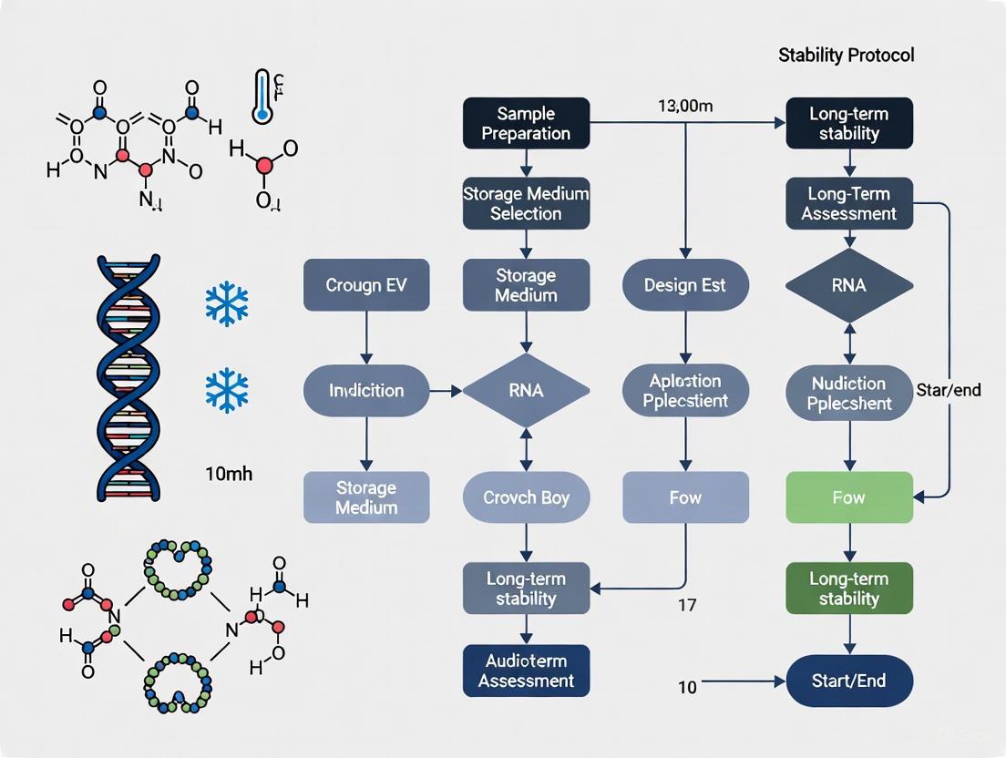
The following diagram illustrates the two primary pathways that lead to RNA degradation, highlighting its intrinsic structural vulnerabilities.
Quantitative Impact of Key Factors on RNA Stability
Table: Factors Influencing RNA Degradation Rates
| Factor | Mechanism of Action | Effect on RNA Stability |
|---|---|---|
| Divalent Cations (Mg²⁺, Ca²⁺) | Catalyze strand scission via in-line hydrolysis, especially at temperatures >80°C [2] [4]. | Significant decrease. Chelation with EDTA is critical for stabilization. |
| pH Level | Alkaline conditions (pH >8) increase availability of hydroxide ions, activating the 2'-OH group and accelerating hydrolysis [1]. | Stability decreases as pH increases. Neutral to slightly acidic conditions are preferred. |
| Temperature | Increases molecular energy, accelerating both enzymatic and chemical degradation. Each 10°C increase can double degradation rate. | Major decrease. For long-term storage, -80°C is required; samples should always be kept on ice during handling [4] [3]. |
| RNase Contamination | Enzymatic cleavage of phosphodiester bonds. RNases are stable and require strong chemical agents for deactivation [2]. | Rapid and complete degradation. A zero-tolerance policy for RNases is necessary. |
Experimental Protocols for Ensuring RNA Integrity
Protocol 1: Creating and Maintaining an RNase-Free Workspace
Principle: Prevent the introduction of RNases into samples through rigorous environmental control [2] [4] [3].
Procedure:
- Designate an RNA-only zone: Set aside a dedicated bench area, pipettes, and centrifuge for RNA work.
- Surface decontamination: Before use, thoroughly clean all surfaces (benches, pipettors, tube racks) with an RNase-decontaminating solution (commercial sprays or towelettes) or a 0.1% DEPC-treated solution. Note: Autoclaving alone is insufficient.
- Use disposable materials: Whenever possible, use sterile, disposable plasticware (tubes, tips) that are certified RNase-free.
- Treat non-disposables:
- Glassware: Bake at 180-250°C for at least 4 hours.
- Plasticware: Soak in 0.1 M NaOH / 1 mM EDTA for 2 hours, then rinse thoroughly with DEPC-treated water.
- Personal precautions: Always wear gloves and a lab coat. Change gloves frequently, especially after touching potentially contaminated surfaces like door handles or computer keyboards.
Protocol 2: Optimal Storage and Handling of RNA Samples
Principle: Minimize both enzymatic and chemical degradation pathways during storage [2] [4] [3].
Procedure:
- For Short-Term Storage (up to 1 month):
- Resuspend purified RNA in RNase-free water containing 0.1 mM EDTA or TE buffer (10 mM Tris, 1 mM EDTA, pH ~7.5).
- Store at -80°C in single-use aliquots.
- For Long-Term Storage (over 1 month):
- The most stable method is to store the RNA as a precipitate in a solution of 0.3 M sodium acetate (pH 5.2) and 70% ethanol at -20°C or -80°C. The low temperature, presence of alcohol, and slightly acidic pH all inhibit degradation.
- Alternatively, store aliquots in TE buffer at -80°C.
- Handling:
- Always keep RNA samples on ice when not in storage.
- Avoid repeated freeze-thaw cycles. Determine the required amount and create single-use aliquots upon initial purification.
- When thawing, gently mix by flicking the tube. If necessary, briefly vortex at low speed.
Protocol 3: Testing for RNase Contamination in Reagents
Principle: Proactively detect RNase contamination in lab-prepared buffers and water sources to prevent experimental failure [2] [5].
Procedure:
- Prepare a test solution: Mix a small aliquot (e.g., 35 µL) of the reagent to be tested with a known intact RNA (e.g., 5 µg).
- Set up controls:
- Positive Control: RNA + RNase-free water.
- Test Reaction: RNA + reagent.
- Protected Reaction: RNA + reagent + a broad-spectrum RNase inhibitor (e.g., 40 U RNasin).
- Incubate: Incubate all samples at 37°C for 1 hour to overnight.
- Analyze: Run the samples on a denaturing agarose gel or an Agilent Bioanalyzer.
- Intact RNA bands in all samples indicate the reagent is RNase-free.
- Degraded RNA (smear) in the "Test Reaction" but not in the "Protected Reaction" confirms RNase contamination in the reagent.
The workflow below summarizes the key steps for testing reagents.
The Scientist's Toolkit: Key Reagent Solutions
Table: Essential Reagents for RNA Stabilization and RNase Control
| Reagent | Function & Mechanism | Example Applications |
|---|---|---|
| DEPC (Diethyl Pyrocarbonate) | An alkylating agent that covalently modifies histidine residues in RNases, irreversibly inactivating them. Used to treat water and buffers [3]. | Making RNase-free water and solutions (except for Tris buffers, which it reacts with). |
| RNase Inhibitor Proteins | Proteins that non-covalently bind to and inhibit a broad spectrum of RNases (e.g., RNase A, B, C) [5] [6]. | Protecting RNA during enzymatic reactions like RT-PCR, in vitro transcription, and cDNA synthesis. |
| Guanidine Isothiocyanate | A powerful chaotropic agent that denatures and inactivates RNases upon cell lysis. A key component in many RNA isolation kits [4] [7]. | RNA extraction from cells and tissues; component of lysis buffers in spin-column and organic extraction methods. |
| RNA Stabilization Solutions (e.g., RNAlater) | Aqueous solutions that rapidly permeate tissues/cells to stabilize and protect RNA by inactivating RNases. Allows samples to be stored at room temperature for short periods or at 4°C/-20°C for longer periods [7]. | Preserving RNA integrity in tissues immediately after dissection, especially when processing many samples. |
| Chelating Agents (EDTA, Citrate) | Bind divalent cations (Mg²⁺, Ca²⁺), preventing metal-catalyzed hydrolysis of the RNA backbone [2] [4]. | Component of RNA storage buffers (e.g., TE buffer) and lysis buffers; essential in reactions involving heat denaturation of RNA. |
Ribonucleases (RNases) constitute a class of enzymes that catalyze the degradation of RNA into smaller components and are found in all domains of life, including viruses [8]. Their extreme stability and relentless activity make them a pervasive threat in laboratories working with RNA and Extracellular Vesicles (EVs). For researchers focused on the long-term storage of RNA and EV samples, understanding and mitigating RNase activity is not merely a best practice but a fundamental requirement for achieving reliable, reproducible results. This guide provides targeted troubleshooting advice and FAQs to help you safeguard your valuable samples throughout your research workflow.
FAQ: Understanding the RNase Threat
This section answers the most common and critical questions regarding RNase stability and its implications for your research.
Q1: Why are RNases considered such a significant threat in RNA and EV research?
RNases are a major concern due to their ubiquity, remarkable stability, and catalytic efficiency.
- Ubiquity: RNases are found in almost all living organisms and are abundant in skin, hair, and bodily secretions. They are also common environmental contaminants [4].
- Stability: Many RNases, like RNase A, are exceptionally hardy. For instance, RNase A is so stable that one common purification method involves boiling a crude cellular extract to denature all other enzymes, leaving RNase A active [8].
- Cofactor Independence: Most RNases do not require cofactors to function, meaning they can remain active in various environments and even in purified water if introduced [4].
- Direct Impact on EVs: For EV research, RNase activity can degrade the RNA cargo within vesicles, compromising studies on EV-based communication, biomarker discovery, and therapeutic development [9] [10].
Q2: What are the primary sources of RNase contamination in my lab?
The main sources can be categorized as follows:
- Endogenous: Originating from within your own biological sample. When cells or tissues are lysed, their native RNases are released [4].
- Exogenous: Introduced from the external environment. Key sources include:
Q3: How does the stability of RNases impact long-term sample storage?
The stability of RNases means that the threat of RNA degradation persists throughout the storage period. Without proper stabilization, RNA within samples or purified RNA preparations can degrade even at low temperatures, though the rate is slowed. This is why simply freezing samples is often insufficient; stabilization agents that inactivate RNases are critical for preserving an accurate snapshot of the RNA profile at the time of collection, which is essential for longitudinal studies and biobanking [11] [4] [12].
Q4: What is a Ribonuclease Inhibitor and when should I use it?
A Ribonuclease Inhibitor (RI) is a protein that binds to certain RNases with extremely high affinity, effectively neutralizing their activity [13]. It is a crucial tool for protecting RNA during in vitro manipulations.
- Function: Recombinant RI binds tightly and irreversibly to RNases like RNase A, B, and C, forming a stable complex that prevents RNA degradation [14].
- When to Use: RI is indispensable in enzymatic reactions involving RNA, such as cDNA synthesis, in vitro transcription, and RT-PCR [14]. It is typically added directly to the reaction mix to safeguard the RNA template.
- Important Note: RI is a cytosolic protein and is primarily used in vitro. It is not a substitute for proper sample handling and storage protocols for whole tissues or biofluids.
Troubleshooting Guide: Common RNase-Related Problems and Solutions
| Problem | Potential Cause | Solution |
|---|---|---|
| Degraded RNA samples (e.g., smeared gel, low RIN) | RNase contamination during handling or from non-sterile supplies; Inadequate sample stabilization before storage [4]. | Use RNase-free consumables; clean surfaces with RNase-deactivating reagents; wear gloves; use stabilization reagents (e.g., RNAlater) for tissues immediately upon collection [11] [4]. |
| Inconsistent RT-qPCR or RNA-seq results | Variable RNA degradation due to RNase activity, often from repeated freeze-thaw cycles of samples or RNA extracts [4]. | Aliquot RNA samples into single-use portions; avoid repeated thawing; store at -70°C or lower for long-term preservation [4]. |
| Low yield in cDNA synthesis or IVT | RNase degradation of the RNA template during the enzymatic reaction [14]. | Include a recombinant RNase Inhibitor in the reaction mixture according to the manufacturer's instructions (e.g., 1-2 U/µL) [14]. |
| Loss of EV RNA cargo or function | Degradation during biofluid storage prior to EV isolation [9] [10]. | For urine, add protease inhibitors and store at -80°C [9] [10]. For milk, remove cells and fat prior to storage to avoid contamination from stress-induced EVs [9]. |
Quantitative Data: RNase Degradation Kinetics and Storage Stability
Understanding the quantitative aspects of RNA degradation and stability under various conditions is vital for planning long-term storage. The following table summarizes key data from research.
Table 1: RNA Degradation Kinetics and Stability in Different Conditions
| Condition | Degradation Rate / Stability Outcome | Experimental Context & Key Findings |
|---|---|---|
| Room Temperature (Stabilized) | Extrapolated degradation rate of 0.7–1.3 cuts/1000 nt/century [12]. | RNA dried with a stabilizer in anoxic, anhydrous capsules. Major finding: atmospheric humidity is a primary degradation factor. |
| Long-term Tissue Storage | RNA Integrity Numbers (RIN) >9 after 2 years, 7 months [11]. | Mouse tissues stored in RNAlater at -20°C. Gene expression profiles showed very high correlation (R=0.994) with fresh-frozen controls. |
| Urine Storage (for EVs) | EV yield decreases within 2 hours of collection; optimal recovery at -80°C [9] [10]. | Protease inhibitors improve yield. Vortexing after thawing from -80°C restored 100% EV-associated protein recovery. |
Table 2: Affinity of Ribonuclease Inhibitor (RI) for Various Ribonucleases
| Ribonuclease | Equilibrium Dissociation Constant (Kd) | Binding Affinity |
|---|---|---|
| Angiogenin (ANG) | 7.1 x 10⁻¹⁶ M [13] | Highest affinity |
| RNase 2 (EDN) | 9.4 x 10⁻¹⁶ M [13] | Extremely high affinity |
| RNase 4 | 4.0 x 10⁻¹⁵ M [13] | Very high affinity |
| RNase A | 4.4 x 10⁻¹⁴ M [13] | Very high affinity |
Experimental Protocols for Validating Storage Methods
Protocol 1: Validating a Room-Temperature RNA Storage Technology
This protocol is based on a study that demonstrated long-term room-temperature RNA stability [12].
Objective: To evaluate the integrity of RNA stored in a stabilizer within an airtight, anhydrous environment over time and at elevated temperatures.
Materials:
- Purified RNA sample.
- RNA stabilizer (commercial or as described in the study).
- Air-tight stainless-steel minicapsules or equivalent.
- Equipment: Agilent 2100 Bioanalyzer or similar for RNA Integrity Number (RIN) calculation, Thermocycler for accelerated aging.
Method:
- Sample Preparation: Mix the RNA with the chosen stabilizer solution.
- Encapsulation: Pipette the RNA-stabilizer mixture into minicapsules and seal them tightly to create an anoxic and anhydrous environment.
- Accelerated Aging: For short-term validation, subject encapsulated samples to elevated temperatures (e.g., 90°C) for defined periods. The degradation rate follows the Arrhenius law, allowing extrapolation to room-temperature stability.
- Analysis:
- At each time point, retrieve samples and resuspend the RNA in RNase-free water.
- Analyze RNA integrity using the Bioanalyzer to generate RIN values.
- Perform downstream functional assays like reverse transcription-quantitative PCR (RT-qPCR) to compare Cycle quantification (Cq) values between stored and fresh RNA.
Expected Outcome: Properly stabilized and encapsulated RNA should show minimal degradation (high RIN, minimal change in Cq values) even after accelerated aging, predicting stability for decades at room temperature [12].
Protocol 2: Evaluating Biofluid Storage for EV-RNA Preservation
This protocol outlines the assessment of pre-processing storage conditions for EV-containing biofluids like urine and plasma [9] [10].
Objective: To determine the impact of different storage temperatures and additives on the yield and RNA content of isolated EVs.
Materials:
- Freshly collected biofluid (e.g., urine, plasma).
- Protease inhibitor cocktail.
- Storage conditions: -80°C, -20°C, 4°C.
- Low-speed centrifuge.
- EV isolation kit (e.g., based on precipitation, size-exclusion chromatography).
- Nanoparticle Tracking Analysis (NTA) instrument or similar for EV concentration.
- RNA isolation kit and Bioanalyzer.
Method:
- Aliquoting: Divide the fresh biofluid into multiple aliquots.
- Additive Conditions: Add protease inhibitors to some aliquots, leave others untreated.
- Storage: Store aliquots under different conditions (-80°C, -20°C, etc.) for a set duration (e.g., 1 week).
- EV Isolation: After storage, thaw samples (vortex thoroughly if frozen) and isolate EVs using your chosen method.
- Analysis:
- EV Yield: Use NTA to determine the concentration and size distribution of isolated particles.
- RNA Assessment: Isolate RNA from the EV fraction and analyze its integrity (RIN) and quantity.
Expected Outcome: Samples stored at -80°C with protease inhibitors are expected to show the highest EV yield and best-preserved RNA integrity, while storage at higher temperatures or without additives will likely result in reduced yield and degraded RNA [9] [10].
Research Reagent Solutions: Essential Tools for RNase Management
The following table lists key reagents and tools essential for combating RNases in a research setting.
Table 3: Key Reagents for RNase Inhibition and RNA Stabilization
| Reagent / Tool | Function & Application |
|---|---|
| Recombinant RNase Inhibitor | Protects RNA from degradation during in vitro enzymatic reactions like cDNA synthesis and IVT. Binds irreversibly to RNases [14]. |
| RNAlater Stabilization Solution | An aqueous, non-toxic reagent that permeates tissues to stabilize and protect RNA immediately upon collection. Allows for short-term non-frozen storage and long-term storage at -20°C [11]. |
| RNA Preserve | A liquid stabilizer for various samples (tissue, bacteria, soil). Enables room-temperature storage and shipping for periods from days to weeks [15]. |
| RNase-Deactivating Reagents | Sprays and solutions used to decontaminate work surfaces, glassware, and equipment to create an RNase-free environment [4]. |
| DEPC-treated Water | Diethyl pyrocarbonate (DEPC) treatment inactivates RNases in water, making it safe for use in RNA-related experiments [4]. |
Workflow Visualization: Safeguarding Your Samples
The following diagram illustrates the critical decision points and recommended practices for protecting your RNA and EV samples from RNases throughout the experimental workflow.
RNase Mitigation Workflow: This diagram outlines the key steps and critical decisions for protecting RNA and EV samples from RNases, from collection through to analysis.
Frequently Asked Questions (FAQs) on EV Membrane Integrity
FAQ 1: What are the primary sources of physical stress that can damage EV membranes during handling? The primary sources of physical stress include freeze-thaw cycles, excessive pressure during concentration, and exposure to inappropriate storage temperatures [16] [17]. During isolation, techniques like tangential flow filtration (TFF) require careful monitoring of flow rate and pressure, as high shear forces can deform or rupture vesicles [17]. Furthermore, aggressive centrifugation or passing samples through narrow-gauge needles can cause mechanical shearing, compromising membrane integrity [4].
FAQ 2: Why are repeated freeze-thaw cycles detrimental to EV integrity? Repeated freeze-thaw cycles can cause EVs to rupture, aggregate, and lose their functional cargos [16] [17]. The formation and melting of ice crystals generate mechanical forces that physically disrupt the lipid bilayer. This damage leads to a decrease in particle number, a change in size distribution, and the degradation of encapsulated RNA and proteins [17]. To ensure reproducibility in downstream assays, it is critical to aliquot EVs into single-use volumes to avoid repeated thawing [17].
FAQ 3: How does improper storage temperature affect the stability of isolated EVs? Isolated EVs resuspended in phosphate-buffered saline (PBS) are unstable at 4°C for extended periods, exhibiting a loss of particle number and surface markers [16] [17]. For long-term storage, -80°C or lower is recommended [16]. However, even at -80°C, standard freezing without cryoprotectants can be damaging. Lyophilization (freeze-drying) with specialized buffers presents a more stable alternative for room-temperature storage, preserving vesicle structure and function [17].
FAQ 4: What are the best practices for concentrating large volumes of EV-containing conditioned media without causing damage? Tangential Flow Filtration (TFF) is a recommended method for concentrating large volumes (>250 mL) as it is a scalable and gentle process that preserves EV integrity [17]. Key best practices include:
- Using membranes with appropriate molecular weight cut-offs (e.g., 100 kDa, 300 kDa).
- Monitoring flow rate and pressure to avoid high shear forces that can deform vesicles.
- Avoiding over-concentration, which can increase sample viscosity and lead to aggregation [17].
Troubleshooting Common EV Integrity Issues
Problem: Low Yield and Particle Aggregation After Isolation
| Observation | Potential Cause | Solution |
|---|---|---|
| Low particle count after isolation [17] | EV aggregation due to over-concentration or abrasive isolation techniques | Use gentle isolation methods like Size-Exclusion Chromatography (SEC); avoid high-pressure systems [17] |
| Visible precipitate in sample [17] | Aggregation from repeated freeze-thaw or freezing without cryoprotectants | Aliquot EVs into single-use volumes; use cryoprotectant buffers (e.g., EVSafe Storage Buffer) for freezing [17] |
| Inconsistent results in functional assays [17] | Damage to surface markers from physical stress, affecting recipient cell interaction | Minimize processing steps that introduce shear forces; validate surface markers with flow cytometry post-isolation [17] |
Problem: Degradation of RNA Cargo
| Observation | Potential Cause | Solution |
|---|---|---|
| Degraded RNA in extracted EV cargo [17] | Membrane rupture from physical stress, exposing RNA to RNases | Use RNase-free reagents and techniques; avoid mechanical force like vortexing [4] [17] |
| Poor RNA quality from stored EVs [16] | Slow hydrolysis and enzymatic activity during storage | For long-term storage, lyophilize EVs with stabilizers or store at -80°C in single-use aliquots [16] [17] |
Experimental Protocols for Assessing Membrane Integrity
Protocol 1: Using Tunable Resistive Pulse Sensing (TRPS) for Size and Concentration Analysis
Purpose: To accurately measure the size distribution, concentration, and zeta potential of EV samples to identify aggregation or fragmentation resulting from physical stress [17].
Methodology:
- Instrument Calibration: Use lyophilized EV standards or silica nanoparticles to calibrate the Exoid or similar TRPS instrument [17].
- Sample Preparation: Dilute the isolated EV sample in a particle-free electrolyte solution to achieve an appropriate concentration for measurement.
- Measurement: Load the sample into the instrument. TRPS works by measuring the change in electrical resistance (pulse) as each individual particle passes through a nanopore. The size of the pulse correlates with the particle's size, and the frequency of pulses indicates concentration [17].
- Data Analysis: Analyze the data for:
- Mean/Modal Size: A significant shift may indicate swelling or fragmentation.
- Polydispersity: An increase suggests a heterogeneous population, potentially from aggregation or breakdown.
- Particle Concentration: A drop may indicate aggregation or membrane rupture.
Protocol 2: Flow Cytometry with Fluorescent Membrane Stains
Purpose: To validate the integrity of the EV lipid bilayer and detect the presence of canonical surface proteins, confirming the isolation of intact vesicles rather than protein aggregates or debris [17].
Methodology:
- Staining: Incubate the EV sample with a membrane-permeant fluorescent stain, such as ExoBrite True EV Membrane Stain, which is designed to stain intact membranes with minimal induction of aggregation [17].
- Surface Marker Labeling (Optional): Combine the membrane stain with fluorophore-conjugated antibodies against common EV surface markers (e.g., CD63, CD81, CD9) for a more comprehensive profile [17].
- Analysis: Run the sample on a flow cytometer capable of detecting nanoparticles. The presence of a double-positive population (membrane stain + surface marker) confirms intact EVs.
- Troubleshooting: Compare stained samples with unstained controls. A low signal for membrane stain may indicate a high proportion of ruptured vesicles.
Diagram: Mechanisms of Physical Stress on EV Integrity
The following diagram illustrates how different types of physical stress lead to the compromise of extracellular vesicle integrity.
The Scientist's Toolkit: Key Reagent Solutions
The following table details essential reagents and kits used in EV research to preserve membrane integrity and ensure sample quality.
| Item | Function | Specific Use Case |
|---|---|---|
| EV-Depleted FBS [17] | Cell culture supplement | Provides essential growth factors while removing contaminating bovine EVs that could skew experimental results during EV production. |
| EVSafe Storage Buffer [17] | Cryoprotectant buffer | Maintains EV stability during freezing at -80°C, prevents aggregation, and preserves particle size distribution with high recovery (>95%). |
| EVSafe Lyophilisation Buffer [17] | Freeze-drying stabilizer | Enables long-term, ambient-temperature storage of EVs by preserving vesicle structure and function during the lyophilization process. |
| RNase-free reagents & kits [4] | Contamination prevention | Protects vulnerable RNA cargo from degradation by ubiquitous RNase enzymes during EV lysis and RNA extraction. |
| ExoBrite Membrane Stain [17] | Fluorescent dye | Selectively stains intact EV membranes for flow cytometry with minimal aggregation compared to traditional dyes like DiI or PKH. |
FAQs: Understanding Cryodamage and Lipid Bilayers
Q1: What are the primary mechanisms of cryodamage during the freezing of biological samples? Cryodamage primarily occurs through two key mechanisms: the formation, growth, and recrystallization of ice crystals, and the accompanying increase in solute concentration. Ice crystals can cause fatal mechanical injury to cells and subcellular structures like lipid bilayers. The formation of extracellular ice leads to cellular dehydration and osmotic pressure damage, while intracellular ice, which forms at higher cooling rates, is almost always lethal [18].
Q2: How do ice crystals specifically damage lipid bilayers? Lipid bilayers themselves can act as ice-nucleating agents, initiating damaging ice formation at temperatures well above the homogeneous freezing point of pure water. Molecular dynamics simulations show that phospholipid bilayers at the interface with supercooled water can facilitate ice nucleation. This ice formation can cause mechanical disruption and phase transitions within the membrane structure, compromising its integrity [19].
Q3: Why is the lipid bilayer a critical target for cryoprotective strategies? Membranes are a primary target for cryodamage. During cooling, lipid bilayers undergo phase transitions and structural changes that have been associated with cold shock damage. Small polar cryoprotectant molecules like DMSO can modulate the hydration layer of membranes, changing their properties at subzero temperatures and generating a high tolerance against the harmful effects of ice recrystallization [20].
Q4: How does cryodamage differ between slow freezing and rapid cooling (vitrification) methods? The mechanisms of injury differ significantly between these approaches [18]:
- Slow Freezing: Primarily causes extracellular ice formation, leading to cellular dehydration and solute concentration effects.
- Rapid Cooling/Vitrification: Aims to achieve an ice-free, glassy state. However, its main limitation is the potential formation of ice nucleation and devitrification (ice recrystallization) during the warming process, which can be fatal.
Q5: What role do lipid bilayers play in initiating ice formation? Research indicates that the complex chemical and structural factors of lipid bilayers make them potent ice-nucleating agents. This finding is a crucial first step in pinpointing the origin of extracellular ice nucleation, with major implications for understanding and improving cryopreservation protocols [19].
Troubleshooting Guide: Common Cryopreservation Issues
| Problem | Likely Cause | Recommended Solution |
|---|---|---|
| Low post-thaw cell viability (Slow freezing) | Excessive dehydration and solute damage from slow, extracellular ice formation. | Optimize cooling rate for your cell type; increase CPA concentration gradually; use controlled-rate freezing [18]. |
| Low post-thaw cell viability (Vitrification) | Devitrification and ice recrystallization during warming; CPA toxicity. | Increase warming rate; use lower toxicity CPAs (e.g., synthetic polymers); employ ice inhibitors [18]. |
| Intracellular ice formation | Cooling rate is too high, preventing water from exiting the cell. | Reduce the cooling rate to allow for sufficient cellular dehydration [18]. |
| Membrane rupture after thawing | Mechanical damage from ice crystals and/or loss of membrane integrity due to lipid phase transitions. | Use membrane-stabilizing CPAs like DMSO; modulate membrane composition (e.g., increase sterol content) [20]. |
| Contamination of EVs with lipoproteins (co-isolation) | Lipoproteins and EVs share similar physical properties like density and size. | Employ purification techniques that combine multiple principles (e.g., density gradient ultracentrifugation or affinity enrichment) to better separate EVs from contaminants [21]. |
Experimental Protocols & Data
Protocol: Assessing Cryoprotectant Efficacy on Membrane Integrity via FRAP
This protocol uses Fluorescence Recovery After Photobleaching (FRAP) to investigate how cryoprotective agents (CPAs) influence plasma membrane fluidity, a key factor in cryotolerance [20].
Key Research Reagent Solutions:
- Fluorescent Lipid: DiOC₁₈ for labeling the plasma membrane.
- Cryoprotective Agents: e.g., DMSO, ethylene glycol.
- Membrane Modulators: Methyl-β-cyclodextrin (MβCD) loaded with cholesterol or ergosterol to alter membrane sterol content.
- Viability Stain: Propidium iodide (PI) to test membrane integrity post-thaw.
Methodology:
- Cell Culture and Treatment: Use a standard cell line like HeLa cells.
- Treat one group with your chosen CPA (e.g., 15% DMSO + 15% ethylene glycol).
- For a comparative approach, treat another group with cholesterol-loaded MβCD (e.g., 10 mM for 60 mins) to artificially increase membrane sterol content without CPAs.
- Membrane Labeling: Label the living cells with the fluorescent lipid DiOC₁₈.
- FRAP Analysis:
- Select a small, uniform section of the plasma membrane on a confocal microscope and bleach it with a high-intensity laser pulse.
- Monitor the recovery of fluorescence into the bleached area over time.
- Calculate the diffusion speed and the mobile fraction of lipids.
- Correlation with Cryotolerance:
- Subject the treated cells to a rapid freeze-thaw cycle (e.g., in Open Pulled Straws in liquid nitrogen, rewarmed at 37°C).
- Assess plasma membrane integrity immediately by staining with propidium iodide (PI). Cells with compromised membranes will show red nuclear fluorescence.
Expected Outcome: Cells treated with CPAs or enriched with sterols will show the presence of an immobile lipid fraction in the membrane and significantly higher plasma membrane integrity after thawing compared to untreated controls [20].
Protocol: Comparing EV Isolation Methods for Purity and Yield
This protocol compares different extracellular vesicle (EV) isolation techniques from small plasma volumes, which is critical for ensuring high-quality, uncontaminated samples for long-term storage and research [21].
Methodology Overview:
- Sample Preparation: Collect human plasma (e.g., 100 μL aliquots) and pre-clear by centrifugation.
- EV Isolation (in triplicate): Isolate EVs using multiple methods based on different principles:
- Ultracentrifugation (UC): Traditional method pelleting EVs via high g-forces.
- Density Gradient UC (DGUC): Separates particles by density for higher purity.
- Size Exclusion Chromatography (SEC): e.g., qEV columns; separates by size.
- Polymer Precipitation: e.g., ExoQuick, Total Exosome Isolation kits; reduces EV solubility.
- Electrostatic Interaction: e.g., MagResyn SAX beads; binds negatively charged EVs.
- Affinity Enrichment: e.g., MagCapture beads; isolates phosphatidylserine-positive (PS+) EVs.
- Post-Isolation Processing: Concentrate and buffer-exchange all samples into PBS using 10 kDa molecular weight cutoff filters to ensure consistency.
- Characterization:
- Particle Analysis: Use Nanoparticle Tracking Analysis (NTA) for particle concentration and size distribution.
- Purity Assessment: Use Simple Western to detect canonical EV markers (e.g., CD9, CD81) and contaminants (e.g., albumin, ApoA1).
- Proteomic Profiling: Use LC-MS/MS for bottom-up proteomics to assess proteome coverage.
Summary of Quantitative Data from Comparative EV Isolation [21]:
| Isolation Method | Principle | Avg. Particle Size (nm) | Key Purity Indicators | Proteome Coverage |
|---|---|---|---|---|
| Ultracentrifugation (UC) | Centrifugation | Wider distribution | Moderate contamination | Good |
| Density Gradient UC (DGUC) | Density | Wider distribution | Low contamination | Good |
| qEV Column (SEC) | Size | ~100-200 nm | Low to moderate contamination | Moderate |
| Polymer Precipitation | Solubility | Wider distribution | High contamination | Lower |
| MagNet / MagCap | Affinity / Electrostatic | Narrowest distribution | Highest purity | Highest |
Diagrams: Mechanisms and Workflows
Cryodamage Mechanisms During Freezing and Thawing
Cryoprotectant Mechanism of Action on Lipid Bilayers
Troubleshooting Guides
Guide 1: Troubleshooting RNA Degradation in Storage
- Problem: RNA samples show signs of degradation (e.g., smeared bands on gel, low RIN scores) after storage.
- Question: Are divalent cations and improper temperature the cause?
- Investigation & Solution:
- Assess Storage Buffer: Check if your storage buffer (e.g., TE buffer) contains EDTA or another chelating agent to sequester divalent cations like Mg²⁺ and Ca²⁺. If not, the RNA is vulnerable to metal-catalyzed hydrolysis [4].
- Check Water Purity: Ensure that nuclease-free, ultrapure water is used to prepare storage solutions, as tap or low-grade purified water can be a source of metal ions [4].
- Verify Storage Temperature: Confirm that RNA aliquots are stored at -70°C or below for long-term preservation. Storage at -20°C is insufficient for prolonged periods and accelerates degradation [22].
- Minimize Freeze-Thaw Cycles: Divide RNA into small, single-use aliquots. Repeated freezing and thawing can reactivate RNases and promote hydrolysis [4].
Guide 2: Troubleshooting EV Aggregation and Cargo Loss
- Problem: Isolated EV preparations show aggregation, a change in size distribution, or loss of RNA cargo upon thawing.
- Question: Did temperature fluctuations during storage cause this damage?
- Investigation & Solution:
- Audit Freezing Protocol: Avoid storing EVs at -20°C. For long-term storage, freeze EV samples at a constant -80°C. Rapid freezing (snap-freezing in liquid nitrogen) is recommended before transfer to -80°C to prevent ice crystal formation [23].
- Eliminate Freeze-Thaw Cycles: Subjecting EVs to multiple freeze-thaw cycles significantly decreases particle concentration, impairs bioactivity, and increases size due to aggregation. Always store in single-use aliquots [23].
- Consider Stabilizers: For critical samples, add cryoprotectants like trehalose to the EV suspension before freezing. This helps maintain vesicle integrity and prevents membrane fusion [23].
- Check Storage Medium: Note that EVs stored in native biofluids (e.g., plasma, cell culture supernatant) often demonstrate better stability than those purified and resuspended in simple buffers [23].
Frequently Asked Questions (FAQs)
FAQ 1: Why are divalent cations like Mg²⁺ so problematic for RNA stability? The primary reason is their role in catalyzing RNA strand cleavage. The 2'-hydroxyl group on the ribose sugar of RNA can act as a nucleophile, attacking the adjacent phosphodiester bond. Divalent cations, particularly Mg²⁺, stabilize the transition state of this reaction, significantly accelerating hydrolysis and breaking the RNA backbone [1]. This makes them potent catalysts of RNA degradation.
FAQ 2: What is the recommended long-term storage temperature for RNA and EVs? For both RNA and EVs, -70°C to -80°C is the recommended temperature for long-term storage [23] [4] [22]. Storage at -20°C leads to significantly faster degradation of RNA and promotes aggregation and loss of function in EVs [23].
FAQ 3: How do different divalent cations compare in their effect on RNA stability? The destabilizing effect of a divalent cation is determined by its charge density (ζ). Cations with higher charge density are more effective at stabilizing the folded structure of RNA, but also more effectively catalyze its degradation when folded structure is not a factor. The following table summarizes the properties of common Group IIA cations [24]:
Table: Influence of Divalent Cations on RNA Folding Stability
| Cation | Ionic Radius (Å) | Charge Density (ζ, e/ų) | Relative Stabilization of Folded RNA |
|---|---|---|---|
| Mg²⁺ | 0.65 | 0.055 | Highest |
| Ca²⁺ | 0.99 | 0.038 | High |
| Sr²⁺ | 1.13 | 0.027 | Medium |
| Ba²⁺ | 1.35 | 0.020 | Low |
FAQ 4: How many freeze-thaw cycles can my RNA or EV samples tolerate? Ideally, zero. Each cycle inflicts cumulative damage. For RNA, freeze-thaw cycles can lead to the reactivation of RNases and physical shearing [4]. For EVs, even a single cycle can cause a noticeable decrease in particle concentration and RNA content, with multiple cycles leading to extensive aggregation and functional loss [23]. Aliquotting is the most effective countermeasure.
Data Presentation
Table 1: Quantitative Impact of Temperature and mRNA Length on Stability
| Factor | Experimental Condition | Key Finding | Experimental Method |
|---|---|---|---|
| Temperature | Analysis of activation energy (Ea) for degradation | Ea = 31.5 kcal/mol normalized per phosphodiester backbone [25] | Thermodynamic analysis |
| mRNA Length | Comparison of different nucleotide lengths | Longer mRNA transcripts are negatively correlated with stability [25] | Integrity analysis via capillary electrophoresis |
| Storage Duration | EVs stored at -20°C vs. -80°C | EVs at -20°C showed significant aggregation and size increase after one month; -80°C preserved integrity [23] | Nanoparticle Tracking Analysis (NTA), Western Blot, functional assays |
Experimental Protocols
Protocol 1: Measuring the Effect of Divalent Cations on RNA Hydrolysis
Objective: To quantify the rate of RNA strand cleavage catalyzed by different divalent cations.
Sample Preparation:
- Prepare a solution of a defined, homogenous RNA transcript (e.g., 1-2 kb in length) in a chelating buffer (e.g., 10 mM Tris-HCl, 1 mM EDTA, pH 7.5).
- Dialyze the RNA extensively against a large volume of chelator-free buffer (e.g., 10 mM Na-Hepes, pH 7.0) to remove all divalent cations [24].
Reaction Setup:
- Aliquot the dialyzed RNA into separate tubes.
- To each tube, add a chloride salt of a divalent cation (MgCl₂, CaCl₂, SrCl₂, BaCl₂) to a final concentration of 1-10 mM. Include a no-cation control.
- Incubate all reactions at a defined temperature (e.g., 37°C or 50°C) for a set period (e.g., 0, 1, 2, 4, 8 hours) [1].
Analysis:
- Stop the reactions by adding excess EDTA (50 mM final concentration).
- Analyze the RNA integrity on a denaturing agarose or polyacrylamide gel. Intact RNA will appear as a sharp band, while degraded RNA will appear as a smear.
- Quantification: Use techniques like capillary electrophoresis (e.g., Bioanalyzer) to calculate the RNA Integrity Number (RIN) or the percentage of intact full-length transcript remaining [25].
Protocol 2: Evaluating Temperature-Induced EV Aggregation
Objective: To assess the physical stability of EVs under different storage temperatures and freeze-thaw cycles.
EV Isolation & Preparation:
- Isolate EVs from a cell culture supernatant (e.g., MSC-conditioned media) using a standardized method like size-exclusion chromatography or ultracentrifugation [23].
- Resuspend the purified EV pellet in a neutral buffer (e.g., PBS) or PBS with 5% trehalose.
Storage Conditions:
- Divide the EV suspension into multiple aliquots.
- Temperature Groups: Store aliquots at 4°C, -20°C, and -80°C. Include a subset snap-frozen in liquid nitrogen before storage at -80°C.
- Freeze-Thaw Groups: Subject a separate set of aliquots (stored at -80°C) to 1, 3, and 5 freeze-thaw cycles. Thaw cycles should be performed rapidly in a 37°C water bath [23].
Analysis:
- Concentration & Size: Use Nanoparticle Tracking Analysis (NTA) to measure the particle concentration and mode/mean size before and after storage. An increase in size indicates aggregation.
- Morphology: Use transmission electron microscopy (TEM) to visually inspect for vesicle deformation, rupture, or fusion.
- Cargo Integrity: Isplicate RNA from the treated EVs and analyze miRNA or mRNA content using qRT-PCR or Bioanalyzer to assess cargo preservation [23].
Pathway and Workflow Visualizations
RNA Hydrolysis Catalyzed by Divalent Cations
EV Storage and Stability Assessment Workflow
The Scientist's Toolkit: Research Reagent Solutions
Table 2: Essential Reagents for Stabilizing RNA and EV Samples
| Reagent / Material | Function / Purpose | Key Consideration |
|---|---|---|
| EDTA (Ethylenediaminetetraacetic acid) | Chelates divalent cations (Mg²⁺, Ca²⁺), preventing metal-catalyzed RNA hydrolysis [4]. | Standard component of RNA storage buffers (e.g., TE buffer). |
| RNAstable or RNAprotect | Commercial stabilization reagents that protect RNA integrity at room temperature by inhibiting RNases and hydrolysis [4]. | Useful for sample transport or short-term storage without freezing. |
| Trehalose | A disaccharide cryoprotectant that stabilizes lipid bilayers and proteins. Helps prevent EV aggregation and membrane fusion during freezing [23]. | Preferable to DMSO for EVs as it is non-cytotoxic and doesn't interfere with downstream applications. |
| RNase-free Water | Ultrapure, nuclease-free water for preparing storage buffers and resuspending RNA. Ensures no introduction of external RNases or metal ions [4]. | A critical baseline for all molecular biology workflows involving RNA. |
| PAXgene Tubes | Specialized blood collection tubes containing additives that immediately stabilize intracellular RNA profiles upon drawing blood [4]. | Essential for clinical biobanking and accurate gene expression studies from blood. |
Proven Protocols: Step-by-Step Storage Methods for Different Sample Types
A technical guide to safeguarding your precious RNA and EV samples
Ensuring the stability of biological samples like RNA and Extracellular Vesicles (EVs) is a cornerstone of reproducible research. The choice between -20°C for short-term needs and -80°C for long-term preservation is critical, impacting everything from sample integrity to experimental costs. This guide provides evidence-based protocols and troubleshooting tips to navigate this essential aspect of your work.
Troubleshooting Common Storage Problems
Q: My RNA samples underwent multiple freeze-thaw cycles. How might this affect my downstream analysis? A: Multiple freeze-thaw cycles are a significant source of RNA degradation. They can lead to:
- RNA Degradation: The repeated formation and melting of ice crystals can physically shear RNA molecules, resulting in fragmented RNA.
- Altered Gene Expression Data: Degraded RNA can skew results in sensitive applications like qRT-PCR and RNA sequencing, leading to inaccurate 3'/5' ratios and loss of transcript detection [26] [11].
- Solution: Always aliquot RNA into single-use portions to avoid repeated freezing and thawing. Consider room-temperature storage technologies for working aliquots [26] [27].
Q: I stored my purified EV samples at -20°C, and now I notice aggregation. What happened? A: Storage of purified EVs at -20°C is often suboptimal and can lead to:
- Vesicle Aggregation and Fusion: The -20°C environment can damage the EV lipid membrane, causing vesicles to clump together or merge, which alters their size distribution and concentration [23].
- Loss of Bioactivity: Membrane damage and aggregation can lead to the leakage of internal cargo (proteins, RNA) and impair the EV's functional activity in recipient cells [10] [23].
- Solution: For any storage beyond a few days, store purified EVs at -80°C. The use of cryoprotectants like trehalose can also help maintain membrane integrity [23].
Q: My samples are stored at -80°C, but the freezer is nearing capacity and is opened frequently. Should I be concerned? A: Yes, frequent opening of an -80°C freezer can compromise sample integrity.
- Temperature Fluctuations: Each opening introduces warm, moist air, causing the internal temperature to spike and promoting frost buildup. This subjects samples to partial thawing and refreezing, similar to the damaging effects of freeze-thaw cycles [28].
- Increased Degradation Rate: Even transient warming can increase the rate of hydrolytic and enzymatic degradation processes.
- Solution: Implement an organized sample management system. Use racks and maps for quick retrieval, keep a detailed inventory to minimize door-open time, and ensure the freezer is well-maintained with a clean condenser [28].
Storage Condition Comparison Tables
RNA Storage Conditions and Outcomes
| Sample Type | Storage Condition | Duration | Key Outcomes & Integrity Metrics | Recommended For |
|---|---|---|---|---|
| Purified Total RNA [26] | Room Temp (in RNAstable) | 4 weeks | RIN: 9.70 ± 0.00; OD 260/280: ~2.02; Microarray data identical to -80°C control [26] | Sample shipping; short-term working aliquots |
| Purified Total RNA [27] | Room Temp (in anhydrous capsules) | Theoretical: decades | Model predicts ~1 cut per 1000 nucleotides per century; stable Cq values in qPCR [27] | Ultra-long-term archiving (years) |
| Tissue for RNA [11] | RNAlater at -20°C | >2.5 years | RNA Integrity Number (RIN) > 9; gene expression profiles highly correlated with frozen samples (R=0.994) [11] | Long-term tissue preservation prior to RNA extraction |
| Purified Total RNA [26] | -80°C (standard) | Long-term | RIN > 9.7; considered the "gold standard" for frozen RNA preservation [26] [11] | Long-term master stock storage |
EV Storage Conditions and Outcomes
| Sample Type / Source | Storage Condition | Duration | Key Outcomes & Integrity Metrics | Recommended For |
|---|---|---|---|---|
| Purified EVs (general) [23] | -80°C | Long-term (months) | Best preservation of concentration, size, morphology, RNA content, and bioactivity [23] | Long-term storage of purified EVs |
| Purified EVs (general) [23] | -20°C | >1 week | Significant particle aggregation, size increase, and potential loss of bioactivity [23] | Not recommended |
| Urine (for EV isolation) [10] | -80°C | 1 week | 100% EV-associated protein recovery with vortexing; higher yield than -20°C [10] | Storage of biofluid prior to EV isolation |
| MSC-derived EVs [23] | -80°C | 1 month | No significant change in uniform size, integrity, or bioactivity [23] | Storage of therapeutic EV candidates |
| Plasma (for EV isolation) [10] | -80°C | 20 months | Decrease in EV yield over time [10] | Note: Biofluid storage can impact yield |
Detailed Experimental Protocols
Protocol 1: Long-Term Room Temperature Storage of RNA using a Stabilizing Matrix
This protocol is adapted from studies evaluating RNAstable, a commercial product that protects RNA by embedding it in a dry, anhydrobiotic matrix at room temperature [26].
- Key Research Application: This method is ideal for maintaining the integrity of purified RNA for microarray analysis and other sensitive downstream applications without a cold chain.
Essential Materials:
- RNAstable tubes or plates (Biomatrica)
- Purified RNA sample (e.g., 250 ng/µl in RNase-free water)
- SpeedVac vacuum concentrator (without heat)
- Sealed moisture barrier bags with desiccant
Step-by-Step Method:
- Preparation: Aliquot your purified RNA sample (e.g., 20 µl) directly into the tube containing the RNAstable stabilizer.
- Drying: Place the tubes in a SpeedVac and dry without heat for approximately 30-60 minutes, or until the sample is completely dry.
- Storage: Transfer the dried samples into a sealed moisture barrier bag with a desiccant pack. Store the bag at room temperature, protected from light.
- Recovery: To use the sample, rehydrate it by adding the original volume of RNase-free water (e.g., 20 µl). Mix gently and use directly in downstream applications like spectrophotometry, bioanalyzer analysis, or microarray labeling without further purification [26].
Critical Steps and Troubleshooting:
- Incomplete Drying: Ensure the sample is completely dry before sealing for storage, as residual moisture can lead to degradation.
- Humidity Control: Always store the dried samples with a desiccant to maintain an anhydrous environment, which is crucial for long-term stability [27].
- Compatibility: The rehydrated sample is compatible with many downstream applications without purification, but it is advisable to validate this for highly sensitive assays.
Protocol 2: Preservation of Tissue Samples for RNA Using RNAlater
This protocol describes the use of RNAlater to stabilize RNA in fresh tissues, preventing degradation during collection and prior to RNA extraction [11].
- Key Research Application: Preserving gene expression profiles in tissues when immediate freezing in liquid nitrogen is not practical.
Essential Materials:
- RNAlater Tissue Collection Solution (Thermo Fisher Scientific)
- Freshly dissected tissue samples (< 0.5 cm in one dimension)
- 1.5-2.0 ml microcentrifuge tubes
Step-by-Step Method:
- Collection: Immediately upon dissection, place the tissue sample into a 5x volume of RNAlater solution (e.g., 0.5 g tissue in 2.5 ml RNAlater).
- Permeation: Incubate the tube at 4°C for several hours (or overnight) to allow the solution to fully permeate the tissue.
- Long-Term Storage: After permeation, the sample can be stored at -20°C for several years. The solution does not solidify, allowing for easy handling and subsampling without thawing [11].
- RNA Extraction: When ready, remove the tissue from RNAlater and proceed with standard RNA isolation protocols (e.g., using TRIzol or silica-membrane kits). The RNA yield and quality (RIN > 9) will be comparable to that from flash-frozen tissue [11].
Critical Steps and Troubleshooting:
- Tissue Size: The tissue piece must be small enough for RNAlater to penetrate quickly; otherwise, the inner core may degrade.
- Delay in Preservation: For best results, submerge the tissue in RNAlater as quickly as possible after dissection to minimize any pre-stabilization degradation.
Protocol 3: Cryopreservation of Purified Extracellular Vesicles (EVs)
This protocol outlines best practices for storing purified EVs to maintain their structural and functional integrity, based on a systematic review of storage conditions [23].
- Key Research Application: Long-term storage of therapeutic EV candidates or EV samples for downstream functional studies and omics analyses.
Essential Materials:
- Purified EV sample (in PBS or similar buffer)
- Cryogenic vials
- -80°C Freezer
- (Optional) Cryoprotectant (e.g., trehalose)
Step-by-Step Method:
- Aliquoting: Aliquot the purified EV preparation into single-use cryogenic vials to avoid repeated freeze-thaw cycles.
- Cryoprotection (Optional): For enhanced stability, consider adding a cryoprotectant like trehalose to a final concentration of 5-10% (w/v) before freezing. This helps protect the EV membrane [23].
- Freezing: Rapidly freeze the aliquots by placing them directly at -80°C. The use of a controlled-rate freezer is ideal but not always necessary.
- Storage: Maintain the samples at a constant -80°C. Avoid storing EVs at -20°C, as this leads to aggregation and loss of function [23].
- Thawing: When needed, thaw the EV aliquot quickly in a 37°C water bath and place it on ice. Gently mix by pipetting or inverting before use. Do not vortex vigorously.
Critical Steps and Troubleshooting:
- Freeze-Thaw Cycles: Avoid multiple freeze-thaw cycles at all costs. Each cycle causes a dramatic decrease in particle concentration, impairs bioactivity, and increases aggregation [23].
- Storage Buffer: EVs stored in their native biofluid (e.g., plasma) often have better stability than those purified and stored in simple buffers like PBS. If possible, characterize EVs soon after isolation from the biofluid [23].
Visual Guide: Sample Storage Workflow
This diagram outlines the decision-making process for choosing the optimal storage strategy for your RNA and EV samples.
Visual Workflow for RNA and EV Sample Storage
The Scientist's Toolkit: Essential Research Reagents & Materials
| Item | Function / Application | Key Consideration |
|---|---|---|
| RNAlater [11] | Stabilizes RNA in tissues and cells at collection; enables storage at 4°C, 25°C (short-term), or -20°C (long-term). | Prevents the need for immediate flash-freezing. Ideal for field work and multi-site studies. |
| RNAstable [26] | Matrix for room-temperature storage of purified RNA in a dry state. Protects against degradation for weeks. | Eliminates cold chain for RNA storage and shipping. Samples are recovered by simple rehydration. |
| Anhydrous Minicapsules [27] | Air- and water-tight containers for RNA storage under an anhydrous, anoxic atmosphere. | Enables theoretical room-temperature storage for decades by eliminating atmospheric humidity. |
| Trehalose [23] | A cryoprotectant used to stabilize EV membranes during freezing at -80°C, reducing aggregation and preserving function. | A non-toxic alternative to DMSO for protecting lipid bilayers from cryo-damage. |
| -80°C Freezer | Primary workhorse for long-term storage of RNA master stocks, purified EVs, and biofluids intended for EV isolation. | Requires consistent power and maintenance. Organize samples to minimize temperature fluctuations. |
| SpeedVac Concentrator | Used to dry down RNA samples in the presence of stabilizers like RNAstable for room-temperature storage. | Using no-heat or low-heat settings is critical to avoid heat-induced RNA degradation during drying. |
Key Takeaways for Optimal Sample Storage
- For RNA: The paradigm is shifting from purely cold-chain reliance. While -80°C remains the gold standard for long-term archives, RNAlater (-20°C) is superior for tissue preservation, and novel room-temperature technologies now offer robust, cost-effective solutions for purified RNA [26] [11] [27].
- For EVs: -80°C is unequivocally required for any meaningful medium- to long-term storage of purified EVs. Storage at -20°C is detrimental, leading to aggregation and functional loss. Minimize freeze-thaw cycles by aliquoting [10] [23].
- Universal Rule: Plan, aliquot, and minimize temperature fluctuations. Whether working with RNA or EVs, proactive sample management is the most effective strategy to ensure integrity and the reproducibility of your research outcomes.
Frequently Asked Questions (FAQs)
Q1: What is the fundamental principle behind using liquid nitrogen for flash-freezing? Liquid nitrogen flash-freezing works by leveraging the extreme cold temperature of liquid nitrogen (-196°C / -320°F) to rapidly absorb heat from a biological sample [29]. When the liquid nitrogen changes from a liquid to a gas, it absorbs a significant amount of energy (about 199 kJ/kg) from its surroundings [29]. This rapid heat transfer causes the water within the sample to freeze almost instantaneously, preventing the formation of large, disruptive ice crystals and halting biological and chemical degradation processes [29] [30].
Q2: Why is flash-freezing crucial for preserving RNA integrity in research samples? RNA is particularly vulnerable to degradation by RNases, which are ubiquitous and stable enzymes [4]. The rapid temperature drop achieved by liquid nitrogen flash-freezing instantly inactivates these RNases, "locking" the RNA in its native state at the moment of collection [4] [3]. This prevents degradation that would otherwise occur during slower freezing and is essential for obtaining accurate gene expression profiles in downstream applications [4].
Q3: What is the difference between vapor phase and liquid phase storage in a liquid nitrogen freezer? Liquid nitrogen freezers offer two primary storage methods, each with distinct advantages [29]:
| Storage Phase | Temperature Range | Key Advantages | Key Considerations |
|---|---|---|---|
| Vapor Phase | -135°C to -190°C [29] | Reduces cross-contamination risk; preferred for biological specimens [29]. | Temperature gradient exists (warmer at top); requires careful sample placement [29]. |
| Liquid Phase | Consistent -196°C [29] | Uniform, ultra-low temperature; eliminates temperature gradients [29]. | Higher risk of cross-contamination; consumes more nitrogen [29]. |
Q4: What are cryoprotective agents (CPAs) and when are they needed? Cryoprotective Agents (CPAs) are substances used to protect cells and tissues from freezing damage (cryoinjury) caused by ice crystal formation and osmotic stress during the freezing process [31]. They are often essential for complex biological samples like tissues, organelles, or certain cell types. CPAs are categorized as:
- Permeating CPAs: These can enter cells (e.g., Dimethyl sulfoxide (DMSO), Glycerol) and help prevent intracellular ice formation [31].
- Non-Permeating CPAs: These do not enter cells (e.g., polymers like hydroxyethyl starch, sugars) and work by increasing the solute concentration outside the cell, drawing water out and reducing the chance of intracellular ice formation [31].
The choice and concentration of CPA must be optimized for each sample type to balance protection against potential toxicity [31].
Troubleshooting Guide
| Problem | Potential Causes | Recommended Solutions |
|---|---|---|
| Low RNA Yield/Quality After Thawing | RNase activation or degradation during slow/pre-freezing handling [4] [32]. | Flash-freeze samples immediately after collection [4] [3]. Use RNase-free tubes and tools. For tissues, grind under liquid nitrogen before homogenization [4]. |
| Sample Cross-Contamination | Direct contact with liquid nitrogen during storage [29]. | Store samples in the vapor phase of the liquid nitrogen freezer instead of submerging them in the liquid [29]. Ensure all sample containers are tightly sealed. |
| Low Cell Viability Post-Thaw | Intracellular ice crystal formation damaging cellular structures [31]. | Use an appropriate Cryoprotective Agent (CPA), such as DMSO or glycerol [31]. Optimize the freezing protocol (cooling rate) for your specific cell type [31]. |
| Cracks or Breaks in Storage Tubes | Liquid nitrogen entering improperly sealed containers and expanding rapidly upon warming [29]. | Use cryogenic vials designed for low temperatures and ensure they are tightly sealed before immersion. Avoid using microfuge tubes not rated for liquid nitrogen storage. |
| High Liquid Nitrogen Consumption | Frequent opening of freezer lid, poor insulation, or using liquid phase storage [29]. | Minimize how long the freezer chamber is open. Ensure the freezer's seals and vacuum insulation are intact. Consider switching to vapor phase storage, which typically uses less nitrogen [29]. |
The Scientist's Toolkit: Essential Research Reagents & Materials
| Item | Function in Flash-Freezing & Storage |
|---|---|
| Liquid Nitrogen | Primary cryogenic fluid for rapid freezing and maintaining long-term storage at -196°C [29]. |
| Cryoprotective Agents (CPAs) | Protect cellular integrity from ice crystal damage during freeze-thaw cycles [31]. Examples: DMSO, Glycerol. |
| RNase Inhibitors | Protect RNA from degradation by RNase enzymes during sample preparation prior to freezing [3]. |
| RNA Stabilization Reagents | Chemically stabilize RNA in tissues or cells at room temperature before freezing (e.g., RNAlater) [4]. |
| Cryogenic Vials | Specially designed tubes that can withstand extreme temperatures without cracking [29]. |
| Double-Walled Vacuum Chambers | Highly insulated storage containers that minimize heat transfer and reduce liquid nitrogen evaporation [29]. |
Experimental Protocol: Standard Flash-Freezing Procedure for Tissue Samples
This protocol is designed for stabilizing RNA in freshly excised tissue samples.
Materials Needed:
- Liquid nitrogen in a dedicated dewar or shallow container [29]
- Cryogenic vials [29]
- Pre-cooled mortar and pestle or cryo-mill [4]
- Forceps, aluminum foil or weighing boats
- Labels and a permanent marker
- Personal protective equipment (insulated gloves, lab coat, face shield) [29]
Method:
- Preparation: Pre-cool the mortar and pestle by adding a small amount of liquid nitrogen. Label cryogenic vials and place them on a rack accessible near the liquid nitrogen.
- Sample Collection: Excise the tissue as quickly as possible.
- Rapid Freezing:
- Option A (Direct Immersion): For smaller tissues (<1 cm³), use forceps to gently dip the sample directly into the liquid nitrogen for 10-30 seconds until fully frozen. Transfer immediately to a pre-cooled cryovial and place it on dry ice or directly into a -80°C freezer before long-term storage [3].
- Option B (Flash-Freezing on a Platform): For smaller or more fragile tissues, place a small boat made of aluminum foil or a weighing boat on the surface of the liquid nitrogen to let it cool. Place the tissue sample on this chilled platform to freeze. This prevents the violent boiling that can occur with direct immersion.
- Grinding (Optional but Recommended for RNA): For optimal RNA extraction, grind the frozen tissue to a powder while keeping it submerged in liquid nitrogen. Add the frozen tissue to the pre-cooled mortar and grind vigorously with the pestle until a fine powder is formed.
- Transfer and Storage: Use a pre-cooled spatula to transfer the powdered tissue to a labeled cryogenic vial. Close the vial tightly and immediately transfer it to a pre-cooled box in a vapor-phase liquid nitrogen freezer or a -80°C freezer for long-term storage [29] [3].
- Documentation: Record all sample details, including storage location, date, and any specific freezing conditions.
Frequently Asked Questions
Q1: What is the primary mechanism of action for RNAlater and TRIzol?
- RNAlater is an aqueous, non-toxic solution that rapidly permeates tissues to inactivate RNases and stabilize RNA, allowing samples to be stored without immediate freezing [33].
- TRIzol is a mono-phasic solution of phenol and guanidine isothiocyanate that denatures proteins and RNases upon contact. It disrupts cells and separates RNA into the aqueous phase during a subsequent chloroform addition and centrifugation step [34].
Q2: My RNA pellet is invisible after precipitation with TRIzol. What should I do? An invisible pellet often indicates a very low RNA concentration [34]. You can:
- Precipitate at lower temperatures: After adding isopropanol, precipitate at 4°C or -20°C for 10–30 minutes [34].
- Use a carrier: Add a carrier such as glycogen, linear polyacrylamide, or salmon sperm DNA to improve the recovery of nucleic acids [34] [22].
- Avoid decanting: To prevent losing the pellet, use pipetting to remove the supernatant instead of decanting [34].
Q3: How do I handle tissue samples for RNA stabilization in the field where liquid nitrogen is not available? RNAlater is ideal for this purpose [33] [35]. The standard protocol is:
- Excise the tissue and quickly cut it into pieces less than 0.5 cm in one dimension to allow for penetration.
- Submerge the tissue completely in 5-10 volumes of RNAlater (e.g., 2.5 mL for a 0.5 g sample).
- The sample can then be stored at 4°C for about a month, at 25°C for a week, or at -20°C indefinitely before RNA extraction [33] [36].
Q4: The aqueous phase has an abnormal color after phase separation with TRIzol. What does this mean? An abnormal color (e.g., yellow-brown, pink, or red) is often sample-specific [34]:
- Blood-rich samples: Hemoglobin can cause yellowing or turbidity. Pre-wash tissues with PBS to reduce blood content [34].
- Lipid-rich tissues: Lipids can carry pigments into the aqueous layer. Centrifuge the homogenate before chloroform addition to remove the lipid layer from the top [34].
- High salt/protein content: This can cause premature phase separation. Ensure you are not exceeding a 1:10 sample-to-TRIzol ratio and consider increasing the TRIzol volume [34].
Q5: My RNA is contaminated with genomic DNA. How can I remove it? DNA contamination is a common issue in RNA isolation [37]. The most effective solution is to include a DNase I treatment step [22] [37]. Many commercial RNA isolation kits offer an "on-column" DNase digestion step for this purpose, which is efficient and avoids the need for extra clean-up steps [35]. For samples in TRIzol, ensure you are carefully pipetting only the aqueous phase and not disturbing the interphase, which contains DNA [37].
Troubleshooting Common Issues
| Problem | Possible Cause | Solution |
|---|---|---|
| Low RNA Yield [22] [37] | Incomplete homogenization; sample not fully lysed. | Ensure thorough homogenization; use mechanical disruption (e.g., bead beater) for tough tissues/cells. |
| RNA Degradation [22] [37] | RNase activity during sample handling; slow stabilization. | Stabilize samples immediately upon collection; add β-mercaptoethanol (BME) to lysis buffer; avoid freeze-thaw cycles. |
| DNA Contamination [37] | Inefficient separation; acidic phenol pH not maintained. | Perform an on-column or post-elution DNase I treatment; ensure proper technique when aspirating aqueous phase in TRIzol. |
| Abnormal A260/280 Ratio (Low Protein Contamination) [22] | Protein carryover; incomplete separation. | Re-purify the RNA with another round of phenol-chloroform or silica column; do not overload the kit capacity. |
| Abnormal A260/230 Ratio (Salt/Organic Contaminant Carryover) [37] | Guanidine salt or organic compound carryover. | Perform additional ethanol washes; for silica columns, wash with 70-80% ethanol; ethanol-precipitate the RNA to desalt. |
| No Interphase in TRIzol Separation [34] | Insufficient mixing after chloroform addition; very low sample input. | Vortex thoroughly after chloroform addition until the mixture appears milky; proceed with caution for low-input samples and use a carrier. |
| Gelatinous or Colored RNA Pellet [34] [22] | Contamination with polysaccharides or proteoglycans (common in plants, insects). | For TRIzol, use a high-salt precipitation step (0.8 M sodium citrate & 1.2 M NaCl) with isopropanol to keep contaminants soluble. |
Experimental Protocols for Sample Stabilization and RNA Isolation
RNA Stabilization and Isolation from Tissues using RNAlater
This protocol is ideal for preserving RNA integrity when immediate RNA extraction is not possible [33] [36].
- Materials: RNAlater Solution, RNase-free tools, PBS (optional), RNA isolation kit (e.g., silica-column based or TRIzol).
- Procedure:
- Tissue Harvesting: Excise tissue promptly. Rinse briefly in PBS if heavily contaminated with blood, and blot dry.
- Dissection: Cut the tissue into small pieces (<0.5 cm in one dimension) to allow RNAlater to penetrate.
- Immersion: Submerge the tissue in 5-10 volumes of RNAlater (e.g., 0.5 g tissue in 2.5 mL RNAlater).
- Storage: Incubate overnight at 2-8°C, then store the sample at -20°C or -80°C indefinitely.
- RNA Isolation: Remove tissue from RNAlater. Proceed with standard RNA isolation protocols. Most tissues can be homogenized directly in lysis buffer. Harder tissues may need freezing in liquid N₂ and grinding [33] [36].
Total RNA Isolation using TRIzol Reagent
This is a robust, universal phenol-guanidine isothiocyanate-based method for a wide variety of samples [34] [22].
- Materials: TRIzol Reagent, Chloroform, Isopropanol, 75% Ethanol (in DEPC-treated water), RNase-free water.
- Procedure:
- Homogenization: Homogenize tissue or cells in TRIzol (e.g., 1 mL per 50-100 mg tissue). Incubate for 5 minutes at room temperature to dissociate nucleoprotein complexes.
- Phase Separation: Add 0.2 mL of chloroform per 1 mL of TRIzol. Cap the tube securely, vortex vigorously for 15 seconds, and incubate at room temperature for 2-3 minutes. Centrifuge at 12,000 × g for 15 minutes at 4°C.
- RNA Precipitation: Transfer the colorless upper aqueous phase to a new tube. Add an equal volume of isopropanol and mix. Incubate at room temperature for 10 minutes (or at -20°C for low concentration samples) to precipitate the RNA. Centrifuge at 12,000 × g for 10 minutes at 4°C.
- RNA Wash: Remove the supernatant. Wash the RNA pellet with 75% ethanol by vortexing and centrifuging at 7,500 × g for 5 minutes at 4°C.
- RNA Redissolving: Air-dry the pellet briefly (do not over-dry) and redissolve the RNA in RNase-free water by pipetting and incubating at 55-60°C for 10-15 minutes if necessary [34] [22].
Reagent Comparison and Selection Guide
The table below summarizes the key characteristics of the three stabilization reagents to guide your experimental choice.
| Feature | RNAlater | TRIzol | RNAprotect / DNA/RNA Shield |
|---|---|---|---|
| Primary Mechanism | Inactivates RNases by rapid permeation [33] | Denatures proteins and RNases with phenol/guanidine [34] | Inactivates nucleases and protects nucleic acids at ambient temperature [35] |
| Ideal For | Stabilizing tissue morphology; field collection; long-term archiving [33] | Difficult-to-lyse samples; simultaneous isolation of RNA, DNA, and protein [34] | Sample collection in field; transport of infectious samples; stabilizing both DNA and RNA [35] |
| Sample Storage | 4°C (1 month), 25°C (1 week), -20°C (indefinitely) [33] | Homogenate can be stored at -80°C for long periods [36] | Room temperature for weeks [35] |
| Key Advantage | Preserves histology; no need for immediate freezing [33] | High yield and integrity; versatile for multiple sample types [34] | Room-temperature stabilization; safe for shipping [35] |
| Consideration | Tissue must be trimmed for penetration; may make homogenization harder [37] [36] | Toxic phenol; requires careful phase separation [34] [37] | Proprietary formulation; cost |
Stabilization and Storage of Extracellular Vesicle (EV) Samples
Within the context of long-term storage for research, preserving Extracellular Vesicles (EVs) presents unique challenges. EVs are sensitive to freezing and storage conditions, which can lead to aggregation, cargo loss, and reduced functionality [38] [39].
- Best Practices for EV Storage:
- Temperature: For long-term storage, -80°C is recommended [38].
- Buffer: Storing EVs in phosphate-buffered saline (PBS) alone can cause damage and particle loss. The use of specialized buffers is strongly advised [39].
- Stabilizing Additives: Adding cryoprotectants like trehalose and proteins like bovine serum albumin (BSA) or human serum albumin (HSA) to PBS-HEPES buffer has been shown to significantly improve the stability of EVs, protecting their physical integrity and cargo [39].
- Freeze-Thaw Cycles: Avoid multiple freeze-thaw cycles, as they decrease particle concentration, impair bioactivity, and cause aggregation [38].
- State of Isolation: Evidence suggests that storing EVs in their native biofluid offers more stability than storing them as purified isolates in buffer [38].
The Scientist's Toolkit: Essential Research Reagent Solutions
| Item | Function |
|---|---|
| RNAlater Stabilization Solution | An aqueous, non-toxic reagent for stabilizing and protecting RNA in fresh tissues and cells without immediate freezing [33]. |
| TRIzol Reagent | A mono-phasic solution of phenol and guanidine isothiocyanate for the effective lysis of samples and subsequent separation of RNA, DNA, and protein [34]. |
| DNA/RNA Shield (e.g., RNAprotect) | A reagent that immediately stabilizes nucleic acids at room temperature upon collection, inactivating nucleases and protecting against degradation [35]. |
| Chloroform | Used in conjunction with TRIzol for phase separation, partitioning DNA to the interphase and RNA to the aqueous phase [34]. |
| Glycogen | Acts as a carrier to precipitate and visualize nanogram quantities of nucleic acids, improving recovery of low-yield RNA [34] [22]. |
| DNase I, RNase-free | An enzyme that digests contaminating genomic DNA to ensure pure RNA samples for downstream applications like RT-qPCR [37] [35]. |
| Trehalose | A cryoprotectant used in optimized storage buffers for Extracellular Vesicles (EVs) to help maintain vesicle integrity and prevent aggregation during freezing [39]. |
| β-Mercaptoethanol (BME) | A reducing agent added to lysis buffers to inactivate RNases by breaking disulfide bonds, helping to prevent RNA degradation during isolation [37]. |
This technical support guide addresses the critical challenge of maintaining the structural and functional integrity of extracellular vesicles (EVs) during long-term storage. Effective preservation is paramount for reliable research and therapeutic applications, as improper storage can lead to EV aggregation, cargo loss, and diminished bioactivity. This resource provides evidence-based, practical guidance on employing two common cryoprotectants—trehalose and dimethyl sulfoxide (DMSO)—within the broader context of optimizing methods for long-term RNA and EV sample storage.
Cryoprotectant Comparison and Selection
The choice of cryoprotectant significantly impacts the stability of EVs. The table below summarizes key findings from comparative studies.
Table 1: Comparative Evaluation of Trehalose and DMSO for EV Preservation at -80°C
| Cryoprotectant | Recommended Concentration | Impact on EV Physicochemical Properties | Impact on Drug Delivery Efficiency | Key Advantages | Reported Limitations |
|---|---|---|---|---|---|
| Trehalose | 25 mM [40] | Better preservation of particle concentration and size distribution compared to PBS control; reduced aggregation [40]. | Preserved drug loading capability and delivery efficiency similar to freshly isolated EVs [40]. | Non-cytotoxic; effective stabilizer for phospholipids and proteins; does not readily pass through cell membranes [41]. | As a non-permeable agent, it primarily protects the external membrane and may be less effective without intracellular delivery [40] [41]. |
| DMSO | 6% [40] | Significant alterations in size, shape, and cargo composition observed during storage in PBS; performance as a cryoprotectant was not top-ranked [40]. | Drug delivery efficiency was not as well preserved as with trehalose [40]. | Permeable cryoprotectant that can penetrate membranes and protect intracellular structures [40]. | Potential cytotoxicity; can inhibit specific downstream cellular processes [23]. |
Experimental Protocols for EV Preservation
Protocol 1: Preservation of Glioblastoma U87-derived EVs with Cryoprotectants
This protocol is adapted from a study evaluating the stability of small and large EVs [40].
- EV Isolation: Culture Glioblastoma U87 cells in appropriate medium. Iserve EVs from the cell culture supernatant via differential ultracentrifugation or other validated methods.
- Cryoprotectant Preparation: Prepare stock solutions of the cryoprotectants in phosphate-buffered saline (PBS):
- 30% (v/v) Glycerol (Gly)
- 6% (v/v) DMSO
- 25 mM Trehalose (Tre)
- PBS alone serves as the control.
- EV Storage: Resuspend the isolated EV pellets in the different storage buffers (PBS, Gly-PBS, DMSO-PBS, Tre-PBS). Aliquot the EV suspensions into cryovials.
- Storage Conditions: Store the aliquots at -80°C for the desired duration (e.g., up to 10 weeks) [40].
- Post-Thaw Analysis: Thaw samples rapidly in a 37°C water bath. Assess stability using:
- Nanoparticle Tracking Analysis (NTA): For particle concentration and size distribution.
- Dynamic Light Scattering (DLS): For particle size and zeta potential.
- Transmission Electron Microscopy (TEM): For morphological assessment.
- Western Blot: For EV marker detection (e.g., CD81, TSG101) [40] [42].
- Functional Assays: Load EVs with a drug like Doxorubicin (Dox) to test drug loading capability and delivery efficiency through in vitro cytotoxicity studies [40].
Protocol 2: Workflow for Handling Archival Frozen Tissues for RNA Quality in EV Research
This protocol provides guidance on handling frozen tissues, which is relevant for ensuring high-quality RNA in tissue-derived EVs [43].
Troubleshooting FAQs
Q1: Why did my EV sample aggregate after thawing from -80°C storage? Aggregation is a common issue often linked to the storage buffer. Storing EVs in PBS alone can lead to significant alterations in their physicochemical properties, including aggregation [40]. To mitigate this, use a cryoprotectant like 25 mM trehalose, which has been shown to better preserve particle concentration and reduce aggregation compared to PBS [40]. Additionally, avoid multiple freeze-thaw cycles, as these are known to increase EV size and cause aggregation [23].
Q2: My EV yield seems low after freeze-thaw. What could be the cause? Multiple freeze-thaw cycles are a major cause of reduced EV yield. Studies have shown that subjecting EVs to several freeze-thaw cycles decreases particle concentrations and RNA content [23]. For long-term storage, ensure samples are aliquoted into single-use volumes before freezing at -80°C to avoid the need for repeated thawing [23] [42]. Furthermore, the freezing process itself can cause vesicle rupture; using cryoprotectants like trehalose helps stabilize the EV membrane during freezing and thawing [40] [41].
Q3: How can I preserve the RNA cargo inside my EVs during long-term storage? The stability of the RNA cargo is closely tied to the overall stability of the EV. Storage at -80°C is crucial for preserving RNA content [23]. Using trehalose as a cryoprotectant can help maintain RNA integrity within EVs during storage [40]. For RNA work in general, it is critical to use RNase-free supplies and reagents to prevent degradation [44]. When handling the source material (e.g., tissues for EV isolation), adding preservatives like RNALater during thawing and minimizing processing delays can significantly improve RNA quality [43].
Q4: Is DMSO or trehalose a better cryoprotectant for my EV-based drug delivery application? Evidence suggests that trehalose may be superior for this specific application. In a direct comparison, EVs stored in 25 mM trehalose at -80°C better preserved their drug loading capability and delivery efficiency, performing similarly to freshly isolated EVs. In contrast, EVs stored with 6% DMSO did not maintain drug delivery efficiency as effectively [40]. Trehalose also offers the advantage of being non-cytotoxic, which is a critical consideration for therapeutic applications [41].
The Scientist's Toolkit: Essential Research Reagents
Table 2: Key Reagents for EV Preservation and Analysis
| Reagent / Kit | Primary Function | Application Note |
|---|---|---|
| Trehalose | Non-permeable cryoprotectant that stabilizes lipid bilayers and proteins by replacing water molecules [40] [41]. | Use at 25 mM in PBS for EV storage. Effective for maintaining drug delivery function [40]. |
| DMSO | Permeable cryoprotectant that penetrates membranes to prevent intracellular ice crystal formation [40]. | Use at 6% in PBS. Potential cytotoxicity should be considered for downstream functional studies [40] [23]. |
| RNALater Stabilization Solution | Preserves RNA in tissues, cells, or lysates by inhibiting RNases [43] [44]. | Useful for stabilizing RNA in source materials before EV isolation, especially for archival frozen tissues [43]. |
| TRIzol Reagent | Monophasic solution of phenol and guanidinium thiocyanate for simultaneous solubilization and separation of nucleic acids, proteins, and lipids [43] [44]. | Used for RNA extraction from cells or tissues; an alternative to RNALater for sample stabilization via flash freezing [44]. |
| CD63 / CD81 / TSG101 Antibodies | Protein markers for the identification and validation of EVs via Western Blot [40] [42]. | Recommended to use at least three positive markers (e.g., CD9, CD81, CD63) and one negative marker (e.g., calnexin) for EV characterization [42]. |
| BCA Assay Kit | Colorimetric assay for quantifying total protein concentration [40]. | A common method for quantifying isolated EVs based on protein content. Can be affected by contaminating proteins [42]. |
FAQs: Addressing Core Concerns in Sample Storage
Q1: Why is it critical to minimize freeze-thaw cycles for biological samples? Repeated freeze-thaw cycles degrade sample integrity by causing physical stress that damages membranes, denatures proteins, and fragments nucleic acids. For extracellular vesicles (EVs), multiple freeze-thaw cycles decrease particle concentrations, reduce RNA content, impair bioactivity, and increase particle size and aggregation [23]. For RNA, each freeze-thaw cycle exposes samples to potential RNase activity and hydrolysis, significantly degrading quality [4].
Q2: What is the optimal storage temperature for EV and RNA samples? For long-term storage, -80°C is universally recommended. EVs stored at -80°C best preserve particle concentration, RNA content, morphology, and biological functionality compared to higher temperatures [23] [45]. Isolated RNA should also be stored at -70°C for long-term preservation [4]. For short-term storage of EVs (a few days to a week), 4°C may be acceptable, though -80°C is still preferred [45].
Q3: How do freeze-thaw cycles specifically affect EV characterization? Studies demonstrate that each freeze-thaw cycle progressively damages EVs. Observed effects include:
- Decreased particle concentration and increased aggregate formation [23]
- Reduced RNA content and impaired bioactivity [23]
- Membrane deformation, vesicle enlargement, and fusion visible via electron microscopy [23]
- Changes in protein composition and altered biological functions [10]
Q4: What are the best practices for aliquoting to prevent freeze-thaw damage?
- Plan Aliquots by Usage: Prepare aliquots in volumes that will be fully utilized in a single experiment [46].
- Process Immediately: Aliquot samples directly upon receipt or thawing [46].
- Use Proper Containers: Utilize sterile, sealable containers appropriate for your storage temperature [46].
- Flash-Freeze: Snap-freeze aliquots in liquid nitrogen or at -80°C before moving to long-term storage [4].
- Implement FIFO Inventory: Use a "first-in, first-out" system and clear labeling to manage aliquot usage [46].
Q5: What protective agents can enhance sample stability during freezing?
- EV Stabilizers: Trehalose and other cryoprotectants help maintain EV integrity, though DMSO should be used cautiously due to potential cytotoxicity [23].
- RNA Protection: Use RNase inhibitors and stabilization reagents (e.g., RNAprotect, DNA/RNA Protection Reagent) during sample collection and storage [4] [47].
- Protease Inhibitors: Essential for urine and other biofluid samples to prevent EV and protein degradation [9].
Troubleshooting Guide: Common Scenarios and Solutions
| Problem | Possible Cause | Solution |
|---|---|---|
| Low EV Yield/Recovery | Sample degradation from multiple freeze-thaw cycles [23] | Aliquot source material upon receipt; avoid thawing entire stock for small uses [46] |
| EV Aggregation | Improper freezing rate or temperature fluctuations [23] | Use rapid freezing; maintain consistent -80°C storage; avoid storage in frost-free freezers [23] |
| Degraded RNA | RNase contamination or repeated thawing [4] | Use RNase-free reagents and plastics; create single-use RNA aliquots [4] [47] |
| Inconsistent Experimental Results | Uneven aliquot composition or freeze-thaw damage [46] [48] | Thoroughly mix original sample before aliquoting; standardize freeze-thaw protocols [46] |
| Loss of EV Bioactivity | Damage to membrane proteins or cargo from freezing [10] [23] | Add cryoprotectants like trehalose; minimize storage time at 4°C [23] |
Quantitative Data: Freeze-Thaw Impact on Biomarkers
The table below summarizes the effects of repeated freeze-thaw cycles on various sample types and analytes, as reported in the literature.
Table 1: Documented Impact of Freeze-Thaw Cycles on Sample Integrity
| Sample Type | Analyte | Number of Freeze-Thaw Cycles | Observed Effect | Source |
|---|---|---|---|---|
| Plasma/Serum | Various Proteins (IFN-γ, IL-8, VEGF-R2) | Up to 5 cycles | No significant concentration change | [48] |
| Plasma/Serum | MMP-7, TNF-α, VEGF | 3-5 cycles | Significant concentration changes (up to ~15% increase for some) | [48] |
| Plasma | Proteome (MALDI-TOF MS) | >2 cycles | Increasing changes in peak intensity | [49] |
| EVs (General) | Particle Concentration & RNA Content | Multiple cycles | Decreased concentration & RNA content | [23] |
| RNA | Integrity | 5 cycles | ~30% integrity loss | [50] |
| RNA | Integrity | 9 cycles | >50% integrity loss | [50] |
| Semen EVs | Anti-HIV Activity | After prolonged storage | Significant decrease in biological activity | [10] |
Experimental Protocols: Key Methodologies
Protocol 1: Optimal Aliquoting of FBS for Cell Culture Application: Preventing contamination and maintaining growth factor activity in fetal bovine serum.
- Preparation: Run the laminar flow hood for 15-30 minutes. Clean all surfaces with 70% ethanol. Gather pre-sterilized 50-ml conical tubes, pipettes, and personal protective equipment [46].
- Thawing and Mixing: Thaw the original FBS bottle completely in a refrigerated water bath. Gently but thoroughly mix the entire bottle to ensure homogeneous distribution of components [46].
- Aseptic Transfer: Working quickly and with proper aseptic technique, pipette the FBS into the labeled conical tubes. Avoid unnecessary movements and close each tube promptly [46].
- Labeling: Clearly label each tube with content, date, batch number, and passage number [46].
- Freezing: Immediately transfer the aliquots to a -80°C freezer for long-term storage [46].
Protocol 2: Storage and Thawing of Isolated Extracellular Vesicles Application: Preserving EV concentration, morphology, and functional properties for downstream analysis.
- Isolation: Isolate EVs from your chosen source (e.g., cell culture media, biofluids) using your standard method (e.g., ultracentrifugation, size-exclusion chromatography) [23].
- Resuspension/Aliquoting: Resuspend the final EV pellet in a suitable buffer, such as PBS. For long-term storage, immediately divide the suspension into single-use aliquots in sterile, cryogenic vials [23] [45].
- Cryopreservation: For maximum stability, flash-freeze the aliquots by placing them directly at -80°C. The use of cryoprotectants like trehalose can be tested for specific EV types [23].
- Thawing: When needed, thaw an aliquot rapidly in a 37°C water bath. Once thawed, keep it on ice and use immediately. Avoid re-freezing previously thawed aliquots [23].
Sample Integrity Workflow
Freeze-Thaw Damage Mechanism
The Scientist's Toolkit: Essential Research Reagent Solutions
Table 2: Key Reagents for Sample Preservation
| Reagent | Primary Function | Application Notes |
|---|---|---|
| Trehalose | Cryoprotectant that stabilizes lipid bilayers and proteins [23] | Use for EV preservation; helps prevent fusion and aggregation during freezing [23] |
| Protease Inhibitor Cocktails | Inhibit proteolytic enzyme activity [9] | Essential for urine samples; add immediately upon collection to prevent EV degradation [9] |
| RNase Inhibitors | Protect RNA from degradation by ribonucleases [4] | Use during RNA extraction and in storage buffers; crucial for maintaining RNA integrity [4] |
| DNA/RNA Protection Reagent | Chemically stabilizes nucleic acids at room temperature [47] | Use for tissue samples prior to disruption; allows for temporary storage without freezing [47] |
| EDTA | Chelates divalent cations (e.g., Mg²⁺) [4] | Reduces metal-catalyzed RNA hydrolysis; component of many storage buffers [4] |
| Dimethyl Sulfoxide (DMSO) | Penetrating cryoprotectant [10] | Use with caution for EVs; can be cytotoxic and interfere with downstream applications [10] |
Troubleshooting Guides
Blood Plasma/Serum EV Workflow Troubleshooting
| Problem | Possible Cause | Solution |
|---|---|---|
| Low EV yield | Low starting volume of plasma/serum | Increase the original sample volume prior to isolation; switch to a larger purification column if necessary [51]. |
| Inefficient EV Lysis for Western Blot | EVs are tougher to lyse than cells; RIPA buffer is too dilute. | Use a higher final concentration of RIPA buffer (e.g., 5x) on non-precious samples to check for improved results [51]. |
| High contamination of soluble proteins | Isolation method does not provide sufficient purity. | Use a purification column (e.g., Gen 2 qEV column) that removes ~99% of soluble proteins for more accurate results [51]. |
| Smearing of CD63 in Western Blot | CD63 is highly glycosylated, leading to a range of apparent molecular weights. | This is normal. For a clearer band, try detecting another tetraspanin, such as CD9 or CD81 [51]. |
Cell Culture-Derived EV Workflow Troubleshooting
| Problem | Possible Cause | Solution |
|---|---|---|
| Poor band resolution in Western blot | Suboptimal gel concentration; overloading of wells; voltage too high. | Optimize gel pore size for target protein size; load a smaller amount of sample; run the gel longer at a lower voltage [52]. |
| Protein degradation (smearing) | Protease activity; improper denaturation. | Add protease inhibitors during lysis. For Western blot, ensure samples are properly denatured with SDS and a reducing agent before loading [51] [52]. |
| Inaccurate flow cytometry results | Improper compensation; reliance on outdated isotype controls. | Use automated compensation with single-color controls that meet brightness and background criteria. Use Fluorescence Minus One (FMO) controls instead of isotype controls for setting positivity [53]. |
Tissue-Derived EV Workflow Troubleshooting
| Problem | Possible Cause | Solution |
|---|---|---|
| Inefficient EV release from tissue | Physical barriers of solid tissue. | Perform thorough tissue homogenization using a method suitable for your tissue type (e.g., mechanical disassociation, enzymatic digestion) before proceeding with EV isolation. |
| Co-isolation of non-vesicular particles | Tissue debris and lipoproteins can be co-isolated. | Combine differential centrifugation with a density gradient or size-exclusion chromatography to improve purity [38]. |
| Aggregation of EVs after isolation | Subjecting EVs to multiple freeze-thaw cycles. | Aliquot purified EVs into single-use portions to avoid repeated freeze-thaw cycles [38]. |
EV Storage and Handling Troubleshooting
| Problem | Possible Cause | Solution |
|---|---|---|
| Loss of EV integrity and function after storage | Suboptimal storage temperature; lack of cryoprotectants. | Store EVs at a constant -80°C. For improved stability, add stabilizers like trehalose or store in native biofluids rather than purified in buffers [38]. |
| Decreased particle concentration and RNA content | Exposure to multiple freeze-thaw cycles. | Minimize freeze-thaw cycles. Electron microscopy reveals vesicle enlargement, fusion, and membrane deformation after substandard storage [38]. |
| EV aggregation | Slow freezing process; lack of cryoprotectants. | Use rapid freezing procedures. The addition of stabilizers like trehalose can help maintain EV integrity [38]. |
Frequently Asked Questions (FAQs)
Q: Should I reduce my EV protein samples for Western blotting? A: It depends on your protein and antibody. Some antibodies only recognize the reduced form of a protein, while others require the non-reduced form. As a general guide, cytosolic proteins may require reducing conditions, while membrane-spanning proteins like tetraspanins may not. The best approach is to test both conditions with a non-precious sample [51].
Q: My Western blot shows no bands at all. What should I check first? A: First, run a sample you know should work, like a recombinant version of your protein or a positive control lysate. If that fails, confirm your electrophoresis setup is correct (e.g., power supply is on, electrodes are connected). If the control works, the issue lies with your sample—it may be degraded or the concentration may be too low [52].
Q: What is the single most important factor for improving band resolution in a gel? A: The gel concentration is the most critical factor. You must select a gel with a pore size optimized for the specific size range of the proteins or nucleic acids you are trying to separate [52].
Q: How can I confirm that my EV isolation was successful before antibody detection? A: Use a reversible membrane stain, like Ponceau S, on your Western blot membrane after transfer. This allows you to confirm that proteins are present in your blot before you proceed with more time-consuming antibody steps [51].
Q: For flow cytometry, why are Fluorescence Minus One (FMO) controls preferred over isotype controls? A: Isotype controls rely on flawed assumptions about antibody binding and can be misleading. FMO controls are better because they accurately reveal the spread of fluorescence into a detector channel due to spillover from all the other fluorochromes in your panel, allowing for correct gate placement [53].
Experimental Workflow for EV Processing
EV Storage Decision Pathway
Research Reagent Solutions
| Reagent/Kit | Function in EV Research | Key Considerations |
|---|---|---|
| RIPA Lysis Buffer | Breaks down EV lipid bilayer to release internal proteins for analysis (e.g., Western blot) [51]. | EVs can be tough to lyse; may require higher-than-standard concentrations (e.g., 5x RIPA). Always add protease (and phosphatase) inhibitors [51]. |
| Protease Inhibitors | Prevents degradation of EV-associated proteins by proteases during the isolation and lysis process [51]. | Essential for maintaining protein integrity. Should be added fresh to lysis and storage buffers. |
| Phosphatase Inhibitors | Preserves post-translational modifications, such as phosphorylation, on EV proteins [51]. | Crucial if the research focus is on cell signaling pathways. |
| Size-Exclusion Chromatography (SEC) Columns (e.g., qEV) | Isolates EVs based on size, separating them from smaller soluble proteins and contaminants [51] [38]. | Provides a good balance of purity and yield. Newer Gen 2 columns offer higher purity (~99% soluble protein removal) [51]. |
| Trehalose | A cryoprotectant stabilizer that helps maintain EV structural and functional integrity during freezing and storage [38]. | Helps prevent vesicle rupture, cargo loss, and aggregation. A key additive for long-term storage stability. |
| Fluorescence Minus One (FMO) Controls | Critical controls for flow cytometry that help accurately define positive populations for dim markers and in complex panels [53]. | Superior to outdated isotype controls. They account for fluorescence spillover spreading into the channel of interest. |
| Single-Color Controls | Beads or cells stained with a single fluorochrome, used to calculate compensation for flow cytometry [53]. | Must be at least as bright as the experimental sample and have matched fluorochromes for accurate automated compensation. |
Solving Common Problems: Maximizing Recovery and Stability
This guide provides essential protocols and troubleshooting advice for establishing a workspace that protects your valuable RNA and extracellular vesicle (EV) samples from ribonuclease (RNase) contamination.
Frequently Asked Questions (FAQs)
1. Why is a dedicated workspace necessary for RNA work? RNA is highly susceptible to degradation by RNases, which are exceptionally stable enzymes found ubiquitously in the environment, including on skin, hair, dust, and laboratory surfaces [4] [2] [54]. Using a dedicated, clean area minimizes the risk of introducing these contaminants, which is crucial for preserving RNA integrity during experiments and for the long-term stability of stored RNA and EV samples [4] [55].
2. How often should I decontaminate my RNA workspace? A proactive schedule is recommended to maintain an RNase-free environment [2]:
- Daily: Use RNase-free consumables and reagents.
- Weekly: Thoroughly clean lab benchtops, pipettors, and tube racks with an RNase decontamination solution.
- Monthly: Test water sources and bench-prepared reagents for RNase contamination.
- As Needed: Decontaminate specific equipment, like electrophoresis tanks, before use for RNA procedures [2].
3. Can I use a laminar flow hood for my RNA work? Yes, a laminar flow hood can help maintain a clean area by providing HEPA-filtered, ISO Class 5 (Class 100) air, which reduces the introduction of dust and airborne particulates that can carry RNases [55]. It is critical to distinguish that a laminar flow hood is designed for product protection, not personnel safety. For work with biohazardous materials, a Biological Safety Cabinet (BSC) of the appropriate class must be used [55].
4. What is the best way to store purified RNA for long-term research? For long-term storage, purified RNA should be divided into small, single-use aliquots to avoid repeated freeze-thaw cycles [4]. These aliquots are best stored in a solution containing a chelating agent like EDTA (e.g., TE buffer or nuclease-free water with 0.1 mM EDTA) at -70°C to -80°C [4] [44] [2]. Storing RNA as a salt/alcohol precipitate at -20°C is also an effective method for long-term preservation [2].
Troubleshooting Guides
Problem: Consistent RNA Degradation in All Samples
| Potential Cause | Investigation | Solution |
|---|---|---|
| RNase contamination on work surfaces or equipment | Inspect cleaning logs and decontamination schedules. | Decontaminate the entire workspace and all equipment (pipettes, racks, etc.) with a commercial RNase decontamination reagent. Use UV light (under 5 minutes) for additional decontamination where possible [44] [55]. |
| Contaminated reagents or consumables | Test water and buffers for RNase activity using a sensitive fluorescent assay [44]. | Use only certified RNase-free water, buffers, and plasticware. Open packages in your clean workspace without touching the interiors [4] [55]. |
| Improper sample handling | Review lab practices: Are gloves changed frequently? Are tubes kept closed and on ice? | Institute strict protocols: always wear gloves, change them often, and keep samples on ice during processing [4] [44]. |
Problem: RNA Degradation After Storage or Freeze-Thaw
| Potential Cause | Investigation | Solution |
|---|---|---|
| Improper storage conditions | Check the storage temperature and buffer composition. | For short-term storage (up to a few weeks), -20°C is acceptable, but for long-term stability, store RNA at -70°C to -80°C in a slightly basic TE buffer (pH 7.5) or a citrate buffer (pH 6.0) to minimize hydrolysis [44] [2]. |
| Multiple freeze-thaw cycles | Review sample handling records. | Aliquot RNA into single-use volumes to avoid repeated freezing and thawing [4]. |
| Absence of a protective chelating agent | Verify the storage buffer recipe. | Resuspend or store RNA in a buffer containing 1 mM EDTA to chelate divalent cations (like Mg²⁺) that catalyze RNA strand scission [4] [2]. |
Experimental Protocols
Protocol 1: Daily Decontamination of the RNA Workstation
Objective: To eliminate RNases from all surfaces and equipment before starting RNA-related work.
Materials:
- RNase decontamination solution (e.g., RNaseZap or RNase-X)
- 70% ethanol or RNase-free water
- Lint-free wipes (dedicated to RNA work)
- RNase-free gloves
Method:
- Put on a clean lab coat and fresh RNase-free gloves.
- Spray all work surfaces—benchtop, pipettors, tube racks, and the exterior of equipment—generously with the RNase decontamination solution.
- Wipe the surfaces thoroughly with a lint-free wipe.
- Rinse by spraying the surfaces with 70% ethanol or RNase-free water and wipe again [44].
- Allow the surfaces to air dry before use.
Protocol 2: Creating an RNase-Free Workspace from Scratch
Objective: To establish a dedicated, low-contamination area for RNA and EV research.
Method:
- Designate a Space: Select a low-traffic area of the lab, preferably a separate bench or cabinet, to be used exclusively for RNA work [4] [54].
- Equip with Dedicated Tools: Provide a dedicated set of pipettes, tips, tubes, and other equipment that will never be used for general molecular biology work involving RNases [4] [55].
- Control the Air: If possible, use a workspace with positive air pressure and HEPA filtration to minimize the influx of dust-borne RNases [55].
- Organize and Label: Clearly mark the area and all equipment "For RNase-Free Use Only." Store certified RNase-free consumables in this area, and open packages carefully to avoid contamination [55].
- Establish a Cleaning Routine: Implement and document the decontamination schedule as outlined in the FAQs above [2].
Research Reagent Solutions
Table: Essential Reagents for Maintaining an RNase-Free Workspace
| Item | Function | Example Products / Notes |
|---|---|---|
| RNase Decontamination Solution | Sprays or wipes that chemically inactivate RNases on non-porous surfaces. | RNaseZap, RNase-X [44] [2]. Corrosive to some metals; surfaces should be rinsed after use [55]. |
| RNase Inhibitor Proteins | Added to enzymatic reactions (e.g., RT-PCR) to bind and inhibit specific RNases. | Human Placenta or Murine RNase Inhibitor. Effective against RNase A, B, C. Requires DTT for activity [2] [54]. |
| Certified RNase-Free Consumables | Pre-sterilized, certified tubes, tips, and plasticware that are guaranteed to be free of RNase activity. | Purchased from reputable suppliers. Use filter tips to prevent aerosol contamination [4] [2] [56]. |
| RNase-Free Water & Buffers | Solvents and solutions certified or treated to be free of RNases. | Can be purchased or made by treating with Diethyl Pyrocarbonate (DEPC), though DEPC is incompatible with Tris buffers [44] [54]. |
| RNase Detection Kits | Fluorescent assays to detect and quantify RNase contamination in water, buffers, or on surfaces. | RNaseReveal Activity Assay Kit. Useful for quality control [44]. |
Workflow Diagram
The following diagram illustrates the logical workflow for establishing and maintaining an RNase-free workspace, integrating key steps for both setup and ongoing maintenance.
Frequently Asked Questions (FAQs)
Q1: What is the single most critical factor for preserving RNA integrity during the freeze-thaw process? A1: The most critical factor is avoiding repeated freeze-thaw cycles. Each cycle significantly degrades RNA integrity, especially in larger tissue aliquots. For small tissue samples (≤100 mg), thawing on ice is recommended, while larger samples (250-300 mg) are better preserved by thawing at -20°C to maintain a higher RNA Integrity Number (RIN) [57].
Q2: My frozen tissue was stored without preservatives. Can I still recover high-quality RNA? A2: Yes. Studies show that adding an RNA preservation solution like RNALater during the thawing process can significantly rescue RNA quality. For archival tissues frozen without preservatives, thawing on ice in the presence of RNALater has been shown to help maintain RNA integrity (RIN ≥ 8) [57].
Q3: How do freeze-thaw cycles affect extracellular vesicles (EVs) isolated from solid tissues? A3: Solid tissue-derived EVs (ST-EVs) are particularly vulnerable. Improper handling during tissue dissociation and processing can degrade the tissue and EVs, altering their molecular cargo and compromising downstream analysis. It is crucial to minimize freeze-thaw events and follow standardized protocols to ensure the reliability of ST-EV research [58].
Q4: What is an acceptable RNA Integrity Number (RIN) for reliable downstream analysis? A4: While requirements can vary by application, a RIN of ≥ 7 is often considered the minimum for many analyses. However, for more sensitive techniques like RNA sequencing, a higher RIN (≥ 8) is preferable. Optimized protocols can consistently achieve RIN values of 8 or higher, even in challenging tissues [59] [57].
Q5: For how long can RNA in a lysis buffer be stored before extraction? A5: RNA in a guanidinium thiocyanate-based lysis buffer (like MagMAX) is stable for extended periods under cold storage. Data shows minimal degradation at -80°C and 4°C for up to 52 weeks. At room temperature (21°C), RNA can typically be stored for up to 12 weeks without significant degradation, making this a viable option for field studies or transport [60].
Troubleshooting Guides
Guide 1: Poor RNA Yield or Integrity After Thawing Frozen Tissues
Problem: Low RNA concentration or degraded RNA after extracting from previously frozen tissue.
| Possible Cause | Recommended Action | Preventive Measures for Future |
|---|---|---|
| Multiple freeze-thaw cycles [57] | Check the sample's freeze-thaw history. If possible, use a fresh aliquot. | Aliquot RNA or tissue into single-use quantities before initial freezing. |
| Inefficient thawing method [57] | For small tissue pieces (≤100 mg), ensure thawing is always performed on ice. For larger pieces, thaw at -20°C overnight. | Incorporate the correct thawing method into your standard operating procedure (SOP). |
| Lack of RNase inhibition during thawing [57] | For tissues frozen without preservatives, add RNALater or a similar stabilization solution during the thawing step. | Always use an RNA preservation solution upon initial sample collection if possible. |
| Large tissue aliquot size [57] | For residual tissue, aseptically dissect a small piece (≤30 mg) for RNA extraction and re-freeze the remainder. | Before initial freezing, portion tissues into small aliquots (≤30 mg) compatible with extraction kits. |
Guide 2: Instability of Lipid Nanoparticles (LNPs) or Extracellular Vesicles (EVs)
Problem: Loss of particle concentration, increased aggregation, or degradation of encapsulated RNA in LNPs or EVs after freeze-thaw.
| Possible Cause | Recommended Action | Preventive Measures for Future |
|---|---|---|
| Physical stress from freezing [61] | Avoid freezing aqueous LNP/EV formulations if possible. If freezing is necessary, use cryoprotectants like sucrose or trehalose. | Lyophilization (freeze-drying) with appropriate cryoprotectants can create a stable solid powder for long-term storage [61]. |
| Chemical degradation of lipids [61] | Analyze particle size and polydispersity post-thaw. A significant increase indicates instability. | Store samples in an inert atmosphere and use antioxidants in formulations to minimize lipid oxidation. |
| Co-isolation of contaminants from tissue [58] | For ST-EVs, use multiple characterization techniques (e.g., NTA, TEM, western blot) post-isolation to confirm EV identity and purity. | Optimize tissue dissociation protocols to minimize cell lysis and use rigorous separation techniques to enrich for EVs. |
The following tables consolidate key experimental data on how freeze-thaw cycles and storage conditions impact RNA quality.
Table 1: Impact of Thawing Conditions and Tissue Mass on RNA Integrity
| Tissue Type | Thawing Condition | Tissue Mass | Key Finding (RNA Integrity Number - RIN) | Citation |
|---|---|---|---|---|
| Rabbit Kidney | On Ice | 70-100 mg | Maintained marginally higher RIN (RIN ≥ 7) | [57] |
| Rabbit Kidney | On Ice | 250-300 mg | Significantly lower RIN (5.25 ± 0.24) | [57] |
| Rabbit Kidney | -20°C | 250-300 mg | Significantly higher RIN (7.13 ± 0.69) | [57] |
| Human Dental Pulp | RNAlater (all) | 10-15 mg | Achieved optimal RNA quality in 75% of samples; Mean RIN: 6.0 ± 2.07 | [59] |
| Human Dental Pulp | Snap Freezing | 10-15 mg | Achieved optimal RNA quality in 33% of samples; Mean RIN: 3.34 ± 2.87 | [59] |
Table 2: Long-Term RNA Stability in Lysis Buffer at Various Temperatures
| Storage Temperature | Maximum Storage Duration with Minimal Ct Value Change* | Fold Loss in RNA Detection at Extended Storage | Citation |
|---|---|---|---|
| -80°C & 4°C | Up to 52 weeks | Minimal (Ct change < 3.3) | [60] |
| 21°C (Room Temp) | Up to 12 weeks | ~100-1000 fold loss by 36 weeks | [60] |
| 32°C | Up to 4 weeks | ~100-1000 fold loss by 8 weeks; most tissues unquantifiable by 52 weeks | [60] |
*Minimal change is defined as a Ct value increase of <3.3, equivalent to less than a 10-fold loss in detectable RNA.
Experimental Protocols
Protocol 1: Rescuing RNA from Archival Frozen Tissues (without Preservatives)
This protocol is adapted from optimization studies on frozen rabbit, human, and murine kidney tissues [57].
Key Materials:
- Cryopreserved tissue sample
- RNALater stabilization solution
- Liquid Nitrogen and pre-cooled mortar & pestle
- RNase-free microcentrifuge tubes, scissors, and tweezers
Workflow:
- Cryogenic Smashing: Using a mortar and pestle pre-cooled with liquid nitrogen, gently smash the frozen tissue block into a fine powder. Keep the tissue submerged in LN to prevent thawing.
- Weighing: Quickly transfer the powdered tissue to a pre-chilled weigh boat and aliquot the desired amount (recommended: 10-30 mg) into a pre-labeled tube.
- Thawing with Preservative: Immediately add a sufficient volume of RNALater (e.g., 300 µL for a 10-30 mg aliquot) to the tube while the tissue is still frozen.
- Incubate on Ice: Vortex briefly and incubate the tube on ice for 45 minutes to allow the preservative to fully penetrate the tissue as it thaws.
- Proceed with Extraction: After incubation, the sample is ready for RNA extraction using your standard kit. Remove the RNALater solution before adding the lysis buffer.
The workflow for this protocol is summarized below:
Protocol 2: Evaluating RNA Stability in Lysis Buffer for Field Studies
This protocol is based on research using MagMAX Lysis/Binding Solution Concentrate to store rodent tissues and blood [60].
Key Materials:
- Fresh tissue or blood sample
- MagMAX Lysis/Binding Solution Concentrate (or similar GITC-based buffer)
- Homogenizer (e.g., Geno/Grinder) with appropriate tubes and beads
- Isopropanol
Workflow:
- Homogenize in Lysis Buffer: Immediately upon collection, homogenize the tissue sample in a 10x volume of MagMAX Lysis/Binding Solution Concentrate. For blood, mix with the lysis buffer.
- Centrifuge: Centrifuge the homogenate at low speed (e.g., 100 × g for 5 minutes) to remove particulate debris.
- Aliquot: Pre-aliquot the homogenized lysate into single-use volumes.
- Store: Store the aliquots at the desired temperature (4°C, 21°C, etc.). For long-term storage, -80°C is optimal.
- Extract RNA: When ready for extraction, add isopropanol (150 µL to 250 µL lysate) to the sample and proceed with your chosen RNA extraction kit.
The Scientist's Toolkit: Essential Research Reagents & Materials
Table 3: Key Reagents for RNA and EV Preservation During Freeze-Thaw
| Item | Function & Application |
|---|---|
| RNALater / RNAlater-ICE | An RNA stabilization solution that permeates tissues to inhibit RNases. Used to preserve RNA in fresh or frozen tissues during thawing, eliminating the need for immediate flash-freezing [59] [57]. |
| GITC-based Lysis Buffers (e.g., MagMAX, TRIzol) | Contain guanidinium thiocyanate, a potent chaotropic agent that denatures RNases and protects RNA from degradation. Ideal for field studies or infectious samples as it also inactivates viruses [60]. |
| Cryoprotectants (Sucrose, Trehalose) | Disaccharides used in formulations (e.g., for mRNA-LNPs or EVs) to protect against ice crystal formation and physical stress during freezing and freeze-drying [61]. |
| Liquid Nitrogen | Used for "snap freezing" or vitrification, instantly halting all biological activity. Essential for long-term biobanking of tissues and cells to preserve native RNA and molecular profiles [58] [57]. |
| Cryogenic Tanks & Vials | Specialized equipment for the safe, long-term storage of samples at ultra-low temperatures (e.g., -150°C in vapor-phase LN₂), ensuring sample integrity over many years [62] [63]. |
Frequently Asked Questions (FAQs)
1. Why do my EVs aggregate after freezing, and how does this affect my research? EV aggregation after freezing is primarily caused by the formation of ice crystals, which can disrupt the vesicle membrane and force particles into close proximity, leading to clumping. This aggregation reduces the effective concentration of available EVs, alters their size distribution, and can impair their biological function, such as the ability to be taken up by target cells, ultimately compromising experimental reproducibility and therapeutic efficacy [23] [64].
2. What is the best temperature for the long-term storage of EVs to prevent aggregation? For long-term storage, -80°C is widely recommended [23]. Systematic reviews conclude that constant storage at -80°C best preserves EV quantity, cargo integrity, and bioactivity compared to higher temperatures like -20°C. Storage at -20°C has been shown to lead to significant particle aggregation and size increase [23].
3. I've already frozen my EVs and they are aggregated. Can I fix this? Yes, aggregated EVs can often be dispersed. Water-bath sonication has been proven effective. One study demonstrated that sonication at 40 kHz, 100 W for 15 minutes significantly increased EV concentration and reduced the number of aggregated particles. In contrast, regular pipetting was not effective and could even promote re-aggregation after sonication [64].
4. Besides temperature, what other factors can I control to minimize aggregation during storage? Two key factors are the storage matrix and handling procedures.
- Storage Buffer: Isolating EVs directly into phosphate-buffered saline (PBS) is common, but adding cryoprotectants can help. Specialized commercial storage buffers are available that claim to prevent aggregation and preserve particle size distribution at -80°C. Lyophilization (freeze-drying) with protective buffers is also an emerging method for ambient-temperature storage [65].
- Freeze-Thaw Cycles: Avoid multiple freeze-thaw cycles at all costs. Subjecting EVs to repeated freezing and thawing decreases particle concentrations, impairs bioactivity, and increases aggregation. Always aliquot EVs into single-use volumes before the initial freeze [23] [65].
5. Are EVs stored in biofluids less prone to aggregation than purified EVs? Yes, evidence suggests that storage in native biofluids offers improved stability over purified EVs resuspended in simple buffers like PBS. The native biological matrix may provide a protective environment that helps maintain EV integrity [23].
Troubleshooting Guide: Addressing EV Aggregation
| Problem | Possible Causes | Recommended Solutions | Preventative Measures |
|---|---|---|---|
| High aggregation after thawing (increased particle size, low yield) | Damaging ice crystal formation during freezing; multiple freeze-thaw cycles. | Use water-bath sonication (e.g., 40 kHz, 100 W, 15 min) to disperse aggregates [64]. | Store at -80°C; use cryoprotectant buffers; aliquot to avoid freeze-thaw cycles [23] [65]. |
| Loss of biological activity in functional assays | Membrane deformation or cargo loss from suboptimal freezing; aggregation preventing cellular uptake. | Re-isolate EVs using size-exclusion chromatography (SEC) to remove aggregates and debris. Test sonicated EVs in uptake assays [64]. | Optimize the freezing rate; use a controlled-rate freezer if possible. Confirm storage temperature consistency. |
| Variable results between aliquots of the same sample | Inconsistent handling; partial thawing during handling; improper aliquoting. | Re-disperse the sample thoroughly after thawing (via gentle sonication) and re-aliquot. | Use single-use aliquots; standardize thawing protocols (e.g., always thaw on ice); maintain meticulous records [9]. |
Experimental Data & Protocols
Quantitative Impact of Storage on EV Integrity
The following table summarizes key findings from recent studies on how storage conditions affect EV parameters, providing a basis for your own experimental comparisons.
| Storage Condition | Impact on EV Concentration | Impact on EV Size/Aggregation | Impact on Function | Source / Study Details |
|---|---|---|---|---|
| -70°C for 3 hours | Significant decrease | Significant increase in particles >400 nm | Reduced cellular uptake by macrophages | [64] - NTA & FE-SEM on purified EVs |
| -80°C for 1 month | No significant change reported | No significant change in uniform size; maintained integrity | Bioactivity preserved | [23] - hUC-MSC derived EVs |
| -20°C for 1 month | Not specified | Significant aggregation and size increase | Impaired bioactivity | [23] - hUC-MSC derived EVs |
| Multiple Freeze-Thaw Cycles | Decreased | Increased size and aggregation | Impaired bioactivity; decreased RNA content | [23] - Systematic Review |
| Sonication of Frozen EVs | Significant increase post-sonication | Significant reduction in aggregation | Restored cellular uptake efficiency | [64] - 40 kHz, 100 W, 15 min |
Detailed Protocol: Dispersing Aggregated EVs via Water-Bath Sonication
This protocol is adapted from a 2025 study that successfully restored the functionality of aggregated EVs [64].
Principle: Low-power sonication in a water bath induces vibrational energy that separates aggregated vesicles without causing significant damage to their membranes.
Materials:
- Aggregated EV sample (thawed on ice)
- Bench-top water-bath sonicator
- Ice bucket
- Timer
Method:
- Preparation: Ensure the water in the sonication bath is at room temperature.
- Sonication: Place the tube containing the aggregated EV sample into the water bath. Ensure the water level is above the liquid level in the tube.
- Treatment: Sonicate the sample for 15 minutes at a power setting of 40 kHz / 100 W.
- Cooling: Immediately after sonication, place the sample tube on ice.
- Verification: Analyze the post-sonication sample using Nanoparticle Tracking Analysis (NTA) or Tunable Resistive Pulse Sensing (TRPS) to confirm the reduction in aggregate size and increase in single-particle concentration.
Note: Avoid performing regular pipetting on the sample after sonication, as this has been shown to cause re-aggregation [64].
Workflow Visualization: Managing EV Aggregation
The diagram below outlines the key decision points and recommended actions for preventing and mitigating EV aggregation in your research workflow.
Research Reagent Solutions
The following table lists key reagents and tools mentioned in research for mitigating EV aggregation.
| Reagent / Tool | Function / Application | Key Benefit / Note |
|---|---|---|
| EVSafe Storage Buffer (Commercial) | Maintains EV stability during freezing at -80°C. | Reported to prevent aggregation and preserve particle size distribution with >95% recovery [65]. |
| EVSafe Lyophilisation Buffer (Commercial) | Formulated for freeze-drying EVs for ambient storage. | Enables long-term storage without cold chain; requires reconstitution before use [65]. |
| Trehalose (Cryoprotectant) | Disaccharide that stabilizes membranes during freezing. | A natural stabilizer shown in studies to help EVs maintain integrity; can be added to storage buffers [23]. |
| Water-Bath Sonicator | Instrument for dispersing aggregated EVs post-thaw. | Effective at specific parameters (e.g., 40 kHz, 100 W); a lab essential for troubleshooting [64]. |
| Size-Exclusion Chromatography (SEC) Columns | Re-isolation/purification of EVs to remove aggregates. | Can be used to clean up aggregated samples, separating single vesicles from larger aggregates [65]. |
Troubleshooting Guides
Guide 1: Troubleshooting Stabilizer Interference in EV-Based Applications
Extracellular Vesicles (EVs) are sensitive to storage conditions and stabilizing reagents. The table below outlines common problems and verified solutions.
| Problem | Root Cause | Recommended Solution | Compatible Downstream Applications |
|---|---|---|---|
| EV Aggregation & Size Increase [23] | Multiple freeze-thaw cycles; Storage at -20°C instead of -80°C. | Implement single-use aliquots; Store at -80°C; Use rapid freezing protocols. | Functional bioassays, in vivo delivery, uptake studies. |
| Reduced RNA Content & Bioactivity [23] | Vesicle rupture and cargo leakage due to suboptimal freezing/storage. | Add cryoprotectants (e.g., trehalose); Store in native biofluids where possible instead of purified buffers. | RNA sequencing, miRNA profiling, functional modulation of recipient cells. |
| Membrane Deformation & Fusion [23] | Mechanical damage from ice crystals during slow freezing. | Optimize freezing rate; Use vitrified storage matrices with trehalose [66]. | Flow cytometry, microscopy, targeting and fusion assays. |
Detailed Protocol: Assessing EV Integrity Post-Storage [23]
- Sample Preparation: Isolate EVs from your source (e.g., cell culture supernatant, biofluid).
- Stabilizer Addition: Aliquot EVs into three conditions: (a) No additive (control), (b) with trehalose (e.g., 5-10% w/v), (c) with another stabilizer like sucrose for comparison.
- Storage Simulation: Subject all aliquots to at least one freeze-thaw cycle (freeze at -80°C for 12 hours, thaw on ice).
- Integrity Analysis:
- Concentration & Size: Analyze via Nanoparticle Tracking Analysis (NTA) or Tunable Resistive Pulse Sensing (TRP). A significant increase in size or decrease in concentration indicates aggregation or degradation.
- Morphology: Use Transmission Electron Microscopy (TEM) to check for membrane integrity and vesicle shape.
- Cargo Integrity: Extract RNA and assess quantity and quality (e.g., RIN number) using a bioanalyzer.
- Functional Assay: Perform a recipient cell assay (e.g., uptake assay or cytokine release) to confirm bioactivity is retained.
Guide 2: Troubleshooting Stabilizer Interference in mRNA-Based Applications
The stability of mRNA is critical, and its interaction with stabilizers must be carefully managed to avoid interference with delivery and expression.
| Problem | Root Cause | Recommended Solution | Compatible Downstream Applications |
|---|---|---|---|
| mRNA Chemical Degradation (Hydrolysis) [66] [67] [68] | Presence of water; alkaline pH; catalytic metal ions (e.g., Cu²⁺, Fe²⁺). | Use lyophilization with internal trehalose; Employ chelating agents (e.g., EDTA); Maintain neutral pH buffers. | In vivo transfection, mRNA vaccine development, protein replacement therapy. |
| RNase Contamination [67] | RNases introduced from reagents, equipment, or user error. | Use RNase-free reagents and consumables; Include RNase inhibitors in formulations; Wear gloves. | All mRNA applications, especially in vitro transcription/translation. |
| Reduced In Vivo Transfection Efficiency [66] | External lyoprotectants not co-delivered with mRNA to cells; Oxidative stress in transfected cells. | Use a dual-function trehalose strategy (loaded internally within Lipid Nanoparticles (LNPs)). | Systemic drug delivery, targeted therapies. |
| Particle Aggregation & mRNA Leakage [67] | Physical shock and vibration during handling/transport. | Avoid vigorous shaking; Use secure packaging; Formulate with stable ionizable lipids in LNPs. | All LNP-based delivery systems. |
Detailed Protocol: Testing mRNA-LNP Stability Post-Lyophilization [66] [67]
- Formulation:
- Prepare mRNA-LNPs with trehalose as an external lyoprotectant (mixed outside) and as an internal lyoprotectant (co-loaded within the LNP core).
- Lyophilization:
- Freeze the formulations and then lyophilize to create a dry powder.
- Storage:
- Store the lyophilized powder at 4°C for accelerated stability testing.
- Post-Storage Analysis:
- Colloidal Stability: Rehydrate particles and measure size and polydispersity index (PDI) via Dynamic Light Scattering (DLS). Maintain original characteristics.
- mRNA Integrity: Use capillary electrophoresis or agarose gel electrophoresis to check for intact mRNA bands and the absence of degraded fragments.
- In Vitro/In Vivo Function: Transfert cells (e.g., HEK293T) and measure protein expression (e.g., via luciferase assay). Compare to fresh, non-lyophilized controls.
Frequently Asked Questions (FAQs)
FAQ 1: What is the single most critical factor for the long-term storage of EVs and RNA?
For both EVs and RNA, temperature is paramount. Long-term storage at -80°C is widely recommended to preserve structural integrity, cargo content, and bioactivity [23] [67]. Storage at -20°C is insufficient for EVs, leading to aggregation and size increases, and is suboptimal for long-term mRNA storage [23] [67].
FAQ 2: Why is trehalose a preferred stabilizer, and how can its delivery method cause interference?
Trehalose is preferred because it functions via the "vitrification theory," forming a stable glassy matrix that immobilizes the product, preventing ice crystal formation and mechanical damage [66]. It can also replace water molecules and form hydrogen bonds with mRNA, enhancing chemical stability [66]. Interference can occur when trehalose is only used externally. It may protect the LNP's structure but fail to stabilize the mRNA chemically. More critically, external trehalose is not efficiently co-delivered into cells with the LNPs, missing its opportunity to mitigate oxidative stress in the transfected cell, which leads to a gap between in vitro stability and in vivo efficacy [66]. The solution is a dual-function strategy where trehalose is also loaded inside the LNP.
FAQ 3: We see good particle integrity after storage, but our EV's functional effect is lost. What could be wrong?
This is a classic sign of cargo degradation due to stabilizer interference or poor storage. Your storage protocol may preserve the vesicle's physical structure but fail to protect its labile internal cargo (e.g., RNA, proteins) [23]. Ensure you are using stabilizers like trehalose and storing at -80°C. Avoid multiple freeze-thaw cycles, as they are particularly detrimental to RNA content and bioactivity, even if particle concentration seems stable [23].
FAQ 4: Are there any advantages to storing EVs in their native biofluid versus a purified buffer?
Yes. Evidence suggests that storing EVs in their native biofluid (e.g., cell culture supernatant, serum) offers improved stability compared to purified EVs resuspended in simple buffers like PBS [23]. The complex matrix of the biofluid may act as a natural cryoprotectant, reducing aggregation and preserving function.
Research Reagent Solutions
The following table details key reagents used to stabilize RNA and EV samples for long-term storage.
| Reagent / Material | Function / Rationale |
|---|---|
| Trehalose | A disaccharide that acts as a cryoprotectant and lyoprotectant. Forms a vitrified matrix to prevent ice crystal damage and can replace hydrogen bonds with water to stabilize mRNA structure [23] [66]. |
| RNase Inhibitors | Enzymes that specifically bind to and neutralize RNases. Critical for protecting mRNA from enzymatic degradation during isolation, handling, and storage [67]. |
| EDTA (Ethylenediaminetetraacetic acid) | A chelating agent that binds divalent metal ions (e.g., Mg²⁺, Ca²⁺). This prevents metal-ion catalyzed hydrolysis of RNA [67] [68]. |
| Lipid Nanoparticles (LNPs) | A delivery and stabilization system for mRNA. LNPs encapsulate and protect mRNA from RNases and the extracellular environment. Their lipid composition can be optimized for storage stability [66] [67]. |
| TE Buffer | A common buffer (Tris + EDTA) for RNA storage. Tris maintains a stable pH, while EDTA chelates metal ions, collectively reducing base-catalyzed and metal-catalyzed RNA degradation [67]. |
Experimental Workflow & Decision Diagrams
Stabilizer Selection Pathway
EV Storage Integrity Check
Troubleshooting Guides
Troubleshooting Guide 1: RNA Degradation in Cryopreserved Tissues
Problem: Low RNA Yield or poor RNA Integrity Number (RIN) from archival frozen tissues stored without preservatives.
Questions to Investigate:
- What is the thawing protocol?
- What is the tissue aliquot size?
- Were any preservatives added upon thawing?
- How many freeze-thaw cycles has the sample undergone?
Solutions and Explanations:
| Problem Root Cause | Recommended Action | Experimental Basis and Rationale |
|---|---|---|
| Suboptimal Thawing Temperature | Thaw small tissue aliquots (≤ 100 mg) on ice. Thaw larger samples (250-300 mg) at -20 °C overnight [43]. | Thawing frozen rabbit kidney tissues on ice preserved significantly greater RNA integrity compared to room temperature thawing. Larger aliquots thawed on ice showed significantly lower RINs (5.25) than those thawed at -20°C (7.13) [43]. |
| Inadequate Preservation Post-Thaw | Add RNA stabilization reagents (e.g., RNALater, TRIzol) immediately upon thawing [43]. | Preservative-treated tissues showed significantly greater RNA integrity. RNALater performed best in maintaining high-quality RNA (RIN ≥ 8) [43]. |
| Excessive Tissue Aliquot Size | For RNA extraction, use aliquot sizes of 30 mg or less to comply with most commercial kit specifications [43]. | While standard biobank protocols may use 0.5-1 g aliquots, most RNA extraction kits are optimized for ≤ 30 mg inputs. Larger aliquots require thawing and dissection, risking RNA degradation [43]. |
| Multiple Freeze-Thaw Cycles | Minimize freeze-thaw cycles. Aliquot samples to avoid repeated thawing of stock material [43]. | After 3–5 freeze-thaw cycles, tissues showed notably greater variability in RIN, and this effect was more pronounced in larger tissue aliquots [43]. |
| Prolonged Processing Delay | Process samples promptly after thawing. For delays, keep samples in preservative at 4°C [43]. | A significant difference in RIN was observed between 120-minute and 7-day processing delays (9.38 vs. 8.45) [43]. |
Troubleshooting Guide 2: HPLC Column Overload
Problem: Peak tailing, fronting, or shifting retention times during analysis.
Questions to Investigate:
- What is the mass of the analyte injected onto the column?
- What is the volume of the sample injection?
- Is the analyte ionized or neutral under the analytical conditions?
- What is the strength of the injection solvent relative to the mobile phase?
Solutions and Explanations:
| Problem Root Cause | Recommended Action | Experimental Basis and Rationale |
|---|---|---|
| Mass Overload | Reduce the mass injected onto the column. For neutral compounds, approximate maximum loadings for a standard 150 x 4.6 mm column is ~15 µg [69]. | Mass overload saturates the stationary phase, causing a "shark fin" peak shape and reduced retention time. The saturation point is drastically lower for ionized analytes [69]. |
| Volume Overload | Keep injection volume below 15% of the peak volume of the first eluting peak of interest [69]. | Excessive injection volume carries analyte molecules through a significant part of the column, leading to peak broadening and later elution times [69]. |
| Overload of Ionized Analytes | For ionizable compounds, suppress ionization via pH control or use a mixed-mode stationary phase [69]. | The loadability of ionized analytes can be 10-50 times lower than for neutral compounds on conventional reversed-phase columns due to electrostatic repulsion [69]. |
| Injection Solvent Too Strong | Use a sample solvent that is weaker than or equal to the mobile phase strength [69]. | A strong injection solvent (e.g., 100% organic) can cause severe peak shape distortion as the sample focuses poorly at the column head [69]. |
Frequently Asked Questions (FAQs)
FAQ 1: How does long-term storage affect Extracellular Vesicles (EVs) for research? Storage temperature and duration significantly impact EV stability. For long-term preservation, storing EVs at -80 °C is widely recommended, as it better maintains particle concentration, RNA content, morphology, and biological functionality compared to -20 °C. Multiple freeze-thaw cycles should be avoided, as they decrease particle concentrations, impair bioactivity, and cause aggregation. The addition of stabilizers like trehalose can help maintain integrity, and storage in native biofluids (e.g., plasma) offers improved stability over purified EVs in buffers [23].
FAQ 2: What is a simple way to calculate the maximum injection volume to avoid volume overload in HPLC? You can estimate the maximum injection volume by calculating your peak's volume. First, determine the peak width in minutes (w_b). Then, multiply this by the flow rate (e.g., mL/min) to get the peak volume. Your injection volume should be less than 15% of this calculated peak volume [69].
FAQ 3: Can RNA integrity predict the viability of stored biological samples? Yes, for some sample types. In long-term preserved bean seeds, a strong positive correlation was found between the RNA Integrity Number (RIN) and germination potential (GP). The difference in RIN (ΔRIN) between aged seeds and a fresh control was a significant indicator of physiological quality, highlighting RNA integrity's potential as a molecular marker for sample viability [70].
FAQ 4: What is First Pass Yield (FPY) and why is it important? First Pass Yield (FPY) is a manufacturing metric that measures the percentage of units produced correctly without any rework the first time through a process. The formula is: FPY = (Quality Units / Total Units Produced) x 100. A high FPY indicates a well-optimized, efficient process with minimal waste (materials, labor, machine time), which leads to lower production costs and higher reliability [71].
Experimental Protocols for Key Scenarios
Protocol 1: Optimized RNA Extraction from Cryopreserved Tissues
This protocol is adapted from studies on frozen rabbit kidney tissues and validated on human and murine tissues [43].
Objective: To obtain high-quality RNA (RIN ≥ 7) from cryopreserved tissues previously stored without preservatives.
Materials:
- Cryopreserved tissue sample
- RNALater Stabilization Solution or TRIzol Reagent
- LN₂-precooled mortar and pestle
- RNase-free scissors, tweezers, and microcentrifuge tubes
- Hipure Total RNA Mini Kit (or equivalent)
Workflow:
- Cryogenic Smashing: For a large frozen tissue block, place it into an LN₂-precooled mortar and gently smash it into smaller pieces using a pestle under continuous LN₂ coverage.
- Weighing: Weigh out a tissue aliquot. For optimal results with commercial kits, aim for ≤ 30 mg [43].
- Preservative Addition: Transfer the aliquot to a tube containing 750 µL of RNALater (or TRIzol). Keep the tube on ice.
- Controlled Thawing:
- Processing: After thawing and tissue softening, immediately proceed with RNA extraction according to your chosen kit's instructions. Minimize any further delays.
Protocol 2: Determining Mass Overload in HPLC
This protocol helps diagnose and resolve mass overload, a common issue in analytical chromatography [69].
Objective: To identify the maximum analyte mass that can be loaded without causing peak distortion.
Materials:
- HPLC system with UV/UV-Vis detector
- Standard column (e.g., 150 x 4.6 mm)
- Analyte of interest, dissolved in a solvent no stronger than the mobile phase
Workflow:
- Establish Baseline: First, run a standard injection that produces a clean, symmetrical peak with good retention.
- Increase Concentration: Increase the concentration of the analyte (or injection volume) significantly—e.g., by a factor of 10 or 20.
- Observe Effects: Inject the high-concentration sample and observe the chromatogram for signs of overload:
- Right-sided "shark fin" peak shape.
- Drastic reduction in retention time.
- Titrate Down: Systematically reduce the injected mass (e.g., by factors of 2 or 10) with subsequent injections.
- Identify Threshold: The optimal loading mass is the highest mass injected before you observe a decrease in retention time and a loss of peak symmetry. For ionizable compounds, remember this threshold will be much lower than for neutral compounds.
| Variable | Condition | Observed Effect on RNA Integrity (RIN) |
|---|---|---|
| Thawing Temperature | Room Temperature (RT) | Significantly lower RNA integrity |
| Ice | Significantly greater RNA integrity (p < 0.01) | |
| -20 °C (for large aliquots) | Higher RIN (7.13) for 250-300 mg aliquots vs. ice (5.25) | |
| Preservative Use | None (Neat control) | Lowest RNA integrity |
| RNALater | Best performance (RIN ≥ 8) | |
| TRIzol / RL Lysis Buffer | Improved integrity vs. control | |
| Tissue Aliquot Size | ≤ 30 mg | Maintained RIN ≥ 8 |
| 70-150 mg | Marginally higher RIN (≥7) with correct thawing | |
| 250-300 mg | Significantly lower RINs, highly sensitive to thaw method | |
| Processing Delay | 120 minutes | RIN = 9.38 |
| 7 days (in RNALater at 4°C) | RIN = 8.45 |
| Column Dimension (length x i.d. mm) | Theoretical Loading Estimate (mg) |
|---|---|
| 150 x 4.6 | 15 |
| 100 x 4.6 | 10 |
| 50 x 4.6 | 5 |
| 100 x 2.1 | 0.2 |
| 50 x 2.1 | 0.1 |
| 30 x 2.1 | 0.06 |
Visualized Workflows and Relationships
RNA Rescue from Cryopreserved Tissue
HPLC Column Overload Diagnosis
The Scientist's Toolkit: Research Reagent Solutions
| Reagent / Material | Function in Yield Optimization Context |
|---|---|
| RNALater Stabilization Solution | An RNA-stabilizing reagent that penetrates tissues to inhibit RNases. Crucial for adding during thawing of cryopreserved samples to rescue RNA quality [43]. |
| TRIzol Reagent | A monophasic solution of phenol and guanidine isothiocyanate. Effectively denatures proteins and protects RNA during homogenization, suitable for both fresh and frozen tissues [43]. |
| Size-Exclusion Chromatography (SEC) Columns (qEV) | Designed for purifying extracellular vesicles (EVs) from low-volume biofluids like plasma (100 µL–2 mL), minimizing contaminants and preserving EV integrity for downstream analysis [72]. |
| Trehalose | A disaccharide sugar that acts as a stabilizer. When added to purified extracellular vesicles (EVs) before storage, it helps to maintain vesicle integrity and prevent aggregation during freezing [23]. |
| Mixed-Mode HPLC Stationary Phases | HPLC columns containing both hydrophobic and electrostatic functional groups. They improve the loadability and peak shape for ionized analytes, which are prone to overloading on standard C18 phases [69]. |
Ensuring Quality: How to Validate and Compare Storage Outcomes
Troubleshooting Guide: A260/A280 and A260/A230 Ratios
A critical step in RNA quality control is the assessment of sample purity using UV spectrophotometry. Deviations from expected ratios often indicate specific contaminants that can interfere with downstream applications. The table below outlines common issues, their causes, and recommended solutions.
| Problem | Possible Cause | Solution |
|---|---|---|
| Low A260/A280 ratio (< 1.8) | Protein contamination [73] [74] | - Ensure thorough homogenization in a chaotropic lysis solution (e.g., guanidinium) [73].- Use an additional phenol-chloroform extraction step to remove proteins [74]. |
| Low A260/A230 ratio (< 2.0) | Contamination by chaotropic salts, phenol, or other organic compounds [74] | - Perform an additional ethanol precipitation step to remove salts [74].- Ensure complete removal of all wash buffers during column-based purification. |
| Irregular or Inconsistent Ratios | pH fluctuations or wavelength accuracy drift in the spectrophotometer [75] | - Use the same RNase-free buffer (e.g., THE RNA Storage Solution) to elute and dilute all samples being compared [73] [75].- Use the same spectrophotometer for comparative studies [75]. |
Troubleshooting Guide: RNA Integrity Number (RIN)
The RIN is an algorithm-based assessment of RNA integrity, crucial for the success of sensitive downstream applications like RNA-Seq. The following table guides the interpretation of RIN values and the actions to take with degraded samples.
| Problem | Possible Cause | Solution |
|---|---|---|
| Low RIN Value (General Degradation) | Activity of RNases during sample collection or handling [73] [75] | - Stabilize samples immediately upon collection: Homogenize in a chaotropic lysis solution, flash-freeze in liquid nitrogen, or use a stabilization solution like RNAlater [73].- Use RNase-free reagents, tubes, and tips. Decontaminate surfaces with a solution like RNaseZap [73].- Change gloves frequently and avoid speaking over open tubes [74]. |
| Low RIN & Low Yield from Specific Tissues | Tissues inherently high in RNases (e.g., pancreas) or lipids (e.g., brain) [73] | - For difficult tissues, use a more rigorous, phenol-based RNA isolation method like TRIzol Reagent [73]. |
| RIN is Acceptable, but Downstream Application Fails | - Residual genomic DNA contamination [73]- The specific RIN requirement was not met for the application [76] | - Perform on-column DNase digestion during the RNA isolation protocol for more efficient DNA removal [73] [75].- Consult application-specific RIN requirements before proceeding (see Table 1 in this guide). |
Frequently Asked Questions (FAQs)
Q1: What are the ideal A260/A280 and A260/A230 ratios for pure RNA? For a pure RNA sample, the ideal A260/A280 ratio is between 1.8 and 2.1 and the ideal A260/A230 ratio is approximately 2.0 [74] [75]. Ratios outside these ranges suggest contamination that may inhibit enzymatic reactions in downstream steps.
Q2: What is an acceptable RIN score for my experiment? The acceptable RIN score depends on the downstream application. The following table summarizes the general guidelines [76]:
| Application | Minimum Recommended RIN |
|---|---|
| RNA Sequencing (RNA-Seq) | 8 - 10 [76] [75] |
| Microarray Analysis | 7 - 10 [76] |
| Gene Expression Arrays | 6 - 8 [76] |
| RT-qPCR | 5 - 6 [76] |
Q3: My RNA has a good RIN but a low A260/A280 ratio. Should I still use it? Proceed with caution. A low A260/A280 ratio indicates protein contamination, which can interfere with reverse transcriptase and polymerase enzymes in applications like RT-qPCR and RNA-Seq [74]. It is recommended to clean up the RNA sample (e.g., by ethanol precipitation) or re-isolate the RNA with special attention to protein removal before using it in critical experiments.
Q4: How should I store purified RNA to maintain its quality for long-term studies?
- For short-term storage, RNA can be stored at -20°C [73] [74].
- For long-term storage, aliquoting RNA and storing it at -80°C is highly recommended to prevent damage from multiple freeze-thaw cycles and accidental RNase contamination [73] [74]. Store RNA in a slightly alkaline, certified RNase-free buffer [73].
Q5: How does RNA quality impact EV research? In EV research, high-quality RNA is crucial for accurately analyzing the functional cargo (e.g., miRNA, mRNA) contained within vesicles. Degraded RNA will skew transcriptomic data and lead to incorrect biological interpretations. Furthermore, the EVs themselves are sensitive to storage conditions. To preserve the integrity of both EVs and their RNA cargo, store isolated EVs at -80°C, avoid multiple freeze-thaw cycles, and consider using stabilizers like trehalose [23].
Experimental Protocols
Protocol for Assessing RNA Quality and Integrity
This protocol describes the standard workflow for a comprehensive assessment of RNA sample quality, combining spectrophotometry and microfluidic capillary electrophoresis.
Title: RNA Quality Control Workflow
Detailed Methodology:
Spectrophotometric Analysis (Purity & Concentration):
- Use a UV-Vis spectrophotometer (e.g., Thermo Scientific NanoDrop) [73].
- Use 1-2 µL of RNA sample diluted in the same elution buffer used for the blank measurement [73] [75].
- Record the concentration, A260/A280, and A260/230 ratios. Pure RNA should have an A260/A280 of 1.8-2.1 and an A260/230 of ~2.0 [74] [75].
- For higher specificity in quantification, especially with contaminated samples, use a fluorometric method like the Qubit Fluorometer [73] [75].
Microfluidic Capillary Electrophoresis (Integrity):
- Use a system like the Agilent 2100 Bioanalyzer or TapeStation with the appropriate RNA kit (e.g., RNA 6000 Nano) [77] [75].
- Load 1 µL of the RNA sample according to the manufacturer's instructions.
- The instrument separates RNA fragments by size and generates an electropherogram and a gel-like image.
- The software automatically calculates the RIN by analyzing the entire electrophoretic trace, including the 28S and 18S ribosomal RNA peaks, the baseline, and the presence of smaller fragments [77] [76].
Protocol for Long-Term Storage of RNA and EV Samples
Ensuring the stability of biological samples is fundamental for reproducible long-term research. This protocol outlines best practices for storing RNA and Extracellular Vesicles (EVs).
Title: Sample Storage Strategy
Detailed Methodology:
Aliquoting:
Storage Buffer and Conditions:
Storage Temperature:
- -80°C is Recommended: This is the gold standard for long-term storage of both RNA and EVs, effectively preserving integrity and functionality for years [73] [23].
- -20°C is Acceptable for Short-Term: Suitable for RNA that will be used within a few weeks, but is not ideal for EVs as it can lead to aggregation [73] [23].
- Room Temperature (Innovative Methods): For RNA, advanced technologies like lyophilization (freeze-drying) of EV-rich samples [78] or encapsulation in inert gas (e.g., RNAshell) can ensure stability for extended periods without refrigeration [79].
The Scientist's Toolkit: Key Research Reagent Solutions
The following table lists essential reagents and kits commonly used in RNA and EV research for isolation, quality control, and storage.
| Product / Reagent | Function / Application | Key Features |
|---|---|---|
| PureLink RNA Mini Kit [73] | Total RNA isolation from most sample types. | Column-based method; fast and convenient; compatible with on-column DNase digestion. |
| TRIzol Reagent [73] [78] | RNA isolation from difficult samples (high in nucleases or lipids). | Phenol-guanidinium based method; rigorous and effective for challenging tissues. |
| RNaseZap RNase Decontamination Solution [73] | Surface decontamination. | Efficiently eliminates RNases from benchtops, pipettors, and equipment. |
| RNAlater Tissue Collection: RNA Stabilization Solution [73] | Sample stabilization before RNA extraction. | Inactivates RNases in fresh tissues/cells, allowing temporary storage at room temp or 4°C. |
| Agilent 2100 Bioanalyzer / TapeStation [77] [75] | RNA integrity and quantity analysis. | Microfluidic capillary electrophoresis; provides RIN and visual electropherogram. |
| THE RNA Storage Solution [73] | Long-term storage of purified RNA. | Certified RNase-free buffer; minimizes base hydrolysis of RNA. |
| DNase I (RNase-free) [75] | Removal of contaminating genomic DNA. | Essential for applications sensitive to DNA contamination (e.g., RNA-Seq, qRT-PCR). |
Troubleshooting Guides
NTA Troubleshooting Guide: Common Issues and Solutions
| Problem | Possible Cause | Solution |
|---|---|---|
| High background noise/particle count [80] | Contaminants (protein aggregates, salt precipitates) or suboptimal sample dilution [80] | Centrifuge sample to remove aggregates; ensure proper sample dilution in a particle-free buffer [80]. |
| Inconsistent size/concentration readings between instruments [80] | Variation in instrument settings (camera, laser, analysis parameters) and lack of calibration [80] | Establish and adhere to a standard operating procedure (SOP); regularly calibrate instruments with standardized particles [80]. |
| Low fluorescent signal in fluorescence NTA [80] | Inefficient labeling, inappropriate dye, or photobleaching [80] | Optimize fluorescent labeling protocol (dye concentration, incubation time); use fresh dye aliquots and confirm laser/filter compatibility [80]. |
| EV aggregation [23] | Suboptimal storage or freeze-thaw cycles [23] | Store EV samples at -80 °C; avoid multiple freeze-thaw cycles; consider adding cryoprotectants like trehalose [10] [23]. |
TEM Troubleshooting Guide: Common Issues and Solutions
| Problem | Possible Cause | Solution |
|---|---|---|
| Poor sample dispersion or clumping [81] | Improper resuspension or aggregation of EVs during preparation [81] | Resuspend EV pellet thoroughly but gently in a suitable buffer (e.g., 0.9% NaCl or DPBS); avoid vortexing [81]. |
| Membrane deformation or vesicle rupture [23] | Damage from electron beam or harsh chemical staining [23] | Use appropriate staining techniques (e.g., negative stain); optimize beam intensity to prevent sample damage [23]. |
| Low particle count on grid [81] | Insufficient EV concentration applied to the grid [81] | Concentrate the EV sample or apply a larger volume to the grid during preparation [81]. |
Frequently Asked Questions (FAQs)
General Characterization
Q1: Why is it important to use both NTA and TEM for EV characterization? A1: NTA and TEM provide complementary information. NTA offers rapid, quantitative data on hydrodynamic size, size distribution, and concentration of particles in solution [80]. TEM provides high-resolution, qualitative images of EV morphology and integrity, confirming the presence of lipid bilayers and revealing damage or aggregation [81]. Using both techniques together gives a more comprehensive view of your EV sample.
Q2: How does NTA differ from Dynamic Light Scattering (DLS) for EV size analysis? A2: While both techniques analyze Brownian motion, NTA tracks and measures individual particles, providing number-based size distribution and particle concentration [80]. DLS measures the collective scattering of all particles in a sample, which can be dominated by larger particles, and typically reports an intensity-based size distribution without concentration data [80]. NTA is generally more suitable for polydisperse EV samples [80].
Sample Preparation and Handling
Q3: What are the best practices for storing EV samples to maintain integrity for characterization? A3: For long-term storage, -80 °C is recommended to best preserve EV concentration, size, and cargo [10] [23]. Avoid multiple freeze-thaw cycles, as this can cause aggregation, increase measured size, and decrease particle concentration [23]. Storage in native biofluids or the use of cryoprotectants like trehalose can also improve stability [23].
Q4: How can I ensure my sample is free of RNase contamination for RNA-containing EV studies? A4: Use certified RNase-free tubes, tips, and reagents [44]. Decontaminate surfaces and equipment with specialized RNase decontamination solutions [44]. Always wear fresh gloves, and consider using RNase inhibitors during processing to protect your samples [44].
Data Analysis and Interpretation
Q5: What does an increase in EV size and concentration in NTA after a freeze-thaw cycle typically indicate? A5: This typically indicates EV aggregation [23]. Freezing and thawing can cause vesicles to fuse or clump together, which is detected by NTA as larger particles and can lead to an apparent increase in concentration of these larger aggregates [23].
Q6: How can I differentiate EVs from other non-vesicular particles in my sample? A6: The light-scattering mode of NTA cannot differentiate between EVs, protein aggregates, or other nanoparticles [80]. Using fluorescence-mode NTA with dyes that label EV-specific markers (e.g., tetraspanins like CD63, CD81) can help identify and quantify the EV subpopulation within a complex mixture [80]. TEM can also visually identify the classic cup-shaped morphology of intact EVs [81].
Table 1: Impact of Storage Conditions on EV Parameters
| Storage Condition | Impact on EV Concentration | Impact on EV Size | Impact on RNA Content/Cargo | Key Evidence |
|---|---|---|---|---|
| -80 °C (Long-term) | Minimal decrease or stable [23] | Stable; minimal change [23] | Well-preserved [23] | Recommended for long-term preservation across multiple studies [10] [23]. |
| -20 °C | Significant decrease [23] | Increase due to aggregation [23] | Potential degradation [23] | Shows significant particle aggregation and size increase compared to -80 °C [23]. |
| Multiple Freeze-Thaw Cycles | Decreased [23] | Increased [23] | Decreased (e.g., RNA content) [23] | Impairs bioactivity and increases aggregation [23]. |
| Liquid Nitrogen (-196 °C) | Conflicting data (may be less than -80 °C) [23] | May cause reduction or disruption [23] | Limited data | Less commonly used; some studies report better outcomes at -80 °C [23]. |
Experimental Protocols
Protocol 1: Nanoparticle Tracking Analysis (NTA) for EV Concentration and Size
This protocol is adapted for characterizing EVs isolated from cell culture media or biofluids [80].
Sample Preparation and Dilution
- Thaw the EV sample on ice if frozen.
- Dilute the sample in a particle-free, isotonic buffer (e.g., DPBS) to achieve a concentration within the ideal detection range of the NTA instrument (typically 10^8 - 10^9 particles/mL) [80]. The optimal dilution factor must be determined empirically.
Instrument Calibration and Setup
- Calibrate the NTA instrument (e.g., NanoSight NS500) using standardized latex beads of known size (e.g., 100 nm) according to the manufacturer's instructions [80].
- Set the laser to the appropriate wavelength (e.g., 405 nm, 488 nm) and adjust the camera level to clearly visualize individual particles without over-saturation [80].
- Maintain a constant measurement temperature (e.g., 25 °C).
Data Acquisition
- Load the diluted sample into the instrument chamber using a sterile syringe.
- Record five independent 60-second videos, ensuring the particle count per frame is between 20 and 100 for optimal tracking. Flush the chamber with buffer between samples.
Data Analysis
- Analyze all videos using the instrument's software (e.g., NTA 3.2 Software) with consistent detection threshold and screen gain settings.
- Report the mean and mode of the particle size distribution and the particle concentration from all technical replicates.
Protocol 2: Negative Stain Transmission Electron Microscopy (TEM) for EV Morphology
This protocol is used to visualize the morphology and integrity of isolated EVs [81].
Grid Preparation
- Use a continuous carbon-coated EM grid. subject it to negative glow discharge (e.g., 25 mA for 15 seconds) to render the surface hydrophilic [81].
Sample Application and Staining
- Apply 3-5 µL of the EV sample onto the discharged grid and allow it to adsorb for 1-2 minutes [81].
- Blot away excess liquid carefully with filter paper.
- Immediately add a drop of 1-2% uranyl acetate solution (or other negative stain) to the grid for 30-60 seconds.
- Blot away the excess stain and allow the grid to air-dry completely.
Imaging and Analysis
- Image the grid using a transmission electron microscope operated at an appropriate accelerating voltage (e.g., 80 kV).
- Capture images at various magnifications (e.g., 20,000x to 100,000x) to assess EV morphology, size, and heterogeneity.
- For manual or automated quantification of EV diameter, use software such as ImageJ (with plugins like "EVfinder") [81].
Experimental Workflow and Signaling
EV Characterization Workflow
Research Reagent Solutions
Table 2: Essential Reagents and Kits for EV Characterization
| Reagent / Kit | Function / Application |
|---|---|
| DPBS (Dulbecco's Phosphate-Buffered Saline) [81] | An isotonic buffer used for diluting EV samples for NTA and resuspending pellets for TEM [81]. |
| Total Exosome Isolation Reagent [81] | A precipitation-based chemical solution for rapid isolation of EVs from serum, plasma, and other biofluids [81]. |
| Uranyl Acetate Solution (1-2%) | A common negative stain used in TEM to provide contrast by embedding around EVs, revealing their structure [81]. |
| Fluorescent Lipophilic Dyes (e.g., for CD63) | Used to label EV membranes for fluorescence-mode NTA, allowing differentiation of EV subpopulations from non-vesicular particles [80]. |
| RNase Decontamination Solution [44] | Used to clean work surfaces and equipment to eliminate RNases, which is critical for preserving RNA cargo within EVs [44]. |
| RNase-free Tubes and Tips [44] | Certified to be free of RNases, preventing degradation of RNA during EV handling and storage [44]. |
| Trehalose [23] | A cryoprotectant that can be added to EV samples before freezing to help maintain vesicle integrity and prevent aggregation during storage [23]. |
What are Functional Assays and Why are They Important?
Functional assays are experimental techniques used to quantitatively measure specific biological activities of cells, such as their ability to proliferate, migrate, adhere, or invade. Unlike methods that simply identify the presence of biomarkers, these assays reveal how cells actually behave, providing direct insight into their bioactivity and aggressive potential. In cancer research, functional assays directly measure phenotypic behaviors related to metastasis, allowing researchers to categorize cell aggression without needing to first identify tissue-specific biomarkers [82].
The epithelial-to-mesenchymal transition (EMT) is a key process in cancer metastasis where tightly connected, immobile epithelial cells differentiate into more migratory mesenchymal cells. This transition increases cell invasiveness, enhances resistance to apoptosis, and promotes reorganization of the cell cytoskeleton. A hallmark of EMT is the loss of cell polarity and cell adhesion junctions, contributing to tumor aggression through increased cell migration and detachment from the primary tumor site. Functional assays effectively capture these critical changes in cell behavior [82].
Connecting Sample Integrity to Functional Assays
The reliability of any functional assay result is fundamentally dependent on the quality and integrity of the starting biological samples. Proper handling and storage of RNA and extracellular vesicles (EVs) is crucial for maintaining the biological activity you need to measure in functional assays.
RNA Stability: RNA is notoriously prone to degradation due to its chemical structure. When working with RNA, it's essential to use RNase-free supplies and solutions, wear clean gloves, and decontaminate work surfaces. For long-term storage, RNA should be kept at -70°C in a solution with pH no higher than 7.5. Products like RNAlater can stabilize RNA in tissues, cells, or lysates, allowing samples to be handled at room temperature, shipped, or subjected to multiple freeze-thaw cycles without RNA degradation [44].
EV Preservation: Extracellular vesicles are sensitive to environmental conditions, and optimal storage protocols are crucial for maintaining their structural, molecular, and functional integrity. Data indicates that rapid freezing procedures and constant storage at -80°C best preserve EV quantity and cargo. Multiple freeze-thaw cycles should be avoided as they decrease particle concentrations, RNA content, impair bioactivity, and increase EV size and aggregation. The addition of stabilizers like trehalose can help maintain EV integrity [23].
Troubleshooting Guides
Common Experimental Challenges and Solutions
Table: Troubleshooting Functional Assays
| Problem | Potential Causes | Solutions |
|---|---|---|
| High variability in migration assay results | Inconsistent initial wound size in scratch assays; variable cell density | Use standardized tools for creating wounds; ensure uniform cell seeding density; include technical replicates |
| Poor cell adhesion in detachment assays | Compromised cell viability; improper coating of culture surfaces; serum starvation effects | Check cell viability before assays; optimize surface coating conditions; minimize starvation period |
| Inconsistent proliferation data | Uneven cell seeding; edge effects in microplates; bacterial/fungal contamination | Use electronic counters for accurate seeding; use specialized microplates that minimize edge effects; maintain sterile technique |
| Degraded RNA from stored samples | RNase contamination; improper storage temperature; repeated freeze-thaw cycles | Use RNase-free reagents and supplies; store RNA at -70°C; aliquot samples to avoid repeated freeze-thaw cycles [44] |
| EV aggregation and cargo loss | Suboptimal freezing rate; inappropriate storage buffer; multiple freeze-thaw cycles | Use rapid freezing; store at -80°C; add stabilizers like trehalose; avoid repeated thawing [23] |
Impact of Sample Storage on Functional Assay Outcomes
Table: Sample Storage Conditions and Their Effects on Functional Assays
| Storage Parameter | Optimal Condition | Effect on Functional Assays | Supporting Evidence |
|---|---|---|---|
| RNA Storage Temperature | -70°C long-term; 4°C or -20°C short-term (≤3 weeks) | Preserves transcriptome profiles essential for maintaining native cell behavior in assays | RNA remains stable in RNAlater at -20°C for >2.5 years with preserved gene expression patterns [11] [44] |
| EV Storage Temperature | -80°C constant temperature | Maintains EV concentration, morphology, and bioactivity for functional studies | EVs stored at -80°C preserve size, concentration, and RNA content better than -20°C; liquid nitrogen may cause membrane disruption [23] |
| Freeze-Thaw Cycles | Minimize (preferably 0) for both RNA and EVs | Multiple cycles decrease RNA integrity and EV bioactivity, affecting downstream functional readouts | Multiple freeze-thaw cycles decrease EV particle concentrations, RNA content, and impair bioactivity [23] |
| Stabilizing Additives | RNAlater for RNA; Trehalose for EVs | Maintains sample integrity during handling and storage, preserving native biological activity | RNAlater prevents RNA degradation; Trehalose helps EVs maintain integrity during storage [11] [44] [23] |
Frequently Asked Questions (FAQs)
Q: Why should I use both migration and adhesion assays in my cancer aggression studies? A: Research has shown that one functional metric alone is not sufficient to categorize cancer cell lines accurately. Both migration and adhesion metrics are necessary to identify functional trends and properly place cells on the spectrum of metastasis. Studies have revealed that cell lines with low metastatic potential are often more aggressive through wound closure migration, while cells with high metastatic potential tend to be more aggressive through loss of cell adhesion. This trend appears independent of tissue origin, indicating a fundamental relationship between metastatic potential and the predominant type of cancer aggression [82].
Q: How many replicates should I include in my functional assays? A: The number of replicates depends on the assay type and variability, but generally:
- Technical replicates: At least 3-5 per condition to account for procedural variability
- Biological replicates: Minimum of 3 independent experiments to account for biological variation For high-content assays like Transwell migration, ensure adequate sampling across membrane areas. For animal studies, power analysis should determine appropriate group sizes based on expected effect sizes.
Q: What is the optimal storage duration for EVs before functional assays? A: Evidence suggests that ultra-low storage at -80°C preserves EV integrity and function for short-term storage (more than 1 week) and is appropriate for long-term preservation. One study showed that HEK293T and MSC-derived EVs preserved their size, concentration, morphology, and RNA/protein content better when stored at -80°C versus -20°C for up to 26 weeks [23].
Q: How does RNA quality affect functional assay outcomes? A: RNA quality directly impacts the reliability of gene expression data that may correlate with functional assay results. Degraded RNA can lead to inaccurate transcript quantification, potentially masking relationships between gene expression and cellular behaviors like migration or proliferation. The RNA Integrity Number (RIN) should be >7 for most applications, with higher values (RIN >9) ideal for sensitive assays [11] [44].
Q: Can I use functional assays without specific biomarker knowledge? A: Yes. Functional assays provide a tissue-agnostic quantification method that relates cell metastatic potential to physical metrics rather than tissue-specific biomarker expression. This approach offers powerful clinical relevancy for future predictive tools of cancer metastasis without needing to first identify specific biomarkers for particular cancer types [82].
Detailed Experimental Protocols
Wound Healing/Scratch Assay for Migration Analysis
Principle: This method measures cell migration by creating a "wound" in a confluent cell monolayer and monitoring closure over time [82].
Step-by-Step Protocol:
- Seed cells in a multi-well plate and culture until 90-100% confluent.
- Create a straight scratch using a sterile pipette tip (200 μL tip recommended).
- Gently wash with PBS to remove detached cells and debris.
- Add fresh medium with appropriate treatments or controls.
- Mark reference points on the plate bottom for consistent imaging positions.
- Take initial images (time 0) and at regular intervals (e.g., every 6-24 hours).
- Maintain plates in a tissue culture incubator between imaging sessions.
Data Analysis:
- Measure wound area at each time point using image analysis software (e.g., ImageJ).
- Calculate migration velocity as: (Initial area - Final area) / Time
- Normalize data to control conditions for comparative analysis.
Technical Notes:
- Consistent scratch width is critical for reproducibility
- Serum concentration should be optimized as it affects migration rates
- Avoid scratching too aggressively to prevent substrate damage
Cell Detachment Assay for Adhesion Strength
Principle: This assay quantifies cell adhesion by measuring resistance to detachment under controlled shear stress, mimicking forces experienced during intravasation in metastasis [82].
Step-by-Step Protocol:
- Seed cells on appropriate culture surfaces and allow adherence (typically 24 hours).
- Place the cell-cultured surface in a parallel plate flow chamber apparatus.
- Connect to a perfusion system with pre-warmed culture medium.
- Initiate flow at low shear stress (e.g., 0.5-1 dyn/cm²) and gradually increase.
- Monitor detachment microscopically or by collecting effluent.
- Quantify detached cells at each shear stress interval.
Data Analysis:
- Calculate percentage of adherent cells at each shear stress level.
- Determine the shear stress required to detach 50% of cells (τ₅₀).
- Compare τ₅₀ values between experimental conditions.
Technical Notes:
- Ensure laminar flow conditions throughout the experiment
- Control for temperature and pH fluctuations
- Include reference cell lines with known adhesion properties
CCK-8 Proliferation Assay
Principle: This colorimetric method measures cell viability and proliferation based on the reduction of tetrazolium salt by cellular dehydrogenases [83].
Step-by-Step Protocol:
- Seed cells in 96-well plates (e.g., 1000-2000 cells/well for HCT116 and HCT15 colon cancer cells).
- Allow cells to adhere overnight under standard culture conditions.
- Apply experimental treatments for desired duration.
- Add 10 μL of CCK-8 reagent directly to each well.
- Incubate plates for 2 hours at 37°C.
- Measure absorbance at 450 nm using a microplate reader.
Data Analysis:
- Subtract background absorbance from blank wells.
- Calculate relative cell viability: (Absorbance of treated cells / Absorbance of control cells) × 100%
- Generate dose-response curves for drug treatments.
Technical Notes:
- Optimize cell seeding density for each cell type
- Avoid bubble formation when adding reagent
- Ensure consistent incubation time before reading
Sample Integrity to Functional Analysis Workflow
Research Reagent Solutions
Table: Essential Reagents for Functional Assays and Sample Preservation
| Reagent/Category | Specific Examples | Function/Application | Storage Considerations |
|---|---|---|---|
| RNA Stabilization | RNAlater | Stabilizes and protects cellular RNA in intact, unfrozen tissue samples; eliminates need for immediate processing or freezing in liquid nitrogen [11] | Store at room temperature initially; samples can be stored at -20°C indefinitely (tissue doesn't freeze), at 4°C for up to a month, or at 25°C for up to a week [11] |
| EV Stabilization | Trehalose | Cryoprotectant that helps maintain EV integrity during freezing and storage; reduces aggregation and membrane damage [23] | Add before freezing; compatible with -80°C storage; concentration needs optimization for different EV types |
| Cell Proliferation | CCK-8 Reagent | Tetrazolium salt reduced by cellular dehydrogenases to form formazan dye; measures cell viability and proliferation [83] | Store at 4°C protected from light; stable for approximately 6 months; avoid freeze-thaw cycles |
| RNA Isolation | mirVana RNA Isolation Kit, TRIzol, Column-based kits | Extracts high-quality RNA from various sample types including fresh tissues and FFPE samples [11] [44] | Store at room temperature; isolated RNA should be stored at -70°C long-term |
| EV Isolation | Precipitation kits, Size exclusion chromatography, Ultracentrifugation | Isolates EVs from biofluids and conditioned media while preserving structure and function [23] | Store according to manufacturer recommendations; isolated EVs best stored at -80°C with cryoprotectants |
PAQR3 Tumor Suppression Pathway in Colon Cancer
Validation and Quality Control Measures
Sample Quality Assessment
RNA Quality Control:
- RNA Integrity Number (RIN): Use algorithms that evaluate the entire electrophoretic trace of RNA samples. RIN values range from 0 (highly degraded) to 10 (highly intact). For functional studies, aim for RIN >7 [11].
- UV Spectrophotometry: Assess RNA purity using A260/A280 ratio (target ~2.0) and A260/A230 ratio (target >2.0). Be aware that ratios can vary with pH, so use consistent solvent conditions [44].
- Fluorescent Dye-Based Assays: For more accurate RNA quantification, use sensitive fluorescent methods like the AccuBlue Broad Range RNA Quantitation Kit, which offers greater sensitivity and linearity than A260 measurement [44].
EV Quality Control:
- Particle Concentration and Size: Use nanoparticle tracking analysis to confirm EV size distribution (typically 30-150nm for exosomes) and concentration.
- Morphology Assessment: Employ electron microscopy to verify vesicle structure and membrane integrity.
- Western Blotting: Confirm presence of EV markers (CD63, CD9, CD81) and absence of contaminants.
Functional Assay Controls and Normalization
Positive Controls:
- For migration assays: Use cell lines with known high migratory capacity (e.g., MDA-MB-231 for breast cancer)
- For proliferation assays: Include serum-stimulated cells as positive control
- For adhesion assays: Use cells with documented adhesion properties
Normalization Methods:
- Normalize migration data to initial wound area
- Express proliferation data as percentage of control
- Calculate adhesion as percentage of initially attached cells
Statistical Considerations:
- Perform power analysis to determine appropriate sample sizes
- Use appropriate statistical tests based on data distribution and experimental design
- Account for multiple comparisons when testing multiple hypotheses
By implementing these comprehensive troubleshooting guides, FAQs, and standardized protocols, researchers can enhance the reliability and reproducibility of their functional assays, ultimately strengthening the connections between sample integrity, molecular mechanisms, and cellular behavior in their research.
Frequently Asked Questions (FAQs)
FAQ 1: What is the optimal temperature for long-term storage of purified RNA? For long-term storage, purified RNA should be stored at -70 °C to maintain integrity. RNA is highly susceptible to degradation due to hydrolysis, and this ultra-low temperature halts enzymatic activity. Always divide RNA into small aliquots to avoid repeated freeze-thaw cycles [4].
FAQ 2: What is the best pre-isolation storage condition for urine samples intended for EV analysis? The optimal storage method for urine samples is at -80 °C. For best EV recovery after thawing, use intensive vortexing. The addition of protease inhibitors at the time of collection can also help maximize EV yield and minimize degradation [10].
FAQ 3: How do multiple freeze-thaw cycles affect extracellular vesicles (EVs)? Multiple freeze-thaw cycles are detrimental to EV samples. They can lead to:
- A decrease in particle concentration.
- A loss of RNA content.
- Impaired bioactivity.
- An increase in EV size and aggregation [23].
FAQ 4: Why is storage at -80 °C generally preferred over liquid nitrogen for many EV samples? While both are ultra-low temperatures, -80 °C is often more practical and commonly recommended. Some studies report issues with samples stored in liquid nitrogen, including particle size reduction and greater concentration loss compared to storage at -80 °C [23].
FAQ 5: How can I prevent RNA degradation during sample collection and handling?
- Stabilize Immediately: Use stabilizing reagents (e.g., RNAlater) or flash-freeze samples in liquid nitrogen immediately after collection [59] [4].
- Work Quickly: Minimize processing time to reduce exposure to ubiquitous RNases [4].
- Maintain a Clean Environment: Use a dedicated RNase-free workspace, wear gloves, and use certified RNase-free consumables [4].
Troubleshooting Guides
Table 1: Troubleshooting RNA Degradation and Low Yield
| Problem | Possible Cause | Solution |
|---|---|---|
| Low RNA Yield | High degree of RNA secondary structure (for small RNAs < 45 nt) [84]. | Dilute your sample with 2 volumes of ethanol instead of one during the binding step [84]. |
| Incomplete elution from the column [84]. | Ensure elution water is delivered directly to the center of the column matrix. Consider larger elution volumes or longer incubation times [84]. | |
| Purified RNA is Degraded | Improper storage [84]. | Use purified RNA immediately or store it at -70 °C [84] [4]. |
| RNase contamination during handling [84]. | Work on a clean bench, wear gloves, and use RNase-free tips and tubes. Ensure all kit components are kept tightly sealed [84]. | |
| Low A260/230 Ratios | Residual guanidine salt carry-over from the purification process [84]. | Ensure all wash steps are completed. Be careful that the column tip does not contact the flow-through. If unsure, re-centrifuge [84]. |
| RNA Degradation in Downstream Steps | DNA contamination [84]. | For applications requiring pure RNA, incubate the RNA sample with DNase I and then perform an RNA cleanup protocol [84]. |
Table 2: Troubleshooting EV Integrity and Function
| Problem | Possible Cause | Solution |
|---|---|---|
| Decreased EV Concentration & Increased Aggregation | Multiple freeze-thaw cycles [23]. | Aliquot EV samples into single-use volumes to avoid repeated freezing and thawing [23]. |
| Loss of EV Bioactivity | Prolonged storage of biofluid before EV isolation (e.g., semen) [10]. | Isolate EVs as soon as possible after biofluid collection. For some biofluids, prolonged freezing before isolation can impair critical therapeutic bioactivity [10]. |
| Contamination by Stress-Induced EVs | Storage of unprocessed milk at 4°C or -80°C causing cell death and release of new EVs [10]. | Remove cells and cream from milk before storage to reduce contamination from stress-induced EVs [10]. |
| Reduced EV Recovery from Urine | Storage at sub-optimal temperatures (e.g., -20°C) [10]. | Store urine samples at -80°C. Vortex intensively after thawing to maximize EV recovery [10]. |
Experimental Protocols for Integrity Checks
Protocol 1: Assessing RNA Integrity Post-Storage
Principle: Comprehensively evaluate RNA quality and quantity after extraction from stored samples using spectrophotometry, fluorometry, and capillary electrophoresis.
Materials:
- Nanodrop Spectrophotometer or equivalent
- Qubit Fluorometer with RNA-specific assay kits
- Bioanalyzer or TapeStation system
Method:
- Quantification and Purity (Spectrophotometry):
- Use 1-2 µL of purified RNA.
- Measure absorbance at 260 nm (A260) for concentration and calculate A260/A280 and A260/A230 ratios.
- Interpretation: An A260/A280 ratio of ~2.0 and an A260/A230 ratio of >2.0 indicate pure RNA. Significantly lower ratios suggest protein or chemical contamination [59].
Accurate Quantification (Fluorometry):
- Use the Qubit RNA HS or BR Assay kit.
- Prepare standards and working solution as per manufacturer's instructions.
- Load samples and read fluorescence.
- Interpretation: Fluorometry provides a more accurate concentration measurement than spectrophotometry, as it is specific to RNA and unaffected by contaminants [59].
Structural Integrity (Capillary Electrophoresis):
- Use an RNA Integrity Number (RIN) algorithm-capable instrument (e.g., Bioanalyzer).
- Load 1 µL of RNA sample.
- Interpretation: A sharp, distinct 18S and 28S ribosomal RNA peak profile indicates high integrity. A RIN value of 6.0 or higher is often considered acceptable for many downstream applications, though values above 8.0 are ideal [59].
Protocol 2: Evaluating EV Cargo and Physical Properties After Storage
Principle: Monitor changes in EV concentration, size, morphology, and specific cargo (e.g., RNA, protein) that may occur during storage.
Materials:
- Nanoparticle Tracking Analysis (NTA) instrument
- Transmission Electron Microscope (TEM)
- Protein analysis system (e.g., western blot, mass spectrometry)
- RNA analysis tools (e.g., Bioanalyzer, qRT-PCR)
Method:
- Concentration and Size Distribution (NTA):
- Dilute the EV sample appropriately in a filtered buffer.
- Inject the sample into the NTA instrument and record multiple videos.
- Interpretation: Analyze the data for particle concentration (particles/mL) and mode/mean diameter. A significant increase in size may indicate aggregation, while a decrease in concentration suggests particle loss [23].
Morphological Assessment (TEM):
- Adsorb EVs onto a formvar-carbon coated grid.
- Negative stain with uranyl acetate and visualize under the microscope.
- Interpretation: Look for intact, cup-shaped vesicles. Vesicle enlargement, fusion, or membrane deformation are signs of damage from suboptimal storage [23].
Cargo Integrity Analysis:
- Protein Content: Isolate proteins from EVs and perform western blotting for common EV markers (e.g., CD63, CD9, CD81, TSG101). A loss of markers or the appearance of smeared bands may indicate degradation [23] [10].
- RNA Content: Extract total RNA from EVs and run it on a Bioanalyzer (e.g., Small RNA Kit). A visible miRNA/tRNA profile indicates preserved small RNA cargo. A significant reduction in RNA yield or a degraded profile suggests cargo loss [23].
Table 3: Comparison of RNA Preservation Methods for Dental Pulp Tissue
This table summarizes quantitative data from a systematic comparison of preservation methods for a challenging tissue [59].
| Preservation Method | Average Yield (ng/µl) | Average RNA Integrity Number (RIN) | Success Rate (Optimal Quality) |
|---|---|---|---|
| RNAlater Storage | 4,425.92 ± 2,299.78 | 6.0 ± 2.07 | 75% |
| RNAiso Plus Reagent | ~2,458.29 (calculated) | Data not explicitly stated, but lower than RNAlater | Data not explicitly stated, but lower than RNAlater |
| Snap Freezing (Liquid N₂) | 384.25 ± 160.82 | 3.34 ± 2.87 | 33% |
Table 4: EV-Associated Protein Recovery from Stored Urine Samples
This table summarizes the impact of storage temperature and post-thaw treatment on protein recovery from urine EVs [10].
| Storage Temperature | Post-Thaw Treatment | EV-Associated Protein Recovery |
|---|---|---|
| -20°C | None | 27.4% |
| -80°C | None | 87.4% |
| -20°C | Intensive Vortexing | 86% |
| -80°C | Intensive Vortexing | 100% |
Research Reagent Solutions
Table 5: Essential Materials for Sample Storage and Integrity Analysis
| Item | Function | Example Use Case |
|---|---|---|
| RNAlater Stabilization Solution | Stabilizes and protects RNA in unfrozen tissue by permeating the tissue and inactivating RNases. | Preservation of dental pulp and other tissues at time of collection; allows for storage at 4°C for up to a week or at -20°C/-80°C for long-term storage [59]. |
| HEMAcollect PROTEIN BCT | A specialized blood collection tube that stabilizes plasma proteins at room temperature for extended periods. | Stabilizes draw-time protein concentrations in whole blood for up to 7 days at room temperature, eliminating need for immediate processing [85]. |
| Cryoprotectants (e.g., Trehalose) | Helps to maintain EV integrity during freezing by stabilizing lipid bilayers and preventing ice crystal formation. | Added to purified EV suspensions before storage at -80°C to reduce vesicle rupture, cargo loss, and aggregation [23]. |
| Amber-like Polymers (Cache DNA) | Synthetic polymers that form a solid, hydrophobic shield around nucleic acids at room temperature. | Room-temperature storage of DNA and potentially other nucleic acids, eliminating the need for freezers [86]. |
| PAXgene Tubes | Blood collection tubes containing reagents that stabilize intracellular RNA upon drawing blood. | Maintains the in vivo gene expression profile for accurate downstream transcriptomic analysis from blood samples [4]. |
Workflow and Relationship Diagrams
Diagram 1: RNA Integrity Assessment Workflow
Diagram 2: EV Storage Decision Pathway
Core Concepts: Native Biofluids vs. Purified Buffers
FAQ: What is the fundamental difference between storing EVs in native biofluids versus purified buffers?
Storing extracellular vesicles (EVs) in their native biofluid (e.g., unprocessed plasma, urine, or conditioned cell culture media) means they remain in the original liquid environment in which they were secreted. In contrast, storage in purified buffers (e.g., phosphate-buffered saline (PBS)) occurs after EVs have been isolated and cleaned from this original matrix. The core difference lies in the surrounding environment; native biofluids contain a complex mixture of proteins, electrolytes, and other biomolecules that can provide a stabilizing effect, whereas purified buffers are simple, defined salt solutions that lack these protective components [23] [87].
FAQ: Why is this comparison critical for EV-based research and therapeutics?
The choice of storage medium directly impacts the stability, functionality, and recovery of EVs, which are critical parameters for reproducible experimental results and reliable clinical applications. Using suboptimal conditions can lead to:
- Decreased particle concentration and loss of RNA content [23].
- EV aggregation, membrane deformation, and impaired bioactivity [23] [40].
- Inconsistent data and failed experiments or therapeutic batches [87].
Troubleshooting Guides & FAQs
FAQ 1: I am getting low EV recovery after storage. Is my storage buffer the problem?
Low EV recovery is a common issue, and the storage buffer is very likely a contributing factor. Multiple studies have demonstrated that storing purified EVs in PBS, a commonly used buffer, leads to a significant and rapid drop in particle concentration [87]. This loss can occur within days and is observed at various temperatures, including -80°C [87].
Troubleshooting Steps:
- Confirm the Problem: Compare the nanoparticle tracking analysis (NTA) concentration of your EV sample immediately after isolation and again after a short storage period (e.g., 1 week at -80°C). A significant decrease points to a stability issue.
- Switch your Storage Buffer: If using PBS, transition to a specialized buffer formulation. Evidence suggests that buffers supplemented with stabilizers like trehalose and human albumin (e.g., PBS-HAT) can drastically improve EV recovery after storage [87].
- Consider Minimal Processing: For diagnostic or certain research applications where the native biofluid components are not an interference, avoid purifying EVs into a buffer. Storing the source biofluid itself (e.g., plasma, urine) at -80°C and performing EV isolation post-thaw can yield better recovery [23].
FAQ 2: My EV samples are aggregating after freeze-thaw cycles. How can I prevent this?
Subjecting EVs to multiple freeze-thaw cycles is detrimental and increases aggregation, size, and fusion events, as visible through electron microscopy [23]. This is particularly pronounced when EVs are stored in simple buffers like PBS.
Troubleshooting Steps:
- Aliquot Your Samples: Before freezing, divide your EV preparation into single-use aliquots to avoid the need for repeated thawing of the same stock [23].
- Use Cryoprotectants: Incorporate non-permeable cryoprotectants like trehalose into your storage buffer. Trehalose helps stabilize lipid bilayers by replacing water molecules and preventing fusion and aggregation during freezing [40] [88].
- Optimize Freezing/Thawing Rates: Use rapid freezing methods (e.g., flash-freezing in liquid nitrogen or a -80°C ethanol bath) and thaw samples quickly in a 37°C water bath to minimize the time spent in a partially frozen state [23].
FAQ 3: How does storage affect the biological function of my EVs, and how can I preserve it?
Storage conditions can impair EV function by degrading surface proteins or damaging the membrane, hindering their ability to interact with recipient cells [23] [40].
Troubleshooting Steps:
- Functional Validation: Always include a functional assay (e.g., cell uptake, migration, or proliferation assay) as a readout when optimizing storage conditions, not just physical characterization [23] [40].
- Stabilize with Proteins: Adding stabilizing proteins like bovine serum albumin (BSA) or human serum albumin to the storage buffer has been shown to help maintain EV functionality by preserving surface protein profiles and preventing adhesion to tube walls [87] [89].
- Leverage Native Biofluid Stability: If possible, test the function of EVs isolated from a stored native biofluid (e.g., frozen plasma) against EVs that were purified and then stored in a buffer. The former often retains superior bioactivity [23].
Experimental Data & Protocols
Table 1: Impact of Storage Medium on Key EV Parameters
| Storage Condition | Particle Concentration | Size & Morphology | RNA/Protein Content | Functional Bioactivity |
|---|---|---|---|---|
| Native Biofluids (e.g., Plasma, Urine) | Better preservation when stored at -80°C [23] [10] | Moderate size increase; some aggregation possible [10] | Good preservation of RNA and protein profiles [10] | Generally well-preserved; dependent on biofluid [23] |
| Purified EVs in PBS | Drastic reduction over time, even at -80°C [87] | Significant aggregation, vesicle enlargement, membrane deformation [23] [40] | Decreased RNA content; potential cargo loss [23] [40] | Often impaired [23] [40] |
| Purified EVs in Enhanced Buffers (e.g., with Trehalose, Albumin) | Greatly improved recovery and stability [87] [40] | Minimal aggregation; maintained membrane integrity [87] [40] | Improved preservation of cargo [40] | Effectively maintained [40] |
Table 2: Evaluation of Cryoprotectants in EV Storage Buffers (Based on [40])
| Cryoprotectant | Mechanism of Action | Effect on EV Stability | Considerations |
|---|---|---|---|
| Trehalose (25 mM) | Non-permeable; interacts with phospholipid head groups, replacing water and preventing fusion [40] [88] | Maintained concentration, size, and drug delivery efficiency best after 10 weeks at -80°C [40] | Biocompatible, non-toxic; a leading candidate for therapeutic applications [87] [40] |
| DMSO (6%) | Permeable; penetrates membranes and reduces ice crystal formation [40] | Less effective than Trehalose; may lead to particle aggregation [40] | Potential cytotoxicity and interference with downstream applications [23] [40] |
| Glycerol (30%) | Permeable cryoprotectant [40] | Showed poor performance in maintaining EV size and concentration [40] | Similar cytotoxicity concerns as DMSO [23] |
Detailed Protocol: Testing EV Stability in Different Media
This protocol allows researchers to empirically determine the best storage condition for their specific EV type.
Objective: To compare the stability of EVs stored in native biofluid, PBS, and an enhanced storage buffer (PBS-HAT) over time.
Materials:
- Source of EVs (e.g., cell culture conditioned media, plasma)
- Ultracentrifuge or Size-Exclusion Chromatography (SEC) columns
- Storage buffers: PBS (control), PBS-HAT (PBS with 1% Human Albumin and 25mM Trehalose) [87]
- Cryovials (polypropylene recommended [89])
- -80°C freezer
- Nanoparticle Tracking Analysis (NTA) instrument
- Western blot reagents for EV markers (e.g., CD63, TSG101)
- Functional assay reagents (e.g., cell migration assay)
Methodology:
- EV Isolation:
- Group A (Native Biofluid): Aliquot a portion of the raw biofluid or conditioned media. Do not isolate EVs. Store directly at -80°C.
- Group B & C (Purified EVs): Isolate EVs from the remaining source material using your standard method (e.g., UC or SEC). Resuspend the final EV pellet/pool in two different buffers: Group B: PBS and Group C: PBS-HAT [87].
Storage:
- Divide each group (A, B, C) into multiple single-use aliquots in polypropylene cryovials [89].
- Store all aliquots at -80°C.
Analysis:
- At predetermined time points (e.g., 1 day, 1 week, 1 month, 6 months), thaw one aliquot from each group quickly and analyze.
- Characterization: Measure particle concentration and size via NTA. Examine morphology by TEM. Check for EV marker proteins via Western blot [40].
- Functionality: Perform a relevant biological assay (e.g., endothelial cell wound healing assay [89] or drug delivery efficiency test [40]).
Workflow and Decision Pathway
The Scientist's Toolkit: Essential Research Reagents
Table 3: Key Reagents for EV Storage and Stability Research
| Reagent / Material | Function / Purpose | Example Use Case |
|---|---|---|
| Trehalose | Non-permeable cryoprotectant; stabilizes lipid bilayers by water substitution [40] [88]. | Added to PBS at 25-100mM to prevent EV aggregation and preserve function during frozen storage [87] [40]. |
| Human Serum Albumin (HSA) / Bovine Serum Albumin (BSA) | Stabilizing protein; prevents adsorption of EVs to tube walls and reduces aggregation [87] [89]. | Used at 0.1-1% in storage buffers (e.g., PBS-HAT) to significantly improve particle recovery [87]. |
| HEPES Buffered Saline (HBS) | Alternative storage buffer; may offer better stability than PBS due to different ionic composition [89]. | Used as a diluent or resuspension buffer during EV concentration and for short-term storage [89]. |
| RNAlater / RNAiso Plus | RNA stabilization solution; inhibits RNases to preserve RNA integrity within EVs and tissues [43] [90]. | Preservation of biofluids or tissues prior to EV/RNA extraction; thawing frozen tissues in RNAlater improves RNA quality [43]. |
| Polypropylene Tubes | Sample storage; minimizes adhesion of EVs to tube walls compared to other materials like glass [89]. | Used for all EV aliquoting and long-term storage to maximize recovery [89]. |
| Dimethyl Sulfoxide (DMSO) | Permeable cryoprotectant; reduces ice crystal formation but can be cytotoxic [23] [40]. | Sometimes used at low concentrations (e.g., 6%); generally less effective and more problematic than trehalose for EVs [40]. |
Conclusion
Successful long-term storage of RNA and EVs hinges on a meticulous, multi-faceted strategy that combines an understanding of degradation pathways with rigorously applied protocols. The key takeaways emphasize immediate sample stabilization, consistent storage at -80°C, strict minimization of freeze-thaw cycles, and the strategic use of stabilizing reagents. Crucially, sample quality must be confirmed through comprehensive validation metrics—including RIN for RNA and particle characterization for EVs—before use in critical downstream applications. Future directions point toward the development of novel cryoprotectants, ambient temperature storage technologies, and standardized, universally accepted protocols. Such advancements will be paramount for enhancing the reproducibility of research and accelerating the clinical translation of EV-based diagnostics and RNA therapeutics, ultimately strengthening the foundation of modern biomedicine.
References
- 1. RNA Stability: A Review of the Role of Structural Features and ... [https://pmc.ncbi.nlm.nih.gov/articles/PMC11676819/]
- 2. The Basics: RNase Control [https://www.thermofisher.com/us/en/home/references/ambion-tech-support/nuclease-enzymes/general-articles/the-basics-rnase-control.html]
- 3. Precautions for Handling of RNA [https://lifescience.roche.com/global/en/article-listing/article/precautions-for-handling-of-rna.html]
- 4. Best practices for RNA storage and sample handling [https://www.qiagen.com/us/knowledge-and-support/knowledge-hub/bench-guide/rna/introduction/general-remarks-on-rna-handling-storage-and-stabilization]
- 5. RNasin® Ribonuclease Inhibitor [https://www.promega.com/products/rna-analysis/rnase-inhibitor-rna-protection/rnasin-ribonuclease-inhibitor/]
- 6. Protector RNase Inhibitor Protocol & Troubleshooting [https://www.sigmaaldrich.com/US/en/technical-documents/protocol/genomics/dna-and-rna-purification/protector-rnase-inhibitor?srsltid=AfmBOorLZk0qCA_FBVUCmY-qcZXMRAB3u8bzJO9qU6lGlGDXVU_ZZd1q]
- 7. The Basics: RNA Isolation [https://www.thermofisher.com/us/en/home/references/ambion-tech-support/rna-isolation/general-articles/the-basics-rna-isolation.html]
- 8. - Wikipedia Ribonuclease [https://en.wikipedia.org/wiki/Ribonuclease]
- 9. 7 Considerations for Storing Samples Prior to Extracellular ... [https://www.izon.com/news/7-considerations-for-storing-samples-prior-to-extracellular-vesicle-isolation]
- 10. Preservation and Storage Stability of Extracellular Vesicles ... [https://pmc.ncbi.nlm.nih.gov/articles/PMC6582961/]
- 11. RNA Remains Stable During Long-term Tissue Storage [https://www.thermofisher.com/us/en/home/references/ambion-tech-support/rna-isolation/tech-notes/rna-remains-stable-during-long-term-tissue-storage.html]
- 12. An efficient method for long-term room temperature storage ... [https://www.nature.com/articles/ejhg2013145]
- 13. Ribonuclease Inhibitor: Structure and Function - PMC [https://pmc.ncbi.nlm.nih.gov/articles/PMC2811166/]
- 14. RNase Inhibitor [https://www.synthego.com/products/ivt-enzymes-rna/rnase-inhibitor/]
- 15. RNA Preserve [https://norgenbiotek.com/product/rna-preserve?srsltid=AfmBOopMs-PHsUCfZspHJdEIyq-aLJ2UtxjudnzlnHaAn5F3Ni1pr1O-]
- 16. Impact of Storage Conditions on EV Integrity/Surface ... [https://pmc.ncbi.nlm.nih.gov/articles/PMC9146501/]
- 17. Step-by-Step Extracellular Vesicle/Exosome Workflow ... [https://www.atlantisbioscience.com/blog/ev-exosome-workflow/?srsltid=AfmBOopbil7DnnsGe0IxcGTCZxWct09rJoGNm1rnkudLoHIdYfr1o6eY]
- 18. Ice Inhibition for Cryopreservation: Materials, Strategies, and Challenges - PMC [https://pmc.ncbi.nlm.nih.gov/articles/PMC7967093/]
- 19. Lipid bilayers as potential ice nucleating agents - Physical Chemistry Chemical Physics (RSC Publishing) [https://pubs.rsc.org/en/content/articlelanding/2022/cp/d1cp05465a]
- 20. Modification of cellular membranes conveys cryoprotection to cells during rapid, non-equilibrium cryopreservation - PMC [https://www.ncbi.nlm.nih.gov/pmc/articles/PMC6179263/]
- 21. Comprehensive Comparison of Methods for Isolation ... [https://pmc.ncbi.nlm.nih.gov/articles/PMC12150312/]
- 22. RNA Sample Collection, Protection, and Isolation Support ... [https://www.thermofisher.com/us/en/home/technical-resources/technical-reference-library/nucleic-acid-purification-analysis-support-center/rna-sample-collection-protection-isolation-support/rna-sample-collection-protection-isolation-support-troubleshooting.html]
- 23. Different storage and freezing protocols for extracellular vesicles [https://stemcellres.biomedcentral.com/articles/10.1186/s13287-024-04005-7]
- 24. Charge density of divalent metal cations determines RNA ... [https://pmc.ncbi.nlm.nih.gov/articles/PMC2523262/]
- 25. Factors Affecting Stability of RNA – Temperature, Length, ... [https://www.sciencedirect.com/science/article/pii/S0022354923004987]
- 26. Assessing a novel room temperature RNA storage medium for compatibility in microarray gene expression analysis - PMC [https://pmc.ncbi.nlm.nih.gov/articles/PMC2763582/]
- 27. An efficient method for long-term room temperature storage of RNA - PMC [https://pmc.ncbi.nlm.nih.gov/articles/PMC3925273/]
- 28. 超低温冰箱常见三大故障的排除 [http://dlwtw.com/News-1461477.html]
- 29. How Does a Liquid Nitrogen Freezer Work? [https://westairgases.com/blog/how-liquid-nitrogen-freezer-works/]
- 30. Understanding Flash Freezing: The Science Behind ... [https://getmainelobster.com/blogs/insights/understanding-science-flash-freezing?srsltid=AfmBOoqJfpJXx6uKZmT1RC43Qq4Q4G2dpcyzjMew3TFoZ-h80WwgnR2w]
- 31. Cryopreservation: A Review Article - PMC [https://pmc.ncbi.nlm.nih.gov/articles/PMC9756256/]
- 32. Troubleshooting Guide for Total RNA Extraction & Purification [https://www.neb.com/en-us/tools-and-resources/troubleshooting-guides/troubleshooting-guide-for-total-rna-extraction-and-purification?srsltid=AfmBOopTQ2Z9r6KRFLutMJ8RWh0U_wWugFM0pA57n0WJ4mbZ72XYP5s9]
- 33. RNAlater Solutions for RNA Stabilization and Storage [https://www.thermofisher.com/us/en/home/brands/product-brand/rnalater.html]
- 34. Common Issues and Troubleshooting in RNA Extraction Using ... [https://www.yeasenbio.com/blogs/molecular-biology/common-issues-and-troubleshooting-in-rna-extraction-using-trizol]
- 35. Tips & Tricks For RNA Isolation [https://www.zymoresearch.com/blogs/blog/tips-tricks-for-rna-isolation?srsltid=AfmBOoouPOw20NDBNFDy7mlqaibNIMbQH5gUQjNm6uU_1Bf3s68hApye]
- 36. Biosamples for RNA Extraction [https://www.arraystar.com/service/biosamples-for-rna-extraction/]
- 37. Troubleshooting RNA Isolation [https://bitesizebio.com/2345/troubleshooting-rna-isolation/]
- 38. Different storage and freezing protocols for extracellular ... [https://pmc.ncbi.nlm.nih.gov/articles/PMC11600612/]
- 39. Evaluation of EV Storage Buffer for Efficient Preservation ... [https://www.mdpi.com/1422-0067/24/16/12841]
- 40. Preservation of extracellular vesicles for drug delivery [https://www.sciencedirect.com/science/article/abs/pii/S1773224725002539]
- 41. strategies to use trehalose in cell preservation - PMC [https://pmc.ncbi.nlm.nih.gov/articles/PMC7565924/]
- 42. Q&A of Extracellular Vesicle Research [https://www.creative-proteomics.com/resource/qa-of-extracellular-vesicle-research.htm]
- 43. Optimizing RNA quality in cryopreserved tissues without ... [https://pmc.ncbi.nlm.nih.gov/articles/PMC12584263/]
- 44. Tech Tip: Tips & Best Practices for Working with RNA [https://biotium.com/tech-tips-protocols/tips-best-practices-for-working-with-rna/]
- 45. extracellular vesicles for biomedical applications... Preserving [https://pmc.ncbi.nlm.nih.gov/articles/PMC8281093/]
- 46. Aliquoting FBS: Best Practices, Common Risks & Alternatives [https://www.capricorn-scientific.com/knowledge-center/aliquot-fbs]
- 47. Troubleshooting Guide for Total RNA Extraction & Purification [https://www.neb.com/en-us/tools-and-resources/troubleshooting-guides/troubleshooting-guide-for-total-rna-extraction-and-purification?srsltid=AfmBOorWLv30Wj5bDJq45fDhIjFoPMD56ZQ-7uPmgMrdy6MXZaqHx28o]
- 48. Effect of Repeated Freezing and Thawing on Biomarker ... [https://www.sciencedirect.com/science/article/pii/S2210909915300588]
- 49. Impact of Freeze-thaw Cycles and Storage Time on Plasma ... [https://pmc.ncbi.nlm.nih.gov/articles/PMC2657648/]
- 50. Demonstration of a Frozen Sample Aliquotter to Prepare ... [https://pmc.ncbi.nlm.nih.gov/articles/PMC4077011/]
- 51. A Step-by-Step Guide to (Not) Failing at Extracellular ... [https://www.izon.com/news/a-step-by-step-guide-to-not-failing-at-extracellular-vesicle-western-blotting]
- 52. Troubleshooting Common Electrophoresis Problems and ... [https://www.labx.com/resources/troubleshooting-common-electrophoresis-problems-and-artifacts/5539]
- 53. 5 Best Practices For Accurate Flow Cytometry Results [https://expertcytometry.com/best-practices-for-accurate-flow-cytometry-results/]
- 54. Avoiding Ribonuclease Contamination [https://www.neb.com/en-us/tools-and-resources/usage-guidelines/avoiding-ribonuclease-contamination?srsltid=AfmBOoo1Hw2L797q-NfwcFR6xBk9fPNyjGTcCeuUem2DAVzjEK-N_h5I]
- 55. Setting up for RNA work- additional considerations [https://www.purilogics.com/news/setting-up-for-rna-work-additional-considerations]
- 56. RNase Contamination – Challenges & Solutions [https://www.susupport.com/blogs/knowledge/rnase-contamination-challenges-solutions]
- 57. Optimizing RNA quality in cryopreserved tissues without ... [https://bmcbiotechnol.biomedcentral.com/articles/10.1186/s12896-025-01059-0]
- 58. Recommendations for Studying In Situ Extracellular Vesicles ... [https://pmc.ncbi.nlm.nih.gov/articles/PMC12606037/]
- 59. RNA preservation in human dental pulp for transcriptomic ... [https://pmc.ncbi.nlm.nih.gov/articles/PMC12592039/]
- 60. Effect of temperature and time on RNA detection by RT ... [https://pmc.ncbi.nlm.nih.gov/articles/PMC12232495/]
- 61. Freeze-Drying of mRNA-LNPs Vaccines: A Review - PMC [https://pmc.ncbi.nlm.nih.gov/articles/PMC12389932/]
- 62. Cryogenic Tanks Industry Outlook Report 2025-2034 [https://finance.yahoo.com/news/cryogenic-tanks-industry-outlook-report-135100178.html]
- 63. Top 50 Companies Cryogenic Equipment Market 2025 [https://www.sphericalinsights.com/blogs/top-50-companies-cryogenic-equipment-market-2025-strategic-overview-future-trends-2024-2035]
- 64. Efficient dispersion of aggregated extracellular vesicles [https://www.nature.com/articles/s41598-025-10050-w]
- 65. Step-by-Step Extracellular Vesicle/Exosome Workflow ... [https://www.atlantisbioscience.com/blog/ev-exosome-workflow/?srsltid=AfmBOorVCJbX8SA5KiF8E_SUevR_xpqwJpZ1z1ebKk6I__18xa3q6eCA]
- 66. Trehalose-loaded LNPs enhance mRNA stability and ... [https://www.nature.com/articles/s41541-025-01253-3]
- 67. Improving mRNA Stability [https://rna.bocsci.com/support/mrna-stability-storage-degradation-factors-and-stabilization-strategies.html]
- 68. RNA Stability: A Review of the Role of Structural Features ... [https://www.mdpi.com/1420-3049/29/24/5978]
- 69. Avoiding the Problems Associated with HPLC Column ... [https://www.chromatographyonline.com/view/lcgc-blog-avoiding-problems-associated-hplc-column-overload]
- 70. Long-term preserved bean seeds exhibit high RNA ... [https://www.nature.com/articles/s41598-024-85030-7]
- 71. First Pass Yield: What is it, Formula, and How to Improve [https://www.machinemetrics.com/blog/first-pass-yield]
- 72. Biobanks in Focus: Enabling Translational EV Research [https://www.izon.com/news/biobanks-in-focus-enabling-translational-ev-research]
- 73. Top Ten Ways to Improve Your RNA Isolation [https://www.thermofisher.com/us/en/home/references/ambion-tech-support/rna-isolation/general-articles/ten-ways-to-improve-your-rna-isolation.html]
- 74. RNA Extraction and Quality Control | RNA Lexicon [https://www.lexogen.com/blog/rna-lexicon-rna-extraction-and-quality-control/]
- 75. Evaluating Quality of Input RNA for NGS Library Preparation [https://www.zymoresearch.de/pages/technote-evaluating-quality-of-input-rna-for-ngs-library-preparation]
- 76. What is the RNA Integrity Number (RIN)? [https://www.cd-genomics.com/longseq/resource-rna-integrity-number.html]
- 77. The RIN: an RNA integrity number for assigning integrity ... [https://bmcmolbiol.biomedcentral.com/articles/10.1186/1471-2199-7-3]
- 78. Protocols for Extraction of miRNA from Extracellular ... [https://pmc.ncbi.nlm.nih.gov/articles/PMC11988657/]
- 79. Room temperature storage and shipping of encapsulated ... [https://www.sciencedirect.com/science/article/pii/S016609342500062X?dgcid=rss_sd_all]
- 80. Nanoparticle Tracking Analysis: An Effective Tool to Characterize ... [https://pmc.ncbi.nlm.nih.gov/articles/PMC11477862/]
- 81. Transmission Electron Microscopy-based characterization of ... [https://biosignaling.biomedcentral.com/articles/10.1186/s12964-025-02383-w]
- 82. Functional assessment of migration and adhesion to quantify ... [https://pubs.rsc.org/en/content/articlehtml/2025/sm/d4sm01351d]
- 83. Transcriptomic and Functional Validation Reveals PAQR3/P6 ... [https://pmc.ncbi.nlm.nih.gov/articles/PMC12292340/]
- 84. Troubleshooting Guide for RNA Cleanup [https://www.neb.com/en-us/tools-and-resources/troubleshooting-guides/troubleshooting-guide-for-rna-cleanup?srsltid=AfmBOor6BCZ71gbcGGu44BUNcYYfeJrCybVvMxnzSZBjpMlEttG6TbEb]
- 85. Preserving Protein Integrity to Transform Proteomic Discovery [https://www.the-scientist.com/preserving-protein-integrity-to-transform-proteomic-discovery-73625]
- 86. This MIT spinout is taking biomolecule storage out of the ... [https://news.mit.edu/2025/mit-spinout-cache-dna-takes-biomolecule-storage-out-freezer-0912]
- 87. Identification of storage conditions stabilizing extracellular ... [https://pmc.ncbi.nlm.nih.gov/articles/PMC9206228/]
- 88. Properties governing dry-state stability of RNA in ... [https://www.nature.com/articles/s43246-025-00850-y]
- 89. Influence of short term storage conditions, concentration ... [https://www.sciencedirect.com/science/article/pii/S093964112100343X]
- 90. RNA preservation in human dental pulp for transcriptomic ... [https://www.frontiersin.org/journals/dental-medicine/articles/10.3389/fdmed.2025.1659932/full]