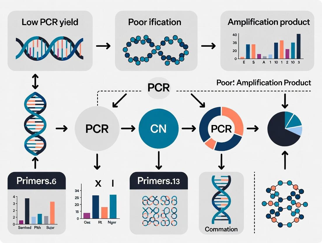
PCR Troubleshooting: Expert Solutions for Low Yield and Poor Amplification in Research and Diagnostics
This comprehensive guide addresses the persistent challenge of low PCR yield and poor amplification, a critical bottleneck in molecular biology, diagnostics, and drug development.
PCR Troubleshooting: Expert Solutions for Low Yield and Poor Amplification in Research and Diagnostics
Abstract
This comprehensive guide addresses the persistent challenge of low PCR yield and poor amplification, a critical bottleneck in molecular biology, diagnostics, and drug development. It provides a systematic framework for researchers and scientists, moving from foundational principles to advanced optimization. The article explores the core biochemistry of PCR failure, presents robust methodological best practices, details a step-by-step troubleshooting workflow for optimization, and finally discusses validation strategies to confirm assay reliability. By synthesizing current best practices and innovative techniques, this resource empowers professionals to achieve consistent, high-quality amplification results essential for downstream applications.
Understanding PCR Failure: The Root Causes of Low Yield and Poor Amplification
Technical Support & Troubleshooting Center
FAQs & Troubleshooting Guides
Q1: What are the definitive quantitative thresholds for defining a 'low yield' PCR amplification curve? A: Definitions are context-dependent (target, sample, instrument) but general benchmarks exist. 'Low yield' is typically indicated by a Cycle Threshold (Ct) value that is significantly later than positive controls or expected values for the assay.
| Parameter | Normal Range | Low Yield / Poor Amplification Indicator |
|---|---|---|
| Cycle Threshold (Ct) | Assay-specific; e.g., 15-30 for abundant targets | Ct > 30-35, or >5 cycles later than positive control |
| ΔRn (Fluorescence Intensity) | Robust plateau > 10^4 RFU | Maximum ΔRn < 1000 RFU |
| Amplification Efficiency (E) | 90-110% (Slope ~ -3.1 to -3.6) | E < 90% or > 110% |
| Plateau Phase Height | High, stable fluorescence | Low, gradual slope, no clear plateau |
Q2: My amplification curve has a normal Ct but a very low plateau (ΔRn). Is this 'poor amplification' and what causes it? A: Yes, a suppressed plateau indicates poor amplification yield despite early detection. Primary causes include:
- Inhibitors in the template: Heparin, EDTA, phenol, humic acid.
- PCR component limitation: Depletion of dNTPs or primers.
- Probe degradation (for qPCR): Hydrolysis probes degrade if contaminated with nucleases.
- Instrument calibration issue: Faulty fluorometer gain settings.
Q3: What is the difference between a late Ct ('low yield') and a failed amplification? A: A late Ct shows a sigmoidal curve with a detectable fluorescence increase above background, just later than optimal. Failed amplification shows no curve—fluorescence remains at baseline or shows nonspecific noise.
Q4: My no-template control (NTC) shows amplification. How does this impact yield analysis? A: NTC amplification invalidates yield calculations from test samples. It indicates contamination (most common) or primer-dimer formation, which consumes reagents and artificially lowers target yield. All results are suspect until contamination is eliminated.
Experimental Protocol: Systematic Diagnosis of Low Yield/Poor Amplification
Objective: To identify the root cause of suboptimal PCR amplification.
Materials: See "The Scientist's Toolkit" below.
Methodology:
- Visual Inspection & Re-analysis: Re-plot raw fluorescence (Rn) vs. cycle. Adjust baseline and threshold settings to ensure correct Ct calling.
- Efficiency Calculation: Perform a 10-fold serial dilution (e.g., 1:10 to 1:10,000) of a positive control template. Run PCR and plot Ct vs. log10(template amount). Calculate efficiency from the slope: Efficiency = (10^(-1/slope) - 1) * 100%.
- Component Titration Test: Set up a matrix of reactions systematically varying one component at a time:
- Primer concentration (50 nM – 900 nM).
- MgCl2 concentration (1.0 mM – 4.0 mM).
- Template amount (1 ng – 100 ng).
- Polymerase units (0.5x – 2x recommended).
- Inhibition Check: Use a known amount of exogenous control DNA (e.g., spike-in) with the sample. A delayed Ct for the spike-in in the test sample vs. water indicates presence of inhibitors.
- Thermal Cycler Verification: Use a calibrated thermocouple to verify block temperature uniformity and gradient accuracy.
Diagnostic Workflow Diagram
Title: PCR Low Yield Diagnostic Troubleshooting Workflow
The Scientist's Toolkit: Research Reagent Solutions
| Item | Function & Rationale |
|---|---|
| High-Fidelity DNA Polymerase | Provides high processivity and fidelity for long or complex templates, improving yield of correct product. |
| PCR Enhancers (e.g., BSA, DMSO, Betaine) | Stabilize polymerase, reduce secondary structure in GC-rich regions, and mitigate mild inhibitor effects. |
| Molecular Biology Grade Water (Nuclease-Free) | Prevents degradation of primers, templates, and probes. Contaminants in poor-quality water are a common cause of failure. |
| dNTP Mix (balanced, pH ~8.4) | Unbalanced dNTP concentrations promote misincorporation and early plateau. Quality is critical for high yield. |
| Validated Positive Control Template | Essential for calculating amplification efficiency and as a benchmark for 'normal' curve shape and Ct. |
| Inhibitor Removal Kit (e.g., silica-column) | For purifying template from challenging samples (blood, soil, formalin-fixed tissue) to remove PCR inhibitors. |
| Calibrated Digital Pipettes | Ensures accurate and precise dispensing of small-volume reaction components, critical for reproducibility. |
| Optical Grade Plate Sealers | Prevents well-to-well contamination and evaporation, which significantly affects reaction yield and consistency. |
Technical Support Center: Troubleshooting PCR Yield and Amplification
Troubleshooting Guides
Issue 1: No or Low PCR Product Yield
- Likely Culprit: Template Degradation or Extremely Low Concentration.
- Investigation Steps:
- Assess Template Integrity: Run the template sample on a 1% agarose gel alongside a high-quality control (e.g., intact genomic DNA). A smear indicates degradation.
- Quantify Template Precisely: Use a fluorescence-based assay (Qubit, PicoGreen) for accurate quantitation of dsDNA, as absorbance (A260) can be skewed by contaminants.
- Perform Serial Dilution Test: Amplify a dilution series of your template (e.g., 100 ng, 10 ng, 1 ng, 0.1 ng) to determine if yield improves at a different concentration.
Issue 2: Non-Specific Amplification (Smearing/Multiple Bands)
- Likely Culprit: Inhibitors in Template Sample or Suboptimal Reaction Conditions.
- Investigation Steps:
- Test for Inhibition: Perform a "spike-in" experiment. Add a known, functional template and its primers to your sample reaction and a clean control reaction. Failure in the sample reaction indicates inhibition.
- Purify Template: Re-purify the template using silica-column or bead-based clean-up kits designed to remove salts, proteins, phenols, or humic acids.
- Optimize Annealing: Perform a thermal gradient PCR to identify the optimal annealing temperature for your primer pair.
Issue 3: Irreproducible Results Between Replicates
- Likely Culprit: Pipetting Error with Low Concentration Template or Inhomogeneous Inhibitors.
- Investigation Steps:
- Template Homogenization: Vortex template stock thoroughly before use and perform a brief spin.
- Master Mix Preparation: Always prepare a common Master Mix (containing enzyme, buffer, dNTPs, primers, water) for all replicates to minimize pipetting variance.
- Use of Carrier: For very dilute templates (<10 pg/µL), consider adding molecular biology-grade carrier RNA or linear acrylamide to the dilution buffer to prevent adsorption to tube walls.
Frequently Asked Questions (FAQs)
Q1: My template is ancient/fixed/from a challenging source (soil, FFPE). How do I maximize my chances of amplification? A: Use a polymerase blend specifically engineered for inhibited and fragmented samples. These often combine a high-processivity enzyme with an antibody-mediated hot-start and enhancers. Also, increase cycle number (up to 45 cycles) and extend elongation time. Prioritize short amplicons (<200 bp).
Q2: I've quantified my DNA with a Nanodrop and it shows a good concentration, but PCR still fails. Why? A: Spectrophotometers (Nanodrop) detect any molecule absorbing at 260nm, including RNA, free nucleotides, and some contaminants. They overestimate DNA concentration and do not detect common inhibitors. Always use a fluorometric assay for critical template quantitation and assess purity via A260/A280 and A260/A230 ratios.
Q3: What is the best method to remove inhibitors from my DNA sample? A: The best method depends on the inhibitor. For common contaminants:
- Polysaccharides/Phenols: Use a CTAB re-purification or specific commercial kits for plant/soil DNA.
- Hemoglobin/Heparin: Use a silica-column kit with inhibitor-removal wash buffers.
- Humic Acid: Use kits specifically validated for environmental samples or add PCR enhancers like BSA (0.1-0.4 µg/µL) or T4 Gene 32 Protein.
Q4: How do I know if my problem is template quantity versus quality? A: Perform the diagnostic experiments summarized in the table below.
| Diagnostic Test | Method | Expected Result for "Quantity Issue" | Expected Result for "Quality Issue" |
|---|---|---|---|
| Template Quantitation | Fluorometric vs. Spectrophotometric | Values correlate; [DNA] is very low. | Fluorometric value is significantly lower than spectrophotometric. |
| Agarose Gel Analysis | Electrophoresis of 100-200 ng template | No smear, but faint or no band. | Visible smearing, no clear high-MW band. |
| Spike-In Control | Amplify known target in your sample | Amplification successful. | Amplification fails or yield is severely reduced. |
| Serial Dilution PCR | Amplify 100ng to 0.1ng of template | Yield gradient correlates with dilution. | No product across all dilutions, or erratic yield. |
| Alternative Polymerase | Use inhibitor-tolerant enzyme | No change in result. | Significant improvement in yield/specificity. |
Experimental Protocols
Protocol 1: Diagnostic Spike-In Test for PCR Inhibitors
- Prepare two 0.2 mL PCR tubes.
- Tube 1 (Test): Combine 15 µL of Master Mix, 1 µL of your target primer pair, 4 µL of your suspect template DNA, and 1 µL of a known, clean control template (e.g., 1 ng/µL λ DNA).
- Tube 2 (Control): Combine 15 µL of Master Mix, 1 µL of your target primer pair, 4 µL of nuclease-free water, and 1 µL of the same known, clean control template.
- Run your standard PCR protocol.
- Analysis: If both tubes show strong bands for the control amplicon, your sample is not inhibitory. If Tube 1 fails or shows a weak control band compared to Tube 2, your sample contains inhibitors.
Protocol 2: Serial Template Dilution for Optimal Concentration Finding
- Starting from your stock, prepare four 10-fold serial dilutions in nuclease-free water (e.g., 10 ng/µL, 1 ng/µL, 0.1 ng/µL, 0.01 ng/µL).
- Label five PCR tubes.
- To each tube, add 19 µL of a standard Master Mix containing primers.
- Add 1 µL of each template dilution to separate tubes (Final amounts: 10 ng, 1 ng, 0.1 ng, 0.01 ng). Include a no-template control (NTC) with water.
- Run your PCR protocol.
- Analyze 5 µL from each reaction on an agarose gel. Identify the dilution giving the strongest, cleanest band for future use.
Visualizations
Title: PCR Failure Troubleshooting Decision Tree
Title: Spike-In Test Workflow for Inhibitor Detection
The Scientist's Toolkit: Research Reagent Solutions
| Item | Function & Rationale |
|---|---|
| Fluorometric DNA Quantitation Kit (e.g., Qubit dsDNA HS/BR) | Provides accurate concentration of intact dsDNA, unaffected by common contaminants like RNA or salts that skew spectrophotometer readings. Essential for low-concentration templates. |
| Inhibitor-Tolerant Polymerase Blends | Enzyme mixes containing additives and polymerases capable of bypassing or withstanding common inhibitors (humic acid, hematin, tannins) found in environmental, forensic, or clinical samples. |
| PCR Enhancers (e.g., BSA, Betaine, T4 GP32) | Molecules that stabilize polymerase, reduce secondary structure in GC-rich regions, or bind non-specific inhibitors, improving yield and specificity from suboptimal templates. |
| Solid-Phase Reversible Immobilization (SPRI) Beads | Magnetic beads used for high-efficiency DNA clean-up and size selection, effectively removing primers, dNTPs, salts, and many organic inhibitors. |
| Molecular Biology-Grade Carrier RNA/DNA | Added to dilution buffers for extremely low-concentration templates (<10 pg/µL) to prevent adsorption to tube surfaces, ensuring accurate pipetting and representation. |
| DNA Integrity Gel Electrophoresis Markers | High-molecular-weight ladders and sample loading dyes that allow clear visualization of template degradation (smearing) versus intact, high-quality DNA. |
Technical Support Center: Troubleshooting PCR Yield and Amplification
FAQ 1: I am experiencing low or no PCR yield. What are the primary reagent-related causes? Low PCR yield is frequently due to reagent integrity or improper composition. The most common culprits are:
- Polymerase Inactivation: Repeated freeze-thaw cycles, improper storage, or contamination with nucleases can inactivate the enzyme.
- dNTP Degradation or Imbalance: dNTPs are susceptible to hydrolysis, and an imbalance in the equimolar ratio can promote misincorporation and early polymerase stoppage.
- Suboptimal Mg²⁺ Concentration: Mg²⁺ is a critical cofactor for polymerase activity. Too little reduces efficiency; too much increases non-specific binding.
- Buffer Inefficacy: Incorrect pH, depleted stabilizing components (e.g., BSA, DTT), or the wrong buffer formulation for your polymerase can severely impact yield.
FAQ 2: How can I systematically troubleshoot reagent-related poor amplification? Follow this diagnostic workflow:
- Prepare Fresh Master Mix: Exclude template DNA to test reagent integrity.
- Test with Control Template: Use a known, high-copy-number template and primer set.
- Titrate Mg²⁺: Perform a gradient PCR (1.0 mM to 4.0 mM in 0.5 mM increments).
- Verify dNTP Concentration: Confirm concentration via spectrophotometry and ensure an equimolar mix.
- Run an Enzyme Activity Assay: Use a standardized assay template provided by the manufacturer.
FAQ 3: What are the optimal storage conditions and stability timelines for core PCR reagents? Adherence to storage protocols is paramount for reagent integrity.
Table 1: Stability and Storage Guidelines for Core PCR Reagents
| Reagent | Recommended Storage | Stable at -20°C (Unopened) | Stable at -20°C (After Thaw/Opened) | Key Integrity Check Method |
|---|---|---|---|---|
| Taq DNA Polymerase | -20°C in glycerol storage buffer | 24 months | 6 months (avoid freeze-thaw) | Activity assay with control amplicon |
| dNTP Mix (100mM) | -20°C in small aliquots, neutral pH | 24 months | 3-6 months (as aliquot) | Spectrophotometry (A260/A280 ~0.8) |
| 10x PCR Buffer (with MgCl₂) | -20°C | 24 months | 12 months | Use with fresh Mg²⁺ titration |
| MgCl₂ Solution (25mM) | -20°C, sterile-filtered | 36 months | 24 months | Avoid precipitation; filter if cloudy |
Experimental Protocol: Mg²⁺ and dNTP Titration for Yield Optimization Objective: Determine the optimal Mg²⁺ and dNTP concentrations for a specific primer-template system. Method:
- Prepare a standard 2X master mix containing polymerase, buffer (without Mg²⁺), primers, and nuclease-free water.
- For Mg²⁺ Titration: Aliquot the master mix. Add MgCl₂ to each tube to create a final concentration series (e.g., 1.0, 1.5, 2.0, 2.5, 3.0, 3.5, 4.0 mM). Keep dNTP concentration constant (e.g., 200 µM each).
- For dNTP Titration: Using the optimal Mg²⁺ concentration from step 2, create a dNTP concentration series (e.g., 50, 100, 200, 400, 800 µM each).
- Add an equal volume of template to each reaction.
- Run the thermocycler with your standard amplification program.
- Analyze products on an agarose gel. The condition producing the brightest, cleanest band of the expected size indicates the optimal concentration.
Diagram: PCR Reagent Integrity Diagnostic Workflow
The Scientist's Toolkit: Key Research Reagent Solutions Table 2: Essential Reagents for PCR Optimization & Integrity Management
| Item | Function in PCR Optimization |
|---|---|
| Hot-Start DNA Polymerase | Minimizes non-specific amplification and primer-dimer formation by requiring thermal activation. |
| PCR-Grade dNTP Mix (100mM, pH 7.0) | Provides stable, nuclease-free, equimolar deoxynucleotide triphosphates as building blocks. |
| MgCl₂ Solution (25-50 mM), Sterile-Filtered | Allows precise titration of the critical polymerase cofactor Mg²⁺. |
| PCR Buffer (10x, Mg²⁺-Free) | Provides optimal pH, ionic strength, and stabilizers without locking in Mg²⁺ concentration. |
| BSA (Bovine Serum Albumin, Molecular Biology Grade) | Stabilizes the polymerase and mitigates inhibition from sample contaminants. |
| DTT (Dithiothreitol, 100mM) | A reducing agent that maintains enzyme activity by preventing oxidation of cysteine residues. |
| Standardized Control Template & Primer Set | Essential control for differentiating between reagent failure and assay design problems. |
| Spectrophotometer/Nanodrop | For accurate quantification and purity assessment (A260/A280) of dNTPs, primers, and template. |
Troubleshooting Guides & FAQs
Q1: My PCR reaction produces multiple unexpected bands or a smear. How can I check if my primers lack specificity? A: This indicates potential off-target binding. Perform an in silico specificity check using tools like NCBI BLAST or Primer-BLAST against the relevant genome database (e.g., human, mouse). Ensure the primer sequence is unique. Experimental validation requires a positive control and optimization of annealing temperature (Ta) using a gradient PCR.
Q2: I suspect primer-dimer formation is consuming my reagents and reducing yield. How do I diagnose and fix this? A: Primer-dimers appear as a low molecular weight smear or band (~30-50 bp) on an agarose gel. To fix this:
- Redesign primers to avoid 3'-end complementarity.
- Increase annealing temperature to reduce spurious binding.
- Use a hot-start polymerase to inhibit activity during setup.
- Lower primer concentration (try 0.1-0.5 µM each) in the reaction.
Q3: How do I assess and mitigate issues from primer secondary structure (hairpins)? A: Use primer analysis software (e.g., OligoAnalyzer, mFold). A strong hairpin with a low ∆G (e.g., < -3 kcal/mol) at the 3' end can severely hinder binding.
- Fix: Redesign the primer to avoid self-complementary stretches, especially at the 3' end. Increasing Ta can also help.
Q4: What is the optimal Tm difference between a primer pair, and how do I calculate it correctly? A: Primers in a pair should have closely matched Tms (within 1-3°C). Use a consistent calculation method. The nearest-neighbor method is most accurate. Software like Primer3 uses this method.
Q5: How does incorrect Tm calculation directly lead to low PCR yield in my research? A: If the calculated Tm is inaccurate, the chosen annealing temperature (Ta) will be suboptimal. If Ta is too high, primers won't bind efficiently. If Ta is too low, primers bind non-specifically, leading to dimers or off-target products. Both scenarios drastically reduce the yield of your desired amplicon.
Table 1: Impact of Common Primer Flaws on PCR Outcome
| Primer Flaw | Typical Gel Result | Primary Effect on Yield | Quick Diagnostic Test |
|---|---|---|---|
| Low Specificity | Multiple bands, smear | Low to moderate | In silico BLAST; Gradient PCR |
| Primer-Dimers | Low MW band/smear | Very low (reagent depletion) | Agarose gel (high %); NTC analysis |
| Strong 3' Hairpin | No product or very low | Very low to none | In silico ∆G analysis |
| Tm Mismatch (>5°C) | Asymmetric or low yield | Low | Re-calculate Tm via NN method |
Table 2: Recommended Parameters for Standard PCR Primer Design
| Parameter | Optimal Range | Critical Note |
|---|---|---|
| Length | 18-25 bases | Shorter for qPCR, longer for complex genomes. |
| Tm (Nearest-Neighbor) | 55-72°C | Pair Tm difference must be ≤ 3°C. |
| GC Content | 40-60% | Avoid long runs of a single nucleotide. |
| 3'-End Stability | ∆G ≥ -3 kcal/mol | Crucial for preventing hairpin formation. |
| Primer Concentration | 0.1-0.5 µM (each) | High conc. promotes dimer formation. |
Experimental Protocols
Protocol 1: In Silico Primer Specificity and Quality Check
- Tool: Access NCBI Primer-BLAST.
- Input: Paste forward and reverse primer sequences in FASTA format.
- Parameters: Select the correct organism database (e.g., RefSeq mRNA, genome). Set PCR product size range.
- Analysis: Examine the "Primer Pair Specificity Checking Results" section. The tool will list all predicted amplicons. Your target should be the top, perfect-match hit.
Protocol 2: Empirical Annealing Temperature Optimization via Gradient PCR
- Setup: Prepare a standard PCR master mix with your primer pair and template.
- Gradient: Use a thermal cycler with a gradient function across the block. Set a temperature range spanning ~10°C below to ~5°C above the calculated lower Tm of your primer pair (e.g., 55°C to 70°C).
- Run: Execute the PCR cycle.
- Analysis: Resolve products on an agarose gel. The optimal Ta is the highest temperature that produces a single, strong band of the correct size.
Protocol 3: Testing for Primer-Dimer Artifacts
- Critical Control: Always include a No-Template Control (NTC) in your experiment.
- Setup: Prepare an identical PCR reaction but use nuclease-free water instead of DNA template.
- Run: Cycle the NTC alongside your test reactions.
- Analysis: Load the NTC product on the gel. Any visible product in the NTC lane is primer-dimer or contamination, indicating problematic primers or conditions.
Visualizations
Title: Primer Design and Validation Workflow
Title: PCR Failure Analysis Based on Gel Result
The Scientist's Toolkit: Research Reagent Solutions
| Item | Function in Addressing Primer Flaws |
|---|---|
| Hot-Start DNA Polymerase | Minimizes non-specific amplification and primer-dimer formation by remaining inactive until the initial denaturation step. Essential for complex templates. |
| Nuclease-Free Water | Used for primer resuspension and reaction setup. Prevents degradation of primers and templates, ensuring reliability. |
| PCR Nucleotide Mix (dNTPs) | Balanced solution of dATP, dCTP, dGTP, dTTP. Quality dNTPs prevent misincorporation, which can be mistaken for specificity issues. |
| MgCl₂ Solution | Critical co-factor for polymerase activity. Concentration optimization (1.5-4.0 mM) is key for improving specificity and yield after primer design. |
| Q5 or Phusion HF Polymerase | High-fidelity polymerases with superior specificity, reducing mispriming and off-target amplification from suboptimal primers. |
| Gradient Thermal Cycler | Allows empirical optimization of the annealing temperature (Ta) in a single run, directly addressing Tm calculation inaccuracies. |
| Agarose Gel Electrophoresis System | The primary method for visualizing PCR product size, purity, and the presence of primer-dimers (via NTC). |
Within the context of correcting low PCR yield and poor amplification, optimizing thermal cycler performance is critical. Inaccurate block temperatures and non-optimal ramp rates directly impact primer annealing, enzyme efficiency, and ultimately, amplicon yield and specificity. This technical support center provides troubleshooting and FAQs to address these core instrument parameters.
Troubleshooting Guides & FAQs
Q1: My PCR yields are consistently low and variable across the block. I suspect temperature inaccuracy. How can I diagnose this? A: This is a classic sign of block temperature uniformity or calibration issues.
- Diagnostic Protocol: Perform a thermal gradient verification test.
- Materials: Use a calibrated NIST-traceable thermocouple reader and fine-gauge thermocouple probes.
- Method: Fill all sample wells with 50 µL of mineral oil or a dummy reaction mix. Insert thermocouple probes into wells representing the four corners and center of the block.
- Run: Program the cycler to hold at three critical temperatures: 95°C (denaturation), 55-60°C (annealing, typical), and 72°C (extension). Hold each for 5 minutes after the block indicates it has reached the setpoint.
- Data Collection: Record the actual temperature from each probe at 30-second intervals during the final 2 minutes of each hold.
- Analysis: Compare actual vs. setpoint temperatures. Calculate mean accuracy and block uniformity (max-min difference). Acceptable performance is typically within ±0.5°C of setpoint with <1.0°C variation across the block.
Q2: Can a slow ramp rate between annealing and extension phases cause poor specificity (e.g., primer-dimer)? A: Yes. Excessively slow ramp rates, particularly through critical temperature ranges below the primer annealing temperature, can promote non-specific priming and increase primer-dimer artifacts.
- Solution & Optimization Protocol:
- Determine the actual ramp rate of your instrument between the annealing and extension steps using the manufacturer's specifications or empirical testing.
- Program a two-step thermal profile to mitigate this:
- Step 1: Denaturation (e.g., 95°C for 10-30s)
- Step 2: Combined Annealing/Extension (e.g., 60-72°C for 30-60s). This eliminates the slow ramp through the problematic temperature zone.
- If a three-step profile is necessary, use the maximum permissible ramp rate (e.g., "Max Speed" setting). Test the effect on yield and specificity using a standardized template.
Q3: My amplification of long amplicons (>3 kb) is inefficient. Could ramp rate be a factor? A: Absolutely. For long amplicons, a very fast ramp rate can be detrimental. Rapid temperature changes may prevent proper double-strand separation or enzyme binding.
- Optimization Protocol:
- Reduce the Ramp Rate: Manually set a slower, controlled ramp rate (e.g., 1.5-2.0°C/second) for the transitions between denaturation and annealing, and annealing to extension.
- Increase Extension Time: Ensure extension time is sufficient for the polymerase's processivity (e.g., 1 min/kb).
- Use a Specialized Polymerase: Employ a polymerase mix optimized for long-range PCR, which often includes a balance of enzymes that benefits from more controlled temperature transitions.
Table 1: Common Thermal Cycler Performance Specifications & Impact
| Parameter | Typical Specification | Impact on PCR if Out of Spec | Corrective Action |
|---|---|---|---|
| Temperature Accuracy | ±0.3°C to ±0.5°C at 55°C | Low/No Yield, Variable Results | Calibrate instrument. Use block conditioner. |
| Block Uniformity | <1.0°C difference across block | Inconsistent yields well-to-well | Verify calibration. Ensure lid is sealed. |
| Average Ramp Rate | 2.0°C/s to 5.0°C/s | Poor specificity (slow) or long amplicon failure (fast) | Optimize protocol for target amplicon. |
| Heated Lid Temp | 105°C (for aqueous samples) | Evaporation/condensation, reaction volume loss | Verify and increase lid temperature setting. |
Table 2: Optimization Guide Based on Amplification Problem
| Observed Problem | Suspected Parameter | Recommended Optimization Test |
|---|---|---|
| Low Yield, High Specificity | Temperature Accuracy (Annealing) | Perform thermal verification at annealing temp. |
| High Yield, Low Specificity | Ramp Rate (too slow) | Increase ramp rate or use 2-step profile. |
| Primer-Dimer Formation | Ramp Rate through low temps | Use "hot-start" enzyme, increase ramp rate. |
| Failure of Long Amplicons | Ramp Rate (too fast), Temp Accuracy | Reduce ramp rate to 1.5-2°C/s, verify 72°C accuracy. |
Experimental Protocols
Protocol: Empirical Measurement of Actual Ramp Rate Objective: To determine the true ramp rate of a thermal cycler between two setpoints. Materials: Data logging thermocouple system, thermal block calibration tool or tube with mock reaction. Method:
- Place the thermocouple probe securely in a well, immersed in 50 µL of mineral oil or solution.
- Program a method: 50°C hold for 1 min, then ramp to 95°C, then hold.
- Start data logging at a high frequency (e.g., 1-2 readings/second).
- Start the thermal cycler run.
- Analysis: From the logged data, identify the time point when the temperature first reaches 51°C (start of ramp) and when it first reaches 94°C (end of ramp). Calculate the rate: Ramp Rate (°C/s) = (94-51)°C / (Time Elapsed in seconds).
Protocol: Two-Step vs. Three-Step PCR Comparison for Specificity Objective: To evaluate the effect of thermal profile on amplicon specificity. Method:
- Template: Use a standardized genomic DNA or plasmid template at a limiting concentration (e.g., 10 ng).
- Primers: Use a primer pair known to be prone to primer-dimer formation.
- Master Mix: Use a standard Taq polymerase mix.
- Run Duplicate Reactions: Program two identical cyclers (or one with two different protocols):
- Profile A (3-Step): 95°C 30s, 55°C 30s, 72°C 30s. Use standard ramp rates.
- Profile B (2-Step): 95°C 30s, 60°C 60s. Use standard ramp rates.
- Run for 30 cycles.
- Analysis: Analyze products on a high-resolution agarose gel (2-3%) or bioanalyzer. Compare band sharpness and the intensity of the low molecular weight primer-dimer smear.
Diagrams
Title: Decision Tree for PCR Yield & Specificity Issues
Title: Three-Step vs. Two-Step Thermal Profile Ramp Comparison
The Scientist's Toolkit: Research Reagent Solutions
| Item | Function in Optimization Context |
|---|---|
| NIST-Traceable Thermocouple Calibrator | Provides gold-standard temperature measurement to verify and calibrate thermal cycler block accuracy. |
| Block Conditioner Tubes/Plates | Ensures thermal mass is consistent during verification tests, simulating real reaction conditions. |
| High-Fidelity/Long-Range PCR Enzyme Mix | Engineered polymerases with enhanced processivity and tolerance to sub-optimal ramp conditions for difficult amplicons. |
| Hot-Start Taq Polymerase | Remains inactive until a high-temperature step, preventing primer-dimer formation during slow ramp-up or setup. |
| PCR Additives (e.g., Betaine, DMSO) | Can lower DNA melting temperature and improve strand separation, mitigating effects of slight temperature inaccuracy. |
| Standardized Validation Template (e.g., Genomic DNA Control) | Provides a consistent baseline to compare yield and specificity across different thermal profiles or instruments. |
Best Practices for Robust PCR: Protocols to Ensure High-Yield Amplification
Technical Support Center
Troubleshooting Guides & FAQs
Q1: My DNA extraction yield from whole blood is consistently low. What could be the cause? A: Low yield from whole blood is often due to incomplete lysis of leukocytes or inefficiency in DNA binding to the chosen matrix. Ensure the lysis buffer contains a potent detergent (e.g., SDS) and a proteinase K digestion step (at 56°C for >30 minutes) is included. For silica-membrane columns, verify that ethanol concentration in the binding mixture is correct (typically 40-50%). Low yields can also result from using outdated or improperly stored anticoagulants (EDTA is preferred over heparin). A modified protocol using an increased initial blood volume and proportionally scaled-up lysis/binding reagents can improve yield.
Q2: I suspect my extracted nucleic acids contain PCR inhibitors. How can I confirm and remedy this? A: Confirm inhibition using a spike-in control assay. Add a known quantity of a control DNA template and its specific primer set to your PCR reaction alongside your sample. If amplification of the control is suppressed, inhibitors are present. Common remedies include:
- Additional Purification: Perform a silica-column clean-up or use bead-based purification.
- Dilution: Dilute the template (1:5, 1:10) to reduce inhibitor concentration, though this also dilutes the target.
- Additive Use: Include PCR additives like bovine serum albumin (BSA, 0.1-0.5 µg/µL) or T4 gene 32 protein (gp32), which can bind inhibitors.
- Alternative Polymerase: Use a inhibitor-resistant polymerase blend designed for forensic or environmental samples.
Q3: My RNA has a low A260/A280 ratio (<1.8), indicating protein contamination. What step likely failed? A: A low A260/A280 ratio suggests residual protein/phenol from the extraction. This commonly occurs during the phase separation in phenol-chloroform methods. Ensure you do not aspirate any of the interphase or organic layer. For column-based methods, ensure all wash buffers contain the correct ethanol concentration and that washes are performed thoroughly. An additional chloroform back-extraction (for aqueous phase methods) or a second column wash with an 80% ethanol solution can remedy this.
Q4: How do I prevent genomic DNA contamination in my RNA samples? A: Always include an on-column DNase I digestion step. The protocol is: after loading the RNA onto the silica membrane, apply a DNase I incubation mix (e.g., 10 µL DNase I, 70 µL RDD buffer from Qiagen) directly onto the membrane and incubate at room temperature for 15 minutes. Then proceed with wash steps. For manual methods, use acid-phenol:chloroform at pH 4.5, which partitions DNA to the interphase/organic phase, leaving RNA in the aqueous phase.
Q5: My DNA/RNA integrity number (RIN/DIN) is poor. How can I improve sample integrity? A: Poor integrity is primarily due to endogenous or exogenous RNase/DNase activity. For RNA:
- Use fresh, ice-cold lysis buffers containing strong denaturants (guanidinium isothiocyanate).
- Homogenize tissue samples immediately in lysis buffer. Keep samples on ice.
- Use RNase-free tubes, tips, and water. For DNA from tissues, ensure rapid processing or flash-freeze in liquid nitrogen. Consider using a lysis buffer with EDTA to chelate metal ions required for nuclease activity.
Experimental Protocols
Protocol 1: Phenol-Chloroform-Isoamyl Alcohol (25:24:1) Extraction for Genomic DNA from Cultured Cells
- Pellet 1-5 x 10^6 cells. Lyse in 500 µL of Lysis Buffer (10 mM Tris-HCl pH 8.0, 100 mM EDTA, 0.5% SDS) with 2 µL of RNase A (20 mg/mL). Incubate at 37°C for 1 hour.
- Add 10 µL of Proteinase K (20 mg/mL). Mix and incubate at 56°C overnight or for at least 4 hours.
- Cool to room temperature. Add an equal volume (≈500 µL) of PCI (25:24:1). Vortex vigorously for 30 seconds.
- Centrifuge at 12,000 x g for 10 minutes at 4°C.
- Carefully transfer the upper aqueous phase to a new tube. Add 0.5 volumes of 7.5 M ammonium acetate and 2 volumes of 100% ethanol. Mix by inversion.
- Spool out the DNA precipitate with a glass rod or pipette tip. Wash in 1 mL of 70% ethanol.
- Air-dry the pellet for 10 minutes. Resuspend in 100 µL of TE buffer (10 mM Tris-HCl, 1 mM EDTA, pH 8.0).
Protocol 2: Silica-Membrane Column-Based Total RNA Extraction from Animal Tissue
- Homogenize 30 mg of tissue in 600 µL of RLT Plus lysis buffer (Qiagen) with 1% β-mercaptoethanol using a rotor-stator homogenizer.
- Centrifuge the lysate at 12,000 x g for 3 minutes. Transfer supernatant to a new tube.
- Add 1 volume (≈600 µL) of 70% ethanol. Mix thoroughly by pipetting.
- Apply the mixture (including any precipitate) to an RNeasy Plus column placed in a 2 mL collection tube. Centrifuge at ≥8000 x g for 30 seconds. Discard flow-through.
- On-Column DNase Digestion: Mix 10 µL DNase I with 70 µL Buffer RDD. Apply directly to the column membrane. Incubate at room temp for 15 minutes.
- Add 500 µL Buffer RPE (with ethanol) to column. Centrifuge for 30 sec. Discard flow-through.
- Add 500 µL Buffer RPE. Centrifuge for 2 minutes to dry membrane.
- Transfer column to a 1.5 mL collection tube. Elute RNA with 30-50 µL RNase-free water. Centrifuge for 1 minute.
Protocol 3: Magnetic Bead-Based PCR Inhibitor Removal Clean-up
- To 50 µL of contaminated DNA sample, add 50 µL of magnetic bead suspension (e.g., SPRI beads). Mix thoroughly by pipetting.
- Incubate at room temperature for 5 minutes.
- Place tube on a magnetic stand until supernatant clears (≈2 minutes).
- Carefully remove and discard the supernatant.
- With tube on magnet, wash beads twice with 200 µL of freshly prepared 80% ethanol. Incubate 30 seconds per wash. Remove all ethanol.
- Air-dry beads for 5-10 minutes until cracks appear.
- Remove from magnet. Elute DNA in 30 µL of low-EDTA TE buffer or nuclease-free water. Mix well. Incubate 2 minutes.
- Place back on magnet. Transfer purified supernatant to a new tube.
Table 1: Comparison of Nucleic Acid Extraction Method Efficiencies
| Method | Sample Type | Average Yield | Average A260/A280 | Average RIN/DIN | Time to Complete | Cost per Sample |
|---|---|---|---|---|---|---|
| Phenol-Chloroform | Cultured Cells (5x10^6) | 15-25 µg DNA | 1.75-1.85 | DIN: 7-8 | 4-6 hours | Low |
| Silica Column | Whole Blood (200 µL) | 0.5-1.5 µg DNA | 1.80-1.95 | DIN: 8-9 | 30-45 min | Medium |
| Magnetic Beads | Bacterial Culture (1 mL) | 2-5 µg DNA | 1.85-2.00 | DIN: 8-9 | 20-30 min | Medium |
| Guanidinium-Based Column | Mouse Liver (30 mg) | 20-40 µg RNA | 1.90-2.10 | RIN: 8.5-10 | 40-60 min | Medium |
| Salting-Out | Buccal Swab | 0.2-2 µg DNA | 1.60-1.80 | DIN: 6-7 | 1.5 hours | Very Low |
Table 2: Common PCR Inhibitors and Mitigation Strategies
| Inhibitor Class | Source | Effect on PCR | Mitigation Strategy |
|---|---|---|---|
| Heparin | Blood collection tubes | Binds polymerase | Use EDTA tubes; Additional purification |
| Hemoglobin/Heme | Blood lysates | Degrades polymerase | Silica column wash; Add BSA (0.4 µg/µL) |
| Humic Acids | Soil/Plant extracts | Binds Taq & template | Dilution; Use PVPP in lysis; Inhibitor-resistant enzymes |
| Polysaccharides | Plant/Fecal samples | Increases viscosity, inhibits | CTAB in lysis; Dilution; Extensive washing |
| Phenol | Organic extraction carryover | Denatures polymerase | Ensure careful phase separation; Ethanol precipitation |
| Ionic Detergents (SDS) | Lysis buffer carryover | Denatures polymerase | Ensure <0.005% final conc.; Use non-ionic detergents |
| Urea/GuSCN | Lysis buffer carryover | Inhibits polymerase | Dilution; Ethanol precipitation; Column washing |
The Scientist's Toolkit
Table 3: Essential Reagents for Nucleic Acid Extraction & QC
| Item | Function | Example(s) |
|---|---|---|
| Lysis Buffer | Disrupts cells, inactivates nucleases, and releases nucleic acids. | ATL, RLT (Qiagen); DNAzol, TRIzol; Guanidinium thiocyanate-based buffers. |
| Proteinase K | Broad-spectrum serine protease that digests proteins and aids in lysis. | Used in tissue and cell lysis protocols, often at 56°C. |
| RNase A | Ribonuclease that specifically degrades RNA to remove RNA from DNA preps. | Added during genomic DNA extraction. |
| DNase I | Endonuclease that cleaves DNA to remove genomic DNA from RNA preps. | Used in on-column or in-solution digestion (RNA extraction). |
| Silica Membrane/Matrix | Selectively binds nucleic acids in high-salt, chaotropic conditions; released in low-salt. | Core of spin-column kits (e.g., Qiagen, Macherey-Nagel). |
| Magnetic Beads (SPRI) | Paramagnetic particles that bind nucleic acids for size selection and purification. | Used in automated and manual high-throughput protocols. |
| Binding Buffer | High-salt, chaotropic solution promoting nucleic acid adsorption to silica/magnetic beads. | Contains guanidine HCl or isothiocyanate. |
| Wash Buffer | Ethanol-containing solution that removes salts and contaminants without eluting NA. | Typically contains 70-80% ethanol in a Tris or salt buffer. |
| Elution Buffer | Low-ionic-strength, slightly alkaline solution (TE or water) that releases NA from matrix. | TE buffer (10 mM Tris, 1 mM EDTA, pH 8.0) or nuclease-free water. |
| Nucleic Acid QC Dye | Fluorescent dye that binds NA for quantification and integrity assessment. | PicoGreen (dsDNA), RiboGreen (RNA), Qubit assays. |
Visualizations
Diagram 1: Decision Tree for PCR Amplification Failure Analysis
Diagram 2: Silica Column Nucleic Acid Extraction Workflow
Diagram 3: Sources of PCR Inhibitors in Sample Prep
Primer Design Guidelines and Tools for Optimal Specificity and Efficiency
Troubleshooting Guides and FAQs
Q1: Why is my PCR yield low despite using standard primer design software? A: Low yield often stems from primers with secondary structures or suboptimal melting temperatures (Tm). Ensure forward and reverse primers have a Tm within 1°C of each other (ideally 60-65°C). Check for self-dimers or hairpins using tools like OligoAnalyzer. A common culprit is a 3'-end stability that is too high or too low; the ΔG of the 3'-end five nucleotides should be between -4 and -9 kcal/mol for efficient initiation.
Q2: My gel shows non-specific bands (primer-dimer or multiple products). How can I improve specificity? A: This indicates low primer specificity. First, verify primer length is 18-30 bases. Second, increase the annealing temperature in increments of 1-2°C during optimization. Third, use a touchdown PCR protocol. Most critically, re-evaluate primer specificity by performing a rigorous BLAST search against the relevant genome database to avoid off-target binding.
Q3: How do I design primers for difficult templates (e.g., high GC-content or repetitive sequences)? A: For GC-rich templates (>65%), include PCR additives in your master mix and design primers with a slightly higher Tm (e.g., 68-72°C). For repetitive sequences, place the 3'-end of the primer in a unique region if possible, and consider using a lower annealing temperature to accommodate reduced specificity at the 5'-end.
Q4: What are the critical parameters to check when designing primers for quantitative PCR (qPCR)? A: For qPCR, amplicon length should be short (80-150 bp for optimal efficiency). Primers must span an exon-exon junction when using cDNA to avoid genomic DNA amplification. The primer efficiency (E) should be 90-110%, corresponding to a slope of -3.1 to -3.6 in your standard curve. Always validate with a melt curve to ensure a single, specific product.
Q5: My primers worked for standard PCR but fail in multiplex PCR. What guidelines should I follow? A: Multiplexing requires stringent design to prevent cross-hybridization. All primers in the reaction must have closely matched Tms (within 2°C). Use software dedicated to multiplex design (e.g., Multiplex Manager, PrimerQuest) to check for inter-primer interactions. It is often necessary to adjust primer concentrations empirically for balanced amplification.
Q6: How can I troubleshoot a consistently failed PCR from a newly designed primer set? A: Follow this systematic protocol:
- In silico Check: Re-run specificity check with updated database.
- Template Quality: Run a positive control with a known template and primer set.
- Annealing Temperature Gradient: Perform a gradient PCR from 5°C below to 5°C above the calculated Tm.
- Component Check: Prepare a fresh master mix and verify Mg2+ concentration.
- Simplified Re-design: If all else fails, re-design primers for a different, shorter amplicon within the same target region.
Table 1: Optimal Primer Design Parameters for High-Yield, Specific PCR
| Parameter | Optimal Range | Consequence of Deviation |
|---|---|---|
| Length | 18-30 nucleotides | <18 bp: Reduced specificity; >30 bp: Increased cost, potential secondary structures |
| Melting Temp (Tm) | 60-65°C (within 1°C for primer pair) | Mismatched Tm: One primer inefficiently binds, reducing yield |
| GC Content | 40-60% | Low GC: Low Tm, poor specificity; High GC: Stable secondary structures |
| 3'-End Stability (ΔG) | -4 to -9 kcal/mol (last 5 bases) | Too stable: Mispriming; Too unstable: Poor initiation |
| Amplicon Length | 100-500 bp (standard); 80-150 bp (qPCR) | Long amplicons: Reduced efficiency and yield |
| Specificity Check | ≥2 mismatches in last 5 bases at 3' end | Fewer mismatches: High risk of off-target amplification |
Table 2: Troubleshooting Low PCR Yield: Common Causes & Solutions
| Observed Problem | Potential Cause | Recommended Action |
|---|---|---|
| Very low/no yield | Primer secondary structures | Analyze with OligoAnalyzer; re-design primers |
| Too high annealing temperature | Perform a temperature gradient PCR | |
| Poor template quality/purity | Check template on gel; re-purify | |
| Non-specific bands/multiple products | Low primer specificity | Perform BLAST; increase annealing temp; use touch-down PCR |
| Primer-dimer formation | Check for 3'-end complementarity; use hot-start polymerase | |
| High baseline in qPCR | Primer-dimer formation | Re-design primers; optimize primer concentration |
| Inconsistent replicate results | Poor primer quality/ degradation | Order new, HPLC-purified primers |
Experimental Protocols
Protocol 1: Systematic Primer Design and Validation Workflow
- Sequence Retrieval: Obtain target sequence from a trusted database (e.g., NCBI Nucleotide). Note splice variants if designing for cDNA.
- In silico Design: Use tools like Primer-BLAST (NCBI) or Primer3. Set parameters from Table 1. For qPCR, select "Exon Junction Spanning" option.
- Specificity Analysis: Run the Primer-BLAST function or a standalone BLASTn search against the appropriate refseq genome to ensure uniqueness.
- Dimer & Hairpin Analysis: Input sequences into OligoAnalyzer (IDT) or equivalent. Acceptable ΔG for hairpins is > -3.0 kcal/mol; for dimers, > -5.0 kcal/mol.
- Ordering: Order primers with standard desalting purification. For qPCR or difficult assays, opt for HPLC purification.
- Wet-Lab Validation: Begin with an annealing temperature gradient PCR (e.g., Tm ±5°C). Analyze products on a 2% agarose gel for single, correctly sized band.
Protocol 2: Touchdown PCR for Enhancing Specificity
- Prepare a standard PCR master mix with your primers and template.
- Set the thermocycler program: Initial denaturation (95°C for 3 min).
- Cycling Stage 1 (Touchdown): 10 cycles of: Denaturation (95°C for 30s), Annealing (start 5-10°C above estimated Tm for 30s, decrease by 0.5-1.0°C per cycle), Extension (72°C for 1 min/kb).
- Cycling Stage 2 (Standard): 25-30 cycles of: Denaturation (95°C for 30s), Annealing (use final, lowered Tm from Stage 1 for 30s), Extension (72°C for 1 min/kb).
- Final Extension: 72°C for 5 min.
- Analyze 5 µL of product by gel electrophoresis.
Visualizations
Title: PCR Troubleshooting Decision Tree for Low Yield
Title: Primer Design and Validation Workflow
The Scientist's Toolkit: Research Reagent Solutions
Table 3: Essential Reagents for Robust PCR Primer Testing and Optimization
| Reagent/Material | Function/Benefit | Example/Notes |
|---|---|---|
| High-Fidelity Hot-Start Polymerase | Reduces non-specific amplification and primer-dimer formation during reaction setup. Higher fidelity minimizes PCR-induced errors. | Taq DNA Polymerase, Q5 High-Fidelity DNA Polymerase. |
| PCR Additives (for difficult templates) | Enhance amplification of GC-rich, long, or complex templates by reducing secondary structure or stabilizing polymerase. | DMSO (1-5%), Betaine (0.5-1.5 M), GC Enhancer. |
| Gradient Thermocycler | Allows empirical determination of optimal annealing temperature in a single run, critical for validating new primer sets. | Essential equipment per Protocol 1. |
| Agarose Gel Electrophoresis System | Standard method for visualizing PCR product size, yield, and specificity. Use 2-3% gels for small amplicons. | Includes gel tank, power supply, imaging system. |
| qPCR Master Mix with Intercalating Dye | For validating primer efficiency and specificity in quantitative applications. Dye allows melt curve analysis. | SYBR Green-based mixes. |
| HPLC-Purified Primers | Higher purity than standard desalted primers, crucial for sensitive applications (qPCR, multiplex) to reduce failed experiments. | Specify "HPLC Purification" when ordering. |
| Nuclease-Free Water | Used to dilute primers and prepare master mixes. Prevents degradation of primers and template. | Certified nuclease-free. |
Technical Support Center
Troubleshooting Guides & FAQs
Q1: My PCR yields are consistently low. I suspect my master mix preparation is the issue. What are the most common pipetting errors, and how can I correct them? A: Low yield often stems from inaccurate liquid handling. Common errors and corrections:
- Error: Not pre-wetting the pipette tip. This causes sample retention in the tip.
- Fix: Aspirate and dispense the reagent 2-3 times before taking the final volume.
- Error: Using the wrong pipette for the volume (e.g., using a 1000 µL pipette for 2 µL).
- Fix: Always use a pipette where the desired volume is within 35-100% of the pipette's range.
- Error: Inconsistent plunger pressure and speed.
- Fix: Use smooth, consistent motions. For positive displacement pipettes, depress the plunger consistently.
Q2: How can I definitively rule out contamination as the cause of my poor or nonspecific amplification? A: Follow this diagnostic protocol:
- Run a No-Template Control (NTC): A reaction with all components except nucleic acid template. Any amplification here indicates contaminating DNA/amplicons in your reagents or water.
- Run a No-Amplification Control: A reaction without polymerase. Any signal in qPCR indicates fluorescent contamination.
- Segregate Workspaces: Use separate, dedicated areas for pre-PCR (reagent prep, master mix assembly), template addition, and post-PCR analysis. Use dedicated equipment and lab coats for each.
- Use UV and Decontamination Reagents: Regularly irradiate workstations with UV light and use dUTP/UDG systems to degrade carryover amplicons.
Q3: What is the optimal order for assembling a master mix to ensure homogeneity and stability? A: The recommended assembly order is:
- Nuclease-free water (largest volume)
- Reaction Buffer (with MgCl2 if separate)
- Nucleotide mix (dNTPs)
- Forward and Reverse Primers
- Optional: Additives (BSA, DMSO, etc.)
- DNA Polymerase (add last to avoid exposing it to potential harsh conditions) Vortex the mix gently after adding all components, then centrifuge briefly. Aliquot the master mix into individual tubes before adding template.
Q4: My amplification efficiency is suboptimal. How do I troubleshoot the master mix components? A: Follow this systematic component check:
| Component | Potential Issue | Diagnostic Test | Corrective Action |
|---|---|---|---|
| MgCl2 Concentration | Too low: poor yield; Too high: nonspecific bands. | Perform a MgCl2 gradient (1.0mM - 4.0mM). | Optimize concentration in 0.5mM increments. |
| Primer Concentration | Too low: low yield; Too high: primer-dimer. | Perform a primer matrix (50nM - 900nM each). | Optimize concentration; typically 200-500nM final. |
| dNTPs | Degraded or inaccurate concentration. | Run a fresh aliquot from a different stock. | Use fresh, high-quality dNTPs; typical conc. is 200µM each. |
| Polymerase | Inactive due to repeated freeze-thaw or thermal inactivation. | Test a fresh aliquot of enzyme. | Aliquot enzyme, avoid freeze-thaw cycles, keep on ice. |
| Template Quality | Inhibitors present (phenol, ethanol, salts). | Dilute template or repurify. | Use spectrophotometry (A260/280 ~1.8) and gel electrophoresis. |
Experimental Protocol: Master Mix Optimization for Low Yield
Objective: To systematically optimize master mix components to correct low PCR yield. Materials: See "The Scientist's Toolkit" below. Method:
- Prepare a base master mix excluding the component to be optimized (e.g., MgCl2).
- Aliquot the base master mix into 8 PCR tubes.
- Spike in the variable component at a range of concentrations (e.g., MgCl2 at 1.0, 1.5, 2.0, 2.5, 3.0, 3.5, 4.0, 5.0 mM final concentration).
- Add an equal amount of template to each tube.
- Run the PCR with your standard cycling protocol.
- Analyze results via gel electrophoresis or qPCR. Plot yield (band intensity or Cq) vs. concentration to find the optimum.
- Repeat the process for primer concentration using a matrix approach.
The Scientist's Toolkit: Research Reagent Solutions
| Item | Function | Key Consideration |
|---|---|---|
| High-Fidelity DNA Polymerase | Enzymatically amplifies DNA with low error rates. Essential for cloning and sequencing. | Check proofreading activity and processivity for long amplicons. |
| dNTP Mix | Provides the nucleotide building blocks (dATP, dCTP, dGTP, dTTP) for new DNA strands. | Use balanced, ultrapure mixes to prevent misincorporation. |
| 10X Reaction Buffer | Provides optimal ionic conditions (KCl, (NH4)2SO4) and pH stabilization for polymerase activity. | Often supplied with the enzyme; MgCl2 may be separate or included. |
| 25mM MgCl2 Solution | Cofactor for polymerase; concentration critically affects primer annealing and product specificity. | The most common component requiring optimization. |
| Nuclease-Free Water | Solvent for reactions; must be free of nucleases and contaminating DNA/RNA. | Do not substitute with DEPC-treated water meant for RNA work. |
| PCR Additives (e.g., DMSO, BSA) | DMSO reduces secondary structure in GC-rich templates; BSA stabilizes enzymes and binds inhibitors. | Optimize concentration (typically 1-10% v/v for DMSO). |
Diagnostic Workflow for Low PCR Yield
Master Mix Assembly Protocol
Technical Support Center
Troubleshooting Guide: Low PCR Yield & Poor Amplification
Issue: Non-specific amplification (multiple bands on gel).
- Root Cause: Annealing temperature is too low, allowing primers to bind to non-target sequences.
- Solution: Perform an annealing temperature gradient test. Increase the optimal annealing temperature in 1-2°C increments. Verify primer specificity using in silico tools (e.g., NCBI Primer-BLAST).
- Protocol: See "Annealing Temperature Gradient Optimization Protocol" below.
Issue: Low yield or no product.
- Root Cause 1: Annealing temperature is too high, preventing primer binding.
- Solution: Perform a downward annealing temperature gradient. Decrease temperature in 1-2°C increments from the calculated Tm.
- Root Cause 2: Insufficient number of cycles for low-copy-number templates.
- Solution: Increase cycle number from a standard 30-35 to 38-40 cycles. Monitor for plateau phase effects and increased non-specific background.
- Protocol: See "Cycle Number Optimization Protocol" below.
Issue: Smear or high-molecular-weight artifacts.
- Root Cause: Excessive cycle number leading to polymerase error accumulation and primer-dimer formation.
- Solution: Reduce cycle number. Optimize primer concentration and ensure precise thermocycler calibration.
Frequently Asked Questions (FAQs)
Q1: How do I determine the starting point for my annealing temperature gradient? A1: Calculate the melting temperature (Tm) of both primers using your polymerase's recommended formula (often the modified Breslauer method). Set the gradient range to span approximately 5-10°C below to 5°C above the lower primer's Tm.
Q2: When should I increase PCR cycle numbers, and what are the risks? A2: Increase cycles when amplifying rare targets (<100 copies). The primary risk is entering the reaction's plateau phase, where reagents are depleted, errors accumulate, and non-specific products can outcompete the desired amplicon. It is not a substitute for proper temperature optimization.
Q3: My gradient experiment shows product at a wide temperature range. Which temperature should I choose? A3: Select the highest temperature within the range that yields a strong, specific band. This maximizes stringency and minimizes off-target binding.
Q4: How do annealing temperature and cycle number interact? A4: A suboptimal annealing temperature (too low) often necessitates fewer cycles to avoid non-specific product accumulation before the target amplicon can outcompete it. Conversely, an optimal, stringent annealing temperature may allow for slightly higher cycle numbers if needed for low-abundance targets without a severe increase in background.
Table 1: Effect of Annealing Temperature Gradient on PCR Yield and Specificity
| Annealing Temp. (°C) | Relative Yield (%) | Specificity (Band Clarity) | Recommended Use Case |
|---|---|---|---|
| Tm - 5°C | 100 | Poor (smearing/multiple bands) | Low-specificity enrichment (e.g., cloning degenerate products) |
| Tm - 2°C | 95 | Moderate | When primer Tm is inaccurate; re-optimization required. |
| Calculated Tm | 85 | Good | Standard, high-specificity amplification for robust templates. |
| Tm + 2°C | 70 | Excellent | High-complexity genomes (e.g., human, mouse) to reduce off-target binding. |
| Tm + 5°C | 30 | Excellent (but low yield) | Verification of specific product; may require increased cycles. |
Table 2: Effect of Cycle Number on Low-Copy DNA Target Amplification
| Cycle Number | Relative Yield (from 100 copies) | Notes & Artifacts |
|---|---|---|
| 25 | 10% | Often insufficient for low-copy targets. |
| 30 | 40% | Standard for robust targets (>1000 copies). |
| 35 | 100% (Plateau) | Optimal for many diagnostic/qPCR endpoints. |
| 40 | 105% | Marginal yield increase; primer-dimer & nonspecific background may become visible. |
| 45 | 100% | Yield plateaus; nonspecific artifacts and smearing increase significantly. |
Experimental Protocols
Protocol 1: Annealing Temperature Gradient Optimization
- Design: Set up a single master mix containing all PCR components (template, primers, dNTPs, buffer, polymerase).
- Dispense: Aliquot equal volumes into 8 PCR tubes.
- Thermal Cycler Programming: Program the cycler with a gradient function across the block. Set a range from 5°C below to 5°C above the calculated primer Tm.
- Cycling: Run standard denaturation and extension steps. The annealing step will use the gradient.
- Analysis: Run all products on an agarose gel. Identify the temperature producing the brightest, single band of the correct size.
Protocol 2: Cycle Number Optimization for Low-Yield Reactions
- Setup: Prepare a single, large-volume master mix for low-copy-number template amplification.
- Dispense: Aliquot into 5 identical tubes.
- Cycling: Program the thermal cycler with a standard protocol (including optimized annealing temperature from Protocol 1), but set different final cycle numbers for each tube (e.g., 30, 33, 36, 39, 42).
- Hold: End all reactions with a final extension and 4°C hold.
- Analysis: Analyze products by gel electrophoresis and/or quantitative methods (e.g., qPCR, fluorometry). Plot yield vs. cycle number to identify the point of diminishing returns (plateau).
Diagrams
Title: PCR Parameter Optimization Workflow
Title: PCR Product Yield Across Cycle Phases
The Scientist's Toolkit: Research Reagent Solutions
| Item | Function in Optimization |
|---|---|
| High-Fidelity DNA Polymerase | Enzyme with proofreading ability to reduce errors during increased cycle numbers. Essential for cloning. |
| Hot-Start Taq Polymerase | Prevents non-specific primer extension during reaction setup, improving specificity, especially in gradient tests. |
| dNTP Mix (balanced) | Deoxynucleotide solution provides building blocks for DNA synthesis. Imbalanced mixes cause polymerase errors. |
| MgCl₂ Solution (separate) | Critical co-factor for polymerase activity. Its concentration affects primer annealing and stringency; often titrated. |
| PCR Enhancers (e.g., DMSO, BSA) | Additives that can help amplify difficult templates (high GC%, secondary structure) by lowering strand separation temperature. |
| Gradient Thermal Cycler | Instrument capable of generating a precise temperature gradient across its block for simultaneous testing of annealing temperatures. |
| Quantitative PCR (qPCR) System | For precise, real-time monitoring of amplification efficiency and determination of optimal cycle number before plateau. |
Technical Support Center: Troubleshooting Low PCR Yield and Poor Amplification
FAQs & Troubleshooting Guides
Q1: My PCR yields are consistently low or absent, even with a positive control. I am using a complex genomic DNA template. Which advanced technique should I prioritize? A1: Implement Touchdown PCR. This is particularly effective for complex templates where non-specific binding and primer-dimer formation compete with the target amplicon. By starting with an annealing temperature above the calculated Tm and gradually decreasing it, you favor specific primer binding in the early, critical cycles, thereby "locking in" the correct product before non-specific amplification can dominate.
Q2: I see a strong, unwanted product band (or smear) below my target band on the gel. Hot-start polymerase did not fully solve this. What should I do? A2: This indicates persistent non-specific priming or secondary structure. Combine Hot-Start Polymerase with additives.
- DMSO (typically 2-5%) helps disrupt secondary structures in GC-rich templates.
- Betaine (0.5-1.5 M) equalizes the contribution of GC and AT base pairs, promoting uniform melting and reducing secondary structure.
- Protocol Adjustment: Use a Hot-Start polymerase and include a "manual hot-start" step: hold the reaction at 95°C for 5 minutes after the initial denaturation to ensure complete enzyme activation before cycling begins. Add DMSO or betaine to the master mix.
Q3: I am amplifying a long (>5kb) or GC-rich (>70%) target. The yield is very poor. What additive combination is recommended? A3: GC-rich and long amplicons benefit from a combination of additives that reduce DNA secondary structure and stabilize the polymerase.
- Primary Additive: Use Betaine at 1 M final concentration.
- Secondary Additive: Add DMSO at 3-5% (v/v) with caution, as higher concentrations can inhibit Taq polymerase. Test a concentration gradient.
- Enhancer: Include BSA (0.1 μg/μL) to stabilize the polymerase and bind inhibitors that may be present in the template.
- Polymerase Choice: Use a high-fidelity, processive polymerase blend specifically optimized for long-range PCR.
Q4: When using multiple additives, how do I maintain reaction efficiency without inhibiting the polymerase? A4: Additives have optimal and often synergistic concentration ranges. Exceeding these can inhibit the reaction. Refer to the table below for standard working concentrations and prepare a master mix that sequentially adds each component, with the polymerase added last. Always run a control with no additive and with single additives to assess their individual effects.
Table 1: Common PCR Additives: Functions, Concentrations, and Applications
| Additive | Primary Function | Typical Working Concentration | Best For | Caution |
|---|---|---|---|---|
| DMSO | Disrupts DNA secondary structure; reduces Tm. | 2-5% (v/v) | GC-rich templates, reducing non-specific bands. | Can inhibit Taq at >10%. |
| Betaine | Equalizes base-pair stability; reduces DNA melting temperature. | 0.5 - 1.5 M | GC-rich templates, long amplicons, reduces sequence bias. | High concentrations can be inhibitory. |
| BSA | Binds inhibitors; stabilizes polymerase. | 0.1 - 0.5 μg/μL | Crude template prep (e.g., direct colony PCR), inhibitor-prone samples. | May interfere with downstream applications if not removed. |
| Formamide | Strong denaturant; lowers Tm significantly. | 1-5% (v/v) | Extremely GC-rich, stubborn secondary structures. | Potent inhibitor; use low concentrations first. |
Experimental Protocol: Optimizing a Stubborn PCR Using Combined Techniques
Objective: Amplify a 2.3 kb GC-rich (72%) region from mammalian genomic DNA with high specificity and yield.
Master Mix (50 μL Reaction):
- Nuclease-free H₂O: to 50 μL final volume.
- 10X High-Fidelity PCR Buffer: 5 μL.
- dNTP Mix (10 mM each): 1 μL.
- Forward Primer (10 μM): 1.25 μL.
- Reverse Primer (10 μM): 1.25 μL.
- Template DNA (100 ng): 2 μL.
- Additive Cocktail:
- 5 M Betaine: 10 μL (1 M final)
- DMSO: 1.5 μL (3% v/v final)
- BSA (10 μg/μL): 0.5 μL (0.1 μg/μL final)
- Hot-Start High-Fidelity DNA Polymerase: 0.5 μL (e.g., 1 unit/μL).
Touchdown PCR Cycling Program:
- Initial Denaturation & Hot-Start: 98°C for 2 min.
- Touchdown Phase (10 cycles):
- Denature: 98°C for 10 sec.
- Anneal: Start at 72°C for 15 sec, decrease by 1°C per cycle.
- Extend: 72°C for 2 min 30 sec.
- Standard Amplification (25 cycles):
- Denature: 98°C for 10 sec.
- Anneal: 62°C for 15 sec.
- Extend: 72°C for 2 min 30 sec.
- Final Extension: 72°C for 5 min.
- Hold: 4°C.
The Scientist's Toolkit: Research Reagent Solutions
| Reagent / Material | Function in PCR Optimization |
|---|---|
| Hot-Start DNA Polymerase | Prevents non-specific primer extension during reaction setup by requiring thermal activation, drastically improving specificity and yield. |
| Betaine (Molecular Biology Grade) | Homogenizes melting temperatures, crucial for amplifying regions with extreme GC content or secondary structure. |
| PCR-Grade DMSO | A potent secondary structure destabilizer, often used synergistically with betaine for difficult templates. |
| Molecular Biology Grade BSA | Acts as a stabilizer and competitor, binding phenolic compounds and other inhibitors common in crude sample preps. |
| High-Fidelity PCR Buffer | Often contains optimized salt concentrations and may include proprietary enhancers for complex or long amplicons. |
| Gradient Thermal Cycler | Essential for empirically determining the optimal annealing temperature when designing or troubleshooting a new assay. |
Diagram 1: Troubleshooting Logic for Poor PCR Amplification
Diagram 2: Mechanism of Additive Action in PCR
PCR Troubleshooting Guide: A Step-by-Step Workflow to Diagnose and Fix Amplification Issues
Troubleshooting Guide & FAQs
Q1: My agarose gel shows a faint or absent target band, but non-specific primer-dimer bands are strong. What's wrong? A: This indicates poor amplification efficiency, often due to suboptimal primer design or annealing temperature. Redesign primers with tools like Primer-BLAST to avoid secondary structures and dimers. Perform a temperature gradient PCR (e.g., 58°C to 68°C) to determine the optimal annealing temperature. Ensure template quality and use a hot-start polymerase to reduce non-specific amplification at setup.
Q2: My melt curve analysis shows multiple peaks or a broad, single peak. How do I interpret this? A: Multiple peaks often indicate non-specific amplification (e.g., primer dimers or unwanted products) or contamination. A broad peak can suggest heterogeneous products or poor PCR efficiency. Verify primer specificity via in silico PCR. For SYBR Green assays, run a post-PCR gel to correlate melt peaks with products. Ensure correct dye saturation and use a recommended melt curve ramp rate (e.g., 0.5°C/step).
Q3: In fragment analysis (e.g., for microsatellites or NGS libraries), I see excessive stutter peaks or low signal intensity. How can I fix this? A: Stutter peaks are common with repetitive sequences but can be minimized by optimizing PCR cycle number and Mg²⁺ concentration. Low signal may be due to poor primer labeling, inefficient purification, or insufficient template. Use high-quality fluorescently labeled primers and size-standard cocktails. Clean up PCR products with magnetic beads or columns before capillary electrophoresis.
Q4: My quantitative PCR (qPCR) standard curve has low efficiency (<90% or >110%). What steps should I take? A: This typically points to issues with pipetting accuracy, inhibitor presence, or primer-probe problems. Prepare fresh, serial dilutions of the standard in the same matrix as the sample. Check for inhibitors by spiking a control. Verify primer and probe concentrations (typically 100-900 nM and 50-250 nM, respectively). Ensure the amplicon length is short (80-150 bp).
Q5: The gel shows a smeared product instead of a crisp band. What causes this and how is it resolved? A: Smearing can result from excess template, too many PCR cycles, degraded reagents (especially polymerase), or gel electrophoresis issues (e.g., old buffer, high voltage). Titrate template DNA (1-100 ng for genomic). Limit cycles to 30-35. Prepare fresh TAE/TBE buffer for gel running. Include a positive control to rule out enzyme activity problems.
Experimental Protocols
Protocol 1: Optimizing Annealing Temperature via Gradient PCR
- Prepare Master Mix: For a 25 µL reaction: 1X PCR buffer, 200 µM dNTPs, 0.5 µM each primer, 1.5 mM MgCl₂, 0.5 units of hot-start DNA polymerase, 10-50 ng template DNA.
- Set Gradient: Program thermocycler with a gradient spanning at least 10°C (e.g., 55°C to 65°C) during the annealing step.
- Run PCR: Initial denaturation: 95°C for 3 min; 35 cycles of: 95°C for 30 sec, gradient annealing for 30 sec, 72°C for 1 min/kb; final extension: 72°C for 5 min.
- Analyze: Run products on a 2% agarose gel with a DNA ladder. Select the temperature yielding the brightest, specific band.
Protocol 2: Performing High-Resolution Melt (HRM) Curve Analysis
- qPCR Setup: Use a 10-20 µL reaction with a saturating DNA-binding dye (e.g., LCGreen Plus, SYBR Green). Ensure precise, uniform pipetting.
- Cycling Conditions: After standard qPCR amplification, add a melt step: 95°C for 30 sec, cool to the determined heteroduplex formation temperature (often 65°C for 1 min), then heat from 65°C to 95°C with continuous fluorescence acquisition (0.1-0.2°C/step).
- Data Analysis: Use instrument software to normalize and temperature-shift melt curves. Compare curve shapes between samples and controls.
Data Presentation
Table 1: Troubleshooting Common Gel Electrophoresis Issues
| Symptom | Possible Cause | Recommended Action | Expected Outcome |
|---|---|---|---|
| No bands | Failed PCR, incorrect buffer | Check reagent viability, run positive control | Clear positive control band |
| Faint target band | Low yield, poor amplification | Optimize Mg²⁺ (1-4 mM test), increase cycles | Brighter specific band |
| Multiple bands | Non-specific binding | Increase annealing temp, use touchdown PCR | Single, dominant band |
| Smear across lane | Excess template, degraded DNA | Titrate template (1-100 ng), use fresh DNA | Crisp, defined bands |
Table 2: qPCR Melt Curve Analysis Interpretation
| Melt Curve Profile | Likely Interpretation | Follow-up Experiment | Impact on Thesis (Low Yield Context) |
|---|---|---|---|
| Single, sharp peak | Specific amplification | Proceed with quantification | Confirms yield issue is not specificity-related |
| Multiple peaks | Non-specific products or dimers | Run agarose gel, redesign primers | Identifies primer design as root cause of poor yield |
| Broad, shallow peak | Heterogeneous products, poor dye saturation | Check primer quality, optimize dye concentration | Suggests reaction conditions inhibit uniform amplification |
| Shifted peak Tm | Sequence variant, SNP | Sequence the product | May explain poor primer binding and low yield |
The Scientist's Toolkit
Table 3: Key Research Reagent Solutions for Amplification Assessment
| Item | Function | Example/Note |
|---|---|---|
| Hot-Start DNA Polymerase | Reduces non-specific amplification during reaction setup by requiring heat activation. | Essential for high-fidelity PCR in yield optimization. |
| DNA-Binding Dye (SYBR Green) | Intercalates into dsDNA for real-time quantification and melt curve analysis. | Use at optimized concentration to avoid inhibition. |
| Fluorescent Size Standard | Provides precise fragment sizing in capillary electrophoresis. | Required for automated fragment analysis systems. |
| PCR Purification Kit (Magnetic Beads) | Removes primers, dNTPs, and salts post-amplification for clean downstream analysis. | Critical before fragment analysis or sequencing. |
| Low-Range DNA Ladder (100-1000 bp) | Allows accurate sizing of amplicons on agarose gels. | Use for standard PCR product verification. |
| DMSO or Betaine Additive | Reduces secondary structure in GC-rich templates, improving yield and specificity. | Add at 3-10% v/v to overcome amplification failure. |
Visualizations
Title: Decision Workflow for PCR Product Assessment
Title: Agarose Gel Verification Protocol & Decision Tree
Troubleshooting Guides & FAQs
Q1: My PCR yield is consistently low. How do I determine if it's a template DNA issue? A1: Low PCR yield often originates from template problems. Follow this systematic check:
- Re-extraction: Re-isolate DNA from your original sample using a different, validated kit (e.g., silica-column based for higher purity). Old or degraded reagents can cause extraction failure.
- Quantification & QC: Re-quantify the new extract using both UV spectrophotometry (NanoDrop) and a fluorometric assay (Qubit). Compare the values.
- NanoDrop A260/A280 ratio: Ideal is ~1.8. A ratio <1.7 indicates protein/phenol contamination.
- NanoDrop A260/A230 ratio: Ideal is 2.0-2.2. A ratio <1.8 indicates salt, guanidine, or ethanol carryover.
- Dilution Test: Perform a dilution series (e.g., 1:1, 1:5, 1:10) of your template in nuclease-free water or TE buffer. PCR inhibitors are often diluted out, while amplifiable template remains.
Q2: My DNA quantification seems fine, but PCR still fails. What should I do? A2: Accurate concentration does not guarantee amplifiability. Use these protocols:
- Alternative Sample Type Validation: If your target is a challenging sample (e.g., FFPE tissue, soil, plant), use a parallel sample type known to work well (e.g., fresh-frozen tissue, pure culture) as a positive control for your entire PCR workflow.
- Inhibition Test (Spike-in Control): Add a known amount of a control DNA template (e.g., from a different species, a plasmid) to your PCR reaction with and without your sample DNA. If amplification fails only in the reaction containing your sample, a potent inhibitor is present.
Q3: What are the most effective methods to overcome PCR inhibition from complex samples? A3: See the table below for comparative strategies.
Comparative Data on Mitigation Strategies for Inhibited Templates
| Strategy | Principle | Typical Use Case | Expected Outcome/Consideration |
|---|---|---|---|
| Simple Dilution | Reduces inhibitor concentration below critical threshold. | Mild inhibition (e.g., from salts, humic acids). | May reduce sensitivity; requires sufficient initial template. |
| Re-extraction with Specialist Kits | Employs resins or buffers designed to bind specific inhibitors (e.g., polyphenols, polysaccharides). | Plant tissues, forensic samples, soil, food. | Higher purity but potentially lower yield. Kit selection is sample-specific. |
| Use of PCR Enhancers | Additives (BSA, betaine, DMSO) stabilize polymerase or reduce secondary structure. | Samples with complex secondary structure or mild, unknown inhibitors. | Optimization of enhancer type and concentration is required. |
| Column-based Purification | Post-extraction clean-up to remove residual contaminants. | Samples with known ethanol or salt carryover. | Involves an extra step and some DNA loss. |
| Switch to Inhibitor-resistant Polymerases | Engineered polymerases tolerate common inhibitors better than Taq. | Crude lysates, direct PCR from blood or tissue. | Increased cost per reaction; buffer conditions may differ. |
Experimental Protocols
Protocol 1: Diagnostic Dilution Series for Inhibition
- Prepare a 10 ng/µL stock of your purified DNA sample.
- Serially dilute this stock in nuclease-free water to create the following concentrations: 5 ng/µL, 1 ng/µL, 0.2 ng/µL.
- Set up identical 25 µL PCR reactions using 2 µL from each dilution as template (i.e., 20 ng, 10 ng, 2 ng, 0.4 ng total input).
- Include a no-template control (NTC) with water.
- Run PCR. A pattern of stronger amplification at higher dilutions is indicative of inhibition in the original stock.
Protocol 2: Post-Extraction Silica Column Clean-up
- Combine DNA extract with 5 volumes of Binding Buffer (e.g., guanidine HCl/isopropanol).
- Apply mixture to silica spin column. Centrifuge at ≥10,000 x g for 30 seconds. Discard flow-through.
- Wash with Wash Buffer (e.g., ethanol/salt solution). Centrifuge. Discard flow-through. Repeat wash step.
- Centrifuge empty column for 2 minutes to dry membrane.
- Elute DNA in 30-50 µL Elution Buffer (10 mM Tris-HCl, pH 8.5) or nuclease-free water. Centrifuge.
Visualizations
Title: Troubleshooting Flowchart for Low PCR Yield
Title: Workflow for Obtaining Clean Template from Complex Samples
The Scientist's Toolkit: Research Reagent Solutions
| Reagent/Material | Primary Function in Template Check |
|---|---|
| Silica-membrane Spin Columns | Selective binding and washing of DNA to remove salts, proteins, and organic contaminants during re-extraction or clean-up. |
| Guanidine Hydrochloride (GuHCl) | Chaotropic agent in binding buffers; disrupts hydrogen bonding, denatures contaminants, and promotes DNA binding to silica. |
| PCR Enhancers (e.g., BSA, Betaine) | Bovine Serum Albumin (BSA) binds to and neutralizes common inhibitors like polyphenols. Betaine reduces DNA secondary structure and stabilizes polymerase. |
| Inhibitor-resistant DNA Polymerases | Engineered enzymes (e.g., from Thermus thermophilus or archaeal variants) with higher tolerance to blood, humic acid, heparin, and ionic detergents. |
| Fluorometric DNA Quantification Dye | Dye that binds specifically to dsDNA (e.g., PicoGreen), providing an accurate concentration measurement unaffected by common contaminants that skew UV absorbance. |
| Alternative Lysis Buffers | Sample-specific buffers containing CTAB (for plants), proteinase K (for tissues), or lysozyme (for bacteria) to improve initial cell disruption and inhibitor neutralization. |
Troubleshooting Guide: FAQs on Low PCR Yield & Poor Amplification
Q1: My qPCR assay shows late or inconsistent Cq values, suggesting poor efficiency. I suspect my primers are the issue. What are the first in-silico checks I should perform?
A: Begin with a comprehensive in-silico re-evaluation. Key parameters to check are:
- Self-Complementarity & Hairpins: Primers should have minimal self-complementarity, especially at the 3' end (ΔG > -2 kcal/mol is preferred).
- Primer-Dimer Formation: Analyze potential cross-dimers between forward and reverse primers. Free energy (ΔG) of dimer formation should be > -5 kcal/mol.
- Melting Temperature (Tm): Primer Tm should be between 58-62°C, with a difference of ≤ 2°C between forward and reverse. Probe Tm should be 8-10°C higher.
- Secondary Structure: Re-analyze the target amplicon region for new SNP data or alternative splice variants that may affect binding.
- Specificity: Re-BLAST the primer/probe sequences against the most recent genomic database to ensure specificity, especially for closely related homologs or pseudogenes.
Q2: After in-silico analysis, I've decided to re-design my primer/probe set. What is the optimal workflow to follow?
A: Follow a systematic re-design protocol:
- Re-target: Select a new amplicon region (75-150 bp) avoiding known SNPs, repetitive elements, and secondary structure.
- Design Rules: Apply stringent design rules: primer length (18-24 bp), GC content (40-60%), avoid runs of identical nucleotides.
- Probe Selection: For hydrolysis probes (TaqMan), ensure no G at the 5' end, keep it shorter than primers, and place it close to the forward primer.
- In-silico Validation: Use multiple algorithms (e.g., Primer-BLAST, OligoAnalyzer) for cross-validation of specificity, secondary structure, and thermodynamic properties.
- Order & Validate: Order new primers/probes with HPLC purification. Validate experimentally with a standard curve to calculate exact efficiency (90-110%) and R² value (>0.99).
Q3: What specific thermodynamic parameters are critical for probe design, and how do I optimize them?
A: Probe stability is crucial for specific binding and reporter cleavage. Key parameters are summarized below:
| Parameter | Optimal Target Value | Reason & Impact |
|---|---|---|
| Length | 15-30 nucleotides | Shorter probes increase specificity but decrease Tm. |
| Tm | 68-72°C | Should be 8-10°C higher than primer Tm to ensure probe binds first. |
| 5' Base | Avoid Guanine (G) | G quenches the fluorophore, reducing baseline signal. |
| GC Content | 40-60% | Ensures stable binding without excessive Tm. |
| Internal Stability | High 5' end, lower 3' end | Facilitates efficient 5' nuclease activity of the polymerase. |
Q4: How do I use in-silico tools to predict and avoid primer-dimer formation during re-design?
A: Utilize thermodynamic algorithms. Follow this protocol:
- Tool: Use tools like OligoAnalyzer (IDT) or Primer3Plus.
- Input: Enter the sequences of your new forward and reverse primers.
- Analysis: Run the "Heterodimer" analysis function.
- Interpretation: Examine the predicted ΔG (kcal/mol). A more negative ΔG indicates a stronger, more stable dimer. Re-design if the 3' end of either primer shows strong complementarity (>4 consecutive bases) or if the overall dimer ΔG is more negative than -5 kcal/mol.
- Visualization: The tool will output an alignment of the predicted dimer structure for inspection.
Q5: I am working with a complex, GC-rich target. What specific re-design strategies and reagent solutions should I consider?
A: GC-rich targets require specialized approaches. Standard primers may form secondary structures or promote mispriming.
Detailed Experimental Protocol for GC-Rich Target Re-design:
- Amplicon Re-selection: Use bioinformatics tools (e.g., UCSC In-Silico PCR) to identify a less GC-dense region if possible.
- Primer Design: Aim for a slightly higher Tm (62-65°C). Deliberately place a C or G at the 3' end ("GC clamp") to enhance specificity of initiation.
- Reagent Solution - PCR Additives: Incorporate PCR enhancers in your master mix.
- Thermocycling Optimization: Implement a two-step PCR protocol (combine annealing/extension at 68-72°C) or a slow ramp rate (e.g., 1°C/sec) to improve specificity.
- Validation: Run a temperature gradient (e.g., 60-72°C) to empirically determine the optimal annealing temperature for the new primer set.
Research Reagent Solutions Toolkit
| Item | Function & Relevance to Re-design |
|---|---|
| High-Fidelity DNA Polymerase | Provides superior accuracy for cloning sequenced amplicons and often includes proofreading activity, beneficial for complex templates. |
| PCR Enhancer Cocktails (e.g., DMSO, Betaine, GC-Rich Enhancers) | Reduce secondary structure in template and primers, improve yield and specificity for GC-rich or complex targets. |
| HPLC-Purified Primers & Probes | Essential for qPCR. Removes truncated oligonucleotides that can cause high background noise and inefficient reactions. |
| Nuclease-Free Water & TE Buffer | Critical for resuspending and diluting primers/probes. Prevents degradation and ensures accurate concentration. |
| Digital PCR (dPCR) Master Mix | For absolute quantification without a standard curve. Useful for validating new primer/probe sets on rare or complex samples. |
| In-Silico Design Software (e.g., Primer-BLAST, Beacon Designer) | Enables comprehensive analysis of specificity, secondary structure, and thermodynamic parameters before synthesis. |
Workflow & Relationship Diagrams
Title: Primer Re-design Workflow for Low Yield PCR
Title: Key Thermodynamic Parameters for qPCR Probe Design
Technical Support Center: Troubleshooting Low PCR Yield & Poor Amplification
This support center provides targeted guidance for researchers within the broader thesis context of Correcting low PCR yield and poor amplification. The following FAQs address common issues encountered during systematic optimization of critical reaction components.
FAQ 1: Why does my PCR yield remain low despite using standard protocols? Low yield often stems from non-optimal concentrations of core reaction components. Standard protocols provide a starting point, but each primer-template system has unique requirements. Systematic, one-variable-at-a-time optimization of Mg²⁺, dNTPs, and primer concentrations is essential to identify the ideal conditions for your specific reaction.
FAQ 2: How do I know if my Mg²⁺ concentration is suboptimal? Signs include:
- Low Yield/No Product: Mg²⁺ acts as a cofactor for Taq polymerase. Too little reduces enzyme activity.
- Non-specific Bands/Smearing: Excess Mg²⁺ stabilizes DNA duplexes, reducing enzyme fidelity and promoting mis-priming.
- Product Size Inconsistency: Improper Mg²⁺ can affect processivity.
FAQ 3: What are the typical effects of incorrect dNTP concentration?
- Too High ( > 200 µM each): Can reduce fidelity by promoting misincorporation. Excess dNTPs chelate Mg²⁺, effectively lowering the free Mg²⁺ available for the polymerase.
- Too Low ( < 20 µM each): Leads to premature termination of synthesis, low yield, and may cause the polymerase to stall.
FAQ 4: How can primer concentration impact amplification efficiency?
- Too High ( > 1 µM each): Increases chance of primer-dimer formation and non-specific amplification, consuming reagents.
- Too Low ( < 0.1 µM each): Limits template saturation, leading to low product yield, especially in later cycles.
Quantitative Optimization Data Tables
Table 1: Recommended Optimization Ranges for Reaction Components
| Component | Typical Starting Concentration | Optimization Range | Key Function & Optimization Impact |
|---|---|---|---|
| MgCl₂ (Mg²⁺) | 1.5 mM | 0.5 mM – 5.0 mM (in 0.5 mM increments) | Cofactor for Taq polymerase. Critical for enzyme activity, fidelity, and primer annealing. |
| dNTP Mix | 200 µM each | 50 µM – 300 µM each (in 50 µM increments) | Building blocks for new DNA strands. Concentration affects yield, fidelity, and Mg²⁺ availability. |
| Primers (Forward & Reverse) | 0.2 µM each | 0.1 µM – 1.0 µM each (in 0.1-0.2 µM increments) | Target sequence identifiers. Concentration affects specificity, primer-dimer formation, and yield. |
Table 2: Troubleshooting Symptoms & Component Adjustments
| Observed Problem | Possible Culprit | Initial Corrective Adjustment |
|---|---|---|
| No product or very faint band | Low Mg²⁺, Low dNTPs, Low primers | Increase Mg²⁺ concentration incrementally. |
| Non-specific bands/smearing | High Mg²⁺, High primers | Decrease Mg²⁺ concentration. Decrease primer concentration. |
| Primer-dimer formation | High primers, Low annealing temp | Decrease primer concentration. Optimize annealing temperature. |
| Low yield with high specificity | Low primers, Low dNTPs, Low Mg²⁺ | Increase primer and/or dNTP concentration moderately. |
| Inconsistent yield between replicates | Unstable Mg²⁺ source, Pipetting error | Use fresh, calibrated MgCl₂ stock. Ensure master mix is used. |
Detailed Experimental Protocols
Protocol 1: Systematic Mg²⁺ Concentration Optimization
Objective: To determine the optimal MgCl₂ concentration for maximal specific yield.
- Prepare a standard PCR master mix containing all components except MgCl₂ and template DNA.
- Aliquot the master mix into 8 PCR tubes.
- Add MgCl₂ stock solution to each tube to achieve final concentrations of: 0.5, 1.0, 1.5, 2.0, 2.5, 3.0, 4.0, and 5.0 mM.
- Add template DNA to each tube.
- Run the PCR using a standard cycling program.
- Analyze products via agarose gel electrophoresis. The concentration giving the brightest specific band with minimal background is optimal.
Protocol 2: dNTP & Primer Co-Optimization Matrix
Objective: To efficiently identify the best combination of dNTP and primer concentrations after Mg²⁺ is set.
- Using the optimized Mg²⁺ concentration, prepare a matrix of 9 reactions.
- Test three dNTP concentrations (e.g., 100, 200, 300 µM each) against three primer concentrations (e.g., 0.2, 0.5, 0.8 µM each) in all combinations.
- Keep all other reaction components constant.
- Run PCR and analyze by gel electrophoresis. Select the condition with the best balance of high specific yield and purity.
Visualizations
Diagram 1: PCR Component Interaction & Optimization Logic
Diagram 2: Mg^{2+} Role in PCR Mechanism
The Scientist's Toolkit: Research Reagent Solutions
| Reagent/Material | Function in Optimization | Key Consideration |
|---|---|---|
| MgCl₂ Stock Solution (25-50 mM) | Provides the divalent cation cofactor essential for Taq polymerase activity and primer annealing. | Use a high-quality, certified nuclease-free stock. Concentration must be verified for accuracy. |
| dNTP Mix (10 mM each) | Provides the deoxynucleotide triphosphate building blocks for DNA synthesis. | Use a balanced, pH-stable mix. Avoid freeze-thaw cycles to prevent degradation. |
| Custom Oligonucleotide Primers | Specifically anneal to flank the target DNA sequence for amplification. | Resuspend and dilute accurately. Verify concentration spectroscopically (A260). |
| Hot Start Taq DNA Polymerase | High-fidelity polymerase that reduces non-specific amplification prior to thermal cycling. | Critical for optimization experiments to baseline non-specific background. |
| PCR Grade Water (Nuclease-Free) | Solvent for all reactions. Must be free of contaminants that inhibit polymerase. | Use dedicated, certified nuclease-free water, not DEPC-treated. |
| Optimization Buffer (10X, no Mg) | Provides optimal pH, ionic strength, and stabilizers for the reaction. | Using a buffer without Mg allows precise, independent adjustment of Mg²⁺ concentration. |
| Agarose & Gel Electrophoresis System | For visualizing PCR product yield, specificity, and size. | Use appropriate agarose percentage for product resolution. Include DNA ladder. |
Troubleshooting Guides and FAQs
Q1: Our PCR yields are consistently low after multiple runs, and we've verified the reagent mix. Could the thermal cycler itself be the problem? A1: Yes, instrument calibration drift is a common but often overlooked cause. First, perform a temperature verification run using a calibrated external probe in a tube filled with mineral oil. Compare the block's setpoint (e.g., 72°C) to the actual measured temperature over 30 cycles. A deviation exceeding ±0.5°C can significantly impact yield. Second, run a reference dye (e.g., ROX) fluorescence uniformity test if using a qPCR instrument. Variability >10% across wells indicates a optical issue. The protocol involves a plate with a uniform SYBR Green/ROX solution.
Q2: When switching from a standard Taq to a high-fidelity polymerase for cloning, our amplification fails entirely. What are the critical adjustment points? A2: High-fidelity enzymes (e.g., Pfu, Q5) often have different processivity and buffer requirements. Key adjustments are: 1) Extension Time: Increase by 15-20 seconds/kb due to lower processivity. 2) Annealing Temperature: Often needs to be increased by 2-5°C as many HF buffers use proprietary enhancers. 3) Template Input: Reduce to 1-10 ng of genomic DNA to minimize carryover of inhibitors. 4) Primer Design: Ensure 3' ends are robust, as some HF polymerases have reduced tolerance for mismatches. Begin with the manufacturer's recommended protocol as a baseline.
Q3: We are attempting to amplify GC-rich (>70%) targets for sequencing. Standard polymerases fail. What alternative systems and cycling conditions are recommended? A3: GC-rich regions require polymerases with high strand-displacement activity and specialized buffers. Use a polymerase system formulated for GC-rich templates (e.g., KAPA HiFi GC-rich, GC-Rich Resolution Buffer). Implement a two-step or three-step touchdown protocol with a higher denaturation temperature (98-99°C) and the addition of 3-5% DMSO or 1M Betaine. A typical protocol: Initial denaturation: 98°C for 2 min; 10x cycles: 98°C for 20s, 72-65°C (touchdown -0.7°C/cycle) for 30s, 72°C for 45s/kb; 25x cycles: 98°C for 20s, 65°C for 30s, 72°C for 45s/kb; final extension: 72°C for 5 min.
Q4: In multiplex PCR for NGS library prep, we see primer-dimer formation and uneven amplification. How can alternative polymerase properties mitigate this? A4: Primer-dimer formation is exacerbated by polymerase terminal transferase activity (adding non-templated nucleotides) and low annealing specificity. Switch to a "hot-start" polymerase with proofreading activity. The proofreading (3'→5' exonuclease) activity helps degrade primer-dimers. Use a polymerase with minimal terminal transferase activity for cleaner ends. Optimize using a gradient PCR to find the highest possible annealing temperature that maintains yield for all primer pairs. Ensure magnesium concentration is optimized, as excess Mg2+ promotes non-specific binding.
Data Presentation
Table 1: Performance Comparison of Alternative DNA Polymerase Systems
| Polymerase Type | Example Enzymes | Fidelity (Error Rate) | Processivity | Optimal Extension Time/kb | Key Applications | Cost per Reaction (Approx.) |
|---|---|---|---|---|---|---|
| Standard Taq | Taq DNA Pol | ~1 x 10⁻⁴ | High | 60 sec | Routine PCR, genotyping | $0.15 |
| High-Fidelity | Pfu, Q5 | ~1 x 10⁻⁶ | Moderate | 75-90 sec | Cloning, site-directed mutagenesis | $0.80 |
| GC-Rich Specialized | KAPA HiFi GC-rich | ~1 x 10⁻⁶ | High | 90 sec | Amplifying high GC content, complex templates | $1.20 |
| Fast Cycling | SpeedSTAR HS | ~1 x 10⁻⁵ | Very High | 15-30 sec | Rapid diagnostic PCR, high-throughput | $0.50 |
| Long-Range | PrimeSTAR GXL | ~1 x 10⁻⁵ | Very High | 120 sec | Amplicons >10 kb, genomic walking | $1.00 |
Table 2: Thermal Cycler Verification Test Results (Example Dataset)
| Well Position | Set Temp (°C) | Measured Temp (°C) | Deviation (°C) | Fluorescence (ROX, RFU) | Pass/Fail (±0.5°C) |
|---|---|---|---|---|---|
| A1 | 72.0 | 72.1 | +0.1 | 10,245 | Pass |
| B2 | 72.0 | 71.4 | -0.6 | 9,876 | Fail |
| D5 | 72.0 | 72.3 | +0.3 | 10,101 | Pass |
| H12 | 72.0 | 71.3 | -0.7 | 8,954 | Fail |
| Mean | 72.0 | 71.8 | -0.2 | 9,850 |
Experimental Protocols
Protocol 1: Thermal Cycler Block Temperature Verification
- Materials: Calibrated thermocouple probe (NIST-traceable), data logger, PCR tube filled with 50 µL mineral oil, thermal cycler.
- Procedure: a. Place the thermocouple probe into the mineral oil in the tube, ensuring it does not touch the tube walls. b. Secure the tube in the thermal cycler block's center well. c. Program the cycler for one hold at 72.0°C for 10 minutes. d. Start the cycler and simultaneously begin logging temperature data at 10-second intervals. e. After the run, calculate the mean temperature from minute 2 to minute 10. Compare to setpoint.
- Analysis: A mean deviation outside the ±0.5°C specification indicates need for instrument calibration.
Protocol 2: Optimizing PCR with a High-Fidelity Polymerase for Cloning
- Reaction Setup (50 µL):
- 10-100 ng plasmid DNA or 1-10 ng genomic DNA.
- 1X HF or GC Buffer (from manufacturer).
- 200 µM each dNTP.
- 0.5 µM each forward and reverse primer.
- 1 U/µL high-fidelity polymerase (e.g., Q5).
- Nuclease-free water to 50 µL.
- Cycling Conditions: a. Initial Denaturation: 98°C for 30s. b. Denaturation: 98°C for 10s. c. Annealing: Optimize using a gradient from Tm+3°C to Tm-3°C for 30s. d. Extension: 72°C for 20-30s/kb. e. Repeat steps b-d for 35 cycles. f. Final Extension: 72°C for 2 min.
- Post-PCR: Analyze 5 µL on agarose gel. For blunt-end cloning, proceed directly to purification. For TA-cloning, an A-tailing step may be required.
Mandatory Visualization
Diagram Title: Decision Pathway for Low Yield PCR Troubleshooting
Diagram Title: Polymerase System Selection Logic
The Scientist's Toolkit
Table: Research Reagent Solutions for Advanced PCR
| Item | Function | Example Brand/Type |
|---|---|---|
| High-Fidelity DNA Polymerase | Provides proofreading (3'→5' exonuclease) activity for high-accuracy amplification, essential for cloning and sequencing. | Q5 (NEB), Pfu (Agilent), KAPA HiFi |
| GC-Rich Enhancer Buffer | Contains co-solvents (like DMSO) and agents (like betaine) that destabilize secondary structures and lower DNA melting temperature in GC-rich regions. | GC-Rich Resolution Buffer (Roche), 5X GC Buffer (NEB) |
| Hot-Start Polymerase | Engineered to be inactive at room temperature, preventing non-specific primer extension and primer-dimer formation during reaction setup. | Hot Start Taq (Qiagen), Phusion HS (Thermo) |
| dNTP Mix, 10mM each | Deoxyribonucleotide triphosphates (dATP, dCTP, dGTP, dTTP) provide the building blocks for DNA synthesis. | Ultra-pure dNTPs (Thermo) |
| Nuclease-Free Water | Solvent free of RNases, DNases, and proteases to prevent degradation of reaction components. | Molecular Biology Grade Water |
| PCR Reaction Optimizer Kits | Pre-mixed solutions of common enhancers (K+, Mg2+, DMSO, etc.) for systematic optimization without manual titrations. | PCR Optimizer Kit (Invitrogen) |
| Calibrated Temperature Probe | For verifying the accuracy and uniformity of the thermal cycler block, critical for reproducible results. | NIST-traceable thermocouple |
Validating PCR Performance: Ensuring Reproducibility and Reliability for Critical Applications
Technical Support Center
Troubleshooting Guides & FAQs
Q1: My PCR reaction shows very low yield or no product on the gel. What are the primary metrics to check first? A: First, assess the Amplification Efficiency (E). An efficiency between 90-105% (3.6 > slope > 3.1) is ideal. Low yield often correlates with efficiency below 90%. Calculate efficiency from your standard curve. Simultaneously, check the dynamic range of your standard curve; a narrow range (< 4 logs) can indicate issues with template quality or inhibitor presence that limit detection.
Q2: I am getting false-positive signals in my no-template controls (NTC). Which metric does this directly impact and how can I address it? A: This directly compromises Specificity. Non-specific amplification or contamination leads to false positives. To address: 1) Optimize annealing temperature using a gradient PCR. 2) Use hot-start polymerase to prevent primer-dimer formation at setup. 3) Ensure strict spatial segregation of pre- and post-PCR areas and use UV-treated pipettes and dedicated lab coats. 4) Design primers with strict criteria (length 18-22 bp, Tm ~60°C, avoid secondary structure).
Q3: My qPCR assay fails to detect low-abundance targets that I know are present. What should I optimize? A: This is a Sensitivity issue, defined by the Limit of Detection (LoD). To improve sensitivity: 1) Increase input template volume (up to 10% of reaction volume) if inhibitor-free. 2) Use a polymerase master mix optimized for sensitive detection. 3) Increase cycle number cautiously (e.g., to 45 cycles), but be aware of increased background noise. 4) Re-design primers/probe to bind in a highly conserved region of the target.
Q4: How do I properly establish the dynamic range for my new assay, and why is it critical for correcting poor amplification data? A: The Dynamic Range is the span of template concentrations over which the assay provides a linear and quantifiable result. To establish it: 1) Serially dilute (e.g., 10-fold) a known positive template over at least 6 orders of magnitude. 2) Run each dilution in replicate (n≥3). 3) Plot Cq (or Ct) vs. log input. The linear portion (R² > 0.99) defines your dynamic range. A broad dynamic range (≥6 logs) is critical for accurately quantifying both high- and low-yield samples in the same run, allowing for reliable comparison and identification of poor amplification outliers.
| Metric | Ideal Value / Range | Calculation / Definition | Impact on PCR Yield & Amplification |
|---|---|---|---|
| Amplification Efficiency (E) | 90% - 105% | E = (10(-1/slope) – 1) x 100%; From standard curve plot (Cq vs. log input) | Low Yield Directly Linked to Low E. E < 90% indicates reaction inhibition, poor primer design, or suboptimal conditions. |
| Sensitivity (LoD) | As low as 1-10 copies/reaction | Lowest concentration where target is detected in ≥95% of replicates. Determined via probit analysis. | Failure to detect low-copy targets leads to false negatives and incomplete data in low-yield research. |
| Specificity | 100% (No signal in NTC) | Ability to detect only the target sequence. Assessed by melt curve analysis, gel electrophoresis, or sequencing. | False positives compromise data integrity and can lead to incorrect conclusions about amplification success. |
| Dynamic Range | ≥ 6 orders of magnitude | Linear range of the standard curve where R² > 0.99. Span from LoD to the Upper Limit of Quantification (ULOQ). | Narrow range prevents accurate quantification of variable-yield samples, masking the true extent of poor amplification. |
| Standard Curve R² | > 0.99 | Coefficient of determination from linear regression of Cq vs. log input. | R² < 0.99 indicates poor reproducibility, imprecision, and unreliable quantification across samples. |
Experimental Protocols
Protocol 1: Determining Amplification Efficiency and Dynamic Range Objective: To generate a standard curve for calculating PCR efficiency and validating the assay's dynamic range. Materials: Known positive control plasmid or cDNA, nuclease-free water, qPCR master mix, primers, validated qPCR instrument. Method:
- Prepare a 10-fold serial dilution series of the positive control template, spanning at least 6 orders of magnitude (e.g., from 10^6 to 10^1 copies/µL).
- For each dilution, prepare a qPCR reaction mix in triplicate according to your master mix protocol.
- Include three no-template control (NTC) reactions.
- Run the qPCR program using optimized cycling conditions.
- Analysis: The instrument software will plot Cq values against the logarithm of the starting quantity. Perform linear regression. The slope is used to calculate efficiency: Efficiency (E) = [10^(-1/slope) - 1] * 100%. The dynamic range is the concentration range over which the plot remains linear with an R² value > 0.99.
Protocol 2: Assessing Specificity via Melt Curve Analysis Objective: To confirm amplification of a single, specific product. Method:
- Following a SYBR Green qPCR run, initiate the melt curve stage according to instrument guidelines (typically from 65°C to 95°C, with continuous fluorescence measurement).
- Analysis: Plot the negative derivative of fluorescence (-dF/dT) against temperature. A single, sharp peak indicates specific amplification. Multiple peaks indicate primer-dimer formation (lower Tm) or non-specific amplification, compromising specificity.
Visualizations
Title: Troubleshooting PCR Yield with Performance Metrics
Title: Protocol: Determining PCR Amplification Efficiency
The Scientist's Toolkit: Research Reagent Solutions
| Item | Function & Role in Correcting PCR Yield |
|---|---|
| Hot-Start DNA Polymerase | Prevents non-specific amplification and primer-dimer formation during reaction setup by requiring heat activation, directly improving specificity and efficiency. |
| PCR Inhibitor Removal Kit | Purifies sample template (e.g., from blood, soil) by removing humic acids, heparin, etc., that lower efficiency, critical for recovering low-yield samples. |
| Commercial qPCR Master Mix | Optimized buffer system containing dNTPs, polymerase, salts, and often SYBR Green or ROX dye. Provides consistent efficiency and broad dynamic range. |
| Nuclease-Free Water | Solvent for all reagents and dilutions. Prevents RNase/DNase degradation of templates and primers, safeguarding sensitivity. |
| Optical Reaction Plates/Seals | Ensure proper thermal conductivity and prevent evaporation during cycling, which is vital for consistent efficiency and replicate precision. |
| Synthetic Oligonucleotide (gBlock) | Used as a precisely quantifiable standard for generating absolute standard curves, enabling accurate calculation of efficiency and definition of dynamic range. |
| PCR Additives (e.g., DMSO, BSA) | Can help amplify difficult templates (high GC, secondary structure) by relaxing DNA or binding inhibitors, thereby improving efficiency and sensitivity. |
Troubleshooting Guides & FAQs
FAQ 1: Why is my PCR yield low even when my external positive control amplifies normally?
- Answer: This indicates the reaction conditions are fundamentally sound, but there is an issue specific to your sample or its internal control. Probable causes include:
- Sample Degradation: The target nucleic acid in the sample is degraded.
- Inhibitors in Sample: Carryover of salts, heparin, phenol, or humic acids that inhibit the polymerase.
- Inefficient Lysis/Extraction: The nucleic acid was not efficiently liberated or recovered from the sample matrix.
- Internal Control Failure: The spiked-in internal control failed due to pipetting error or reagent instability, giving a false negative for the sample target.
- Troubleshooting Steps:
- Check sample integrity (e.g., RNA Integrity Number, RIN).
- Dilute the sample template to dilute potential inhibitors.
- Re-purify the sample using a column-based or SPRI bead clean-up protocol.
- Verify the internal control is present at the correct concentration and is stable.
FAQ 2: My external negative control shows amplification. What does this mean and how should I proceed?
- Answer: Amplification in the negative control signifies contamination, rendering the run invalid.
- Troubleshooting Steps:
- Identify Source: Common sources are amplicon carryover (from previous runs), contaminated reagents, or contaminated equipment.
- Decontaminate: Clean workspaces and equipment with DNA/RNA decontamination solutions (e.g., 10% bleach, dedicated DNA Away solutions).
- Replace Reagents: Prepare fresh aliquots of all master mix components, especially water and primers/probes. Use new tips and tubes.
- Re-run: The entire experiment must be repeated with fresh reagents and meticulous technique.
FAQ 3: The internal control in my sample is detected, but the target is not. What is the interpretation?
- Answer: This is a true negative result for the target. The successful amplification of the internal control validates that the sample contained amplifiable nucleic acid, the extraction was successful, and no significant inhibitors are present. The assay has worked correctly, and the target is deemed absent or below the limit of detection.
FAQ 4: Both the internal control and target in my sample failed to amplify, but the external controls are fine. What should I do?
- Answer: This suggests a sample-specific inhibition or a processing error unique to that sample well.
- Troubleshooting Steps:
- Re-test: Re-amplify from the original extracted sample.
- Spike-in Test: Dilute the extracted sample 1:10 and re-amplify. If the internal control then works, inhibition is confirmed.
- Re-extract: Perform nucleic acid extraction on the original sample again, paying close attention to protocol adherence.
FAQ 5: How do I determine the acceptable ranges (means, CV%) for my external controls during run monitoring?
- Answer: Acceptable ranges are established during the assay validation phase. Data from multiple runs (typically ≥20) under normal operating conditions are used to calculate statistical parameters.
Table 1: Example of Established QC Ranges for a qPCR Assay
| Control Type | Target | Mean Ct | Acceptance Range (Mean ± 3SD) | Max %CV |
|---|---|---|---|---|
| Positive | Target Gene | 22.5 | 21.0 – 24.0 | 3.5% |
| Negative | NTC | Undetected (≥40) | Undetected | N/A |
| Extraction | IC | 28.8 | 27.5 – 30.1 | 4.0% |
Experimental Protocols
Protocol 1: Determining Assay Efficiency and Dynamic Range
- Prepare a serial dilution (e.g., 1:10) of a known positive template, covering at least 5 orders of magnitude.
- Run each dilution in triplicate on the qPCR instrument.
- Plot the mean Log10(Starting Quantity) against the mean Ct value for each dilution.
- Perform linear regression. The slope is used to calculate efficiency: Efficiency % = [10^(-1/slope) - 1] x 100%.
- Acceptance Criteria: A slope of -3.32 ± 0.1, corresponding to 100% ± 10% efficiency. The linear regression R² should be >0.99.
Protocol 2: Internal Control (IC) Spike-in for Inhibition Monitoring
- Select IC: Choose a non-competitive synthetic DNA/RNA sequence not found in the sample (e.g., from a plant or phage genome). It should be amplified by a separate primer/probe set.
- Add IC: Introduce a known, low copy number (e.g., 10^3 copies/µL) of the IC into the lysis buffer or master mix before nucleic acid extraction.
- Co-extract & Co-amplify: Process the sample through extraction and PCR. The IC will undergo the same procedural steps as the target.
- Interpretation: The Ct value of the IC should be consistent across all samples (± 3 Ct). A significant delay or absence indicates inhibition or extraction failure for that sample.
Protocol 3: Establishing a Run Monitoring Dashboard with Westgard Rules
- For each external control (Positive, Negative, Extraction IC), collect Ct data from at least 20 validation runs.
- Calculate the mean and standard deviation (SD) for each control where applicable.
- For subsequent clinical/research runs, apply Westgard multi-rules:
- 1₂₅: A control value exceeds ±2SD (warning).
- 1₃₅: A control value exceeds ±3SD (reject, systematic error).
- R₄₅: Range between two control measurements exceeds 4SD (reject, random error).
- Any rejected rule triggers a run failure and requires investigation.
Diagrams
Decision Tree for Interpreting PCR Controls
Workflow of Internal Control Co-Extraction & Amplification
The Scientist's Toolkit
Table 2: Research Reagent Solutions for PCR Troubleshooting
| Item | Function & Role in Control Strategy |
|---|---|
| Synthetic DNA/RNA Oligonucleotides | Serve as templates for external positive controls and non-competitive internal controls (IC). |
| dNTP Mix | Building blocks for DNA synthesis; a potential source of contamination if degraded. |
| Thermostable DNA Polymerase | Enzyme critical for amplification; activity can be inhibited by sample contaminants. |
| Master Mix with UDG/UNG | Prevents amplicon carryover contamination by degrading PCR products from previous runs. |
| Inhibitor-Removal Columns/SPRI Beads | Used to clean up sample extracts when inhibition is suspected (e.g., failed IC). |
| Nucleic Acid Decontamination Solution | Essential for cleaning workspaces and equipment to maintain integrity of negative controls. |
| Digital PCR Master Mix | For absolute quantification used in precisely quantifying control material for standard curves. |
| RNAse/DNAse Inactivation Reagents | Protect control and sample integrity during storage and handling. |
Comparative Analysis of Polymerase Fidelity and Processivity for Different Templates
Technical Support Center: Troubleshooting Low PCR Yield & Poor Amplification
FAQ & Troubleshooting Guide
Q1: My PCR yield is consistently low across multiple template types (genomic DNA, plasmid, amplicon). What are the primary culprits? A: Low yield universally often points to suboptimal reaction conditions or component degradation. Follow this systematic check:
- Template Quality/Quantity: Re-measure concentration (ng/µL) and purity (A260/A280 ~1.8-2.0). For genomic DNA, check for fragmentation via gel electrophoresis.
- Polymerase Selection: Standard Taq may perform poorly on complex or GC-rich templates. Refer to Table 1 for polymerase recommendations.
- Reagent Degradation: Aliquot dNTPs to avoid freeze-thaw cycles. Prepare fresh dilution of primers from the stock.
- Cycling Conditions: Ensure the denaturation temperature is appropriate for your template (typically 94-98°C) and that extension time is sufficient (1 kb/min is a standard starting point).
Q2: I am amplifying a long (>5 kb) or high-GC (>70%) template with very poor yield. Which polymerase should I use and what protocol adjustments are critical? A: These templates require high-processivity, GC-tolerant enzymes. Use a specialized polymerase blend (e.g., mixes containing a proofreading polymerase and a processivity-enhancing factor). Critical protocol adjustments include:
- Additives: Include 5% DMSO, 1M Betaine, or GC enhancer solutions.
- Template Denaturation: Use a higher initial denaturation (98°C for 30-60 sec) and incorporate a "hot start" to prevent nonspecific priming.
- Cycling: Use a two-step PCR (combine annealing/extension) at 68-72°C or a slow ramping rate (e.g., 0.5°C/sec) during annealing.
Q3: I need high-fidelity amplification for cloning but am getting low yield. How do I balance fidelity with efficiency? A: High-fidelity (proofreading) polymerases often have lower processivity and may stall. To optimize:
- Increase Template Amount: Use 100-500 ng of genomic DNA.
- Optimize Mg2+ Concentration: Titrate Mg2+ (typically 1.5-3 mM) as proofreading enzymes are sensitive.
- Increase Extension Time: Proofreading polymerases can be slower; increase extension time to 2-3 min/kb.
- Use a Hybrid Protocol: Start with a few cycles using a high-processivity polymerase for initial amplification, then add the high-fidelity polymerase (this requires specialized kit formulations).
Q4: I see smearing or multiple bands specifically when using a complex genomic DNA template. How can I improve specificity and yield? A: This indicates nonspecific priming. Solutions include:
- Touchdown PCR: Start with an annealing temperature 5-10°C above the calculated Tm, then decrease by 0.5-1°C per cycle for the first 10-15 cycles.
- Increase Annealing Temperature: Optimize by testing a gradient from 55°C to 70°C.
- Use a Hot-Start Polymerase: This prevents activity prior to the initial denaturation, reducing primer-dimer and nonspecific amplification.
Experimental Protocols Cited
Protocol 1: Standardized PCR Efficiency Test Across Polymerases
- Objective: Compare yield and fidelity of different polymerases on standardized templates.
- Template Mixture: Prepare separate reactions with: 1) 100 ng human genomic DNA (3 kb target), 2) 10 ng plasmid DNA (1 kb target), 3) 50 ng GC-rich genomic DNA (70% GC, 500 bp target).
- Master Mix (50 µL reaction): 1X Polymerase Buffer, 200 µM each dNTP, 0.5 µM forward/reverse primer, 1.25 U polymerase, nuclease-free water.
- Cycling Conditions: Initial Denaturation: 98°C, 30 sec; 35 cycles of: Denaturation: 98°C, 10 sec, Annealing: 60°C, 15 sec, Extension: 72°C, 1 min/kb; Final Extension: 72°C, 5 min.
- Analysis: Run 10 µL product on 1% agarose gel. Quantify yield using image analysis software. For fidelity, clone 5-10 amplicons per condition and perform Sanger sequencing.
Protocol 2: Processivity Challenge Assay (Long-Range PCR)
- Objective: Evaluate polymerase ability to amplify long fragments.
- Template: High-molecular-weight mouse genomic DNA (>50 kb).
- Targets: Primer sets for 5 kb, 10 kb, 15 kb, and 20 kb fragments from a single locus.
- Master Mix: Use a commercial long-range PCR mix or formulate with a dedicated blend (see Table 1). Include recommended additives.
- Cycling Conditions: Initial Denaturation: 94°C, 2 min; 35 cycles: Denaturation: 94°C, 15 sec, Annealing/Extension: 68°C, 1 min/kb (with a minimum of 4 min); Final Extension: 72°C, 10 min.
- Analysis: Use pulsed-field or standard 0.6% agarose gel electrophoresis to resolve large fragments.
Data Presentation
Table 1: Polymerase Performance Metrics on Different Templates
| Polymerase Type (Example) | Fidelity (Error Rate) | Processivity (Max Reliable Length) | Optimal Template Type | Recommended for Low-Yield Scenario |
|---|---|---|---|---|
| Standard Taq | ~1 x 10⁻⁵ | <3 kb | Simple plasmid, low-GC amplicon | Not recommended for challenging templates. |
| High-Fidelity (e.g., Pfu) | ~1 x 10⁻⁶ | 2-5 kb | Cloning, sequencing, mutagenesis | Use with optimized Mg2+, more template, longer extension. |
| High-Processivity Blends (e.g., proprietary mixes) | ~5 x 10⁻⁶ | >20 kb | Genomic DNA, long amplicons, GC-rich regions | First choice for long or complex templates; use with additives. |
| Hot-Start Taq | ~1 x 10⁻⁵ | <3 kb | Routine PCR with high specificity | Use when primer-dimer/nonspecific binding is the yield issue. |
Table 2: Troubleshooting Matrix for Low Yield
| Observed Problem | Possible Cause (Template-Linked) | Recommended Action | Expected Outcome |
|---|---|---|---|
| No product, all templates | Polymerase inactive, critical component missing | Run a positive control reaction with a known template. Make fresh master mix. | Identification of reagent failure. |
| Low yield, genomic only | Inhibitors (phenol, salts, heparin) present | Purify template again (ethanol precipitation, column clean-up). Dilute template. | Increased yield from purified template. |
| Low yield, long/GC-rich only | Polymerase stalling, incomplete denaturation | Switch to high-processivity blend. Add DMSO/Betaine. Increase denaturation temp/time. | Successful amplification of target. |
| High yield but low fidelity (mutations) | Polymerase with no proofreading used for cloning | Switch to a high-fidelity polymerase. | High yield of accurate product. |
Mandatory Visualizations
Diagram 1: PCR Yield Troubleshooting Decision Tree
Diagram 2: Polymerase Selection Workflow for Optimal Yield
The Scientist's Toolkit: Research Reagent Solutions
| Item | Function & Relevance to Yield |
|---|---|
| High-Processivity Polymerase Blends | Engineered mixes containing proofreading and non-proofreading enzymes with processivity factors. Essential for amplifying long, complex, or GC-rich templates where standard polymerases stall. |
| Proofreading (High-Fidelity) Polymerases | Polymerases with 3’→5’ exonuclease activity (e.g., Pfu, Q5). Critical for cloning and sequencing applications to maintain sequence integrity, though may require optimization for yield. |
| Hot-Start Polymerases | Enzymes rendered inactive at room temperature by antibodies or chemical modification. Prevent nonspecific priming and primer-dimer formation, improving specificity and yield of target amplicon. |
| PCR Additives (DMSO, Betaine, GC Enhancer) | Reduce secondary structure in DNA, lower melting temperatures of GC-rich regions, and promote polymerase processivity. Directly combat yield problems from complex templates. |
| dNTP Mix (Stable, High-Purity) | Deoxyribonucleotide triphosphates are the building blocks. Degraded or imbalanced dNTPs are a common cause of failed or low-yield PCR. Use aliquoted, high-quality stocks. |
| Template Preparation Kits (Inhibitor Removal) | Silica-column or magnetic bead-based kits designed to remove common PCR inhibitors (salts, phenols, proteins) from genomic DNA or cDNA preparations. Purity is paramount for yield. |
| PCR Optimization Kits (Buffer/Mg2+ Screen) | Pre-formatted kits for rapid titration of Mg2+ concentration and buffer pH. Mg2+ is a critical cofactor for polymerase activity; its optimal concentration varies by template and enzyme. |
Digital PCR (dPCR) as a Validation Tool for Absolute Quantitation and Rare Target Detection
Technical Support Center: Troubleshooting dPCR Experiments
Frequently Asked Questions (FAQs)
Q1: My dPCR experiment shows a high rate of failed or empty partitions. What could be the cause? A: High rates of failed partitions (non-amplifying) often indicate issues with partitioning efficiency or reaction setup. Common causes include:
- Inadequate sample mixing: Ensure the digital PCR mix is thoroughly homogenized before partitioning.
- Improper droplet generator or chip priming: Follow manufacturer protocols precisely for chip or droplet generation instrument priming.
- Sample viscosity: High viscosity from excessive genomic DNA or contaminants can hinder clean partitioning. Evaluate DNA purity and consider dilution or additional cleanup.
- Instrument clogging: Check fluidic lines and consumables for obstructions.
Q2: I am observing high coefficients of variation (CV) between replicates in my absolute quantitation assay. How can I improve precision? A: High inter-replicate CV undermines the precision advantage of dPCR. Troubleshoot by:
- Verifying template homogeneity: Vortex and spin down all samples and master mix components thoroughly before partitioning.
- Optimizing template input amount: Target between 10,000 and 100,000 copies per reaction to avoid high CVs associated with very low or very high copy numbers per partition.
- Checking for instrument performance issues: Run manufacturer-recommended performance validation tests on your dPCR system.
Q3: For rare mutation detection, I get sporadic false positives in my no-template controls (NTCs). How do I address this? A: Contamination is a critical issue for rare target detection.
- Implement strict spatial separation: Perform pre- and post-PCR work in physically separated labs with dedicated equipment.
- Use uracil-DNA glycosylase (UDG) and dUTP: Incorporate these into your master mix to carryover contamination from previous PCR products.
- Employ aerosol-resistant pipette tips and decontaminate surfaces with sodium hypochlorite or specialized DNA degradation solutions.
Q4: The calculated copies per microliter from my dPCR run are significantly different from my qPCR results. Which should I trust? A: dPCR is an absolute quantitation method that does not rely on standard curves, making it inherently less prone to certain biases.
- Potential qPCR issues: The qPCR standard curve may be inaccurate, or the sample may contain inhibitors affecting qPCR efficiency more than dPCR.
- Potential dPCR issues: Verify your partition volume is correctly defined in the analysis software. Ensure your assay efficiency is high (~90-110%) by running a serial dilution; low efficiency can cause underestimation.
- Action: Use dPCR as the validation tool to recalibrate the qPCR standard curve for the specific target.
Troubleshooting Guide: Correcting Low Yield/Poor Amplification in dPCR
Issue: Low apparent target count (copies/μL) or poor amplitude separation between positive and negative partitions.
| Symptom | Possible Cause | Diagnostic Experiment | Corrective Action |
|---|---|---|---|
| Low target count, high failed partitions | PCR inhibitors present | Spike a known amount of target into sample vs. water. Compare recovery. | Clean up template with column-based or SPRI bead purification. Dilute template to reduce inhibitor concentration. |
| Poor amplitude separation | Suboptimal annealing temperature | Run a thermal gradient assay on a standard qPCR instrument. | Adjust annealing temperature in dPCR protocol to the identified optimum. |
| Low target count, good separation | Assay efficiency low due to poor primer/probe design | Analyze amplification curves and efficiency via qPCR. | Redesign primers/probe to avoid secondary structures; ensure amplicon size is optimal for dPCR (typically 50-120 bp). |
| Inconsistent results across a plate (chip-based systems) | Uneven thermal transfer | Run a calibration plate with a uniform target. | Ensure the chip/seal is properly seated. Contact instrument support if a thermal gradient is confirmed. |
Experimental Protocol: Validating qPCR Yield with dPCR
This protocol is designed to validate and correct absolute quantitation from a qPCR assay suspected of giving low yield.
Title: Protocol for Absolute Quantitation Validation of a Target Gene Using Droplet Digital PCR.
Principle: A target sequence is partitioned into thousands of nanoliter-sized droplets. After end-point PCR amplification, droplets are analyzed individually to count the number of target molecules present absolutely, without a standard curve.
Materials:
- Template DNA: Sample of unknown concentration, and a reference standard of known concentration (e.g., NIST standard, if available).
- dPCR Supermix: Commercial master mix optimized for droplet formation and stability (e.g., Bio-Rad ddPCR Supermix, QIAGEN dPCR Mastermix).
- Assay: Validated primer and hydrolysis probe set (FAM-labeled) for the target gene. A reference assay (HEX-labeled) for a control gene may be used for multiplexing.
- Droplet Generator and associated consumables (cartridges, gaskets).
- Thermal Cycler with a 96-deep well reaction plate.
- Droplet Reader and analysis software.
Procedure:
- Reaction Setup: On ice, prepare a 22 μL dPCR reaction for each sample and control (No-Template Control - NTC, Positive Control). The reaction contains:
- 11 μL of 2x dPCR Supermix.
- 1.1 μL of 20x primer/probe assay (final 1x).
- Variable volume of template DNA (aiming for ~10,000-80,000 theoretical copies per 20 μL reaction).
- Nuclease-free water to 22 μL.
- Droplet Generation: Load 20 μL of the reaction mix into the sample well of a DG8 cartridge. Load 70 μL of droplet generation oil into the oil well. Place the gasket and run the droplet generator. Carefully transfer the ~40 μL of generated droplets from the droplet well into a clean, semi-skirted 96-well PCR plate. Seal the plate with a foil heat seal.
- PCR Amplification: Run the following thermal cycling protocol on a conventional thermal cycler:
- Step 1: Enzyme activation at 95°C for 10 minutes.
- Step 2: 40 cycles of:
- Denaturation: 94°C for 30 seconds.
- Annealing/Extension: 55-60°C (assay-specific) for 60 seconds.
- Step 3: Enzyme deactivation at 98°C for 10 minutes.
- Hold: 4°C indefinitely. (Ramp rate: 2°C/second standard).
- Droplet Reading: Place the plate in the droplet reader. The instrument will aspirate each sample, read the fluorescence (FAM and HEX) of each droplet, and export data files.
- Data Analysis: Using the manufacturer's software:
- Apply amplitude thresholds to distinguish positive and negative droplet populations for each channel.
- Review the 1D or 2D amplitude plots. The software will apply the Poisson distribution to calculate the absolute concentration (copies/μL) in the original reaction, factoring in the partition volume.
- Compare the dPCR-derived concentration of your unknown sample to the concentration estimated by qPCR.
Visualizations
dPCR Experimental and Analysis Workflow
Troubleshooting Low Amplification Yield Logic Tree
The Scientist's Toolkit: Key Reagent Solutions for dPCR Validation
| Item | Function in dPCR Validation | Key Consideration |
|---|---|---|
| dPCR-Specific Supermix | Provides optimal chemistry for stable droplet formation, efficient amplification in partitioned volumes, and inhibitor tolerance. | Do not substitute with standard qPCR mix. Use the mix recommended for your instrument. |
| Hydrolysis Probes (FAM/HEX) | Target-specific detection with high signal-to-noise ratio, essential for clear binary calling (positive/negative) of partitions. | Validate assays for efficiency (90-110%) prior to dPCR use. Ensure probes are compatible with your master mix. |
| Nuclease-Free Water | Serves as the reaction diluent. Must be ultra-pure to prevent contamination that can cause false positives in rare detection or affect partitioning. | Use a dedicated, certified source. Do not use DEPC-treated water. |
| Droplet Generation Oil | The immiscible oil used to generate uniform, stable water-in-oil emulsions (droplets). | Oil is specific to the droplet generator system. Do not interchange brands. |
| DNA Cleanup Beads (SPRI) | For purifying template DNA to remove PCR inhibitors (humic acids, heparin, etc.) that can cause partition failure and low yield. | Size selection ratio can be adjusted to recover the desired fragment size range. |
| UDG/dUTP System | Enzymatic prevention of carryover contamination. UDG cleaves dU-containing prior amplicons before PCR, critical for rare target assays. | Must be incorporated into the master mix and include an initial incubation step. |
Protocol Standardization and Documentation for Reproducible Research and Regulatory Submissions
Technical Support Center: Troubleshooting Low PCR Yield & Poor Amplification
FAQs & Troubleshooting Guides
Q1: My gel shows very faint or no bands after endpoint PCR. What are the primary causes? A: Low yield is typically caused by suboptimal template quality/quantity, ineffective primer design, or inhibited polymerase activity. Follow the systematic checklist in the table below.
| Issue Category | Specific Parameter | Recommended Check/Action |
|---|---|---|
| Template | Quality (Purity) | A260/A280 ratio: Ensure it is 1.8-2.0. Re-purify if contaminated. |
| Quantity | Use 10-100 ng genomic DNA or 1-10 ng cDNA per 50 µL reaction. | |
| Primers | Design | Check Tm (~55-65°C), ΔTm <5°C, length 18-22 bp, avoid secondary structures. |
| Concentration | Optimize final concentration between 0.1-1.0 µM (0.2 µM is standard start). | |
| Reaction | Mg²⁺ Concentration | Titrate MgCl₂ from 1.5 mM to 4.0 mM in 0.5 mM increments. |
| Denaturation | Ensure initial denaturation at 95°C for 2-5 min is complete. | |
| Cycle Number | Excess Cycles | For high-copy templates, reduce from 35 to 25-30 cycles to avoid plateau. |
| Too Few Cycles | For low-copy templates, increase cautiously up to 40 cycles. | |
| Inhibition | Carryover | Dilute template 1:10 to dilute potential inhibitors. Use a polymerase with inhibitor-resistant buffers. |
Q2: How do I document a PCR optimization experiment for a regulatory audit trail? A: Use a standardized protocol with controlled versions. Below is a detailed methodology for a Mg²⁺ and Annealing Temperature Gradient Optimization experiment.
Experimental Protocol: PCR Optimization for Low-Yield Targets
1. Objective: To systematically determine the optimal MgCl₂ concentration and annealing temperature (Ta) for the amplification of target [INSERT GENE NAME] to achieve sufficient yield and specificity for downstream sequencing.
2. Materials (Research Reagent Solutions):
| Reagent/Material | Function & Critical Specification |
|---|---|
| High-Fidelity DNA Polymerase (e.g., Q5, Phusion) | Provides high processivity and fidelity for complex or GC-rich targets. |
| 25 mM MgCl₂ Stock Solution | Cofactor for polymerase; concentration directly affects yield and specificity. |
| Nuclease-Free Water | Solvent; must be certified PCR-grade to avoid RNase/DNase contamination. |
| dNTP Mix (10 mM each) | Nucleotide building blocks for DNA synthesis. |
| Optimized 10X Reaction Buffer | Supplied with enzyme; provides optimal pH and salt conditions. |
| Template DNA (50 ng/µL) | Quantified via fluorometry; aliquot to avoid freeze-thaw cycles. |
| Primer Pair (10 µM each) | HPLC-purified; sequences verified, stored at -20°C in low-bind tubes. |
| Thermal Cycler with Gradient Function | Essential for running annealing temperature gradients. |
3. Procedure:
- Master Mix Preparation (for 8 reactions + 10% overage): In a sterile 1.5 mL tube on ice, combine:
- 44.0 µL Nuclease-Free Water
- 11.0 µL 10X Reaction Buffer
- 11.0 µL dNTP Mix (10 mM)
- 11.0 µL Primer Forward (10 µM)
- 11.0 µL Primer Reverse (10 µM)
- 2.2 µL DNA Polymerase (e.g., 1 U/µL)
- Aliquot and Add Variables: Aliquot 14 µL of Master Mix into each of 8 PCR tubes.
- Add Mg²⁺ Gradient: Add µL of 25 mM MgCl₂ stock to achieve the final concentrations below. Then add nuclease-free water to bring each tube to a total volume of 19 µL.
- Tubes 1 & 5: 0.0 µL MgCl₂ (1.5 mM final - from buffer)
- Tubes 2 & 6: 0.5 µL MgCl₂ (2.0 mM final)
- Tubes 3 & 7: 1.0 µL MgCl₂ (2.5 mM final)
- Tubes 4 & 8: 1.5 µL MgCl₂ (3.0 mM final)
- Add Template: Add 1 µL of Template DNA (50 ng) to each tube. Mix gently by pipetting. Centrifuge briefly.
- Thermal Cycling: Place tubes in gradient thermal cycler. Use the following program:
- Step 1: Initial Denaturation: 98°C for 30 seconds.
- Step 2: Denaturation: 98°C for 10 seconds.
- Step 3: Annealing: Gradient from 55°C to 68°C for 30 seconds (Set Tube 1-4 on low Ta end, 5-8 on high Ta end).
- Step 4: Extension: 72°C for 30 sec/kb.
- Repeat Steps 2-4 for 35 cycles.
- Step 5: Final Extension: 72°C for 2 minutes.
- Hold: 4°C.
- Analysis: Run 5 µL of each product + loading dye on a 1.5% agarose/EtBr gel at 5V/cm for 45 min. Image with gel documentation system.
4. Documentation: Record all lot numbers, instrument IDs, and raw image files in the electronic lab notebook (ELN). Link to the executed version of this protocol SOP.
Q3: My amplification shows non-specific bands (primer-dimer or multiple products). How do I improve specificity? A: This often requires increasing annealing stringency and/or using specialized polymerase mixes.
- Solution A (Standard): Perform a Touchdown PCR. Start annealing 10°C above estimated primer Tm, decrease by 1°C per cycle for 10 cycles, then continue at the lower temperature for 20 cycles.
- Solution B (High-Fidelity): Use a hot-start, high-fidelity polymerase with a specialized buffer. These enzymes often have 3'-5' exonuclease activity (proofreading) which increases specificity and yield for long or difficult templates.
- Solution C (qPCR Assays): For quantitative assays, use sequence-specific probes (TaqMan) instead of intercalating dyes to eliminate signal from non-specific products.
Conclusion
Achieving consistent, high-yield PCR amplification is not a matter of luck but a systematic application of biochemical understanding, meticulous methodology, structured troubleshooting, and rigorous validation. This guide has outlined a holistic approach: first, comprehending the multifaceted causes of failure; second, implementing robust, contamination-free protocols; third, employing a diagnostic troubleshooting workflow to pinpoint and resolve issues; and finally, validating the optimized assay to ensure it meets the stringent demands of research, diagnostics, and drug development. The future of PCR optimization lies in the integration of intelligent design algorithms, real-time monitoring technologies, and the complementary use of digital PCR for absolute quantification. By mastering these principles, scientists can transform PCR from a potential bottleneck into a reliable cornerstone of their molecular workflows, accelerating discovery and ensuring data integrity.