Optimizing Nucleic Acid Extraction: A Comprehensive Guide to Maximizing Yield, Purity, and Downstream Success
This article provides a systematic guide for researchers and drug development professionals on optimizing nucleic acid extraction, a critical first step in molecular biology and diagnostic workflows.
Optimizing Nucleic Acid Extraction: A Comprehensive Guide to Maximizing Yield, Purity, and Downstream Success
Abstract
This article provides a systematic guide for researchers and drug development professionals on optimizing nucleic acid extraction, a critical first step in molecular biology and diagnostic workflows. Covering foundational principles to advanced applications, it details core methodologies from traditional phenol-chloroform to modern magnetic bead-based automated systems. The content offers practical troubleshooting strategies for common issues like low yield, contamination, and inhibitor removal, and presents a rigorous comparative analysis of extraction techniques based on recent, large-scale clinical evidence. The goal is to empower scientists with the knowledge to select, optimize, and validate extraction protocols that ensure high-quality nucleic acids for sensitive downstream applications like PCR, sequencing, and clinical diagnostics.
The Building Blocks of Success: Core Principles of Nucleic Acid Extraction
Why Yield and Purity are Non-Negotiable for Downstream Applications
Frequently Asked Questions (FAQs)
1. What are the key indicators of DNA and RNA purity, and what are their acceptable ranges? Purity is typically assessed using absorbance ratios measured by a spectrophotometer. For DNA, an A260/A280 ratio of ~1.8 and an A260/A230 ratio of 2.0–2.2 are generally accepted as pure. For RNA, the acceptable A260/A280 ratio is typically 1.8–2.2 [1] [2] [3]. A lower A260/A280 ratio may indicate protein or phenol contamination, while a lower A260/230 ratio suggests the presence of contaminants like salts, carbohydrates, or guanidine [1] [2].
2. Why is assessing both yield and purity crucial before expensive downstream applications? Overlooking DNA yield and purity assessment is a common source of troubleshooting in experiments like PCR and sequencing [4]. Impure or incorrectly quantified DNA can lead to:
- Low-confidence results and failed experiments.
- High costs associated with repeating assays.
- Waste of precious samples that may be irreplaceable [1] [4]. Ensuring high quality prevents these issues and guarantees the reproducibility and accuracy of your research.
3. My DNA has a good A260/A280 ratio but my downstream applications are failing. What could be wrong? The A260/A280 ratio does not provide information about DNA integrity (the size and fragmentation of the nucleic acid strands) [1]. Degraded DNA, which appears as a smear on a gel instead of a tight high-molecular-weight band, can cause failures in applications like long-read sequencing or PCR [1] [3]. It is essential to use methods like agarose gel electrophoresis or instruments like the Bioanalyzer to confirm DNA integrity.
4. How does the source of my biological sample impact the quality of the extracted nucleic acid? The sample source greatly influences the quality and suitability of the DNA for different applications.
- Frozen blood is a prospective collection option that provides high-quality DNA [1].
- Frozen tissue is recommended when diseased tissue is a requisite for the study [1].
- Formalin-Fixed Paraffin-Embedded (FFPE) tissues typically yield more degraded and lower-quality DNA compared to frozen tissues [1].
5. What is the difference between spectrophotometry and fluorometry for quantifying nucleic acids? These two methods provide different information and should be used complementarily.
Table 1: Comparison of Nucleic Acid Quantification Methods
| Feature | Spectrophotometry (e.g., NanoDrop) | Fluorometry (e.g., Qubit with Assay Kits) |
|---|---|---|
| Principle | Measures absorbance of UV light | Fluorescence of dye that binds specifically to nucleic acids |
| Measures | Total nucleic acids (dsDNA, ssDNA, RNA) | Specific nucleic acid type (e.g., dsDNA, RNA) |
| Purity Info | Yes (via A260/A280 & A260/230 ratios) | No |
| Sensitivity | Lower (e.g., 2-5 ng/µL) | Higher (e.g., pg/µL levels) [2] |
| Key Advantage | Fast, requires small volume, assesses purity | Highly specific and accurate for concentration |
For optimal results, use fluorometry for accurate concentration measurement and spectrophotometry for purity assessment [1] [3].
Troubleshooting Guides
Problem: Low Purity (Contaminated Nucleic Acid Sample)
Low purity ratios indicate the presence of contaminants that can inhibit enzymatic reactions in downstream applications.
Table 2: Troubleshooting Low Purity Absorbance Ratios
| Symptom | Potential Contaminant | Solutions |
|---|---|---|
| Low A260/A280 (≤1.6) | Protein or Phenol | • Use additional purification steps (e.g., silica column clean-up) [1]. • Ensure complete removal of organic phases during extraction. |
| High A260/A280 (>1.8 for DNA) | RNA in DNA sample | • Treat DNA sample with RNase A [5]. |
| Low A260/A230 (<<2.0) | Salts, EDTA, carbohydrates, or guanidine | • Perform an ethanol precipitation with a final 70% ethanol wash [1] [2]. • Ensure the elution buffer used is of low-ionic strength (e.g., Tris-HCl, TE buffer) and not water, which can affect the ratio [1]. |
Problem: Low Yield or Degraded Nucleic Acids
Low Yield:
- Cause: Inefficient binding during extraction, over-dilution, or using a sample with low starting material.
- Solutions:
- Optimize binding conditions: For silica-based methods, ensure a high concentration of chaotropic salt and the correct pH. One study showed shifting binding buffer pH from 8.6 to 4.1 increased DNA binding efficiency from 84.3% to 98.2% [6].
- Increase input material: If possible, start with more biological material.
- Use carrier RNA: For very low concentration samples, carrier RNA can help improve recovery in some extraction protocols.
Degraded Nucleic Acids:
- Cause: RNase contamination (for RNA) or physical shearing and nuclease activity (for DNA).
- Solutions:
- Use nuclease-free reagents and consumables.
- Handle samples gently: Avoid vortexing or pipetting high-molecular-weight DNA vigorously [3].
- Store properly: For long-term storage of DNA, use TE buffer and store at -20°C or -80°C [3].
- Work quickly and on ice: Keep samples cold to slow nuclease activity.
Essential Experimental Protocols
Protocol 1: Assessing DNA/RNA Purity and Concentration using Spectrophotometry
This protocol is for instruments like the NanoDrop.
- Blank the instrument using the same elution buffer your nucleic acid is dissolved in (e.g., TE buffer or nuclease-free water).
- Apply 1-2 µL of your sample to the measurement pedestal.
- Record the following values:
- Concentration (ng/µL): Provided by the instrument based on A260.
- A260/A280 Ratio: Target ~1.8 for DNA, 1.8-2.2 for RNA.
- A260/230 Ratio: Target 2.0-2.2 for both.
- Interpret results and proceed with purification if ratios are outside the acceptable range (refer to Table 2).
Protocol 2: Assessing Nucleic Acid Integrity by Agarose Gel Electrophoresis
This method visually confirms the size and integrity of your DNA.
- Prepare a 0.8%-1% agarose gel in 1X TAE or TBE buffer, stained with a fluorescent nucleic acid dye (e.g., GelRed, SYBR Safe).
- Mix a volume of your DNA sample (e.g., 50-100 ng) with a DNA loading dye.
- Load the mixture into the gel well alongside a DNA molecular weight marker (ladder).
- Run the gel at 5-8 V/cm until bands have adequately separated.
- Visualize under UV light.
- Interpret results: High-quality genomic DNA should appear as a single, tight high-molecular-weight band. A smear indicates degradation. RNA samples should show sharp ribosomal RNA bands (28S and 18S for eukaryotic RNA) with a 2:1 intensity ratio [1] [2].
Workflow Visualization
The following diagram illustrates the critical decision points in the nucleic acid quality control workflow.
Nucleic Acid Quality Control Workflow
The Scientist's Toolkit: Research Reagent Solutions
Table 3: Essential Reagents and Kits for Nucleic Acid QC
| Item | Function/Benefit |
|---|---|
| Qubit Fluorometer & dsDNA BR Assay | Provides highly specific and sensitive quantification of dsDNA concentration, unaffected by contaminants like RNA or salts [3]. |
| NanoDrop Spectrophotometer | Rapidly assesses nucleic acid concentration and purity (A260/A280 and A260/230 ratios) using only 1-2 µL of sample [2]. |
| Agilent 2100 Bioanalyzer | Provides a highly sensitive, automated, and quantitative assessment of nucleic acid integrity and size, similar to a digital gel [2] [3]. |
| Silica-Membrane Spin Columns | Standard solid-phase extraction method for purifying nucleic acids, effectively removing contaminants and yielding high-purity eluates [5]. |
| Magnetic Silica Beads | Enable automated, high-throughput nucleic acid extraction with efficient binding and washing, ideal for processing many samples [1] [6]. |
| PicoGreen dsDNA Quantification Reagent | A fluorometric dye used in plate readers for highly sensitive detection of dsDNA, minimizing contributions from RNA and ssDNA [1]. |
This technical support center guide is framed within a broader thesis on optimizing nucleic acid extraction, a critical upstream process that fundamentally impacts the success and accuracy of downstream molecular analyses in research and drug development [6] [7]. The universal four-step process—Lysis, Binding (Purification), Wash, and Elution—forms the foundation of most modern solid-phase extraction methods. This guide provides detailed troubleshooting FAQs and experimental protocols to help researchers maximize nucleic acid yield and purity, thereby enhancing the reliability of applications from PCR to next-generation sequencing.
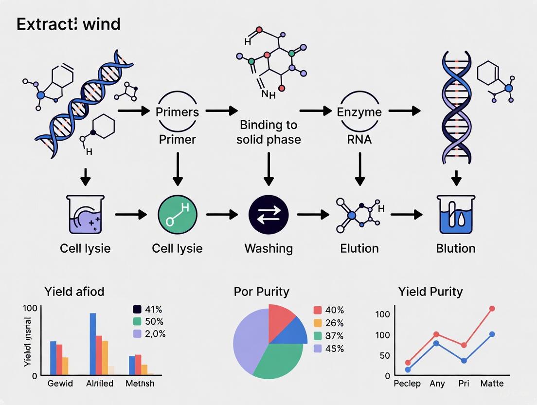
The Core Four-Step Workflow
The following diagram illustrates the universal four-step nucleic acid extraction workflow, from sample input to final elution, including the key actions and objectives at each stage.
Troubleshooting Guides and FAQs
Frequently Asked Questions
Q1: My nucleic acid yield is consistently low. What are the primary factors to investigate?
Low yield can stem from multiple points in the workflow. Systematically check the following:
- Incomplete Lysis: Insufficient lysis is a major cause [8]. Optimize by increasing incubation time with lysis buffer, using a more aggressive lysing matrix (e.g., bead beating for tough tissues), or adding enzymatic digestion with Proteinase K [9] [10].
- Inefficient Binding: Ensure the binding buffer has the correct pH; a lower pH (e.g., ~4.1) can significantly improve DNA binding to silica by reducing electrostatic repulsion [6]. Verify ethanol concentration is correct and that stocks are fresh, as old ethanol can absorb water and skew the working concentration [8]. For high-input samples, increase the volume of the binding matrix (e.g., silica beads) [6].
- Inefficient Elution: Elution efficiency is affected by pH, temperature, and duration [6]. Use a slightly basic elution buffer (e.g., 10 mM Tris-HCl, pH 8.5-9.0) instead of water, as DNA hydrates and dissolves more effectively in buffer. Let the buffer stand on the membrane for a few minutes before centrifugation [8].
Q2: My extracts have low purity (poor A260/A280 or A260/A230 ratios). How can I remove contaminants effectively?
Low purity indicates contamination with protein, salts, or other reagents.
- Protein Contamination (Low A260/A280): This suggests inadequate washing or overloading of the solid phase [8]. Ensure you are not exceeding the binding capacity of your column or beads. Perform the recommended wash steps thoroughly.
- Salt Contamination (Low A260/A230): This is often due to residual chaotropic salts or wash buffers [11] [8]. Ensure wash buffers are prepared with high-quality ethanol. If the problem persists, add an extra wash step with the provided ethanol-based wash buffer. After the final wash, a "dry spin" of the empty column is crucial to remove all residual ethanol [8].
- Specific Sample Inhibitors: For challenging samples like plants (polysaccharides) or blood (heme), use specialized kits that include additives like polyvinylpyrrolidone (PVP) to bind polyphenols or optimized wash buffers to remove specific inhibitors [7] [10].
Q3: I am working with a difficult sample type (e.g., plant, FFPE, blood). What special considerations are needed?
- Plant Tissues: These contain rigid cell walls and secondary metabolites. Use the CTAB (cetyltrimethylammonium bromide) method or a specialized kit with PVP. Grind tissue in liquid nitrogen for efficient lysis [7] [10].
- Formalin-Fixed Paraffin-Embedded (FFPE) Tissues: These require dewaxing and deparaffinization, often with xylene, followed by extended digestion with Proteinase K to reverse cross-links. Automated, non-xylene methods using heating and digestion are also available [10].
- Blood Samples: Use an anticoagulant like EDTA; avoid heparin as it inhibits PCR [9]. For fresh blood, a DNA stabilizer can prevent degradation. Remove protein precipitates by centrifugation before binding to prevent column clogging [9].
Quantitative Data for Method Selection
The table below summarizes key performance metrics from different DNA extraction methods as reported in recent studies, aiding in evidence-based protocol selection.
Table 1: Performance Comparison of DNA Extraction Methods
| Extraction Method | Reported Yield Range | Processing Time | Key Advantages | Common Challenges |
|---|---|---|---|---|
| Phenol-Chloroform [12] | 50-100 ng/µL (from ticks) | Long (>1 hour) | High yield, high purity | Toxic reagents, labor-intensive, safety risks |
| Silica Column [12] [6] | 40-80 ng/µL (from ticks) | Moderate (~25 min) [6] | Good balance of yield and purity, convenient | Can be less efficient with high microbial loads [12] |
| Magnetic Beads [6] [12] | 20-96% of input DNA [6] | Rapid (6-7 min) [6] | Fast, automatable, high throughput | Potential bead carryover, equipment cost [12] |
Experimental Protocols for Optimization
Detailed Methodology: Optimizing Binding Efficiency with Magnetic Silica Beads
This protocol is adapted from the SHIFT-SP method, which focuses on maximizing speed and yield [6].
1. Reagents and Materials:
- Lysis/Binding Buffer (LBB): Contains guanidinium hydrochloride (a chaotropic salt) and Triton X-100, pH adjusted to 4.1.
- Magnetic Silica Beads
- Wash Buffer: Typically an ethanol-based saline solution.
- Elution Buffer (EB): 10 mM Tris-HCl, pH 8.5-9.0.
- Sample: Purified genomic DNA (e.g., from Mycobacterium smegmatis) spiked into LBB for optimization experiments.
2. Equipment:
- Thermonixer or heating block.
- Magnetic separation rack.
- Micro-pipettes.
- qPCR machine for quantification.
3. Step-by-Step Procedure:
- Step 1: Binding.
- Mix the sample (containing 100-1000 ng DNA) with LBB and 10-50 µL of magnetic silica beads in a tube.
- For "tip-based" mixing, repeatedly aspirate and dispense the binding mix for 1-2 minutes. Alternatively, orbital shaking can be used but requires longer incubation (5-15 min) for similar efficiency [6].
- Incubate at 62°C during mixing.
- Step 2: Magnetic Separation and Washing.
- Place the tube on a magnetic rack until the solution clears.
- Carefully aspirate and discard the supernatant.
- Add wash buffer to the bead pellet, resuspend thoroughly and incubate for 30 seconds. Repeat this wash step twice.
- Step 3: Elution.
- After removing the final wash, add the pre-warmed Elution Buffer.
- Resuspend the beads and incubate at 70°C for 1-5 minutes.
- Place the tube back on the magnetic rack and transfer the eluate containing the purified DNA to a new tube.
4. Quantification and Analysis:
- Quantify the extracted DNA using a spectrophotometer or fluorometer.
- To precisely calculate binding and elution efficiency, use a qPCR-based approach [6]:
- Measure the amount of input DNA in the starting sample.
- Measure the amount of DNA left in the supernatant after binding.
- Measure the amount of DNA in the final eluate.
- Binding Efficiency (%) = (Input DNA - Supernatant DNA) / Input DNA × 100
- Elution Efficiency (%) = Eluted DNA / (Input DNA - Supernatant DNA) × 100
The Scientist's Toolkit: Essential Research Reagents
Table 2: Key Reagents for Nucleic Acid Extraction and Their Functions
| Reagent / Material | Core Function | Optimization Tip |
|---|---|---|
| Chaotropic Salts (e.g., Guanidine HCl) [6] [8] | Denature proteins, inactivate nucleases, enable NA binding to silica. | Critical for both lysis and binding steps. |
| Silica Matrix (Membranes/Magnetic Beads) [6] [5] | Solid phase that selectively binds NA in high-salt conditions. | Bead size and surface chemistry impact yield; "tip-based" mixing improves binding kinetics [6]. |
| Proteinase K [5] [10] | Broad-spectrum protease that digests proteins and aids cell lysis. | Most effective in denaturing conditions; essential for tissues and FFPE samples. |
| Ethanol / Isopropanol [5] [8] | Promotes NA binding to silica; used in wash buffers to remove salts. | Use fresh, high-quality stocks; incorrect concentrations impair binding or washing. |
| Elution Buffer (TE Buffer, pH 8-9) [5] [8] | Hydrates and releases NA from the silica matrix in low-salt conditions. | Pre-warming and longer incubation (5 min) increase elution yield, especially for high MW DNA. |
| Inhibitor Removal Additives (e.g., PVP) [7] [10] | Binds to specific contaminants like plant polyphenols. | Add directly to the lysis buffer for samples rich in secondary metabolites. |
Key Workflow Considerations and Pathway
The following diagram outlines the critical decision points and parameters for optimizing each step of the nucleic acid extraction workflow.
The optimization of nucleic acid extraction is a foundational pillar in molecular biology, directly influencing the success of downstream applications in research and drug development. The pursuit of high yield and purity hinges on a detailed understanding of the key chemical agents that facilitate cell lysis, protein denaturation, and nuclease inhibition. This guide provides an in-depth examination of the core reagents—chaotropic salts, EDTA, SDS, and enzymes—framed within the context of troubleshooting and experimental optimization. By elucidating their specific mechanisms and interactions, we aim to empower researchers to systematically refine their protocols and overcome common challenges in nucleic acid purification.
Fundamental Roles and Mechanisms of Action
The following table summarizes the primary functions of these essential chemical agents.
Table 1: Key Chemical Agents in Nucleic Acid Extraction
| Chemical Agent | Primary Function | Mechanism of Action | Typical Concentration/Usage |
|---|---|---|---|
| Chaotropic Salts (e.g., Guanidine HCL, Guanidine Thiocyanate) | Protein denaturation & nucleic acid binding [8]. | Disrupt hydrogen bonds and hydrophobic interactions, denaturing proteins and nucleases; create conditions for nucleic acids to bind to silica membranes [8]. | Component of lysis and binding buffers; often used with ethanol [8]. |
| EDTA (Ethylenediaminetetraacetic acid) | Inhibition of nucleases [13]. | Chelates (binds) metal ions like Mg²⁺ and Ca²⁺, which are essential cofactors for DNase and RNase activity [13]. | 1-10 mM in extraction buffers [13]. |
| SDS (Sodium Dodecyl Sulfate) | Cell lysis and protein denaturation [14]. | Anionic detergent that solubilizes membrane lipids and proteins, aiding in cell lysis; also stimulates Proteinase K activity [14]. | ~0.1-1% (e.g., 10 µL of 10% SDS in a 200 µL lysate) [14]. |
| Enzymes (e.g., Proteinase K) | Digestion of cellular proteins. | Broad-spectrum serine protease that degrades proteins and nucleases, removing contaminants from the nucleic acid preparation [14]. | Concentration varies (e.g., >400 units/mL); incubation at 55°C [14]. |
Synergistic Interactions in a Standard Workflow
The effectiveness of nucleic acid extraction relies on the coordinated action of these reagents. The diagram below illustrates their sequential roles and synergistic relationships in a typical silica spin-column-based protocol.
Troubleshooting Guide: Common Issues and Solutions
This section addresses specific problems related to extraction chemicals, their root causes, and evidence-based solutions.
Table 2: Troubleshooting Common Nucleic Acid Extraction Problems
| Problem | Potential Chemical-Related Cause | Recommended Solution |
|---|---|---|
| Low Yield | Incomplete lysis due to inefficient SDS action or insufficient Proteinase K [15]. | Optimize lysis protocol; ensure correct SDS concentration; verify Proteinase K activity and incubation time/temperature [15]. |
| Inefficient binding to silica matrix [16]. | Ensure binding buffer (containing chaotropic salts) is fresh and mixed with the correct volume/quality of ethanol [8]. | |
| DNA/RNA Degradation | Ineffective nuclease inhibition due to insufficient EDTA or improper sample handling [15]. | Always include EDTA in lysis buffers; keep samples on ice; use nuclease-free consumables [13] [16]. |
| Protein Contamination | Incomplete protein digestion or removal [15]. | Extend Proteinase K digestion time (e.g., by 30 min to 3 hours) after tissue dissolution; ensure SDS is present to stimulate protease activity [15]. |
| Salt Contamination | Carryover of chaotropic salts from binding or wash buffers [15]. | Ensure wash buffers contain the correct ethanol concentration; perform a final "dry spin" with an empty column to evaporate residual ethanol [8] [15]. |
| Inhibition of Downstream Applications | Residual EDTA chelating Mg²⁺ ions essential for PCR [13]. | Use the correct concentration of EDTA (do not over-concentrate); consider a final purification step if excessive EDTA is suspected [13]. |
Frequently Asked Questions (FAQs)
Q1: Why is EDTA so critical in nucleic acid extraction buffers? EDTA plays multiple protective roles. Its primary function is to chelate metal ions like Mg²⁺ and Ca²⁺, which are essential cofactors for DNase and RNase activity. By removing these ions, EDTA significantly reduces nuclease activity, protecting the extracted nucleic acids from degradation [13]. Additionally, it aids in cell lysis by helping to remove metal ions bound to DNA within the cell, improves DNA solubility, and prevents metal-ion-induced precipitation [13].
Q2: My DNA appears intact on a gel, but my PCR fails. Could my extraction chemicals be the cause? Yes. Two common culprits are:
- Residual Chaotropic Salts: Inefficient washing can leave behind chaotropic salts that inhibit the polymerases used in PCR [16] [8]. Ensure wash steps are performed thoroughly.
- Residual EDTA: If the concentration is too high, EDTA can chelate the Mg²⁺ ions in the PCR reaction mix, which are an essential cofactor for Taq polymerase [13]. Using the recommended EDTA concentration in lysis buffers and ensuring proper washing can mitigate this.
Q3: How do chaotropic salts work in silica-based kits? Chaotropic salts have a dual function. First, they denature proteins, including nucleases, by disrupting hydrogen bonds and hydrophobic interactions, thereby stabilizing nucleic acids [8]. Second, they create a high-salt environment that dehydrates nucleic acid molecules and facilitates their selective binding to the silica membrane in spin columns, while proteins and other impurities are washed away [8].
Q4: What is the specific role of SDS when used with Proteinase K? SDS is an anionic detergent that acts synergistically with Proteinase K. It solubilizes cellular membranes and denatures proteins, making them more accessible for digestion by Proteinase K. Furthermore, SDS itself has been shown to stimulate the activity of Proteinase K, leading to more efficient and rapid digestion of cellular proteins [14].
Experimental Data and Protocol Optimization
Quantitative Effects of EDTA and Temperature on DNA Yield
A forensic study on DNA extraction from teeth provides quantitative insights into optimizing EDTA-containing buffers. The research demonstrated that incubating tooth powder with 0.5 M EDTA buffer at different temperatures significantly impacted DNA yield, with optimal results at 56°C [17]. This underscores the importance of temperature optimization during the demineralization and lysis steps when dealing with difficult samples.
Table 3: Effect of EDTA Buffer and Temperature on DNA Yield from Teeth
| Incubation Temperature | Relative DNA Yield | Remarks |
|---|---|---|
| 4°C | Low | Slow demineralization, insufficient for high yield. |
| 18°C | Low | Demineralization remains suboptimal. |
| 37°C | Moderate | Improved yield but not maximal. |
| 56°C | High | Optimal temperature for efficient demineralization and DNA release. [17] |
| 56°C (with distilled water) | Very Low | Highlights the essential role of EDTA. |
Detailed Protocol: Mammalian Tissue Genomic DNA Extraction
The following workflow, adapted from a commercial kit protocol, illustrates the precise points where key chemical agents are utilized to maximize yield and purity [14].
Key Optimizations from the Protocol:
- Tissue Preparation: Mincing tissue into the smallest possible pieces (<25 mg) is critical for efficient lysis and prevents nuclease degradation [15].
- Digestion Conditions: Lysis Buffer containing EDTA is used with Proteinase K and SDS, followed by incubation at 55°C for ~3 hours (or overnight) to ensure complete digestion [14].
- RNA Removal: An optional RNase A digestion step is included to minimize RNA contamination, which is particularly important for DNA-rich tissues like liver and spleen [14] [15].
- Binding Conditions: The addition of Binding Buffer (containing chaotropic salts) and a precise volume of ethanol (200 µL of 96-100%) is essential for maximum binding of DNA to the silica membrane [8] [14].
The Scientist's Toolkit: Essential Research Reagents
This table catalogs the fundamental reagents, their functions, and key considerations for use in optimizing nucleic acid extraction protocols.
Table 4: Essential Reagents for Nucleic Acid Extraction
| Reagent | Function | Key Considerations for Optimization |
|---|---|---|
| Chaotropic Salts (Guanidine HCl/Thiocyanate) | Denatures proteins; enables nucleic acid binding to silica. | Ensure fresh, high-quality ethanol is used in the binding step. Old ethanol stocks can absorb water, reducing effective concentration and binding efficiency [8]. |
| EDTA | Chelates metal ions; inhibits nucleases. | Use at 1-10 mM concentration. Too high a concentration can inhibit downstream PCR by chelating Mg²⁺ [13]. |
| SDS | Ionic detergent for cell lysis and protein denaturation. | Can precipitate in the presence of guanidine isothiocyanate; a subsequent heating step (70°C for 10 min) is often used to re-solubilize it [14]. |
| Proteinase K | Broad-spectrum protease for digesting proteins. | Works effectively in the presence of SDS and EDTA. Incubation temperature (55°C) and time (1-3 hours or more) must be optimized for the sample type [14] [15]. |
| Ethanol | Promotes nucleic acid binding to silica in the presence of chaotropic salts; used in wash buffers. | Must be of high purity (96-100%). Used in wash buffers to remove salts without eluting DNA [8]. |
| RNase A | Degrades RNA to prevent contamination of DNA preparations. | An optional but recommended step for tissues with high RNA content. Ensures accurate DNA quantification and unimpeded downstream applications [14]. |
| Elution Buffer (e.g., Tris-HCl, TE buffer) | Hydrates and releases nucleic acids from the silica membrane. | Slightly basic pH (8-9) is ideal for DNA elution and stability. Allowing the buffer to sit on the membrane for a few minutes before centrifugation can increase yield [8]. |
Nucleic acid extraction is a foundational step in molecular biology, critical for applications ranging from clinical diagnostics to next-generation sequencing. However, the path to obtaining high-yield, high-purity DNA or RNA is often obstructed by the unique biochemical and physical properties of different sample types. Blood contains potent PCR inhibitors, stool is a complex mixture of host and microbial DNA with abundant inhibitors, tissues present structural and enzymatic barriers, and microbial cells have robust walls resistant to lysis. This technical support center provides targeted troubleshooting guides and FAQs to help researchers navigate these sample-specific challenges, all within the broader context of optimizing nucleic acid extraction for superior yield and purity.
Sample-Specific Challenges and Solutions
Blood Samples
Key Challenges: Blood contains potent PCR inhibitors like heme and immunoglobulin, and samples can have low pathogen concentration in a high-background of human DNA [18] [10]. The venepuncture process also introduces contamination risk from skin flora [18].
Optimized Protocol (Magnetic Silica Bead-Based):
- Lysis: Use a lysis binding buffer (LBB) at pH 4.1 rather than pH 8.6. Research shows pH 4.1 achieves 98.2% DNA binding within 10 minutes, compared to 84.3% at 15 minutes for higher pH, by reducing electrostatic repulsion between silica and negatively charged DNA [6].
- Binding: Employ "tip-based" mixing (aspirating and dispensing repeatedly) instead of orbital shaking. For 100ng input DNA, tip-based mixing achieves ~85% binding in 1 minute versus ~61% with orbital shaking [6].
- Elution: Optimize elution buffer pH and temperature. Higher temperatures (62°C) can significantly improve elution efficiency [6].
Troubleshooting FAQ:
- Q: How can I improve DNA yield from low-concentration blood samples?
- A: Implement a method that maximizes binding efficiency. The SHIFT-SP method, which uses tip-based mixing and optimized pH, extracts nearly all nucleic acid in the sample and can process microbes from enriched whole blood for downstream whole genome amplification and sequencing [6].
- Q: My blood DNA extracts show inhibition in downstream PCR. What steps can I take?
- A: Ensure thorough washing steps to remove heme and other inhibitors. Magnetic bead-based methods with optimized chemistries can effectively target genomic DNA while removing sample-specific inhibitors [10].
Stool Samples
Key Challenges: Stool contains complex microbial communities and high levels of PCR inhibitors including bilirubin, complex polysaccharides, and bile salts [10]. The ratio of host to microbial DNA can also be problematic for certain analyses.
Optimized Protocol:
- Homogenization: Use specialized bead tubes in a homogenizer like the Bead Ruptor Elite for mechanical disruption. Optimize speed, cycle duration, and bead type to balance effective disruption with DNA integrity [19].
- Inhibitor Removal: Use specialized kits that incorporate compounds like polyvinylpyrrolidone (PVP) to adsorb polyphenols [10].
- Stabilization: For stored samples, use stabilization media to prevent bacterial growth and degradation. Note that stabilized samples may yield less DNA, so input volume may need adjustment [10].
Troubleshooting FAQ:
- Q: How can I reduce co-extraction of inhibitors from stool samples?
- A: Incorporate multiple wash steps with optimized buffers. Guanidinium thiocyanate-based extractions may result in better inhibitor removal from different sample types than other methods [6].
- Q: My stool DNA yields are low after stabilization in transport media. What should I do?
- A: Consider using two swabs in a single isolation or increasing the input volume. For stool swabs in media, mechanical homogenization may not be needed for microbial DNA identification [10].
Tissue Samples
Key Challenges: Tissues are often fibrous with rigid cell walls that complicate lysis [10]. They also contain abundant nucleases that can degrade DNA, and some tissues are rich in inhibitors like lipids and pigments.
Optimized Protocol:
- Disruption: For fresh/frozen tissue, use freeze-grinding in liquid nitrogen to powder the tissue without repeated freeze-thaw cycles [7].
- Lysis: Use a buffer containing 10mM Tris-Cl (pH 8.0), 100mM EDTA, 0.5% SDS, and 200μg/mL protease K, incubated at 37-55°C until tissue is dissolved [7].
- DNA Protection: Include EDTA in the lysis buffer to chelate metals and inhibit nuclease activity [7] [19].
Troubleshooting FAQ:
- Q: How can I prevent DNA degradation during tissue extraction?
- A: Work quickly, use nuclease inhibitors, and maintain appropriate temperatures. Excessive heating during homogenization can accelerate DNA oxidation and hydrolysis [19].
- Q: What is the best approach for FFPE tissue samples?
Microbial Samples
Key Challenges: Microbial cell walls are robust and resist standard lysis methods. Gram-positive bacteria with thick peptidoglycan layers are particularly challenging. Environmental samples also often contain difficult-to-lyse spores.
Optimized Protocol:
- Lysis: Use bead beating with specialized beads (ceramic, stainless steel) optimized for microbial disruption [19].
- Inhibitor Removal: Guanidinium thiocyanate-based extractions are excellent at denaturing proteins such as DNases and inactivating viruses in samples during NA extraction [6].
- Binding Conditions: For magnetic bead-based methods, ensure proper pH and binding time. Studies show that extending binding time to 2 minutes with increased bead quantity (30-50μL) can achieve >90% binding efficiency for higher DNA inputs [6].
Troubleshooting FAQ:
- Q: How can I improve lysis of tough microbial cells like Gram-positive bacteria?
- A: Implement a combination approach of mechanical and chemical lysis. Bead beating with the Bead Ruptor Elite using optimized bead tubes can efficiently disrupt tough-to-lyse bacterial samples, yielding lysate suitable for downstream analysis [19].
- Q: My microbial DNA extraction from low-biomass samples has high background contamination. How can I address this?
- A: Include appropriate negative controls throughout the process. Studies have shown that bacteria from skin flora and laboratory reagents can contaminate samples, especially in low-microbial biomass environments [18].
Comparative Performance Data
Extraction Method Efficiency
| Extraction Method | Processing Time | Relative DNA Yield | Key Advantages |
|---|---|---|---|
| SHIFT-SP (Bead-Based) [6] | 6-7 minutes | ~100% | Highest speed, automation compatible, efficient for both DNA and RNA |
| Commercial Bead-Based [6] | ~40 minutes | Similar to SHIFT-SP | Established protocol |
| Commercial Column-Based [6] | ~25 minutes | ~50% of SHIFT-SP | Widely accessible |
| Phenol-Chloroform [7] | 60+ minutes | High | High purity, cost-effective for small batches |
| Magnetic Nanoparticles [20] | Not specified | Cost-effective | Cost-effective, developing technology |
Sample-Specific Troubleshooting Guide
| Sample Type | Common Issue | Potential Solution |
|---|---|---|
| Blood [18] [10] | PCR inhibition from heme | Increase wash steps; use inhibitor removal chemistry |
| Stool [10] | Low yield from stabilized samples | Increase input material; extend lysis time |
| Tissue [7] [19] | DNA degradation during processing | Optimize homogenization settings; add nuclease inhibitors |
| Microbes [6] [19] | Incomplete cell lysis | Combine mechanical and chemical lysis methods |
| FFPE Tissue [7] [10] | DNA cross-linking and fragmentation | Incorporate high-temperature protease digestion |
| Plant [7] [10] | Polysaccharide/polyphenol contamination | Add PVP to extraction buffer |
Essential Research Reagent Solutions
| Reagent/Tool | Function | Application Examples |
|---|---|---|
| Magnetic Silica Beads [6] | Nucleic acid binding and separation | SHIFT-SP protocol; automated extraction systems |
| Guanidinium Thiocyanate [6] | Chaotropic salt; denatures proteins, inactivates nucleases | Lysis buffer for blood, microbial samples |
| Polyvinylpyrrolidone (PVP) [10] | Adsorbs polyphenols | Plant extracts; stool samples |
| EDTA (Ethylenediaminetetraacetic acid) [7] [19] | Chelating agent; inhibits nucleases | Tissue lysis buffers; bone demineralization |
| CTAB Buffer [7] | Selective precipitation of nucleic acids | Plant DNA extraction (gold standard) |
| Protease K [7] | Digests proteins; inactivates nucleases | Tissue lysis; FFPE sample processing |
| Magnetic Nanoparticles [20] | Alternative binding matrix for DNA | Cost-effective DNA isolation |
Optimizing nucleic acid extraction across diverse sample types requires both understanding fundamental biochemical challenges and implementing targeted methodological solutions. The protocols and troubleshooting guides presented here provide evidence-based approaches for overcoming the specific barriers presented by blood, stool, tissues, and microbial samples. As extraction methodologies continue to advance—with innovations like the SHIFT-SP method dramatically reducing processing times while maintaining high yields—researchers are better equipped than ever to obtain the high-quality nucleic acids essential for reliable downstream applications in drug development and clinical diagnostics.
A Practical Guide to Modern Nucleic Acid Extraction Techniques
Within the context of optimizing nucleic acid extraction yield and purity, the phenol-chloroform method, often termed organic extraction, maintains a significant legacy position. As molecular biology evolves towards automation and high-throughput systems, understanding this foundational technique remains crucial for researchers, scientists, and drug development professionals. It is historically the most common, tried-and-true method for isolating RNA and removing cellular proteins [21]. The core principle relies on the partitioning of biomolecules between an aqueous phase and an organic phase based on their chemical properties and solubility [22]. Proteins and lipids tend to partition into the organic phase, while nucleic acids, being hydrophilic, remain in the aqueous phase [22]. This process is pH-dependent; for DNA extraction, neutral or slightly alkaline conditions (pH 7-8) are used to ensure DNA remains in the aqueous phase, while acidic conditions favor RNA recovery [21] [22]. Despite the advent of newer methods, organic extraction is still valued for its ability to yield high-purity DNA from complex samples and its cost-effectiveness for large sample volumes [22]. This article details the advantages, limitations, and specific scenarios where this method continues to be the preferred choice for nucleic acid purification.
Core Principles and Comparative Analysis
The Scientific Workflow of Organic Extraction
The organic extraction protocol is a multi-step process designed to separate nucleic acids from other cellular components. The following diagram illustrates the key stages from sample input to final elution, highlighting critical decision points that influence yield and purity.
Diagram 1: The Organic Extraction Workflow. This process involves sample homogenization in a phenol-containing solution, phase separation via centrifugation, careful recovery of the aqueous phase containing nucleic acids, and final precipitation and washing steps to obtain pure DNA or RNA [21] [23].
Quantitative Comparison of Nucleic Acid Extraction Methods
The selection of a nucleic acid extraction method involves trade-offs between yield, purity, scalability, and safety. The following table summarizes the key characteristics of the three primary techniques used in laboratories today, providing a direct comparison for informed decision-making.
Table 1: Comparative Analysis of Common Nucleic Acid Extraction Methods
| Feature | Organic Extraction | Spin Column Method | Magnetic Bead Method |
|---|---|---|---|
| Principle | Liquid-phase partitioning based on solubility in organic solvents [21] [22] | Solid-phase adsorption to a silica membrane [21] | Solid-phase adsorption to silica-coated magnetic beads [21] [6] |
| Typical Yield | High (e.g., 50-100 ng/µL from ticks) [12] | Moderate (e.g., 40-80 ng/µL from ticks) [12] | Variable, can be very high (e.g., 20-70 ng/µL from ticks; ~96% efficiency in SHIFT-SP method) [12] [6] |
| Purity | High, effective protein separation [22] [7] | High [24] [7] | High [24] |
| Throughput | Low, not amenable to automation [21] | High, amenable to 96-well plates [21] | Very high, easily automated [21] [24] |
| Cost | Low (uses common lab reagents) [24] [22] | Moderate (commercial kits) [21] | Higher (specialized beads and equipment) [21] [25] |
| Key Advantage | Gold standard for purity; cost-effective for large volumes [21] [22] | Simple, convenient, ready-to-use kits [21] | Automation, high-throughput, rapid processing [21] [6] [24] |
| Key Disadvantage | Use of toxic chemicals; labor-intensive [21] [22] [23] | Membrane clogging with large/impure samples [21] | Higher cost; bead carryover risk [21] [12] |
The Scientist's Toolkit: Essential Reagents for Organic Extraction
Successful execution of the organic extraction protocol requires a set of specific reagents, each serving a critical function in the separation and purification process.
Table 2: Key Research Reagent Solutions for Organic Extraction
| Reagent | Function | Key Consideration |
|---|---|---|
| Phenol | Denatures proteins by disrupting hydrogen bonds, allowing nucleic acids to remain in the aqueous phase [22]. | pH is critical: acidic for RNA, neutral/alkaline for DNA extraction [21] [22]. |
| Chloroform | Enhances phase separation and promotes partitioning of proteins and lipids into the organic phase [22]. | Often mixed with isoamyl alcohol (24:1) to reduce foaming [22] [23]. |
| SDS (Sodium Dodecyl Sulfate) | Ionic detergent that solubilizes cell and nuclear membranes, facilitating the release of intracellular contents [22] [23]. | Effective in combination with Proteinase K for comprehensive lysis [23]. |
| Proteinase K | Broad-spectrum serine protease that digests and inactivates nucleases, protecting nucleic acids from degradation [7] [23]. | Requires incubation at 37-55°C for several hours for complete digestion [7]. |
| Chaotropic Salt (e.g., Guanidine) | Denatures proteins and inactivates nucleases (e.g., DNases, RNases); used in some lysis buffers [6]. | Guanidinium thiocyanate-based extractions can improve inhibitor removal [6]. |
| Ethanol / Isopropanol | Precipitates nucleic acids from the aqueous solution after extraction, aiding in their recovery and concentration [22] [23]. | Used ice-cold; isopropanol is preferred for precipitating lower concentrations of DNA [23]. |
Troubleshooting Common Experimental Challenges
Even with a well-established protocol, researchers can encounter issues that compromise yield and purity. This section addresses frequent challenges and provides evidence-based solutions.
FAQ 1: An emulsion forms during the phase separation step, preventing a clean separation. How can I resolve this?
Emulsion formation is a common problem, often occurring when samples contain high amounts of surfactant-like compounds such as phospholipids, free fatty acids, proteins, or triglycerides [26].
- Prevention is key: Gently swirl or invert the tube instead of shaking it vigorously. This reduces agitation while maintaining the surface area for extraction [26].
- Salting Out: Add brine or saturated salt solution (e.g., NaCl) to increase the ionic strength of the aqueous layer. This can force the surfactant-like molecules to separate into one phase or the other, breaking the emulsion [26].
- Centrifugation and Filtration: Centrifuging the sample can help isolate the emulsion material. Alternatively, the emulsion can be filtered through a glass wool plug or a highly silanized phase separation filter paper designed to allow one phase to pass through [26].
- Alternative Solvents: Adding a small amount of a different organic solvent can adjust the solvent properties and break the emulsion [26].
- Alternative Method: For samples consistently prone to emulsions, consider Supported Liquid Extraction (SLE), which uses a solid support (e.g., diatomaceous earth) to create an interface for extraction, thereby avoiding emulsion formation altogether [26].
FAQ 2: My final DNA yield is consistently low. What are the potential causes and optimizations?
Low yield can stem from incomplete precipitation, inefficient phase separation, or sample handling errors.
- Ensure Complete Precipitation: Use ice-cold ethanol or isopropanol and ensure sufficient incubation time at -20°C (e.g., overnight) to maximize DNA precipitation [23] [27]. The addition of carriers like glycogen can also improve the recovery of微量 nucleic acids [25].
- Optimize Phase Separation: During the aqueous phase transfer, take extreme care not to disturb the protein interphase. Even a small amount of contamination can lead to protein carry-over that interferes with downstream precipitation [21] [23]. Using phase-lock gel tubes can make this process easier and more reliable [25].
- Verify pH Conditions: For DNA extraction, the phenol must be equilibrated to a neutral or slightly basic pH (7-8). An incorrect pH can cause DNA to partition into the organic phase or interphase, drastically reducing yield [21] [22].
- Improve Lysis Efficiency: For difficult-to-lyse samples (e.g., ticks, plant tissues, Gram-positive bacteria), incorporate a mechanical disruption step such as bead-beating with glass beads. A study on DNA extraction from stool parasites found that adding a bead-beating step to the phenol-chloroform protocol significantly improved DNA yield and subsequent PCR detection rates [27].
FAQ 3: My nucleic acid sample is contaminated with protein. How can I improve purity?
Protein contamination is typically indicated by a low A260/A280 ratio in spectrophotometric analysis.
- Repeat Extraction: The most direct solution is to perform a second round of extraction on the recovered aqueous phase. Add an equal volume of chloroform:isoamyl alcohol (24:1), mix, centrifuge, and carefully recover the aqueous phase. This step removes residual phenol and any remaining protein [23].
- Ensure Proper Initial Ratios: Confirm that the volumes of the sample lysate and the phenol-chloroform mixture are equal. An incorrect ratio can lead to incomplete protein removal [23].
Legacy Uses and Modern Research Applications
Despite its manual nature, organic extraction remains the preferred or required method in several advanced research contexts due to its unparalleled ability to handle complex samples and deliver high yields.
- Virome Studies: The phenol-chloroform DNA extraction protocol is currently the gold standard for studying viral metagenomes (viromes) from diverse environments like human gut, animal microbiomes, and soil [25]. Its effectiveness in extracting viral nucleic acids from these complex samples makes it a benchmark against which newer kit-based methods are validated [25].
- Challenging Sample Types: For samples that are rich in PCR inhibitors or have resilient structures (e.g., tick exoskeletons, fungal spores, plant tissues rich in polysaccharides and polyphenols), organic extraction often provides superior yields. A systematic review on DNA extraction from ticks noted that phenol-chloroform extraction achieved high DNA yields (50-100 ng/µL), albeit with safety risks and longer processing times [12].
- High-Purity Requirements: In applications where the highest possible purity is critical for downstream processes, such as long-read sequencing, cloning, or sensitive enzymatic assays, the rigorous purification offered by organic extraction is often favored over other methods [22] [7]. It is also widely used in proteomic studies for protein extraction and purification, where organic solvents like phenol-acetone are used to denature and precipitate proteins away from contaminants [22].
The following decision tree can help researchers determine when organic extraction is the most appropriate choice for their experimental goals.
Diagram 2: Nucleic Acid Extraction Method Selection Guide. This flowchart assists in selecting the optimal extraction method based on sample type, research priorities, throughput, safety, and budget [21] [12] [25].
Troubleshooting Guides
Common Issues and Solutions for Solid-Phase Extraction (SPE)
| Problem | Common Cause | Solution |
|---|---|---|
| Low Recovery Rate [28] | Target compound is insufficiently retained on the adsorbent. [28] | Adjust the sample pH to neutralize the analyte's charge for better retention on non-polar sorbents; for ion-exchange, ensure pH favors analyte charging. [29] |
| Target compound is not completely eluted. [28] | Use a stronger elution solvent; adjust pH to ionize the compound; add an organic modifier; use multiple elution aliquots with a soak step. [30] [29] | |
| Poor Reproducibility [28] | Column bed dries out before sample loading. [28] | Do not let the cartridge run dry after conditioning; re-condition if unsure. [29] |
| Sample flow rate is too fast. [28] | Process samples at a slower flow rate, typically around 1 mL/min, and as low as 100 µL/min for ion-exchange mechanisms. [30] | |
| Undesirable Purification Effect [28] | Incorrect purification mode or lack of selectivity. [28] | Use a selective sorbent (e.g., ion-exchange > normal phase > reversed-phase); optimize wash and elution solvent strengths. [28] [30] |
| Slow Flow Rate [28] | Sample contains particulate impurities. [28] | Centrifuge or filter the sample solution before loading. [28] |
| Sample viscosity is too high. [28] | Dilute the sample with a compatible solvent before loading. [28] | |
| Analyte Breakthrough | Sorbent is not properly conditioned and equilibrated. [30] | Condition with methanol followed by water or a buffer that matches the sample's pH and ionic strength. [30] [29] |
| Flow rate during loading is too high. [31] | Load the sample at a slow, controlled flow rate without applying pressure initially. [29] |
Common Issues and Solutions for Nucleic Acid Spin Kits
| Problem | Common Cause | Solution |
|---|---|---|
| Low Yield | Insufficient cell or tissue lysis. [32] [33] | Increase digestion or homogenization time; optimize lysis protocol for your sample type. [32] [33] |
| Column is overloaded with sample. [32] [34] | Reduce the amount of starting material to within the kit's specifications. [32] [34] | |
| Inefficient binding of nucleic acids to the silica membrane. [33] | Ensure the binding buffer is correct; optimize incubation time and mixing. [33] | |
| Nucleic Acid Degradation | Action of nucleases in the sample. [32] [33] [34] | Use nuclease-free reagents; work quickly on ice; use RNase inhibitors for RNA; flash-freeze samples after collection. [32] [33] [34] |
| Poor sample storage. [34] | Flash-freeze tissue samples in liquid nitrogen and store at -80°C; use RNA/DNA stabilization reagents. [32] [34] | |
| Salt Contamination (Low A260/230) | Carryover of guanidine salts from binding buffer. [32] | Ensure wash buffers are used; do not let the column contact flow-through; centrifuge again if needed; blot collection tube rim if reused. [32] |
| Protein Contamination (Low A260/280) | Incomplete protein digestion or removal. [32] [34] | Ensure Proteinase K digestion is complete; centrifuge lysate to pellet debris before loading onto the column. [32] [34] |
| DNA Contamination in RNA Preps | gDNA carryover. [32] | Perform an on-column or in-solution DNase I digestion step. [32] |
Frequently Asked Questions (FAQs)
1. How do I choose the right sorbent for my SPE application? The choice of sorbent is critical and depends on your analyte and sample matrix. The general selectivity rule is Ion-Exchange > Normal Phase > Reversed Phase [28]. For non-polar analytes, use reversed-phase sorbents (e.g., C18). For polar compounds, normal-phase sorbents are suitable. For ionizable compounds, ion-exchange sorbents are highly selective [30] [31]. Knowing your analyte's logP/logD and pKa values is invaluable for this decision [30].
2. Why is conditioning and equilibration of the SPE column necessary? Conditioning (typically with methanol or acetonitrile) prepares or "wets" the sorbent surface, ensuring maximum capacity and interaction with your analyte [29]. The subsequent equilibration step (with water or a buffer similar to your sample) ensures the sorbent environment matches the sample solvent. This prevents the sample from being repelled by the sorbent and minimizes the risk of analyte breakthrough [30] [29].
3. My nucleic acid yield is low but the lysis looked complete. What could be wrong? A common cause is inefficient binding of the nucleic acids to the silica membrane. Confirm that your binding buffer is prepared correctly and has the right pH. Also, ensure adequate incubation and mixing time after adding the binding buffer to the lysate to allow the nucleic acids to bind efficiently to the silica matrix [33].
4. My extracted nucleic acids are degraded. How can I prevent this? Degradation is often due to nucleases. Always use nuclease-free tips and tubes. Work quickly and on ice whenever possible. For RNA, use specific RNase inhibitors. When collecting samples, especially tissues, flash-freeze them immediately in liquid nitrogen and store at -80°C to preserve integrity [32] [33] [34].
5. How can I improve the purity of my nucleic acids for sensitive downstream applications? Ensure you are using the correct volumes of wash buffers and that they contain the appropriate ethanol concentration. Before elution, make sure wash buffers are completely removed. Consider an additional wash step or extending the centrifugation time for the final wash. For DNA contamination in RNA, incorporate a DNase I digestion step [32].
Experimental Protocols & Optimization
Method for Optimizing SPE Wash and Elution Solvents
1. Objective: To determine the optimal solvent strength for washing and eluting your target analyte from an SPE cartridge, maximizing recovery while minimizing co-elution of impurities [28] [30].
2. Methodology:
- For Reversed-Phase Mode:
- Load your target compound in an aqueous solution onto the conditioned SPE cartridge.
- Elute the cartridge with a series of methanol-in-water solutions of increasing concentration (e.g., 5%, 10%, 20%, 30%, 40%, 50%, 60%, 70%, 80%, 90%, 100%).
- Collect each eluate fraction separately and analyze them to determine the methanol concentration at which your compound just begins to elute and when it is completely eluted [28].
- For Normal-Phase Mode:
- Load your target compound in a non-polar solvent like n-hexane.
- Elute with a series of n-hexane and methyl tert-butyl ether (MTBE) with increasing MTBE concentration.
- Collect and analyze fractions as above [28].
3. Optimization:
- The wash solvent should have a strength just below the point where your analyte begins to elute. This will remove impurities without losing your target [30].
- The elution solvent should have a strength just above what is required to completely elute your analyte. This avoids using an excessively strong solvent that would elute more impurities [28] [30].
On-Column DNase I Digestion for RNA Purification
1. Objective: To remove genomic DNA contamination during RNA extraction using a spin kit [32].
2. Detailed Workflow:
- After loading the sample lysate onto the spin column and washing it with the appropriate wash buffer, prepare the DNase I reaction mix.
- Pipette the DNase I solution directly onto the center of the silica membrane.
- Incubate the column at room temperature for 15 minutes, as specified in the kit protocol.
- After incubation, perform a wash step with the kit's wash buffer to remove the DNase I enzyme and any digested DNA fragments.
- Proceed with the final wash steps and elution as normal [32].
Key Workflow Diagrams
SPE Method Development Logic
Nucleic Acid Spin Column Workflow
The Scientist's Toolkit: Essential Research Reagents & Materials
| Item | Function & Application |
|---|---|
| C18 Sorbent | A reversed-phase sorbent ideal for extracting non-polar to moderately polar organic compounds from aqueous matrices. A versatile and common choice. [35] [31] |
| Ion-Exchange Sorbents (SAX, SCX) | Selective sorbents for extracting ionizable analytes. SAX (Strong Anion Exchange) for acids; SCX (Strong Cation Exchange) for bases. Offers high selectivity. [28] [35] |
| Silica-based Sorbents | The foundation for many functionalized sorbents (C18, C8, etc.). The silica particle size and pore structure impact efficiency and flow. [35] |
| Proteinase K | A broad-spectrum serine protease used in nucleic acid extraction to digest histones and other cellular proteins, degrading nucleases and helping to release nucleic acids. [32] [34] |
| RNase A & DNase I | Specific nucleases for removing unwanted nucleic acids. RNase A removes RNA from DNA preparations, while DNase I removes genomic DNA from RNA preparations. [32] [34] |
| Binding Buffer (Guanidine Salts) | A key component in spin kits. High concentrations of chaotropic salts (e.g., guanidine thiocyanate) disrupt the hydration shell of nucleic acids, allowing them to bind efficiently to the silica membrane. [32] [34] |
| Wash Buffer (Ethanol) | Typically contains ethanol or another appropriate solvent. It removes salts, metabolites, and other contaminants from the silica membrane while leaving the nucleic acids bound. [32] |
Core Principles of Magnetic Bead-Based Separation
Magnetic bead-based systems utilize superparamagnetic particles to isolate and purify biomolecules like nucleic acids or proteins from complex samples. The core principle involves a magnetic core (often iron oxide) that becomes magnetized only in the presence of an external magnetic field, preventing self-aggregation and allowing redispersion when the field is removed [36]. This magnetic core is surrounded by a protective coating and a functionalized surface with reactive groups (e.g., carboxyl, amine, or streptavidin) that enable specific binding to target molecules [37] [36].
The workflow operates through a series of controlled buffer conditions. Under high-salt, chaotropic conditions (e.g., with guanidine isothiocyanate), nucleic acids bind specifically to the functionalized bead surface. When placed near a magnet, the beads are immobilized against the tube or plate wall, allowing impurities to be washed away. Finally, the purified nucleic acids are eluted in a low-salt buffer or water [37] [38]. This mechanism provides a solid-phase extraction method that eliminates the need for centrifugation, making it particularly suitable for automation and high-throughput applications [39] [38].
Standard Workflow for Nucleic Acid Extraction
The following diagram illustrates the generalized workflow for magnetic bead-based nucleic acid extraction, from sample preparation to final elution.
Detailed Protocol for High-Throughput Nucleic Acid Extraction
Sample Lysis and DNA Binding:
- Add 25 mL of pre-warmed (65°C) CTAB extraction buffer to 30 mL of sample. The buffer should contain chaotropic salts like 1 M guanidine isothiocyanate (GITC) to facilitate DNA binding [37].
- Mix the solution at 45 rpm for 3 hours using a digital rotator to ensure proper interaction between the DNA and magnetic beads [37].
- Centrifuge at 12,000 × g for 10 minutes at 20°C to separate phases. Carefully remove the upper oily phase (supernatant) [37].
DNA Precipitation and Purification:
- Add an acryl carrier solution (5 μL per 1 mL sample), followed by 1/10 volume of 3 M sodium acetate (pH 5.2) and an equal volume of isopropanol [37].
- Incubate overnight at -20°C to precipitate DNA. Centrifuge at 16,000 × g for 20 minutes at 4°C, discard supernatant, and resuspend the pellet in 1 mL of specific solution [37].
- Add 0.2 mg of carboxyl-modified magnetic beads (300 nm diameter) to the aqueous DNA solution, followed by a specific volume of ethanol/isopropanol. Mix gently by inversion and incubate for 10 minutes to facilitate DNA binding [37].
Washing and Elution:
- Separate the beads using a Magnetic Separator Stand and wash twice with 1 mL of 70% ethanol (v/v) to remove impurities [37].
- Air-dry beads at room temperature for 3-5 minutes to evaporate residual ethanol [37].
- Elute DNA by adding 100 μL of TE buffer (10 mM Tris, 1 mM EDTA-2Na, pH 8.0), mixing thoroughly, and incubating at 60°C for 10 minutes. Separate beads until the TE buffer is clear and transfer eluted DNA to a clean storage tube [37].
Troubleshooting Common Experimental Issues
Low Nucleic Acid Yield
- Cause: Suboptimal bead-to-sample ratio or insufficient mixing during binding [40].
- Solution: Ensure accurate pipetting and reagent proportions. Use a sample mixer (e.g., HulaMixer) during incubation steps to maintain consistent suspension and maximize binding surface contact [40].
- Cause: Incomplete elution due to inadequate buffer volume or contact time.
- Solution: Ensure beads are fully resuspended in elution buffer. Pre-warm elution buffer to 60°C and increase incubation time to 10 minutes to enhance DNA release [37].
Poor Nucleic Acid Purity
- Cause: Residual contaminants from incomplete washing [39].
- Solution: Incorporate additional wash steps with 70% ethanol. For challenging samples like tissues, introduce an extra chloroform extraction step during sample preparation to remove lipids and proteins effectively [39].
- Cause: Contaminated reagents or buffers.
- Solution: Prepare fresh ethanol wash buffers daily and filter-sterilize solutions to prevent nuclease or particulate contamination [39].
Inconsistent Results Between Samples
- Cause: Variable bead sedimentation during processing [40].
- Solution: Standardize mixing procedures across all samples. Use an orbital shaker or rotator that provides consistent, gentle agitation to maintain beads in uniform suspension [40].
- Cause: Improper storage or handling of magnetic beads.
- Solution: Store beads according to manufacturer specifications. Avoid repeated freezing and thawing, and vortex beads thoroughly before use to ensure homogeneous dispersion [41].
Beads Not Binding Target Molecules Effectively
- Cause: Degraded or low-quality magnetic beads [41].
- Solution: Check expiration dates and storage conditions. Verify bead integrity and binding capacity using a control sample with known nucleic acid concentration [41].
- Cause: Incorrect buffer pH or composition interfering with binding chemistry [41].
- Solution: Ensure coupling buffers are at correct pH (typically pH 6.0 for carboxyl-modified beads) and concentration. Avoid buffers containing primary amines (like Tris) when using amine-reactive coupling chemistries [37] [41].
Research Reagent Solutions
The table below details essential materials and their functions in magnetic bead-based workflows.
| Reagent/Material | Function in Workflow |
|---|---|
| Carboxyl-Modified Magnetic Beads | Solid-phase support for nucleic acid binding under high-salt conditions; 300 nm size optimal for DNA recovery [37]. |
| Guanidine Isothiocyanate (GITC) Buffer | Chaotropic agent that denatures proteins and facilitates nucleic acid binding to magnetic beads [37]. |
| CTAB (Cetyltrimethylammonium bromide) Buffer | Facilitates transfer of DNA from organic to aqueous phase, particularly useful for complex samples like plant tissues or oils [37]. |
| Proteinase K | Enzyme that digests proteins and nucleases, eliminating contaminants and protecting nucleic acids from degradation [42]. |
| RNAse A | Removes contaminating RNA from DNA preparation, ensuring pure DNA extraction without RNA interference [42]. |
| Ethanol (70%) | Wash solution that removes salts and other impurities while maintaining nucleic acid binding to beads [37]. |
| TE Buffer (Tris-EDTA) | Elution solution that chelates magnesium to inhibit DNase activity and stabilizes nucleic acids for long-term storage [37]. |
| Sodium Acetate (3M) | Facilitates ethanol precipitation of nucleic acids, enhancing recovery during concentration steps [37]. |
Performance Optimization Data
The table below summarizes key performance metrics from optimized magnetic bead-based extraction protocols, demonstrating their efficiency for high-throughput applications.
| Application/Matrix | Recovery Efficiency | Purity (A260/A280) | Key Optimized Parameter |
|---|---|---|---|
| Refined Soybean Oil [37] | 76.37% | ~2.0 (PCR compatible) | 1 M GITC buffer, pH 6.0 with 300 nm carboxyl beads |
| Non-Human Primate Tissues [39] | Significantly increased with modified protocol | ~2.0 (improved with chloroform) | Additional chloroform and ethanol steps |
| PCR Product Purification [38] | Excellent, especially for small fragments | Suitable for sequencing/cloning | Automation-compatible 96-well plate format |
| High-Throughput Automated RNA [39] | Varies by kit; improved with protocol modification | ~2.0 across kits | Standardized protocol for interlaboratory comparisons |
Frequently Asked Questions (FAQs)
What are the advantages of magnetic bead-based systems over spin columns for high-throughput labs?
Magnetic bead systems enable full automation on robotic liquid-handling platforms, support 96- or 384-well plate formats, and eliminate centrifugation steps. This significantly reduces hands-on time while providing excellent recovery rates, particularly for small fragments or low-concentration samples [38].
How can I improve nucleic acid yield from difficult sample types like refined oils or fatty tissues?
Implement a CTAB-based phase separation step before magnetic bead purification. This effectively transfers DNA from the oil phase to the aqueous phase. Additionally, use carboxyl-modified magnetic beads (300 nm) with optimized GITC buffer conditions (1 M, pH 6.0) to maximize recovery from challenging matrices [37].
Why is my nucleic acid purity insufficient for downstream applications like qPCR?
Incorporate additional purification steps such as chloroform extraction to remove co-precipitating contaminants. Ensure wash buffers are fresh and applied in sufficient volumes. For automated systems, verify that the washing steps are thoroughly replacing previous solutions without cross-contamination [39].
Can magnetic bead-based systems be fully automated?
Yes, magnetic bead systems are ideal for automation. Platforms like the KingFisher Flex can be integrated with pipetting robots (e.g., ASSIST PLUS) to create walk-away purification workflows. Specialized modules like MAG and HEATMAG provide magnetic separation with heating capabilities for streamlined processing [43] [39].
How critical is bead size selection for extraction efficiency?
Bead size significantly impacts performance. For DNA extraction from refined oils, 300 nm carboxyl-modified beads demonstrated optimal DNA adsorption compared to 100 nm or 500 nm beads. The larger surface-to-volume ratio of appropriately sized beads enhances biomolecular adsorption efficiency [37] [36].
What quality control measures should I implement for consistent results?
Regularly verify bead binding capacity using control samples. Monitor extraction efficiency with internal positive controls (IPC) like VetMAX Xeno IPC RNA. Track nucleic acid purity through absorbance ratios (A260/A280) and confirm integrity via electrophoretic analysis [39].
In the context of optimizing nucleic acid extraction for molecular diagnostics and research, rapid protocols like boiling and heat-shock methods offer significant advantages in speed and cost-effectiveness. These techniques leverage high temperatures to disrupt cell membranes and denature proteins, facilitating the release of nucleic acids without the need for complex purification systems. Framed within a broader thesis on extraction optimization, this technical support center addresses the practical challenges and methodological refinements necessary to implement these protocols successfully. While ideal for high-throughput screening and resource-limited settings, these methods require precise troubleshooting to ensure yield and purity compatible with downstream applications like PCR and sequencing [44] [45].
Troubleshooting Guides
Common Problems and Solutions for Boiling Protocols
Problem: Low Nucleic Acid Yield
- Potential Causes and Solutions:
- Incomplete Cell Lysis: Ensure thorough sample homogenization prior to boiling. For tough samples like tissue or spores, combine mechanical disruption (e.g., bead beating) with the boiling step [19].
- Suboptimal Boiling Duration or Temperature: Verify the incubation time and temperature. Typical protocols use 95-100°C for 10-15 minutes. Use a calibrated heat block for consistent results [45].
- Insufficient Starting Material: Increase the sample input volume within the limits of the protocol, ensuring it does not exceed the buffer's capacity.
- Nuclease Degradation: Add chelating agents like EDTA (e.g., in TE buffer) to the boiling buffer to inhibit nucleases that may remain active after heating [19].
Problem: PCR Inhibition or Failure
- Potential Causes and Solutions:
- Carry-over of PCR Inhibitors: Boiling methods do not purify nucleic acids. Inhibitors like hemoglobin, polysaccharides, or ionic detergents can co-precipitate. Centrifuge the boiled sample at high speed (e.g., 14,000 rpm for 5 minutes) and use only the supernatant for downstream assays [44].
- Excessive Sample Background: For blood-contaminated samples, a pre-wash with PBS or a dilute detergent solution can reduce interference. Note that magnetic bead methods show superior resistance to hemoglobin inhibition [44].
- Inadequate Dilution: The crude nucleic acid extract may contain high levels of background. Perform a dilution series of the extract to find a concentration that minimizes inhibition in the PCR reaction.
Problem: Inconsistent Results Between Replicates
- Potential Causes and Solutions:
- Non-uniform Heating: Avoid using water baths, which can cause temperature gradients and cross-contamination. Use a calibrated digital dry bath or heat block [45].
- Improper Sample Handling: After boiling, briefly centrifuge the tube to collect all condensation before opening. Always pipette from the same consistent depth of the supernatant.
- Variability in Sample Lysis: Standardize the pre-boiling sample preparation step. For solid tissues, ensure uniform powdering under liquid nitrogen [19].
Frequently Asked Questions (FAQs)
Q1: What are the primary advantages and disadvantages of the boiling method compared to commercial column or bead-based kits?
A1: The boiling method is rapid, cost-effective, and technically straightforward, requiring minimal equipment. It is suitable for PCR-based screening in high-volume settings [45]. However, its main disadvantages are lower purity and higher susceptibility to inhibitors. Studies show that while the boiling method fails to detect HPV when hemoglobin concentration exceeds 30 g/L, magnetic bead methods remain effective even at 60 g/L [44]. Commercial kits provide higher purity nucleic acids, better consistency, and are more automation-friendly, but at a higher cost per sample and with longer processing times.
Q2: How can I improve the purity of my DNA obtained via a boiling protocol without switching to a column-based method?
A2: Several modifications can enhance purity:
- Use of Chelating Resins: Incorporating Chelex-100 resin in the boiling buffer significantly improves yield and purity by chelating metal ions that are cofactors for nucleases. One study on dried blood spots found that a Chelex boiling method yielded significantly higher DNA concentrations than several column-based kits [45].
- Optimized Buffers: Replacing simple water or TE buffer with a specialized buffer containing detergents (e.g., Tween 20) and chelating agents can improve lysis efficiency and nuclease inactivation [45].
- Post-Boiling Purification Steps: A simple alcohol precipitation or a clean-up with pre-made magnetic beads can be added to the protocol to remove contaminants without committing to a full commercial kit [6].
Q3: My downstream quantitative PCR (qPCR) results are variable when using crude boiling extracts. How can I improve reproducibility?
A3: Reproducibility is a known challenge with crude extracts. To improve it:
- Introduce an Internal Control: Spike a known amount of exogenous DNA or an internal amplification control into the sample before lysis. This allows you to distinguish between true target variation and inhibition or extraction inefficiency.
- Normalize Sample Input: Use a standardized metric for input material (e.g., cell count, tissue weight) rather than volume alone.
- Optimize Elution Volume: Consistently using a small, fixed elution volume (e.g., 50 µL) can increase the final DNA concentration and improve pipetting accuracy for downstream setup [45].
Q4: Are boiling methods suitable for RNA extraction?
A4: Boiling is generally not recommended for RNA extraction due to the high susceptibility of RNA to degradation by RNases. Most RNases are very heat-stable and can quickly degrade RNA upon cooling after the boil. RNA extraction requires specialized, RNase-inhibiting reagents and conditions. For rapid RNA extraction, dedicated commercial kits that include guanidinium thiocyanate-based lysis buffers are the preferred choice [46].
Comparative Performance Data
The following tables summarize key quantitative findings from recent studies comparing boiling methods with other extraction techniques.
Table 1: Anti-Interference Performance against Hemoglobin (Simulated Blood Contamination)
| Hemoglobin Concentration (g/L) | Boiling Method (HPV Detection) | Magnetic Bead Method (HPV Detection) |
|---|---|---|
| 20 g/L | Positive | Positive |
| 30 g/L | Negative | Positive |
| 60 g/L | Negative | Positive |
Data adapted from a study comparing nucleic acid extraction methods for HPV genotyping. The magnetic bead method demonstrated significantly higher resistance to PCR inhibitors like hemoglobin [44].
Table 2: Comparative Analysis of DNA Extraction Methods from Dried Blood Spots (DBS)
| Extraction Method | Relative DNA Yield (by qPCR) | Key Characteristics |
|---|---|---|
| Boiling with Chelex-100 Resin | Highest | Cost-effective, rapid, easy, suitable for large-scale studies [45] |
| Roche High Pure Column Kit | High | Significantly higher than other column kits [45] |
| QIAGEN QIAamp DNA Mini Kit | Low | Standardized but costly [45] |
| QIAGEN DNeasy Blood & Tissue Kit | Low | Standardized but costly [45] |
| Boiling with TE Buffer | Not Reported | Simple but may yield lower purity [45] |
Table 3: Large-Scale HPV Detection Rate and Cost-Benefit Comparison
| Parameter | Boiling Method | Magnetic Bead Method |
|---|---|---|
| Positive Detection Rate | 10.02% | 20.66% |
| Cost Relative to Boiling | Baseline (0%) | +13.14% |
| Detection Rate Increase | Baseline (0%) | +106.19% |
Data from a longitudinal large-scale analysis (16,540 cases). The magnetic bead method, while slightly more expensive, provided a dramatically higher detection rate, making it highly cost-effective for clinical diagnostics [44].
Experimental Protocols
Detailed Method: Chelex-100 Boiling Protocol for Dried Blood Spots
This protocol, optimized for DNA extraction from Dried Blood Spots (DBS), is noted for its high yield and cost-effectiveness [45].
Research Reagent Solutions:
- Chelex-100 Resin: A chelating resin that binds metal ions, inactivating nucleases and improving DNA stability.
- PBS (Phosphate-Buffered Saline): A balanced salt solution used for washing and maintaining osmotic balance.
- Tween 20: A non-ionic detergent that aids in cell lysis and washing away impurities.
Procedure:
- Punch and Soak: Punch one 6 mm disk from the DBS and place it in a 1.5 mL microcentrifuge tube. Add 1 mL of 0.5% Tween 20 solution in PBS.
- Overnight Incubation: Incubate the tube at 4°C for a minimum of 8 hours (e.g., overnight) to elute cells from the paper.
- Wash: Carefully remove and discard the Tween20 solution. Add 1 mL of pure PBS to the punch and incubate at 4°C for 30 minutes. After incubation, remove and discard the PBS.
- Chelex Boiling: Add 50 µL of a pre-heated 5% (w/v) Chelex-100 suspension to the punch. Pulse-vortex the mixture for 30 seconds.
- Heat Shock: Incubate the tube at 95°C for 15 minutes. During this incubation, pulse-vortex the tube briefly every 5 minutes.
- Pellet Debris: Centrifuge the tube at 11,000 x g for 3 minutes to pellet the Chelex beads and paper debris.
- Recover Supernatant: Carefully transfer the supernatant (containing the DNA) to a new microcentrifuge tube. For maximum recovery, the centrifugation and transfer steps can be repeated.
- Storage: Store the extracted DNA at -20°C until use. The DNA is suitable for direct use in PCR and qPCR [45].
Detailed Method: Standard Boiling Protocol for Cervical Swabs
This is a simpler protocol used for releasing DNA from cervical swab samples, typically included with commercial PCR kits [44].
Procedure:
- Sample Preparation: Mix the specimen tube thoroughly. Transfer 300 µL into a 1.5 mL microcentrifuge tube.
- Pellet Cells: Centrifuge the tube at 14,000 rpm for 3 minutes. Carefully discard the supernatant.
- Resuspend and Boil: Add 200 µL of nucleic acid extraction reagent (typically containing Chelex or similar agents) to the pellet and mix thoroughly.
- Incubate: Incubate the tube in a 100°C heat block for 15 minutes.
- Clarify: Centrifuge the tube at 14,000 rpm for 5 minutes.
- Recover DNA: Use 5 µL of the clear supernatant as the template for PCR amplification [44].
Workflow and Pathway Diagrams
Protocol Decision Pathway
This diagram outlines the decision-making process for selecting and troubleshooting a rapid nucleic acid extraction protocol, based on sample type and experimental goals.
Boiling Protocol Optimization Parameters
This workflow visualizes the key parameters that can be optimized in a standard boiling protocol to improve nucleic acid yield and quality.
Troubleshooting Guides
This guide addresses common challenges in fully automated nucleic acid extraction platforms, helping you optimize yield and purity for your research and drug development workflows.
FAQ 1: Why is my nucleic acid yield low after automated extraction?
Low yield can stem from several issues, from sample preparation to instrument operation.
| Problem Cause | Specific Examples | Recommended Solution |
|---|---|---|
| Incomplete Lysis | - Large tissue pieces [47] [7].- Insufficient lysis time or incorrect buffer [48]. | - For tissues: Grind frozen samples in liquid nitrogen or cut into smallest pieces [47] [7].- Optimize lysis protocol (mechanical, chemical, enzymatic) for your sample type [48]. |
| Inefficient Binding | - Incorrect binding buffer pH [6].- Insufficient mixing during binding [6].- Overloaded solid phase (beads/column) [47]. | - Use a binding buffer with lower pH (e.g., ~4.1) to enhance silica-DNA binding [6].- Ensure protocol uses vigorous mixing (e.g., tip-based aspiration) for rapid, efficient binding [6].- Do not exceed recommended sample input amounts; reduce for DNA-rich tissues [47]. |
| Nuclease Degradation | - Sample not stored properly; repeated freeze-thaw cycles [48] [47].- DNase/RNase activity in certain samples (e.g., pancreas, liver) [47]. | - Flash-freeze samples in liquid nitrogen and store at -80°C [47].- Work quickly on ice, use nuclease-free consumables, and include RNase inhibitors for RNA [48]. |
FAQ 2: How can I prevent contamination and carryover of inhibitors in my automated workflow?
Carryover of inhibitors or cross-contamination can lead to false negatives or inaccurate downstream results.
| Problem Cause | Specific Examples | Recommended Solution |
|---|---|---|
| Carryover of Inhibitors | - Incomplete washing of magnetic beads/silica membrane [48].- Hemoglobin from blood samples [44].- Polysaccharides/polyphenols from plant samples [49] [7]. | - Follow washing protocols diligently with recommended volumes [48].- For bloody samples, use magnetic bead methods which show superior resistance to hemoglobin interference [44].- For plants, add PVP to lysis buffer and use high-salt buffers to prevent polysaccharide co-precipitation [49] [7]. |
| Cross-Contamination | - Aerosols during pipetting or splashing [47].- Reusing comb-style tips on magnetic rods [50]. | - Use a unidirectional workflow and fresh pipette tips for every sample [48].- Ensure disposable tips are properly installed and decontaminate magnetic rods between runs with 70% alcohol [50]. Use instruments with built-in UV decontamination between runs [50]. |
FAQ 3: Why is my extracted nucleic acid purity poor, and how does it affect downstream applications?
Poor purity, indicated by abnormal absorbance ratios, suggests contamination that can inhibit enzymatic reactions.
| Problem Cause | Specific Examples | Recommended Solution |
|---|---|---|
| Protein Contamination | - Incomplete digestion of sample [47].- Membrane clogged with tissue fibers [47]. | - Extend Proteinase K digestion time [47].- Centrifuge lysate at high speed (e.g., 3 min) to pellet fibers before binding [47]. |
| Salt Contamination | - Carryover of chaotropic salts (e.g., guanidine) from binding/wash buffers [47]. | - Avoid pipetting onto upper column areas; transfer lysate carefully onto the center of the membrane [47]. Ensure complete removal of wash buffers before elution [48]. |
| Organic Solvent Carryover | - Incomplete removal of ethanol from wash buffers [48]. | - Allow sufficient time for wash buffer evaporation before the elution step [48]. |
Experimental Protocols for Optimization
The following detailed protocols are derived from recent research and can be used to validate and optimize performance on your automated platform.
Protocol 1: Quantifying Extraction Efficiency Using qPCR
This method quantifies DNA losses during binding and elution steps to precisely measure extraction efficiency [6].
1. Principle: A known quantity of DNA is spiked into the lysis-binding buffer (LBB). The amount of DNA is quantified in the starting sample, the supernatant after binding (unbound DNA), and the final eluate. This allows calculation of binding efficiency (%) and elution efficiency (%).
2. Key Reagents and Equipment:
- qPCR instrument and reagents
- DNA standard (e.g., purified Mycobacterium smegmatis DNA)
- Lysis-Binding Buffer (LBB) with chaotropic salt (e.g., guanidine thiocyanate)
- Silica-coated magnetic beads and compatible automated extractor
- Nuclease-free TE buffer
3. Step-by-Step Procedure:
- Step 1: Preparation. Spike a known, quantified amount of DNA (e.g., 100 ng) into the LBB. This is your "input sample."
- Step 2: Dilution for qPCR. To accurately quantify DNA in the chaotrope-heavy LBB, dilute the input sample 500-fold in 1X TE buffer. This dilution factor has been shown to effectively eliminate PCR inhibition from guanidine and Triton X-100 [6].
- Step 3: Binding Step. Perform the binding step on the automated platform using the spiked LBB. After the beads are magnetically captured, retain the supernatant.
- Step 4: Measure Unbound DNA. Dilute the supernatant 500-fold in 1X TE buffer and quantify the DNA via qPCR. This represents unbound DNA.
- Step 5: Measure Eluted DNA. Perform the elution step. Quantify the DNA in the final eluate directly or with a minor dilution using qPCR. This is your eluted DNA.
- Step 6: Calculation.
- Binding Efficiency (%) = [(Input DNA - Unbound DNA) / Input DNA] × 100
- Elution Efficiency (%) = (Eluted DNA / (Input DNA - Unbound DNA)) × 100
Protocol 2: Comparing Automated vs. Manual Method Recovery Rates
This protocol provides a head-to-head comparison to benchmark a new automated system against a established manual method [44].
1. Principle: The same sample is split and processed in parallel using the automated magnetic bead method and a manual column-based or boiling method. The yields and purity from both methods are compared.
2. Key Reagents and Equipment:
- Automated nucleic acid extraction system (e.g., magnetic bead-based)
- Manual extraction kit (e.g., silica column or boiling method reagents)
- Spectrophotometer/Fluorometer for quantification
- Set of matched patient samples (e.g., cervical swabs, blood)
3. Step-by-Step Procedure:
- Step 1: Sample Collection and Splitting. Collect a sufficient number of biological samples. For each sample, split the volume equally into two portions.
- Step 2: Parallel Processing. Process one portion according to the automated platform's protocol. Process the other portion using the manual method protocol.
- Step 3: Elution. Elute both sets of nucleic acids in the same volume of elution buffer.
- Step 4: Quantification and Quality Control. Quantify the concentration and assess the purity (A260/A280 and A260/A230 ratios) of all eluates.
- Step 5: Downstream Application Assessment. Perform a relevant downstream assay (e.g., PCR for a specific pathogen like HPV, or a genotyping assay) on all extracts. Compare the detection rates and sensitivity, noting any differences in cycle threshold (Ct) values or false negatives [44].
4. Data Analysis: The following table summarizes key metrics for comparison:
| Metric | Automated Magnetic Bead Method | Manual Boiling/Column Method |
|---|---|---|
| Average Yield (ng/µL) | ||
| Average A260/A280 | ||
| Average A260/A230 | ||
| Positive Detection Rate | e.g., 20.66% [44] | e.g., 10.02% [44] |
| Inhibitor Resistance | e.g., Detects HPV in 60 g/L Hb [44] | e.g., Fails at 30 g/L Hb [44] |
Workflow and Relationship Visualizations
Automated Nucleic Acid Extraction Workflow
Common Failure Points and Solutions Logic
The Scientist's Toolkit: Research Reagent Solutions
This table details key reagents and materials essential for successful nucleic acid extraction on automated platforms.
| Item | Function & Importance | Optimization Tips |
|---|---|---|
| Silica-Coated Magnetic Beads | Solid phase that binds nucleic acids in presence of chaotropic salts, enabling magnetic separation [6] [50]. | Bead quantity must be scaled for high DNA input (>1000 ng) to maintain binding efficiency (>90%) [6]. |
| Lysis-Binding Buffer (LBB) | Contains chaotropic salts (e.g., guanidine) to denature proteins, inactivate nucleases, and promote nucleic acid binding to silica [6]. | pH is critical. A lower pH (~4.1) significantly improves binding efficiency compared to a higher pH (~8.6) [6]. |
| Wash Buffers | Typically ethanol-based; remove salts, proteins, and other contaminants while keeping nucleic acids bound [48] [47]. | Ensure complete removal to prevent ethanol carryover, which can inhibit downstream reactions. |
| Elution Buffer | Low-salt buffer (e.g., TE buffer or nuclease-free water) that disrupts the silica-nucleic acid interaction, releasing pure DNA/RNA [48]. | Heated elution (e.g., 65°C) can increase elution efficiency and final yield [50]. |
| Proteinase K | Broad-spectrum serine protease that digests proteins and nucleases, aiding cell lysis and improving purity [47]. | Amount and incubation time may need optimization for different sample types (e.g., use 3 µL for brain tissue) [47]. |
| RNase A / DNase I | For DNA extraction, RNase A removes contaminating RNA. For RNA extraction, DNase I removes genomic DNA [47]. | Ensure proper incubation time and conditions for complete digestion of unwanted nucleic acids. |
Solving Common Extraction Problems: From Low Yield to Inhibitor Interference
Frequently Asked Questions (FAQs)
Q1: My DNA pellet won't dissolve after extraction. What should I do?
A common reason for DNA pellets not dissolving is overdrying. If the DNA pellet is dried for too long (especially with vacuum suction), it can become very difficult to resuspend [51]. To fix this:
- Limit air-drying of DNA pellets to less than 5 minutes and avoid using vacuum suction devices [51].
- Try adding TE buffer or 8 mM NaOH to the pellet before all the ethanol has completely evaporated. The pellet should become clear after a 5–10 minute incubation. Pipet the solution up and down to aid solubilization [51].
- For severely overdried pellets, place the tube in the refrigerator and pipet the solution periodically until it goes into solution [51].
Q2: I get a low A260/A280 ratio. What does this indicate and how can I fix it?
A low A260/A280 ratio often indicates contamination.
- Phenol contamination: If your tube smells like phenol after extraction, it indicates residual phenol, which increases absorbance at 280 nm. Perform a second ethanol precipitation to remove the phenol and any excess salt [51].
- Protein contamination: If the correct amount of reagent was not used or viscous supernatant was pipetted with the DNA pellet, protein contamination can occur. Re-extract the DNA with your original reagent or with phenol to remove the protein [51].
- If the sample is dissolved in acidic water, the ratio may be artificially low. Try dissolving the DNA in TE buffer and zeroing the spectrophotometer with TE [51].
Q3: My nucleic acid yield is low from a bacterial culture. What could be wrong?
For plasmid DNA preps from bacterial cultures, low yields are often related to culture conditions and lysis efficiency.
- Culture Issues: Avoid using old colonies or allowing the culture to become oversaturated. Inoculate from a fresh colony and grow the culture only until it reaches the late log phase to ensure optimal plasmid replication [52].
- Lysis Problems: A common mistake is being too gentle during the lysis and neutralization steps. After adding the lysis buffer, gently invert the tube continuously for 3 minutes, then immediately add the neutralization buffer and mix by inverting for 1 minute [52].
- Plasmid Copy Number: Be aware that plasmids with large inserts or those that are low-copy number will naturally yield less DNA. You may need to process a larger volume of culture [52].
Troubleshooting Guide: Common Problems and Solutions
The table below outlines frequent issues, their potential causes, and evidence-based solutions to help you diagnose and fix problems in your nucleic acid extraction workflow.
| Problem | Possible Cause | Recommended Solution |
|---|---|---|
| Low Yield / No DNA | Incomplete cell lysis [53] | Optimize lysis protocol: use mechanical homogenization (bead beating) for tough samples, ensure correct lysis buffer, and optimize incubation time/temperature [19] [54]. |
| Inefficient binding to silica matrix [53] | Ensure binding buffer has correct composition and pH; a lower pH (e.g., 4.1) can enhance DNA binding to silica [6]. Optimize mixing; a "tip-based" pipetting method can increase binding efficiency vs. orbital shaking [6]. | |
| Overdried DNA pellet [51] | Limit pellet air-drying to <5 min. Do not use vacuum suction. Rehydrate with TE buffer or 8 mM NaOH before ethanol fully evaporates [51]. | |
| Column overloaded with tissue fibers [55] | For fibrous tissues, centrifuge the lysate at maximum speed for 3 min to remove indigestible fibers before loading onto the column [55]. | |
| DNA Degradation | Nuclease activity [55] | Process samples immediately or flash-freeze in liquid nitrogen. Store at -80°C. Keep samples on ice during preparation. For nuclease-rich tissues (e.g., liver, pancreas), do not thaw before adding lysis buffer [55]. |
| Overly aggressive mechanical homogenization [19] | Balance effective disruption with DNA preservation. Use homogenizers that allow control over speed and use cooling to minimize heat buildup and shearing [19]. | |
| Sample was not stored properly [55] | Do not store tissues long-term at 4°C or -20°C. For long-term storage, flash-freeze with liquid nitrogen and store at -80°C or use stabilizing reagents like RNAlater [55]. | |
| Protein Contamination | Incomplete digestion [55] | Cut tissue into the smallest possible pieces. Extend lysis time by 30 minutes to 3 hours after the tissue appears dissolved [55]. |
| Membrane clogged with tissue fibers or hemoglobin precipitates [55] | Centrifuge lysate to remove fibers. For blood samples with high hemoglobin, reduce Proteinase K lysis time to prevent precipitate formation [55]. | |
| RNA Contamination | Inefficient RNase A digestion [55] | Do not exceed recommended input amounts, as viscous lysates can inhibit RNase A. Extending the lysis time can also improve the efficiency of RNase A digestion [55]. |
| Inhibitors in Eluate | Carryover of guanidine salt or other contaminants [55] [53] | Ensure wash buffers are completely removed before elution. When loading lysate, avoid touching the upper column area with the pipette tip to prevent salt carryover [55]. |
Optimized Experimental Protocols
Optimized Bead-Based Binding Protocol
This protocol, based on the SHIFT-SP method, maximizes nucleic acid binding to magnetic silica beads for high yield and speed [6].
- Binding Buffer: Use a Lysis Binding Buffer (LBB) with a pH of ~4.1. A lower pH reduces the negative charge on silica beads, minimizing electrostatic repulsion with DNA and significantly improving binding efficiency [6].
- Bead Mixing Method: Use a "tip-based" binding method instead of orbital shaking. Repeatedly aspirate and dispense the binding mix (containing beads and sample) for 1-2 minutes. This rapidly exposes the beads to the entire sample, achieving ~85% binding within 1 minute, far more efficient than orbital shaking [6].
- Binding Time and Bead Volume: For standard inputs (~100 ng), 1 minute of tip-based mixing with 10 µL of beads is sufficient. For higher inputs (e.g., 1000 ng), increase the bead volume to 30-50 µL and extend the binding time to 2 minutes to achieve >90% binding [6].
Protocol for Challenging Samples (e.g., Bone, Plant, Gram-Positive Bacteria)
Tough samples require a combination of mechanical and chemical lysis.
- Mechanical Homogenization: Use a bead beater with appropriate beads (e.g., ceramic, glass, or stainless steel). For gram-positive bacteria, glass bead beating with three bead-beating cycles has been shown to increase RNA yields by over 15-fold while maintaining integrity [54]. For tissues like bone, use a powerful homogenizer to physically break through the mineralized matrix [19].
- Chemical Demineralization/Softening: For bone, use EDTA to chelate calcium and demineralize the matrix. Balance is crucial, as EDTA can also inhibit PCR [19]. For plant tissues rich in polysaccharides and polyphenols, use the CTAB method with 2-5% PVP (polyvinylpyrrolidone) in the lysis buffer to adsorb polyphenols and prevent oxidation [7].
- Temperature Control: Perform mechanical homogenization with a system that minimizes heat buildup or uses cryo-cooling to protect nucleic acids from thermal degradation [19].
Workflow Visualization
The following diagram illustrates the logical decision-making process for diagnosing and resolving low nucleic acid yield, integrating the key troubleshooting points from this guide.
Diagnosing Low Nucleic Acid Yield
Research Reagent Solutions
The table below lists key reagents and materials essential for successful nucleic acid extraction, along with their primary functions.
| Reagent / Material | Function in Extraction |
|---|---|
| Silica Magnetic Beads | Solid phase for binding nucleic acids in the presence of chaotropic salts, allowing for magnetic separation and automation [6]. |
| Chaotropic Salts | Denature proteins and facilitate binding of nucleic acids to the silica matrix [6]. |
| Lysis Binding Buffer (LBB) | Lyses cells and provides the optimal chemical environment (pH, salt) for nucleic acid binding to silica. pH is critical for efficiency [6]. |
| Proteinase K | Digest proteins and nucleases, inactivating them and helping to release nucleic acids from complexes [55] [7]. |
| CTAB (Cetyltrimethylammonium bromide) | A detergent used in plant DNA extraction to separate DNA from polysaccharides and polyphenols [7]. |
| EDTA (Ethylenediaminetetraacetic acid) | Chelates divalent cations (Mg2+, Ca2+), which are required cofactors for nucleases, thus inhibiting enzymatic degradation [19] [7]. |
| RNase A | Digests RNA to prevent RNA contamination in genomic DNA preps [55]. |
| Wash Buffer | Typically contains ethanol or alcohol to remove salts, solvents, and other contaminants from the silica matrix without eluting the nucleic acids [51] [55]. |
| Elution Buffer | A low-salt buffer (e.g., TE, 8 mM NaOH) or water that disrupts the interaction between the nucleic acid and the silica matrix, releasing the purified nucleic acid [51]. |
Troubleshooting Guides
Troubleshooting Protein Contamination
Protein contamination is a common issue that can lower the A260/A280 ratio and inhibit enzymatic downstream applications.
Q: My nucleic acid sample has a low A260/A280 ratio (<1.8), indicating protein contamination. What went wrong?
| Problem Cause | Solution |
|---|---|
| Incomplete tissue digestion [56] | Cut tissue into smallest possible pieces. Extend lysis incubation time by 30 minutes to 3 hours after tissue dissolves to degrade residual proteins. |
| Clogged membrane from tissue fibers [56] | Centrifuge lysate at maximum speed for 3 minutes to remove indigestible fibers before binding to silica membrane. For fibrous tissues, do not exceed 12-15 mg input material. |
| High hemoglobin in blood samples [56] [57] | Evidenced by dark red color after lysis. Extend lysis time by 3-5 minutes. For some species, reduce Proteinase K time to prevent precipitate formation. |
| Sample overload [56] | Overwhelmed chemistry leads to protein carryover. Use recommended input amount. Clean up sample with another purification round or ethanol precipitation. |
Troubleshooting Salt Contamination
Carryover of chaotropic salts from lysis or wash buffers can significantly impact downstream reactions.
Q: My nucleic acid eluate has a low A260/230 ratio, suggesting salt carryover. How can I prevent this?
| Problem Cause | Solution |
|---|---|
| Guanidine salt carryover [56] [58] | Avoid pipetting onto upper column area or transferring foam. Close caps gently to avoid splashing. Perform extra washes with 70-80% ethanol. |
| Inadequate washing [58] | Ensure wash buffers contain alcohols. For silica columns, add a few extra washes with 70-80% ethanol to clear salts. For TRIzol preps, wash precipitate with ethanol. |
| Incomplete inhibitor removal [58] | If contamination persists, de-salt via ethanol precipitation. |
Troubleshooting Solvent and Reagent Contamination
Organic solvents from lysis or extraction can co-elute with nucleic acids.
Q: My downstream applications (like PCR) are inhibited, but my purity ratios look good. Could solvent be the issue?
| Problem Cause | Solution |
|---|---|
| Incomplete ethanol evaporation [5] | Ensure final wash buffer is thoroughly removed. Air-dry column or pellet adequately before elution. Do not exceed recommended air-dry time to prevent nucleic acid hyrdolysis. |
| Phenol carryover (acidic pH) [58] | For phenol-based methods, use acidic phenol. Skillfully pipette only the aqueous phase. Re-purify sample if needed. |
| Guanidine inhibition [6] [58] | Chaotropic salts are potent PCR inhibitors. Ensure thorough washing. If using a high-concentration eluate, dilute 500-fold in TE buffer to mitigate effects for qPCR. |
Frequently Asked Questions (FAQs)
Q: Besides these common contaminants, how can I remove pervasive genomic DNA contamination from my RNA samples? The most effective and common method is DNase I digestion [59]. RNase-free DNase I specifically degrades both single- and double-stranded DNA without harming RNA. This can be performed in solution or as an efficient "on-column" digestion during silica-based purification to minimize handling. After digestion, the enzyme must be inactivated with EDTA or removed using a purification column [59].
Q: I've followed all purity advice, but my DNA yield is still low. What are the key factors affecting yield? Low yield often originates from suboptimal binding or elution, or nuclease degradation [56] [57].
- For Binding: Ensure correct pH. A lower pH (e.g., ~4.1) reduces electrostatic repulsion between DNA and silica, significantly improving binding efficiency [6]. Efficient mixing, like pipette tip-based agitation, can also enhance yield and speed compared to orbital shaking [6].
- For Elution: Use a low-ionic-strength elution buffer like TE or nuclease-free water [5]. For silica columns, ensure the elution volume is sufficient to resuspend the pellet completely; using a larger volume can recover more RNA [58].
- For Nucleases: Keep samples frozen and on ice. For DNase-rich tissues (e.g., liver, pancreas), flash-freeze with liquid nitrogen and store at -80°C [56].
Q: What are the best practices for sample storage to prevent degradation and maintain purity?
- Stabilization: Use DNA/RNA stabilizing reagents for blood or RNAlater for tissues to preserve nucleic acids at higher temperatures [56] [57].
- Temperature: Flash-freeze samples in liquid nitrogen and store at -80°C for long-term storage. Avoid repeated freeze-thaw cycles [56].
- Aliquoting: Distribute purified nucleic acids into single-use aliquots to avoid contamination and degradation from repeated handling [57].
- Buffer: Elute or store DNA in TE buffer, as the EDTA chelates magnesium and inhibits nucleases. For EDTA-sensitive applications, use a buffered solution to prevent acid hydrolysis [5].
Optimized Experimental Protocols
Protocol: High-Yield DNA Extraction Using Magnetic Silica Beads
This protocol, adapted from recent research, optimizes for both yield and purity by focusing on binding and elution parameters [6].
- Principle: Nucleic acids bind to silica-coated magnetic beads in the presence of chaotropic salts and are released under low-ionic-strength conditions.
- Reagents:
- Lysis/Binding Buffer (LBB) with Guanidine Thiocyanate (pH adjusted to ~4.1)
- Silica-coated Magnetic Beads
- Wash Buffer (typically ethanol-based)
- Elution Buffer (TE Buffer or Nuclease-free Water)
- Method:
- Lysis: Create a lysate using your sample and LBB.
- Binding: Add silica beads to the lysate. Use a pipette-based "tip mixing" method (aspirating and dispensing repeatedly) for 1-2 minutes. Incubate at 62°C for optimal efficiency [6].
- Washing: Capture beads with a magnet. Discard supernatant. Wash beads twice with Wash Buffer.
- Elution: Resuspend beads in Elution Buffer. Incubate for 5 minutes at 70°C to maximize elution yield. Capture beads and transfer the purified DNA supernatant to a new tube.
Protocol: On-Column DNase I Treatment for RNA Purification
This protocol is integrated into silica-column-based RNA extraction kits to remove genomic DNA contamination effectively [59].
- Principle: While RNA is bound to the silica membrane, DNase I is applied directly to the membrane to digest contaminating DNA, which is then washed away.
- Reagents:
- RNase-free DNase I
- DNase Reaction Buffer (10x: 100mM Tris-HCl, 25mM MgCl₂, 5mM CaCl₂)
- Standard RNA purification kit reagents
- Method:
- Bind RNA: Follow your standard kit protocol to bind RNA to the silica membrane.
- DNase Treatment: Prepare a digestion mix of DNase I and reaction buffer. Pipet the mix directly onto the center of the membrane. Incubate at room temperature for 15 minutes.
- Wash and Elute: Continue with the kit's standard wash steps to remove the DNase enzyme and digestion products. Elute the DNA-free RNA in nuclease-free water.
Workflow Visualization
The following diagram illustrates the core nucleic acid purification workflow, highlighting key contamination sources and control points.
The Scientist's Toolkit: Essential Reagents for High-Purity Extraction
| Reagent/Material | Function in Purification | Key Consideration for Purity |
|---|---|---|
| Chaotropic Salts (e.g., Guanidine HCl/Thiocyanate) [5] [6] | Disrupt cells, inactivate nucleases, and enable nucleic acid binding to silica. | Major contaminant if not washed thoroughly; potent PCR inhibitor. |
| Silica Matrix (Magnetic Beads or Columns) [5] [6] | Solid phase that binds nucleic acids in high-salt conditions, separating them from impurities. | Binding capacity must not be exceeded to avoid protein/DNA carryover. |
| Proteinase K [56] | Broad-spectrum serine protease that digests proteins and nucleases. | Essential for degrading contaminating proteins and nucleases in tissues. |
| RNase A or DNase I [5] [59] | Enzymes that degrade unwanted RNA or genomic DNA contaminants. | RNase A can be added to elution buffer; DNase I requires subsequent inactivation. |
| Ethanol-Based Wash Buffers [5] [58] | Removes salts, solvents, and other contaminants while keeping nucleic acid bound. | Extra washes can resolve salt/organic solvent contamination issues. |
| Low-Salt Elution Buffer (TE or Water) [5] | Disrupts nucleic acid-matrix interaction by creating a low-ionic-strength environment. | EDTA in TE buffer chelates Mg²⁺ and inhibits nucleases but can interfere in some applications. |
Polymerase chain reaction (PCR) inhibition is a major challenge in molecular diagnostics and research, leading to reduced sensitivity or false-negative results. Inhibitors are substances that prevent or impede the amplification of nucleic acids and can originate from the sample itself (e.g., blood, soil, stool) or be introduced during sample preparation. This guide provides targeted protocols for overcoming three common and challenging inhibitors: hemoglobin (from blood), humic substances (from soil/environmental samples), and bile salts (from stool and intestinal contents). The strategies outlined herein are designed to integrate seamlessly into research focused on optimizing nucleic acid yield and purity.
Hemoglobin: Troubleshooting and Protocols
FAQ on Hemoglobin Inhibition
Q1: How does hemoglobin inhibit PCR? Hemoglobin inhibits PCR through two primary mechanisms. First, it directly affects DNA polymerase activity, reducing the enzyme's efficiency in synthesizing new DNA strands [60]. Second, hemoglobin, and its derivative hematin, can quench the fluorescence of dyes commonly used in real-time PCR, such as EvaGreen or passive reference dyes like ROX, leading to inaccurate signal detection and quantification [60].
Q2: My research involves direct PCR from whole blood. What is the major consideration beyond hemoglobin? While hemoglobin is a key inhibitor, Immunoglobulin G (IgG) is another major PCR inhibitor in blood. IgG binds to single-stranded genomic DNA, which can prevent the DNA polymerase from accessing the template, especially during the initial PCR cycles [60]. Therefore, a robust protocol must address both hemoglobin and IgG.
Detailed Protocol: Overcoming Hemoglobin Inhibition
Objective: To extract and purify nucleic acids from whole blood samples for reliable downstream PCR. Principle: This protocol combines a magnetic silica bead-based extraction with optimized binding conditions to maximize nucleic acid yield and purity while minimizing hemoglobin co-purification.
Materials and Reagents:
- Lysis Binding Buffer (LBB): A guanidine-based chaotropic salt buffer. Function: Denatures proteins and facilitates binding of nucleic acids to silica.
- Magnetic Silica Beads: Function: Solid matrix for selective nucleic acid binding.
- Wash Buffer: Typically an ethanol-based solution. Function: Removes salts, proteins, and other impurities without eluting nucleic acids.
- *Elution Buffer (EB): Low-salt buffer (e.g., Tris-EDTA) or nuclease-free water. Function: Provides the optimal pH and ionic conditions to release purified nucleic acids from the silica beads.
Procedure:
- Lysis: Mix the whole blood sample with Lysis Binding Buffer (LBB). Ensure the LBB is adjusted to a low pH (approximately pH 4.1). A lower pH reduces the negative charge on both the silica beads and the DNA backbone, minimizing electrostatic repulsion and significantly improving binding efficiency [6].
- Binding: Add magnetic silica beads to the lysate. For maximum efficiency, use a "tip-based" mixing method (repeatedly aspirating and dispensing the mixture with a pipette) instead of orbital shaking. This method exposes the beads to the entire sample more effectively, achieving ~85% DNA binding within 1 minute, compared to only ~61% with orbital shaking [6]. Incubate at an elevated temperature (e.g., 62°C) to enhance binding kinetics.
- Washing: Place the tube on a magnetic stand to capture the beads. Discard the supernatant containing dissolved hemoglobin and other inhibitors. Wash the beads twice with Wash Buffer to remove residual contaminants.
- Elution: Resuspend the beads in Elution Buffer. The optimal elution temperature and duration should be determined experimentally, as warmer temperatures (e.g., 70-80°C) and longer incubation times (a few minutes) can increase final DNA yield [6].
Additional Considerations:
- Inhibitor-Tolerant Polymerases: Consider using DNA polymerases engineered for high resistance to blood components.
- Sample Dilution: If inhibition persists, dilute the extracted DNA template. This simple step can reduce inhibitor concentration below a critical threshold, though it may also dilute the target DNA [61].
Humic Substances: Troubleshooting and Protocols
FAQ on Humic Acid Inhibition
Q1: What are humic substances and why do they inhibit PCR? Humic substances (including humic and fulvic acids) are complex organic molecules resulting from the decomposition of plant and animal matter in soil and water [61]. They inhibit PCR by directly binding to and inactivating DNA polymerase. Furthermore, they can compete with DNA for binding to silica surfaces during purification and interfere with the electrochemical processes during detection [62] [61].
Q2: My soil-extracted DNA has a brownish tint. What does this indicate? A brown or yellow color in your DNA extract is a strong visual indicator of humic acid contamination. These substances co-extract with nucleic acids and are a major source of inhibition in environmental genomics, metagenomics, and forensic science.
Detailed Protocol: Removing Humic Acids from Soil Extracts
Objective: To obtain high-purity nucleic acids from soil samples, free from humic substance interference. Principle: This protocol exploits the differential solubility of humic acids and peptides/proteins at low pH, combined with a size-based filtration step, to remove humics efficiently [62].
Materials and Reagents:
- Detergent-based Lysis Buffer: Contains SDS or similar. Function: Disrupts microbial cell walls and membranes.
- Trichloroacetic Acid (TCA): Function: Precipitates proteins and humic acids.
- Acidified Solution (pH 2-3): Function: Creates an environment where humic acids are insoluble.
- 10 kDa Molecular Weight Cut-Off (MWCO) Filters: Function: Retains large humic acid molecules while allowing smaller peptides to pass through.
Procedure:
- In-situ Lysis and Precipitation: Lyse the soil sample using a detergent-based buffer. Subsequently, precipitate the crude extract with TCA. This step precipitates a significant amount of protein and some humic material. Note: Do not attempt to remove humics at this protein-level stage, as it can lead to undesirable co-precipitation and loss of nucleic acids [62].
- Protein Digestion and Humic Acid Solubilization: Digest the precipitated pellet with a protease (e.g., Proteinase K) to break down proteins and release nucleic acids. The humic acids will remain in the solution.
- Acid Precipitation of Humics: Acidify the digested solution to a pH between 2 and 3. At this low pH, humic acids become insoluble, while nucleic acids and small peptides remain in solution.
- Size-Exclusion Filtration: Pass the acidified solution through a 10 kDa MWCO filter. Most humic acids have larger molecular weights than the filter's cutoff and will be retained, while the nucleic acids and small peptides are collected in the filtrate [62].
- Final Purification: The filtrate can now be processed using a standard silica-based purification method (e.g., column or beads) to concentrate the nucleic acids and remove the remaining small peptides and salts.
Alternative/Complementary Methods:
- Specialized Kits: Commercial kits like the BcMag One-Step PCR Inhibitor Removal Kit use magnetic beads with proprietary chemistry to selectively capture and remove humic acids, fulvic acids, and other polyphenolic compounds [61].
- XAD Resin Chromatography: The International Humic Substance Society (IHSS) method uses XAD-8 resin to purify fulvic and humic acids, which can be adapted for cleaner nucleic acid extraction [63].
Bile Salts: Troubleshooting and Protocols
FAQ on Bile Salt Inhibition
Q1: Where are bile salts commonly encountered and how do they inhibit PCR? Bile salts are most commonly found in stool and fecal samples [61]. They are biological detergents produced by the liver to emulsify fats. In PCR, their detergent properties can disrupt enzyme function by denaturing the DNA polymerase.
Q2: What is the simplest first step to overcome bile salt inhibition? The most straightforward initial approach is to dilute the DNA extract. Dilution reduces the concentration of bile salts below an inhibitory level. However, this also dilutes the target DNA, which can be a problem for low-abundance targets and may reduce assay sensitivity [61].
Detailed Protocol: Purifying DNA from Bile-Rich Stool Samples
Objective: To extract PCR-amplifiable DNA from stool samples with high bile salt content. Principle: This protocol uses a robust silica-column-based method with a high-salt binding buffer and thorough washing to effectively remove bile salts and other complex inhibitors present in stool.
Materials and Reagents:
- High-Salt Lysis Buffer: Contains high concentrations of chaotropic salts (e.g., guanidine hydrochloride). Function: Ensures efficient binding of DNA to the silica column membrane in the presence of inhibitors.
- Inhibitor Removal Solution: A proprietary solution often provided in stool DNA kits. Function: Preferentially binds to and helps remove non-nucleic acid impurities, including bile salts and complex polysaccharides.
- Silica Spin Columns: Function: The solid-phase matrix for DNA binding, washing, and elution.
- Wash Buffers (Ethanol-based): Function: Remove salts, residual inhibitors, and other contaminants.
Procedure:
- Lysis: Suspend the stool sample in a high-salt lysis buffer. Add an Inhibitor Removal Solution. Vortex thoroughly and incubate at high temperature (e.g., 95°C) to fully dissociate inhibitors from the nucleic acids.
- Initial Precipitation: Centrifuge the lysate to pellet stool debris, proteins, and a significant portion of the inhibitors.
- Binding: Apply the supernatant directly to the silica spin column and centrifuge. The DNA binds to the membrane while the flow-through containing bile salts and other soluble inhibitors is discarded.
- Washing: Wash the column membrane twice with the provided Wash Buffers. It is critical to ensure all ethanol from the wash buffer is evaporated before elution, as carryover ethanol can itself be a PCR inhibitor [61].
- Elution: Elute the DNA in a low-salt elution buffer or nuclease-free water.
Additional Strategy:
- Polymerase Selection: As with other inhibitors, using a DNA polymerase specifically engineered for high tolerance to common inhibitors, including bile salts, can significantly improve amplification success [61].
Comparative Data and Reagent Toolkit
Quantitative Data on PCR Inhibitors
The table below summarizes key inhibitory concentrations and effective removal strategies for hemoglobin, humic acids, and bile salts.
Table 1: Characterization and Mitigation of Common PCR Inhibitors
| Inhibitor | Common Sources | Reported Inhibitory Concentrations | Primary Mechanism of Inhibition | Recommended Removal Strategies |
|---|---|---|---|---|
| Hemoglobin | Whole blood, tissue | 160 - 620 μM [60] | Direct inhibition of DNA polymerase; Fluorescence quenching [60] | Silica-based extraction at low pH (≈4.1); Tip-based mixing; Sample dilution; Inhibitor-tolerant polymerases [60] [6] |
| Humic Substances | Soil, water, sediment | Even low doses are inhibitory [61] | Binding & inactivation of DNA polymerase; Interference with silica binding [62] [61] | Acidification + 10kDa filtration; TCA precipitation; Specialized magnetic bead kits (BcMag); XAD resin [63] [62] [61] |
| Bile Salts | Stool, fecal samples | Varies by sample [61] | Detergent action denatures DNA polymerase [61] | Silica-column purification with inhibitor removal solutions; Sample dilution; Inhibitor-tolerant polymerases [61] |
Research Reagent Solutions
Table 2: Essential Reagents for Combating PCR Inhibitors
| Reagent / Kit | Function / Principle | Application Context |
|---|---|---|
| Magnetic Silica Beads | Solid-phase reversible immobilization of nucleic acids in chaotropic buffers. | High-yield, automatable extraction from blood and other complex samples [6]. |
| Guanidine-based Lysis Buffer | Chaotropic salt that denatures proteins, inactivates nucleases, and promotes nucleic acid binding to silica. | Core component of Boom method extraction; effective against a wide range of inhibitors [6]. |
| BcMag PCR Inhibitor Removal Kit | Magnetic beads with proprietary chemistry to capture inhibitors via "negative chromatography." | Targeted removal of humics, tannins, dyes, and divalent cations from impure DNA samples [61]. |
| Inhibitor-Tolerant DNA Polymerases | Engineered enzyme variants resistant to the effects of common inhibitors. | A simple "kit-free" approach to improve amplification success from difficult samples [61]. |
| XAD-8 / PVP Resin | Non-ionic, macroporous resin that adsorbs humic and fulvic acids. | Purification of nucleic acids from environmental samples per IHSS protocols [63]. |
Visual Workflows and Mechanisms
Inhibitor Mechanisms and Removal Strategy
The following diagram illustrates the primary mechanisms of PCR inhibition for hemoglobin, humic acids, and bile salts, and aligns them with the strategic countermeasures detailed in this guide.
Humic Acid Removal Workflow
This diagram details the specific workflow for removing humic acids from soil samples prior to proteomic or genomic analysis, based on the optimized extraction method.
This technical support center provides troubleshooting guides and FAQs to help researchers maintain nucleic acid integrity, a critical factor in optimizing extraction yield and purity for downstream applications.
FAQs: Core Concepts and Prevention
What are the primary mechanisms of DNA degradation? DNA degradation occurs through several key mechanisms: oxidation (caused by heat or UV radiation), hydrolysis (breakdown by water molecules), enzymatic breakdown (by nucleases), and physical shearing from overly aggressive mechanical processing [19]. RNA is also highly susceptible to enzymatic degradation by RNases.
What is the gold-standard method for preserving fresh tissue samples? Flash-freezing in liquid nitrogen followed by storage at -80°C is widely regarded as the best method for preserving nucleic acid integrity by rapidly halting enzymatic activity [19]. When freezing isn't feasible, chemical preservatives designed to stabilize nucleic acids can be used [19].
How can I physically disrupt tough samples without degrading the DNA? For challenging samples like bone or tough plant matter, a combination of chemical and mechanical methods is often most effective. Using a homogenizer like the Bead Ruptor Elite, with precise control over speed, cycle duration, and temperature, can efficiently lyse cells while minimizing DNA shearing. Using the correct bead type (e.g., ceramic or stainless steel) is also crucial [19].
What is the most sensitive method for assessing RNA integrity? While denaturing agarose gels are common, the Agilent 2100 Bioanalyzer (a microfluidics instrument) is a highly sensitive alternative. It requires only 1 µL of sample (at 10 ng/µL) and provides an RNA Integrity Number (RIN), simultaneously assessing concentration, integrity, and purity [64]. For low-yield samples, alternative stains like SYBR Gold can be used with gels to detect as little as 1 ng of RNA [64].
Troubleshooting Guide: Identifying and Solving Integrity Issues
Problem 1: Low Yield or Degraded Nucleic Acids
| Symptom | Possible Cause | Solution |
|---|---|---|
| Smeared band/no rRNA bands on RNA gel [64] | RNase contamination or improper sample handling. | Use nuclease-free reagents and tubes, maintain cold chain, and use RNase inhibitors. |
| Low 28S/18S rRNA ratio (<2:1) on gel [64] | Partial RNA degradation. | Optimize sample preservation; process immediately or use proper preservatives. |
| Excessive DNA fragmentation [19] | Overly aggressive mechanical lysis or physical shearing. | Optimize homogenization settings (speed, time); use a controlled homogenizer. |
| Low quantification reading across methods | PCR inhibitors (e.g., EDTA, guanidine) carried over from extraction [19] [6]. | Ensure complete removal of wash buffers; include additional wash steps or dilute sample for analysis [6]. |
Problem 2: Downstream Application Failures
| Symptom | Possible Cause | Solution |
|---|---|---|
| Poor PCR/sequencing performance | Use of degraded or impure nucleic acids. | Implement rigorous Quality Control (QC); use capillary electrophoresis to profile sample size and purity [65]. |
| Inconsistent gene expression data | Degraded RNA used in assays like RNA-seq. | Check RNA integrity with a Bioanalyzer or similar tool; only use samples with high RIN values [66]. |
| Failed library preparation | DNA fragments are too short for the intended workflow. | Use fragment analysis to size-select DNA appropriate for your application [19]. |
Quantitative Data for Method Selection
The following table summarizes key findings from a 2025 study comparing a high-yield magnetic bead-based extraction method (SHIFT-SP) to two commercial kits [6].
Table 1. Performance Comparison of Nucleic Acid Extraction Methods [6]
| Extraction Method | Total Processing Time | Relative DNA Yield | Key Optimized Parameters |
|---|---|---|---|
| SHIFT-SP (Bead-based) | 6-7 minutes | ~100% | Binding buffer pH 4.1, "tip-based" mixing, 62°C incubation [6]. |
| Commercial Bead-based Kit | ~40 minutes | ~100% | Standard protocol per manufacturer. |
| Commercial Column-based Kit | ~25 minutes | ~50% | Standard protocol per manufacturer. |
Experimental Protocols for Integrity Assessment
This classic method visually inspects RNA quality.
- Prepare Gel: Cast a 1.5% denaturing agarose gel.
- Load Sample: Mix 200 ng of total RNA with loading dye and load onto the gel alongside RNA size markers.
- Run Gel: Conduct electrophoresis at an appropriate voltage until bands separate sufficiently.
- Stain and Visualize: Stain the gel with ethidium bromide (EtBr), SYBR Gold, or SYBR Green II. Visualize under UV light.
- Interpret Results: Intact eukaryotic RNA shows two sharp bands (28S and 18S rRNA), with the 28S band approximately twice as intense as the 18S band. A smear indicates degradation.
This precise, quantitative protocol uses digital PCR (dPCR) to assess mtDNA health.
- Prepare Probe Mix: For each reaction, combine 2 µL of 5X dPCR Master Mix, 0.5 µL of each 20X probe (e.g., targeting D-Loop, ND1, ND4, and a nuclear reference gene like B2M), and nuclease-free water.
- Add DNA Template: Combine the master mix with DNA (e.g., 1.1 µL from PBMCs or whole blood). The final reaction volume should be 10 µL.
- Load Plate: Add 9 µL of the sample-master mix to a MAP16 dPCR plate well. Add 15 µL of isolation buffer.
- Run dPCR: Seal the plate and run on a QuantStudio Absolute Q system with the following cycling conditions:
- Pre-heat: 96°C for 10 minutes.
- 40 cycles of: 96°C for 5 seconds, 60°C for 30 seconds.
- Analyze Data: Use the instrument's software to calculate key ratios:
- Deletion Load: ND4 / ND1
- mtDNA Integrity: D-Loop / ND1
- Copy Number: ND1 / B2M (nuclear reference)
Workflow Visualization
The following diagram illustrates the critical control points in a nucleic acid workflow to prevent degradation, from sample collection to analysis.
Nucleic Acid Integrity Workflow
Research Reagent Solutions
This table lists key reagents and materials essential for maintaining nucleic acid integrity during research.
Table 2. Essential Reagents for Nucleic Acid Integrity
| Reagent/Material | Function | Example Application |
|---|---|---|
| EDTA | Chelating agent that inhibits metal-dependent nucleases [19]. | Added to lysis and storage buffers to prevent enzymatic DNA degradation. |
| Guanidinium Salts | Chaotropic salt that denatures proteins and nucleases, and facilitates binding to silica [6]. | Core component of many lysis and binding buffers in solid-phase extraction methods. |
| Silica Magnetic Beads | Solid matrix for binding nucleic acids in the presence of chaotropic salts for purification [6]. | Used in high-yield, automated nucleic acid extraction protocols. |
| RNase Inhibitors | Enzymes that specifically bind to and inhibit RNase activity. | Added to RNA lysis buffers and RT-PCR reactions to prevent RNA degradation. |
| SYBR Gold/SYBR Green II | Sensitive fluorescent nucleic acid gel stains [64]. | Detecting low amounts of RNA (as little as 1-2 ng) in gel integrity assays. |
| PAXgene Blood RNA Tubes | Chemical preservatives that stabilize RNA in blood samples at collection [66]. | Pre-analytical stabilization of RNA for gene expression studies from whole blood. |
Frequently Asked Questions (FAQs)
Q1: What are the most critical parameters to optimize for maximizing nucleic acid yield? The most critical parameters are the binding buffer pH, the mode of bead mixing during binding, and the elution temperature and duration. Research shows that adjusting the binding buffer to a lower pH (e.g., 4.1) can increase DNA binding efficiency to over 98% compared to 84% at pH 8.6. Furthermore, using an active pipette "tip-based" mixing method can achieve ~85% binding in just 1 minute, a significant improvement over traditional orbital shaking [6].
Q2: How can I improve RNA purity from complex tissue matrices? For complex tissues, incorporating additional purification steps into commercial kits can dramatically improve results. A 2025 study demonstrated that adding a chloroform extraction step and an extra ethanol wash to standard magnetic bead-based kits significantly enhanced RNA purity (as measured by 260:280 ratios) and extraction efficiency across diverse tissues like brain, heart, and liver [39].
Q3: My downstream applications are sensitive to inhibitors. How can I optimize the lysis and washing steps? Effective inhibitor removal starts with optimized lysis conditions. For tough samples like bone, a combination strategy using chemical agents (e.g., EDTA for demineralization) and controlled mechanical homogenization is effective. However, balance is critical, as over-aggressive mechanical processing can cause DNA shearing, and reagents like EDTA can themselves become PCR inhibitors if not properly removed in subsequent washes [19]. Automated systems that allow for precise control of homogenization parameters can help achieve this balance.
Q4: Are magnetic bead-based methods always superior to column-based ones? While magnetic beads offer advantages in speed and automation potential, the choice depends on your priorities. A direct comparison showed a magnetic bead-based method (SHIFT-SP) was twice as fast as a column-based method with double the DNA yield [6]. However, magnetic beads can sometimes present challenges like bead carryover, which can inhibit downstream enzymatic reactions. Bead-free solid-phase extraction technologies are emerging as alternatives to mitigate this risk [67].
Troubleshooting Guides
Table 1: Troubleshooting Low Yield and Purity
| Problem | Possible Cause | Recommended Solution |
|---|---|---|
| Low Nucleic Acid Yield | Incomplete cell lysis | - Optimize lysis time and temperature.- Use a combination of mechanical and chemical lysis for tough samples [19]. |
| Inefficient binding to matrix | - Lower binding buffer pH to ~4.1 [6].- Use active "tip-based" mixing instead of passive shaking [6].- Increase bead quantity for high-input samples [6]. | |
| Inefficient elution | - Increase elution temperature (e.g., 40°C) [67].- Use a low-salt elution buffer and allow sufficient incubation time [7]. | |
| Poor Purity (Inhibitors Present) | Inadequate washing | - Add an extra wash step [67].- For RNA, add a chloroform extraction and ethanol wash to standard kits [39]. |
| Bead carryover (for magnetic methods) | - Optimize washing on the liquid handler; consider additional centrifugation or switching to bead-free methods [67]. | |
| Degraded Nucleic Acids | Harsh mechanical disruption | - Use a homogenizer that allows precise control of speed and time to minimize shearing [19].- Process samples at controlled, cooler temperatures [19]. |
| Nuclease activity | - Ensure lysis buffer contains adequate chaotropic salts or other nuclease inhibitors [6] [7]. |
Table 2: Optimized Protocol Parameters from Recent Studies
| Optimization Parameter | Standard Condition | Optimized Condition | Effect on Yield/Purity |
|---|---|---|---|
| Binding Buffer pH | pH 8.6 | pH 4.1 | Increased DNA binding from ~84% to ~98% [6] |
| Binding Mixing Mode | Orbital Shaking | Active "Tip-based" Mixing | ~85% binding in 1 min vs. ~61% with shaking [6] |
| Bead Quantity (for 1000 ng DNA) | 10 µL | 30-50 µL | Increased binding from ~56% to ~92-96% [6] |
| RNA Purification | Kit protocol alone | + Chloroform & Ethanol steps | Improved RNA purity and extraction efficiency [39] |
| Cost-Effectiveness | Commercial MNP Kit | Lab-made MNP Protocol | Cost reduced from ~€120 to ~€18 per 96 isolations [68] |
Experimental Workflow & Optimization Pathways
The following diagram illustrates the core workflow for optimizing nucleic acid extraction, highlighting key decision points and parameters to fine-tune for maximum yield and purity.
Diagram Title: Nucleic Acid Extraction Optimization Workflow
Research Reagent Solutions
Table 3: Essential Materials for Optimized Extraction
| Item | Function & Rationale | Example / Specification |
|---|---|---|
| Magnetic Silica Beads | Solid matrix for nucleic acid binding. Surface chemistry and size affect binding capacity and efficiency. | VERSANT sample preparation beads [6]; Functionalized ferrite nanoparticles (e.g., NiFe₂O₄) [68]. |
| Lysis Binding Buffer (LBB) | Facilitates binding of nucleic acids to the silica matrix. pH is critical; a low pH (~4.1) reduces electrostatic repulsion, enhancing yield [6]. | Contains chaotropic salts (e.g., guanidine salts) to denature proteins and inactivate nucleases [6]. |
| Binding Enhancers | Chemicals that improve the interaction between nucleic acids and the binding matrix. | N/A |
| Elution Buffer (EB) | Releases purified nucleic acids from the binding matrix. Low salt concentration and slightly alkaline pH are typical. | Tris-EDTA (TE) buffer or nuclease-free water [7]. |
| Specialty Wash Buffers | Remove impurities without dislodging nucleic acids. Additional ethanol washes can improve RNA purity [39]. | Typically contain ethanol or isopropanol; may contain buffering agents. |
| Proteinase K | Proteolytic enzyme that digests proteins and nucleases, aiding in cell lysis and protecting nucleic acids from degradation. | Incubate at 37-55°C for 1-3 hours for thorough digestion [67]. |
Evidence-Based Selection: Validating and Comparing Extraction Performance
For researchers, scientists, and drug development professionals, establishing a robust validation framework for nucleic acid extraction is fundamental to experimental reproducibility and reliable downstream results. This framework ensures that extracted DNA and RNA meet stringent quality standards required for advanced applications including PCR, next-generation sequencing, and molecular diagnostics [69]. The core pillars of this validation—yield, purity, and inhibitor resistance—provide quantitative and qualitative measures to assess extraction success and identify potential protocol optimizations for specific sample types.
The global nucleic acid extraction kit market, projected to grow at a CAGR of 7.4% from 2025 to 2035, reflects the critical importance of these processes across clinical diagnostics, pharmaceutical development, and academic research [69]. This technical support guide provides a comprehensive framework for validating your extraction workflows, complete with troubleshooting solutions and standardized protocols to address common experimental challenges.
Core Validation Metrics and Measurement
A robust validation framework rests on three fundamental metrics, each providing critical information about the quality and utility of the extracted nucleic acids.
Table 1: Core Validation Metrics for Nucleic Acid Extraction
| Metric | Definition | Measurement Tools | Ideal Values | Impact on Downstream Applications |
|---|---|---|---|---|
| Yield | Quantity of nucleic acid recovered from sample | Spectrophotometry (NanoDrop), Fluorometry (Qubit) | Varies by sample type; sufficient for intended application | Low yield can lead to failed reactions or reduced sensitivity in PCR and sequencing [70] |
| Purity (A260/A280) | Ratio indicating protein contamination | UV Spectrophotometry | ~1.8 for pure DNA; ~2.0 for pure RNA [71] | Deviations indicate contaminant carryover that can inhibit enzymatic reactions [72] |
| Purity (A260/A230) | Ratio indicating organic compound contamination (e.g., guanidine salts) | UV Spectrophotometry | 2.0-2.2 [71] | Low values suggest inhibitor presence that affects PCR efficiency and sequencing library prep [72] |
| Inhibitor Resistance | Ability of extraction to remove substances that interfere with downstream applications | Spiking experiments with known inhibitors; PCR amplification efficiency | >90% recovery in spiked samples | Directly impacts diagnostic sensitivity and research accuracy, particularly in clinical samples [6] |
Advanced Metric: Inhibitor Resistance Assessment
Beyond standard purity ratios, specifically evaluating inhibitor resistance is crucial for challenging samples. Guanidine salts from binding buffers are common contaminants showing strong absorbance at 220-230 nm [72]. A specialized assessment protocol involves:
- Spike-and-Recovery Experiments: Introduce a known quantity of purified nucleic acid into a challenging sample matrix (e.g., blood, plant tissue) pre-extraction. Calculate the percentage recovery post-extraction to quantify inhibitor removal efficiency [6].
- PCR Amplification Efficiency: Use standardized templates (e.g., plasmid DNA) with the extracted nucleic acids. A delay of >1 Cq value compared to the control indicates the presence of PCR inhibitors [71].
- Sample-Specific Challenges: Plant tissues contain polysaccharides and polyphenols; blood samples contain hemoglobin and heparin; forensic samples may have humic acids or indigo dyes [73] [7]. Tailor validation protocols to expected inhibitors in your sample type.
Troubleshooting Guide: FAQs and Solutions
Low Yield
Q: My DNA extraction consistently yields lower quantities than expected from cell culture pellets. What could be causing this?
A: Several factors in the initial processing steps can dramatically reduce yield:
- Cause: Thawing cell pellets too abruptly or resuspending them improperly. Frozen cell pellets should be thawed slowly on ice and released from the tube bottom by flicking. Resuspend gently using cold PBS by pipetting up and down 5-10 times until a uniform suspension is achieved [72].
- Cause: Inefficient binding of nucleic acids to the solid phase (silica membrane or magnetic beads). Ensure the binding buffer has the correct composition and pH. For silica-based methods, a lower pH (e.g., 4.1-4.5) reduces electrostatic repulsion between the negatively charged silica and DNA backbone, significantly improving binding efficiency [6].
- Cause: Overly aggressive mechanical homogenization causing DNA shearing. While effective lysis is crucial, excessive mechanical force can fragment DNA. Optimize homogenization settings (speed, duration, bead type) to balance cell disruption with DNA preservation [19].
Q: I am working with fibrous tissues and getting low yields. How can I improve recovery?
A: Fibrous tissues (muscle, heart, skin) present specific challenges:
- Solution: Ensure complete tissue digestion by cutting samples into the smallest possible pieces and extending Proteinase K digestion time by 30 minutes to 3 hours after the tissue appears dissolved [72].
- Solution: Remove indigestible fibers by centrifuging the lysate at maximum speed for 3 minutes before transferring the supernatant to the binding column. For specific tissues like ear clips and brain, do not exceed 12-15 mg input material to ensure complete fiber removal [72].
DNA/RNA Degradation
Q: My nucleic acids appear degraded on the bioanalyzer. How can I prevent this?
A: Degradation occurs through multiple mechanisms that can be mitigated:
- Cause: Sample not stored properly. Tissues stored long-term at 4°C or -20°C will show degradation. Flash-freeze tissue samples with liquid nitrogen or dry ice and store at -80°C. Alternatively, use stabilizing reagents like RNAlater [72].
- Cause: Nucleases in certain tissues. Organ tissues (pancreas, intestine, kidney, liver) have high nuclease content. Keep samples frozen and on ice during preparation, and process minimal recommended input amounts quickly [72].
- Cause: Excessive heating during homogenization. This accelerates DNA oxidation and hydrolysis. Use homogenizers with temperature control or cryo cooling units to minimize thermal damage during processing [19].
Contamination Issues
Q: My purity ratios (A260/A230) indicate salt contamination. How is this happening?
A: Salt carryover typically occurs during the binding or washing steps:
- Cause: Binding buffer contacting the upper column area. When transferring the lysate/binding buffer mix, avoid touching the upper column area with the pipette tip and pipette carefully directly onto the silica membrane. Avoid transferring any foam, and close caps gently to prevent splashing [72].
- Solution: Implement additional washing steps. If salt contamination is a consistent issue, invert columns a few times with wash buffer as indicated in specific protocols to ensure complete salt removal [72].
Q: I suspect cross-contamination between samples during high-throughput processing. How can I prevent this?
A: Cross-contamination can lead to false positives in sensitive applications:
- Solution: Use aerosol-resistant filter tips and process samples in a unidirectional workflow, physically separating pre-and post-amplification areas [70].
- Solution: Utilize automated nucleic acid extraction systems with closed systems and disposable cartridges, which significantly reduce the risk of cross-contamination between samples [70].
Inhibitor Carryover
Q: My extracts have good yield and A260/A280 ratios, but my PCR reactions still fail. What could be the issue?
A: This pattern suggests carryover of PCR inhibitors not detected by standard purity ratios:
- Cause: Incomplete washing of the solid phase. Ensure wash buffers are used at the recommended volumes and completely removed before elution. Some automated systems offer precise and reproducible washing steps that minimize this user error [70].
- Cause: Sample-specific inhibitors. For plant tissues rich in polysaccharides and polyphenols (like grapevine), add polyvinylpyrrolidone (PVP) to the lysis buffer to adsorb polyphenols [73] [7]. For blood samples with high hemoglobin, optimize lysis time to prevent precipitate formation [72].
Experimental Protocols for Validation
Protocol 1: Comprehensive Extraction Efficiency Workflow
This workflow provides a standardized approach to validate any nucleic acid extraction method, incorporating key quality control checkpoints.
Standardized Extraction Validation Workflow
Procedure:
- Sample Preparation: Begin with precisely quantified starting material. For tissues, cut into smallest possible pieces or grind under liquid nitrogen. Record exact input mass/volume [72] [73].
- Lysis Optimization: Select lysis method based on sample type:
- Mechanical Homogenization: Use bead-beating instruments (e.g., Bead Ruptor Elite) for tough samples (bone, plant). Optimize speed, cycle duration, and bead type to balance disruption with DNA integrity [19].
- Chemical Lysis: Use detergents (SDS, CTAB) for standard cell cultures. For plants, use CTAB buffer with PVP and β-mercaptoethanol to combat polyphenols and polysaccharides [73] [7].
- Enzymatic Lysis: Digest with Proteinase K (10 μL for most samples; 3 μL for brain/kidney) at 55-65°C until completely dissolved [72].
- Nucleic Acid Extraction: Follow kit protocols precisely. For manual column-based methods, ensure consistent pipetting onto the center of silica membranes to prevent salt carryover [72].
- Post-Extraction QC: Quantify using both spectrophotometry (NanoDrop for A260/A280 and A260/A230 ratios) and fluorometry (Qubit for accurate concentration of dsDNA or RNA) [71].
- Inhibitor Resistance Testing: Perform spike-and-recovery by adding known DNA quantities to sample lysates pre-extraction. Calculate percentage recovery post-extraction [6].
- Functional Validation: Test extracts in intended downstream application (e.g., PCR, qPCR, sequencing). Compare Cq values and amplification efficiency with positive controls [71].
Protocol 2: Rapid Extraction Validation for High-Throughput Settings
For laboratories requiring rapid turnaround, the SHIFT-SP (Silica bead-based High-yield Fast Tip-based Sample Prep) method provides a validated alternative:
- Binding Optimization: Use magnetic silica beads with binding buffer at pH 4.1 and "tip-based" mixing (aspirating and dispensing repeatedly) for 1-2 minutes. This achieves >85% binding efficiency within 1 minute [6].
- Rapid Elution: Elute with low-salt buffer (e.g., TE buffer) at 65-70°C for 1 minute to maximize recovery while maintaining nucleic acid integrity [6].
- Quality Assessment: Validate rapid extracts using fluorometric quantification and PCR amplification of a housekeeping gene to confirm absence of inhibitors [6].
Research Reagent Solutions
Table 2: Essential Reagents for Nucleic Acid Extraction Validation
| Reagent/Category | Specific Examples | Function in Validation | Technical Considerations |
|---|---|---|---|
| Lysis Buffers | CTAB Buffer, SDS-Based Lysis Buffer, Commercial Lysis Buffers (from Qiagen, Thermo Fisher) | Cell disruption and nucleic acid release | CTAB ideal for plant tissues; SDS-based for animal tissues; commercial buffers optimized for specific sample types [73] [7] |
| Binding Systems | Silica Membranes (in spin columns), Magnetic Silica Beads | Selective nucleic acid binding in presence of chaotropic salts | Magnetic beads offer automation compatibility; silica membranes are standard for manual protocols [6] [74] |
| Enzymes | Proteinase K, RNase A | Protein digestion and RNA removal (for DNA extraction) | Quantity must be optimized for sample type (e.g., 3 μL for brain tissue vs. 10 μL for other tissues) [72] |
| Wash Buffers | Ethanol-Based Wash Buffers, Commercial Wash Buffers | Remove contaminants, salts, and inhibitors while retaining nucleic acids | Must be completely removed before elution; typically contain ethanol or isopropanol [72] [70] |
| Elution Buffers | TE Buffer, Nuclease-Free Water, Commercial Elution Buffers | Release purified nucleic acids from solid phase | Low salt concentration and slightly alkaline pH (e.g., Tris-HCl, pH 8.0-8.5) improve elution efficiency and storage stability [72] [73] |
| Inhibition Testing Tools | PCR Amplification Kits, Internal Control Templates, Spike DNA | Detect presence of PCR inhibitors in extracted nucleic acids | Use standardized templates and compare Cq values to controls; essential for diagnostic validation [71] [6] |
Method Comparison and Selection Guide
Different extraction methods offer distinct advantages depending on application requirements. The following diagram illustrates the decision-making process for selecting the appropriate extraction technology based on key experimental parameters.
Extraction Method Selection Guide
Table 3: Nucleic Acid Extraction Method Comparison
| Extraction Method | Typical Yield | Processing Time | Cost per Sample | Best For | Limitations |
|---|---|---|---|---|---|
| Phenol-Chloroform | High | 2-4 hours | Low | Protein-sensitive applications, challenging samples [7] | Toxic reagents, technical complexity, difficult automation [7] |
| Silica Column | Moderate to High | 25-40 minutes | Moderate | Routine molecular biology, balanced yield/purity [73] | Potential for column clogging with complex samples, manual processing limits throughput [72] |
| Magnetic Beads | High | 6-40 minutes | Moderate to High | High-throughput applications, automation compatibility [6] [74] | Higher equipment costs, optimization required for different sample types [6] |
| Rapid Protocols (HotShot) | Variable | ~30 minutes | Very Low | Rapid diagnostics, large-scale screening when cost is primary factor [73] | May co-extract inhibitors, not ideal for all sample types, potential variability [73] |
In large-scale clinical studies, the choice of nucleic acid extraction method is a critical determinant of the success and reliability of downstream molecular analyses. The debate between using traditional boiling methods and modern magnetic bead-based techniques is central to optimizing both yield and purity. This technical support center provides a detailed, evidence-based comparison to guide researchers in selecting and troubleshooting these methods, directly supporting the broader thesis that method optimization is foundational to robust nucleic acid research.
Performance Comparison: Key Metrics for Large-Scale Studies
Direct comparative studies provide quantitative data essential for evidence-based protocol selection. The following tables summarize key performance indicators from recent research.
Table 1: Overall Performance and Cost-Benefit Analysis
| Extraction Method | Reported HPV Positive Detection Rate | Anti-Hemoglobin Interference | Reported Cost Increase | Reported Increase in Detection Rate |
|---|---|---|---|---|
| Boiling Method | 10.02% (n=639) [44] | Failed at 30 g/L hemoglobin [44] | (Baseline) | (Baseline) |
| Magnetic Bead Method | 20.66% (n=639) [44] | Effective at 60 g/L hemoglobin [44] | +13.14% [44] | +106.19% [44] |
Table 2: DNA Yield and Accuracy in Pathogen Detection from Whole Blood
| Extraction Method | Type | Accuracy for E. coli | Accuracy for S. aureus |
|---|---|---|---|
| QIAamp DNA Blood Mini Kit | Column-based | 65.0% (26/40) [75] | 67.5% (27/40) [75] |
| K-SL DNA Extraction Kit | Magnetic Bead-based | 77.5% (31/40) [75] | 67.5% (27/40) [75] |
| GraBon System | Automated Magnetic Bead | 76.5% (30/40) [75] | 77.5% (31/40) [75] |
Experimental Protocols for Method Evaluation
To ensure the reproducibility of comparative studies like the one cited above, the following details the core experimental methodology.
Protocol 1: Simulating Anti-Hemoglobin Interference Ability
This protocol assesses the robustness of an extraction method when dealing with blood-contaminated samples, a common challenge in clinical specimens [44].
- Sample Preparation: A whole blood specimen with a hemoglobin (Hb) level of 120 g/L is diluted with distilled water to create a series of concentrations (e.g., 120, 100, 80, 60 ... 0 g/L). Each diluted whole blood sample is then mixed with a known HPV-positive control in a 1:1 ratio, resulting in final hemoglobin concentrations of 60, 50, 40, 30... 0 g/L [44].
- Extraction & Detection: A fixed volume (e.g., 300 µL) of each mixed specimen undergoes parallel nucleic acid extraction using both the boiling and magnetic bead methods. The resulting nucleic acids are then tested using an appropriate downstream detection method, such as a specific HPV-DNA genotyping test, with each sample tested in replicate (e.g., three times) [44].
- Analysis: The fluorescence values or other positive/negative results are recorded. The method's performance is evaluated based on the highest hemoglobin concentration at which the positive control can still be reliably detected [44].
Protocol 2: Paired-Sample Comparison for Clinical Detection Rate
This protocol directly compares the clinical sensitivity of two methods using the same patient samples.
- Sample Collection: Clinical specimens (e.g., cervical swabs) are collected following standardized procedures to minimize pre-analytical variability. For a small-scale comparison, hundreds of specimens (e.g., 639) are required, while large-scale longitudinal analysis may involve tens of thousands of samples [44].
- Paired Extraction: An aliquot from each individual sample is processed using both the boiling method and the magnetic bead method simultaneously [44].
- Downstream Analysis & Statistics: All extracts are subjected to the same downstream molecular detection assay (e.g., PCR for HPV genotypes). The positive detection rates of the two methods are then compared using appropriate statistical tests, such as the McNemar test for paired nominal data [44].
Troubleshooting Guides
Magnetic Bead Method Troubleshooting
Magnetic bead protocols, while efficient, are susceptible to specific errors across different phases. The table below outlines common issues and their solutions [76] [77].
Table 3: Magnetic Bead Method Troubleshooting Guide
| Phase | Common Problem | Root Cause | Solution & Prevention |
|---|---|---|---|
| Binding | Low nucleic acid yield | Inadequate mixing during binding incubation [76]. | Ensure continuous or frequent gentle mixing to keep beads suspended and maximize contact [76]. |
| Suboptimal bead-to-sample ratio [76]. | Titrate bead quantities to match sample DNA content without saturating binding capacity [76]. | ||
| Washing | Poor purity (inhibitors in eluate) | Incomplete supernatant removal after washing [76]. | Carefully aspirate liquid without disturbing the bead pellet. Angle tubes for better separation [76]. |
| Insufficient washing cycles [76]. | Perform the recommended number of washes (typically 2-3). For high-contaminant samples, consider an additional wash [76]. | ||
| Elution | Low final yield | Incomplete bead resuspension during elution [77]. | Vortex or pipette mix vigorously to ensure complete bead dispersal in the elution buffer [77]. |
| Over-drying of magnetic beads [77]. | Avoid over-drying. Leave beads slightly damp after the final wash. Air-dry for 5-10 minutes at room temperature [76] [77]. | ||
| Premature magnetic separation [76]. | Allow the elution incubation to complete fully before applying the magnet to capture beads. |
Boiling Method Troubleshooting
The boiling method, though simple, faces challenges related to purity and inhibitor resistance.
- Problem: Co-purification of PCR inhibitors (e.g., hemoglobin).
- Solution: The boiling method lacks purification steps, making it highly susceptible to inhibitors. When interference is anticipated, switching to a method with wash steps, like magnetic beads, is recommended. For the boiling method, optimizing the starting sample volume and dilution factor may sometimes mitigate inhibition [44] [50].
- Problem: Low nucleic acid yield from complex samples.
- Solution: Ensure complete cell lysis before boiling. For gram-positive bacteria or tough tissues, incorporate a mechanical lysis step (e.g., bead-beating) or a pre-lysis enzymatic treatment to effectively break down cell walls [75].
Frequently Asked Questions (FAQs)
Q1: The magnetic bead method is more expensive. Is the cost increase justified for a large-scale study?
A: While reagent costs are higher, the magnetic bead method can provide a superior overall value. One study found that although the magnetic bead method cost 13.14% more per sample than boiling, it increased the detection rate by 106.19%. This dramatic improvement in sensitivity can lead to more accurate results and reduce costs associated with false negatives or sample re-testing, making it highly cost-effective in a clinical research context [44].
Q2: Can the boiling method be used for automated, high-throughput workflows?
A: The boiling method is inherently difficult to automate effectively due to its reliance on centrifugation steps and the lack of a solid phase to manipulate. Magnetic bead-based methods are the standard for automation because robots can easily manipulate magnetic beads through pre-filled reagents, enabling high-throughput processing that is consistent and reduces labor [78].
Q3: Why is my magnetic bead-based DNA yield low even though the binding seemed efficient?
A: The most common cause of low yield in the magnetic bead workflow is incomplete elution. Ensure you are using a low-salt elution buffer or nuclease-free water, and that the buffer is at a slightly alkaline pH (8.0-8.5). Furthermore, incubate the beads with the elution buffer at an elevated temperature (50-65°C) for several minutes with vigorous mixing to ensure the nucleic acids are fully released from the beads [76] [79].
Q4: How does sample type influence the choice between boiling and magnetic bead methods?
A: Sample type is a critical factor.
- Simple samples: For pure cultures or samples with low inhibitor content, boiling can be a quick and adequate method.
- Complex clinical samples: For blood, tissue, stool, or swab samples that contain high levels of PCR inhibitors (hemoglobin, bile salts, etc.), the magnetic bead method is strongly preferred due to its effective washing steps that remove these contaminants [44] [75] [80].
Workflow Visualization
The Scientist's Toolkit: Key Research Reagent Solutions
The following reagents and kits are central to the experiments cited in this guide.
Table 4: Essential Reagents and Kits for Nucleic Acid Extraction
| Item Name | Type/Component | Function in Experiment |
|---|---|---|
| Chelex 100 Resin [44] [45] | Chelating Resin | Key component in boiling method reagent; chelates metal ions to protect DNA during heating [44] [45]. |
| Tellgenplex HPV27 Test [44] | Downstream Detection Kit | Used for HPV genotyping to evaluate the performance of extracted nucleic acids [44]. |
| qEx-DNA/RNA Virus Kits [44] | Magnetic Bead Kit | Contains lysis/binding buffers and magnetic beads for automated extraction of viral nucleic acids [44]. |
| Proteinase K [77] | Enzyme | Digests proteins during lysis, crucial for efficient release of nucleic from complex samples [77]. |
| DNase I [79] | Enzyme | Can be added to lysate to digest contaminating genomic DNA during RNA extraction protocols [79]. |
| Tween 20 [45] [79] | Detergent | Reduces nonspecific binding and bead aggregation in magnetic bead protocols; used in wash buffers [45] [79]. |
The pursuit of high yield and purity in nucleic acid extraction is a fundamental objective in molecular biology, directly influencing the success of downstream applications like PCR, qPCR, and next-generation sequencing [81]. This analysis directly compares manual column-based methods with automated magnetic bead-based systems, the dominant technology in automation, to provide a structured framework for selecting and troubleshooting protocols within an optimized workflow [78] [81]. The focus on efficiency and consistency provides a critical lens for evaluating these methodologies in modern research and diagnostic settings.
Quantitative Comparison: Automated vs. Manual Column-Based Methods
The choice between manual and automated extraction methods involves balancing multiple factors, including throughput, cost, and required human intervention. The following table summarizes a direct comparison of key parameters based on current technologies.
Table 1: A direct comparison of manual column-based and automated magnetic bead-based nucleic acid extraction methods.
| Parameter | Manual Column-Based Extraction | Automated Magnetic Bead-Based Extraction |
|---|---|---|
| Throughput | Low to Moderate (typically 1-20 samples per run) [82] | High (can process 96 samples or more per run) [81] [83] |
| Hands-on Time | High (requires extensive pipetting and centrifugation) [82] | Minimal (after initial setup, the process is automated) [83] |
| Reproducibility & Consistency | Prone to user variability; lower consistency [81] [82] | High reproducibility due to standardized, automated protocols [81] [83] |
| Contamination Risk | Higher risk due to extensive manual handling and tube opening [81] [82] | Lower risk due to enclosed systems and minimal human contact [81] [84] |
| Startup & Operational Cost | Lower initial investment; higher per-sample labor cost [81] | Significant initial investment in equipment; cost-effective for high-throughput workflows [81] [82] |
| Purity & Yield Consistency | Can achieve high purity but yield may vary between users and batches [82] | Consistently high purity and yield, optimized through pre-set protocols [83] [85] |
| Scalability | Difficult and labor-intensive to scale for large sample volumes [81] | Easily scalable for large sample batches [81] |
| Flexibility & Optimization | High flexibility for protocol adjustments and troubleshooting [78] | Can be less flexible; often optimized for specific kits and protocols [81] |
Experimental Protocols: Comparative Methodologies
To ground this analysis in practical science, below are detailed protocols for a comparative experiment, suitable for generating data on yield, purity, and functionality.
Back-to-Back Comparison Protocol
Objective: To quantitatively compare the yield, purity, and downstream functionality of nucleic acids extracted from identical samples using manual column-based and automated magnetic bead-based methods.
Sample Type Preparation:
- Use a consistent and well-characterized sample type, such as HeLa cell culture, to minimize pre-analytical variability [78]. A defined number of cells (e.g., 1x10^6 cells per sample) ensures a standardized amount of input genetic material.
- Split a single, homogeneous sample preparation into identical aliquots for direct comparison between the two methods.
Manual Column-Based Protocol (e.g., Qiagen DNeasy Blood & Tissue Kit):
- Lysis: Add the sample aliquot to a microcentrifuge tube with Buffer ATL and Proteinase K. Incubate at 56°C until the sample is completely lysed [86].
- Binding: Add Buffer AL and ethanol to the lysate, mix thoroughly, and transfer the mixture to a DNeasy Mini spin column. Centrifuge to bind the DNA to the silica membrane [81].
- Washing: Wash the membrane by centrifuging with Buffer AW1 and then Buffer AW2.
- Elution: Elute the DNA by adding Buffer AE or nuclease-free water to the center of the membrane and centrifuging. The elution volume should be recorded (e.g., 100 µL) [86].
Automated Magnetic Bead-Based Protocol (e.g., KingFisher System with MagMAX Kit):
- Setup: Dispense all reagents (Lysis/Binding solution, wash buffers, elution buffer) and samples into a designated plate or strip tubes according to the kit manufacturer's instructions [83].
- Loading: Place the plate on the instrument and select the pre-programmed protocol.
- Automated Run: The instrument performs all steps: lysis, binding of DNA to magnetic beads, multiple washes to remove impurities, and final elution of purified DNA in the specified volume [83]. The run time is typically 25-60 minutes for a 96-well plate [83].
Downstream Application Assessment: qPCR
Objective: To determine the quality and functionality of the extracted DNA in a key application.
Method:
- Quantify the DNA from both methods using spectrophotometry (e.g., A260/A280 and A260/A230 ratios) and a fluorometric method for accurate concentration measurement [78] [86].
- Perform qPCR amplification of a single-copy gene (e.g., ACTB) using a standardized master mix and a defined amount of DNA template from each extraction method [86].
- Key Metric: Compare the Cycle Threshold (Ct) values. A difference of less than one cycle between the two extraction methods is considered acceptable and indicates comparable DNA quality for PCR [78].
Troubleshooting Guides and FAQs
This section addresses specific, high-impact issues users might encounter, framed within the context of optimizing yield and purity.
Frequently Asked Questions (FAQs)
Q1: When should my lab consider switching from manual column-based methods to automation? A: Consider automation when your lab faces: 1) High sample volumes demanding increased throughput [83]; 2) A need for higher reproducibility and reduced human error [81] [83]; or 3) Critical time constraints where quick processing is essential [83]. Automation is also beneficial for reducing hands-on time by up to 80%, freeing personnel for other tasks [83].
Q2: Does automated extraction always provide better yield and purity than manual methods? A: Not necessarily. An automated method's performance is highly dependent on the optimization of the underlying protocol. A foundational principle is that an automated workflow will rarely outperform a well-executed manual method that uses the same chemistry. The primary advantages of automation are consistency, throughput, and labor savings, not necessarily superior single-sample performance [78]. Before automation, it is crucial to first establish a manual magnetic bead-based method that meets your quality standards to serve as a control [78].
Q3: My automated extraction yields are low. What are the first parameters to check? A: First, investigate the magnetic bead handling:
- Incomplete Binding: Ensure adequate mixing time and intensity during the binding step. Beads must remain fully suspended to allow nucleic acids to bind efficiently [78].
- Over-drying: If beads are dried for too long, nucleic acids can become difficult to rehydrate and elute, drastically reducing yield. Follow the manufacturer's recommended drying times [78].
- Liquid Classes: On liquid handling robots, improperly defined liquid classes for reagents of different viscosities can lead to inaccurate volume transfers, directly affecting yield. Consult the robot or kit manufacturer for correct liquid classes [78].
Troubleshooting Common Issues
Problem: Inconsistent Yields or Purity Between Automated Runs.
- Potential Cause: Improper bead dispersion during wash steps. Magnetic beads can aggregate, trapping impurities and leading to inconsistent results [78].
- Solution: Visually confirm that the automated protocol includes sufficient mixing pulses to fully resuspend and disperse the bead pellet in wash buffers. If using a liquid handler, check that the pipetting steps for washes are vigorous enough to break up aggregates [78].
Problem: High Contamination (e.g., low A260/A230 ratio indicating salt carryover) in Manual Column-Based Methods.
- Potential Cause: Salts from wash buffers trapped in the cap or rim of the spin column tube [78].
- Solution: During the manual process, always change collection tubes after wash steps before the final elution. This is a simple but critical step often overlooked [78].
Problem: Low DNA Purity Affecting Downstream qPCR.
- Potential Cause: Inadequate washing, leaving inhibitors like proteins or salts.
- Solution: For manual columns, ensure wash buffers are prepared correctly and the full volume is passed through the membrane. For automated magnetic beads, verify that the number of wash steps is sufficient for your sample type and that all wash buffer is fully removed in each step [78] [81].
The Scientist's Toolkit: Essential Research Reagent Solutions
Table 2: Key reagents and materials for nucleic acid extraction workflows and their functions.
| Item | Function in the Workflow |
|---|---|
| Lysis Buffer | Contains detergents and salts to break down cell membranes and the nuclear envelope, releasing nucleic acids into solution [78] [81]. |
| Magnetic Beads (e.g., Silica-coated) | The solid phase for binding nucleic acids in automated systems. Their surface allows reversible binding of DNA/RNA in the presence of chaotropic salts, enabling magnetic separation [78] [81]. |
| Proteinase K | An enzyme that digests and denatures proteins, helping to eliminate contaminants and nucleases that could degrade the nucleic acids [82]. |
| Wash Buffers | Typically ethanol-based solutions used to remove salts, detergents, and other impurities while the nucleic acids remain bound to the silica matrix (column or beads) [78] [81]. |
| Elution Buffer (or Nuclease-free Water) | A low-salt aqueous solution (often Tris-EDTA or water) used to rehydrate and release purified nucleic acids from the silica binding matrix [81]. |
| Chaotropic Salts | Chemicals (e.g., guanidine hydrochloride) that disrupt hydrogen bonding, facilitating the binding of nucleic acids to silica surfaces in both column and bead-based methods [81]. |
Workflow Visualization
The fundamental difference between the two main types of automated systems lies in what is moved during the procedure: the beads or the liquid.
Troubleshooting Guides and FAQs
Frequently Asked Questions (FAQs)
Q1: What are the primary cost drivers in a high-throughput nucleic acid extraction workflow? The primary costs are associated with commercial kit reagents, labor time, and the initial investment in automation equipment. Traditional column-based methods and many commercial kits have significant per-sample reagent costs and can be time-intensive. Automated systems using magnetic bead-based protocols, such as the SHIFT-SP or NAxtra methods, drastically reduce hands-on time and can process dozens to hundreds of samples in a single run, offering better long-term cost-efficiency despite a higher initial instrument investment [6] [87].
Q2: How does extraction yield impact downstream diagnostic sensitivity? Extraction yield is directly correlated with downstream sensitivity, especially for samples with low microbial biomass or low viral loads. A high-yield extraction method ensures that a sufficient number of target nucleic acid molecules are presented to the amplification reaction (e.g., PCR). For instance, an efficient extraction is critical for detecting pathogens in sepsis or circulating tumor DNA in oncology, where target molecules are exceptionally rare. Methods that recover nearly all the nucleic acid in a sample, like SHIFT-SP, improve the clinical sensitivity of molecular tests by increasing the probability of detecting low-abundance targets [6] [88].
Q3: My current extraction method has low purity, leading to PCR inhibition. What should I optimize? Low purity often results from inadequate washing steps or inefficient removal of inhibitors. To address this:
- Optimize Wash Buffers: Ensure that wash buffers contain the correct concentration of ethanol to facilitate clean removal of salts and other contaminants without eluting the bound NA.
- Increase Wash Stringency: Consider implementing an additional wash step or increasing the volume of the wash buffer.
- Verify Bead Handling: When using magnetic beads, ensure complete separation and removal of the supernatant after each wash. Protocols like the Boom method (silica-based binding in the presence of chaotropic salts) are excellent at denaturing proteins like DNases and removing inhibitors, but require thorough washing to remove PCR-inhibitory chaotropes like guanidine [6] [89].
Q4: What strategies can I use to scale up from manual, low-throughput extraction to an automated, high-throughput system without compromising yield? The key is to adopt a magnetic silica bead-based platform that is automation-compatible. Methods like SHIFT-SP and NAxtra are designed for liquid handling robots. When scaling up:
- Validate the New Protocol: Directly compare the yield, purity, and downstream application performance (e.g., qPCR Ct values) of the new automated method against your current manual method using a standardized sample.
- Optimize for Your Sample Type: The efficiency of binding and elution can be affected by sample matrix. You may need to adjust sample input volume or lysis conditions for your specific sample type (e.g., nasopharyngeal swabs vs. saliva) [87].
- Leverage Robotics: Automation not only increases throughput but also improves reproducibility by minimizing human error [6] [87].
Troubleshooting Common Extraction Problems
Problem: Low Nucleic Acid Yield
- Cause 1: Inefficient binding to the solid phase.
- Solution: Optimize the binding conditions. Lowering the pH of the lysis/binding buffer (e.g., to pH ~4.1) reduces the negative charge on silica, minimizing electrostatic repulsion with nucleic acids and significantly improving binding efficiency. Furthermore, the mode of bead mixing is critical; a vigorous "tip-based" mixing (repeated aspiration/dispensing) can achieve ~85% binding within 1 minute, compared to only ~61% with gentle orbital shaking for the same duration [6].
- Cause 2: Insufficient sample lysis.
- Solution: For tough-to-lyse samples like microalgae with extensive extracellular matrices, a combination of physical disruption methods (e.g., bead-beating with silica/zirconia beads and freeze/thaw cycles) can increase DNA yield by 300-fold compared to standard kit-based protocols [90].
- Cause 3: Overloading of the binding matrix.
- Solution: For samples with high DNA content, increase the amount of binding matrix. For example, in the SHIFT-SP method, increasing the bead volume from 10 μL to 50 μL raised the binding efficiency for a high-input DNA sample from ~56% to ~96% [6].
Problem: Inconsistent Results Between Samples in a Single Run
- Cause 1: Inconsistent bead mixing during the binding step.
- Solution: Replace manual or orbital shaking with a consistent, automated mixing method. The "tip-based" binding method ensures uniform exposure of the beads to the entire sample, leading to more reproducible binding [6].
- Cause 2: Variable elution volume or temperature.
- Solution: Standardize the elution process. Using a smaller, consistent elution volume (e.g., 80 μL instead of 100 μL) increases the final nucleic acid concentration. Also, ensure elution is performed at a stable, optimized temperature to maximize elution efficiency and consistency [87].
Problem: Poor Downstream Performance (e.g., High Ct in qPCR)
- Cause 1: Co-elution of PCR inhibitors.
- Cause 2: Low yield of the target molecule.
- Solution: If the target is rare (e.g., a pathogen in blood), use a high-yield extraction method to maximize recovery. For ultra-sensitive detection in liquid biopsy, pairing a high-yield extraction with a highly sensitive detection method like digital PCR or BEAMing is often necessary to detect variants at a frequency of 0.01% or lower [88].
Experimental Protocols for Key Methodologies
Protocol 1: The SHIFT-SP (Silica bead-based High-yield Fast Tip-based Sample Prep) Method
This protocol is optimized for speed and high yield, completing extraction in 6-7 minutes [6].
Lysis and Binding:
- Mix the sample with a Lysis Binding Buffer (LBB) at pH 4.1, containing chaotropic salts (e.g., guanidine hydrochloride) and a detergent (e.g., Triton X-100).
- Add magnetic silica beads (30-50 μL for high-DNA samples).
- Critical Step: Perform "tip-based" binding by repeatedly aspirating and dispensing the mixture for 1-2 minutes at 62°C. This ensures rapid and efficient nucleic acid binding.
Washing:
- Immobilize the beads using a magnet and carefully remove the supernatant.
- Wash the beads with a buffer containing ethanol to remove proteins, salts, and other contaminants. Perform this wash step twice to ensure purity.
Elution:
- After removing the final wash buffer, add a low-salt elution buffer (e.g., Tris-EDTA buffer or nuclease-free water).
- Incubate at an optimized temperature for a short duration (e.g., 1-2 minutes) to dissociate the nucleic acids from the beads.
- Resuspend the beads to ensure efficient elution. Separate the beads and collect the eluate containing the purified nucleic acids.
Protocol 2: The EMNE (Eukaryotic Microalgal Nucleic Acids Extraction) Method
This protocol is designed for difficult-to-lyse samples, such as microalgae, but is applicable to other robust cells [90].
Cell Disruption:
- Combine the sample with a lysis buffer and a mixture of silica/zirconia beads with heterogeneous diameters.
- Subject the sample to vigorous bead-beating in a homogenizer.
- Critical Step: Include freeze/thaw cycles (freezing in liquid nitrogen and thawing at high temperature) to mechanically break down tough cell walls and the extracellular matrix.
Nucleic Acid Purification:
- Following disruption, the lysate is processed using a standard silica-based purification method, either through a column or using magnetic beads, to isolate the nucleic acids from the lysate debris.
- The resulting DNA and RNA are of high quality and integrity, suitable for sensitive downstream applications like real-time PCR.
Table 1: Comparison of Nucleic Acid Extraction Methods
| Method Name | Total Time | Binding Efficiency / Yield | Cost Analysis | Key Application |
|---|---|---|---|---|
| SHIFT-SP [6] | 6 - 7 minutes | ~85% binding in 1 min; ~96% with optimized beads | >15% more cost-effective than commercial kits | High-throughput diagnostics; rapid turnaround tests |
| EMNE [90] | Not specified | 300x higher DNA yield than commercial kits; 2x higher RNA yield | Cost-effective; superior to kits by >15% | Tough-to-lyse samples (e.g., Botryococcus braunii) |
| NAxtra [87] | 14 min (automated, 96 samples) | Effective for low-biomass respiratory samples | Low-cost, automatable | Microbiota profiling in low-microbial biomass samples |
| Column-Based Kit [6] | 25 minutes | ~50% of SHIFT-SP yield | Higher reagent cost per sample | General purpose, low-to-mid throughput |
Table 2: Impact of Extraction Parameters on Yield (SHIFT-SP Method)
| Parameter | Condition 1 | Condition 2 | Effect on Yield |
|---|---|---|---|
| Binding Buffer pH [6] | pH 8.6 | pH 4.1 | Binding increased from 84.3% to 98.2% |
| Mixing Mode [6] | Orbital Shaking | Tip-based Mixing | Binding increased from ~47% to ~62% (for 1000 ng input) |
| Bead Volume [6] | 10 μL | 50 μL | Binding increased from ~56% to ~96% (for 1000 ng input) |
Workflow and Relationship Diagrams
Diagram 1: High-Yield NA Extraction Optimization
Diagram 2: Tech Selection for Diagnostic Sensitivity
The Scientist's Toolkit: Research Reagent Solutions
Table 3: Essential Reagents for High-Performance Nucleic Acid Extraction
| Reagent / Material | Function | Considerations for Optimization |
|---|---|---|
| Magnetic Silica Beads | Solid phase for binding and purifying nucleic acids in an automatable format. | Bead size and surface chemistry affect binding capacity and kinetics. Volume must be optimized for sample type and input [6] [87]. |
| Chaotropic Salts (e.g., Guanidine HCl) | Denature proteins, inactivate nucleases, and promote binding of NA to silica. | A key component of Lysis Binding Buffer (LBB). Must be thoroughly washed away as it inhibits PCR [6] [89]. |
| Lysis Binding Buffer (LBB) | Facilitates cell lysis and creates conditions for NA binding to silica. | pH is critical. A lower pH (~4.1) significantly enhances binding efficiency compared to a higher pH (~8.6) [6]. |
| Wash Buffer (with Ethanol) | Removes contaminants, salts, and enzymes from the silica matrix without eluting NA. | Ethanol concentration is crucial for effective cleaning without losing yield. Multiple washes may be needed for dirty samples [6]. |
| Low-Salt Elution Buffer | Disrupts the interaction between NA and silica, releasing pure NA into solution. | Using a smaller elution volume increases final concentration. Temperature and pH can be optimized to maximize elution efficiency [6] [87]. |
This technical support center provides troubleshooting guidance for a core challenge in molecular diagnostics: selecting and optimizing nucleic acid extraction methods to maximize pathogen detection rates. The reliability of downstream applications like PCR is fundamentally dependent on the yield, purity, and integrity of the nucleic acids isolated upstream. This case study focuses on the detection of two critical viral pathogens: high-risk Human Papillomavirus (hrHPV), the primary cause of cervical cancer, and SARS-CoV-2, the virus responsible for the COVID-19 pandemic. The methodologies and principles discussed are foundational and can be adapted for the detection of a wide range of other pathogens.
The experimental data cited herein compares traditional nucleic acid extraction methods with modern alternatives, including simplified pre-denaturation solutions and magnetic bead-based protocols. The objective is to equip researchers with the knowledge to troubleshoot common issues, select appropriate methods for their specific context (e.g., high-throughput screening vs. point-of-care testing), and ultimately improve the accuracy and sensitivity of their molecular assays.
Key Experimental Protocols & Data
Comparative Evaluation of HPV DNA Detection Methods
Background: WHO recommends primary HPV testing for cervical cancer screening. The high volume of samples in population-based screening programs drives the need for simpler, faster, and more cost-effective methods that can replace or supplement traditional nucleic acid extraction [91].
Protocol 1: Traditional Nucleic Acid Extraction with Magnetic Beads
- Method: DNA was extracted from 200 µL of cervical swab sample placed in viral transport medium using the Molgen PurePrep Pathogen DGX kit on an automated PurePrep96 system. The process involves lysis, binding of DNA to magnetic beads, multiple wash steps to remove impurities, and final elution in 50 µL of buffer [91].
- Detection: 2.5 µL of the nucleic acid extract was used in a real-time PCR reaction with the TÜSEB DiaKit HighRisk HPV qPCR diagnostic kit on a CFX96 Touch Real-Time PCR Detection System. Cycling conditions consisted of 40 cycles [91].
Protocol 2: Direct PCR with a Pre-denaturation Solution
- Method: The proprietary PharmaDirect pre-denaturation solution was used to prepare samples without a separate nucleic acid extraction step. The reaction mixture for PCR was prepared by combining 7.5 µL Mastermix, 2.5 µL Primer Mix, 2.5 µL PharmaDirect, and 2.5 µL of the raw cervical swab sample [91].
- Detection: The prepared reaction mixture was tested directly using the same TÜSEB DiaKit and CFX96 instrument. The PCR cycling protocol was adjusted to 35 cycles [91].
Summary of Comparative Data (Clinical Phase, n=1200 samples): The table below summarizes the performance of the direct pre-denaturation method compared to the gold-standard nucleic acid extraction.
| Evaluation Metric | Nucleic Acid Extraction (Gold Standard) | PharmaDirect Pre-denaturation Solution |
|---|---|---|
| Overall Sensitivity | 100% (Reference) | 88.1% [91] |
| HPV16 Sensitivity | 100% (Reference) | 92.3% [91] |
| Mixed Genotype Sensitivity | 100% (Reference) | 73.7% [91] |
| Sample Positivity Rate | 11.9% (143/1200) | 11.4% (137/1200) [91] |
| Rate of Discordant Results | - | 2.3% (28/1200) [91] |
Rapid, High-Yield Magnetic Bead Extraction (SHIFT-SP Protocol)
Background: The "SHIFT-SP" (Silica bead based HIgh yield Fast Tip based Sample Prep) method was developed to drastically reduce extraction time while maximizing nucleic acid yield, which is critical for detecting low-abundance targets [6].
Key Optimized Protocol Steps:
- Binding: The sample is mixed with a low-pH (4.1) Lysis Binding Buffer (LBB) containing chaotropic salts and magnetic silica beads. Binding is achieved through rapid aspirating and dispensing ("tip-based" mixing) for 1-2 minutes at 62°C, rather than slower orbital shaking. This method achieved ~85% binding efficiency in 1 minute [6].
- Washing: The bead-bound nucleic acids are washed with a suitable buffer to remove contaminants and PCR inhibitors.
- Elution: Nucleic acids are eluted in a small volume of elution buffer. Optimizing pH and temperature during this step is critical for achieving high elution efficiency [6].
Performance Comparison: The SHIFT-SP method was compared to two commercial kits. It completed extraction in 6-7 minutes, roughly six times faster than a commercial bead-based method (~40 min) and four times faster than a column-based method (~25 min). Furthermore, SHIFT-SP yielded nearly 100% of the input DNA, double the yield of the commercial column-based method [6].
Extraction-Free mRNA Detection for HPV
Background: Detection of hrHPV E6/E7 oncogene mRNA is a more specific biomarker for precancerous lesions than DNA. However, traditional RNA extraction is complex and equipment-intensive. This protocol describes an extraction-free method for point-of-care or resource-limited settings [92].
Protocol: Extraction-Free RT-RPA for HPV mRNA
- Sample Preparation: Cervical cells are lysed using a simple enzymatic reaction. The crude lysate is then treated with a DNase enzyme to degrade abundant genomic and viral DNA, which is essential for specific mRNA detection since the DNA and RNA sequences are identical [92].
- Amplification & Detection: The treated lysate is directly added to a Reverse Transcription Recombinase Polymerase Amplification (RT-RPA) reaction. RPA is an isothermal amplification method that can be performed with minimal instrumentation. The designed assays specifically target the E7 mRNA of HPV16, HPV18, and HPV45 [92].
- Sensitivity: The assay demonstrated a detection limit of 100 copies of HPV mRNA per reaction, which is on par with the only FDA-approved hrHPV mRNA test [92].
Troubleshooting Guides & FAQs
Frequently Asked Questions (FAQs)
Q1: When should I consider using a direct PCR method (like a pre-denaturation solution) over traditional nucleic acid extraction? A: Direct PCR is best suited for high-volume screening programs where sample throughput, cost, and speed are the highest priorities, and where a slight reduction in sensitivity (approximately 10-15%) is an acceptable trade-off [91]. It is less suitable for samples with very low pathogen load or when detecting mixed infections, where its sensitivity can drop more significantly [91].
Q2: What are the main advantages of magnetic bead-based extraction methods? A: Magnetic bead methods offer several key advantages: they are highly amenable to automation, leading to high throughput and reproducibility; they can achieve very high nucleic acid yield and purity; and they allow for processing larger sample volumes, effectively concentrating the target and improving detection sensitivity for pathogens present at low levels [75] [6].
Q3: My nucleic acid yield is low across all sample types. What is the most critical step to optimize? A: The binding efficiency is often the primary culprit. Ensure your binding buffer is at an optimal low pH (e.g., ~4.1) to reduce electrostatic repulsion between the negatively charged silica and DNA [6]. Also, optimize the mixing method; vigorous and rapid "tip-based" mixing can significantly increase binding efficiency and reduce the required binding time compared to gentle orbital shaking [6].
Q4: I am working with Gram-positive bacteria and getting poor detection results. What should I check? A: The thick peptidoglycan cell wall of Gram-positive bacteria is difficult to lyse. Verify that your lysis protocol is sufficiently vigorous. Automated systems that use motor-driven rotating tips for vigorous vortexing have been shown to outperform gentler tube-mixing methods for Gram-positive organisms like Staphylococcus aureus [75].
Troubleshooting Common Extraction Problems
| Problem | Possible Causes | Recommended Solutions |
|---|---|---|
| Low Nucleic Acid Yield | Incomplete cell lysis [77].Inefficient binding to matrix [77].Over-drying of magnetic beads [77].Incomplete elution [77]. | > Optimize lysis conditions (buffer, time, temperature); use mechanical disruption for tough cells [77] [75].> Ensure proper pH and mixing during binding; avoid overloading the column/beads [77] [6].> For magnetic beads, do not over-dry; leave beads slightly damp after washing [77].> Pre-heat elution buffer; increase incubation time; perform a second elution [77]. |
| Poor Purity (PCR Inhibition) | Incomplete washing of the matrix [77].Carryover of organic solvents (phenol-chloroform) [93].Protein contamination [77]. | > Ensure wash buffers are added in correct volumes and flow through completely [77].> Use phase-lock gel tubes or perform additional chloroform extraction [77].> Add a proteinase K treatment or an additional chloroform extraction step [77]. |
| DNA Degradation/Shearing | Presence of nucleases [77].Overly vigorous physical manipulation (e.g., vortexing) [77].Improper sample storage. | > Use nuclease-free reagents and consumables; add RNase inhibitors for RNA work [77].> Avoid excessive vortexing or pipetting of high-molecular-weight DNA [77].> Store samples and extracts at recommended temperatures (-20°C or -80°C). |
| Inconsistent Results Between Samples | Incomplete bead resuspension [77].Variations in sample volume or starting material.Manual protocol steps with high operator variability. | > Vortex bead suspensions thoroughly to ensure a homogeneous mixture [77].> Standardize the sample input volume and quality.> Where possible, transition to an automated extraction system to improve reproducibility [75]. |
Workflow Visualization & Pathway Diagrams
Method Selection for Pathogen Detection
This diagram outlines a decision pathway for selecting a nucleic acid preparation method based on key experimental requirements.
Magnetic Bead Extraction Workflow (SHIFT-SP)
This diagram illustrates the optimized, rapid workflow of the SHIFT-SP nucleic acid extraction method.
The Scientist's Toolkit: Research Reagent Solutions
The following table details key reagents, kits, and technologies referenced in the case studies and troubleshooting guides.
| Item Name | Type/Function | Key Application Note |
|---|---|---|
| PharmaDirect | Pre-denaturation Solution | Enables direct PCR from cervical swab samples, bypassing nucleic acid extraction. Offers 88.1% sensitivity vs. traditional extraction, ideal for high-throughput screening [91]. |
| Magnetic Silica Beads | Solid-Phase Extraction Matrix | Used in automated, high-yield nucleic acid purification. Binding efficiency is highly dependent on buffer pH and mixing method [6]. |
| Molgen PurePrep Pathogen DGX | Magnetic Bead-based Extraction Kit | Used as a gold-standard automated method for nucleic acid extraction in comparative studies [91]. |
| SHIFT-SP Method | Optimized Rapid Protocol | A magnetic bead-based method emphasizing low-pH binding and rapid "tip-based" mixing to achieve ~85% binding in 1 minute and full extraction in 6-7 minutes [6]. |
| RT-RPA Assays | Isothermal Amplification | Used for extraction-free detection of HPV16, HPV18, and HPV45 E7 mRNA. Suitable for point-of-care testing with sensitivity of 100 copies/reaction [92]. |
| Proteinase K | Proteolytic Enzyme | Digests and removes contaminating proteins during the lysis step, improving nucleic acid purity and yield [77] [94]. |
| Chaotropic Salts (e.g., Guanidine) | Binding Buffer Component | Disrupts hydrogen bonding, denatures proteins, and facilitates binding of nucleic acids to silica matrices [6]. |
| DNase I (RNase-free) | Enzyme | Critical for extraction-free RNA workflows to degrade genomic DNA and prevent false-positive amplification when detecting viral mRNA [92]. |
Conclusion
Optimizing nucleic acid extraction is not a one-size-fits-all endeavor but a critical, sample-dependent process that fundamentally determines the success of downstream molecular applications. The evidence clearly shows that while simpler methods like boiling have a place, magnetic bead-based and automated systems offer superior yield, purity, and resistance to inhibitors, justifying their higher cost in clinical and high-throughput research settings. Future directions will likely see a greater integration of fully automated, closed-system platforms that minimize human error and contamination risk. For the biomedical research community, investing in rigorous extraction optimization and validation is not merely a preliminary step but a foundational strategy for ensuring data reliability, diagnostic accuracy, and the overall advancement of molecular science.
References
- 1. DNA Source Selection for Downstream Applications Based ... [https://pmc.ncbi.nlm.nih.gov/articles/PMC4991598/]
- 2. Methods of RNA Quality Assessment [https://www.promega.com/resources/pubhub/methods-of-rna-quality-assessment/]
- 3. Input DNA/RNA QC [https://nanoporetech.com/document/input-dna-rna-qc]
- 4. Assessment of DNA Yield and Purity: an Overlooked Detail ... [https://www.sciencedirect.com/science/article/abs/pii/S0196439911000560]
- 5. DNA Purification | DNA Extraction Methods [https://www.promega.com/resources/guides/nucleic-acid-analysis/dna-purification/]
- 6. A rapid and high-yield method for nucleic acid extraction [https://www.nature.com/articles/s41598-025-95226-0]
- 7. Advances in DNA Extraction: Methods, Improvement and ... [https://www.cd-genomics.com/blog/dna-extraction-methods-optimization-troubleshooting/]
- 8. A complete guide to how nucleic acid extraction kits work [https://bitesizebio.com/13516/how-dna-extraction-rna-miniprep-kits-work/]
- 9. Troubleshooting DNA Extraction from Blood [https://www.mpbio.com/troubleshooting-dna-extraction-from-blood?srsltid=AfmBOopMQ-PRAPPxvcZM8OW-iMYMsuyBJk1UzZwyirfulWTEPjCdpL0g]
- 10. DNA Extraction Guide: Mastering 7 Essential Sample Types [https://www.thermofisher.com/blog/life-in-the-lab/dna-extraction-guide-mastering-7-essential-sample-types/]
- 11. 10 Common Mistakes in Nucleic Acid Extraction and How ... [https://www.4esci.com/10-common-mistakes-in-nucleic-acid-extraction-and-how-to-avoid-them.html?srsltid=AfmBOorbjh3aIpBw_MATiE4adpuDxGhVved_h2B8ERFS198GZ2nKFxiX]
- 12. Methods for Nucleic Acid Extraction from Ticks [https://pubmed.ncbi.nlm.nih.gov/40238186/]
- 13. what is the function of edta in dna extraction [https://www.irochelating.com/news/what-is-the-function-of-edta-in-dna-extraction.html]
- 14. Genomic DNA Extraction - PureLink [https://www.thermofisher.com/us/en/home/references/protocols/nucleic-acid-purification-and-analysis/dna-extraction-protocols/genomic-dnaextractiion-purelink.html]
- 15. Troubleshooting Guide for Genomic DNA Extraction & ... [https://www.neb.com/en-us/tools-and-resources/troubleshooting-guides/troubleshooting-guide-for-t3010?srsltid=AfmBOoroFJAxlRr3gTLZyMIjjkXkhnfjAbNDbXN4gpOZfrhAROb1MCvb]
- 16. 10 Common Mistakes in Nucleic Acid Extraction and How ... [https://www.4esci.com/10-common-mistakes-in-nucleic-acid-extraction-and-how-to-avoid-them.html?srsltid=AfmBOoo3CCYjGoZSdtJfY_aXhjZjzcvSmuMBtFzVwmtC7FjLJUoyGr_W]
- 17. The Effect of EDTA Buffer and Temperature on DNA ... [https://brieflands.com/journals/amhsr/articles/113043]
- 18. Experimental tests challenge the evidence of a healthy ... [https://pmc.ncbi.nlm.nih.gov/articles/PMC11839906/]
- 19. How to Work with Challenging or Limited Genomic ... [https://blog.omni-inc.com/blog/how-to-work-with-challenging-or-limited-genomic-samples-updated-for-2025]
- 20. Investigation and optimization of DNA isolation efficiency ... [https://www.sciencedirect.com/science/article/pii/S2215017X25000311]
- 21. The top pros and cons of different RNA extraction methods [https://lifescience.roche.com/global/en/article-listing/article/the-top-pros-and-cons-of-different-rna-extraction-methods.html]
- 22. Organic Extraction Method | Thermo Fisher Scientific - ES [https://www.thermofisher.com/us/en/home/life-science/dna-rna-purification-analysis/organic-extraction.html]
- 23. Phenol-Chloroform Extraction - an overview [https://www.sciencedirect.com/topics/biochemistry-genetics-and-molecular-biology/phenol-chloroform-extraction]
- 24. Comparison of the Advantages and Disadvantages ... [https://www.linkedin.com/pulse/comparison-advantages-disadvantages-several-dna-extraction-methods-1jgbf]
- 25. Benchmarking of a time-saving and scalable protocol for ... [https://pmc.ncbi.nlm.nih.gov/articles/PMC11740735/]
- 26. Tips for Troubleshooting Liquid–Liquid Extractions [https://www.chromatographyonline.com/view/tips-troubleshooting-liquid-liquid-extractions]
- 27. Comparative Study of DNA Extraction Methods for the PCR ... [https://pmc.ncbi.nlm.nih.gov/articles/PMC9689707/]
- 28. How to Solve the Common Faults of SPE Cartridge - Hawach [https://www.specartridge.com/how-to-solve-the-common-faults-of-spe-cartridge/]
- 29. SPE - Optimize Your Solid Phase Extraction-Method - Wiki [https://community.agilent.com/technical/consumables/w/wiki/21631/spe---optimize-your-solid-phase-extraction-method]
- 30. The Most Common Mistakes in Solid-Phase Extraction [https://www.chromatographyonline.com/view/most-common-mistakes-solid-phase-extraction]
- 31. 7 Horrible Mistakes You're Making with Solid Phase ... [https://www.biotage.com/blog/7-horrible-mistakes-youre-making-with-solid-phase-extraction]
- 32. Troubleshooting Guide for Monarch Spin RNA Isolation Kit ... [https://www.neb.com/en-us/tools-and-resources/troubleshooting-guides/troubleshooting-guide-for-monarch-spin-rna-isolation-kit-mini-neb-t2110?srsltid=AfmBOor5AaGSUTe9hUgOk40kUuGSODrNLQw58TKZYWOT_zVZl0dJy5af]
- 33. 10 Common Mistakes in Nucleic Acid Extraction and How ... [https://www.4esci.com/10-common-mistakes-in-nucleic-acid-extraction-and-how-to-avoid-them.html?srsltid=AfmBOopZjHtunCzVjpXc-ltJG1OBXBnAZIk0IOe0lVMUfKZuyIPMMlbV]
- 34. Troubleshooting Guide for Genomic DNA Extraction & ... [https://www.neb.com/en-us/tools-and-resources/troubleshooting-guides/troubleshooting-guide-for-t3010?srsltid=AfmBOooE4lZTqZrt5K7UL7V-xzB5euYJoi1lZ3mZ3a_ybCDEuQNxzbq7]
- 35. Important Technical Parameters of SPE Columns - Hawach [https://www.specartridge.com/important-technical-parameters-of-spe-columns/]
- 36. Magnetic Bead Manipulation in Microfluidic Chips for ... [https://pmc.ncbi.nlm.nih.gov/articles/PMC10243203/]
- 37. Magnetic Nanoparticle-Based DNA Isolation from Refined ... [https://pmc.ncbi.nlm.nih.gov/articles/PMC12469460/]
- 38. PCR Purification: Magnetic Beads vs Spin Columns [https://biotech-spain.com/en/articles/pcr-purification-magnetic-beads-vs-spin-columns-which-is-best-for-your-lab-/]
- 39. Standardizing high-throughput RNA extraction: Modifying ... [https://www.sciencedirect.com/science/article/abs/pii/S1056871925007993]
- 40. Dynabeads Cell Isolation and Expansion Support ... [https://www.thermofisher.com/us/en/home/technical-resources/technical-reference-library/cell-culture-support-center/dynabeads-cell-isolation-expansion-support/dynabeads-cell-isolation-expansion-support-troubleshooting.html]
- 41. Why Your Antibody Isn't Binding to Magnetic Beads [https://nanomicronspheres.com/antibody-not-binding-to-magnetic-beads/]
- 42. SimpleChIP Protocol (Magnetic Beads) [https://www.cellsignal.com/learn-and-support/protocols/protocol-chip-magnetic?srsltid=AfmBOooEhT5qkHpT8PoVpLU8kChOk2l3-TqH5f8NuvQakU0N4Qpp-KN6]
- 43. Innovative Modules Simplify Magnetic Bead Purification ... [https://www.labmanager.com/innovative-modules-simplify-magnetic-bead-purification-workflows-31864]
- 44. Comparison of boiling versus magnetic bead techniques in ... [https://pmc.ncbi.nlm.nih.gov/articles/PMC12613369/]
- 45. Comparison and Optimization of DNA Extraction Methods ... [https://www.mdpi.com/2036-7503/17/2/30]
- 46. Optimized SDS-Based Protocol for High-Quality RNA ... [https://pmc.ncbi.nlm.nih.gov/articles/PMC11858800/]
- 47. Troubleshooting Guide for Genomic DNA Extraction & ... [https://www.neb.com/en-us/tools-and-resources/troubleshooting-guides/troubleshooting-guide-for-t3010?srsltid=AfmBOorOVhFi7dfFfHjJuBj_kksUb_KBrdtiRSTjsdvqcXRt4sFQ2hUz]
- 48. 10 Common Mistakes in Nucleic Acid Extraction and How ... [https://www.4esci.com/10-common-mistakes-in-nucleic-acid-extraction-and-how-to-avoid-them.html?srsltid=AfmBOoryDjDw3Jid-e-A7SFEBlstsijad827R2c44bgdh4MO3aNdeu7X]
- 49. How to Extract High-Quality DNA and RNA from Plants [https://biotech-spain.com/en/articles/how-to-extract-high-quality-dna-and-rna-from-plants-principles-challenges-and-best-practices/]
- 50. Common Problems with Nucleic Acid Extraction [https://www.sterilab.co.uk/news/common-problems-with-nucleic-acid-extraction]
- 51. Genomic DNA Purification Support—Troubleshooting [https://www.thermofisher.com/us/en/home/technical-resources/technical-reference-library/nucleic-acid-purification-analysis-support-center/genomic-dna-purification-support/genomic-dna-purification-support-troubleshooting.html]
- 52. 11 Reasons Why Your Plasmid Yield is Low & Easy Fixes [https://bitesizebio.com/13514/boost-plasmid-yield/]
- 53. 10 Common Mistakes in Nucleic Acid Extraction and How ... [https://www.4esci.com/10-common-mistakes-in-nucleic-acid-extraction-and-how-to-avoid-them.html?srsltid=AfmBOooInG3lCKPBejNzW92VIEw8stceFIWBxgxNC40yXxpcyvrvHZ2L]
- 54. An optimised bead beating RNA extraction method for ... [https://www.sciencedirect.com/science/article/pii/S2950194625000731]
- 55. Troubleshooting Guide for Genomic DNA Extraction & ... [https://www.neb.com/en-us/tools-and-resources/troubleshooting-guides/troubleshooting-guide-for-t3010?srsltid=AfmBOorUkDRPD0jqdXT3e5iCPkqiESexSmpNttjunky095COVnR3Am-i]
- 56. Troubleshooting Guide for Genomic DNA Extraction & ... [https://www.neb.com/en-us/tools-and-resources/troubleshooting-guides/troubleshooting-guide-for-t3010?srsltid=AfmBOorYu-RDFReW81ycQGQws8CuAYnn8SK2YZf8mQEyQH6LqkVwsbyX]
- 57. Troubleshooting DNA Extraction from Blood [https://www.mpbio.com/troubleshooting-dna-extraction-from-blood?srsltid=AfmBOooINFFE9LRaAUCQ4lQJhlBuYYhMKmQfur5uQR31j5aTBnRz5Ti_]
- 58. Troubleshooting RNA Isolation [https://bitesizebio.com/2345/troubleshooting-rna-isolation/]
- 59. Methods for removing DNA contamination from RNA samples [https://www.biosyn.com/tew/Methods-for-removing-DNA-contamination-from-RNA-samples.aspx]
- 60. Inhibition mechanisms of hemoglobin, immunoglobulin G, and ... [https://pmc.ncbi.nlm.nih.gov/articles/PMC5857286/]
- 61. PCR Inhibitors: Effects on PCR Reactions and Removal ... [https://bioclone.net/technology-pcr-inhibitors/?srsltid=AfmBOopbL9cRpaxw_7Jn8hXRSeWFfueivL7YXyx89eZzrnUP9S62iU8U]
- 62. Optimized Extraction Method To Remove Humic Acid ... [https://pubmed.ncbi.nlm.nih.gov/28537741/]
- 63. Isolation of IHSS Soil Fulvic and Humic Acids [https://humic-substances.org/isolation-of-ihss-soil-fulvic-and-humic-acids/]
- 64. Is Your RNA Intact? Methods to Check RNA Integrity [https://www.thermofisher.com/us/en/home/references/ambion-tech-support/rna-isolation/tech-notes/is-your-rna-intact.html]
- 65. Advancing Analytical and QC Methods for RNA Therapeutics [https://exothera.world/overcoming-rna-bottlenecks-part-4-advancing-analytical-and-qc-methods-for-rna-therapeutics/]
- 66. Development of an End-to-End Total RNA Sequencing ... [https://www.sciencedirect.com/science/article/abs/pii/S1525157825001436]
- 67. Optimizing Sample Preparation for DNA Extraction [https://dpxtechnologies.com/optimizing-sample-preparation-for-dna-extraction/]
- 68. Investigation and optimization of DNA isolation efficiency ... [https://pmc.ncbi.nlm.nih.gov/articles/PMC12274735/]
- 69. Nucleic Acid Extraction Kit Market [https://www.futuremarketinsights.com/reports/nucleic-acid-extraction-kit-market]
- 70. 10 Common Mistakes in Nucleic Acid Extraction and How ... [https://www.4esci.com/10-common-mistakes-in-nucleic-acid-extraction-and-how-to-avoid-them.html?srsltid=AfmBOorP-9A1kvijq-XYuLQ5NG_lO_z1u5VeV7Z1qRQFxb3xBVNkxdzd]
- 71. Evaluation of extraction methods for co-isolation of nucleic ... [https://link.springer.com/article/10.1007/s00414-025-03641-9]
- 72. Troubleshooting Guide for Genomic DNA Extraction & ... [https://www.neb.com/en-us/tools-and-resources/troubleshooting-guides/troubleshooting-guide-for-t3010?srsltid=AfmBOooprwbTyakhQSnIU2UYWEJrbhXjZUlO1Dgk_ideHuQmtU3U-RnQ]
- 73. An optimized DNA extraction protocol for reliable PCR-based ... [https://pmc.ncbi.nlm.nih.gov/articles/PMC12532460/]
- 74. Nucleic Acid Extraction & Purification 2025 Trends and ... [https://www.archivemarketresearch.com/reports/nucleic-acid-extraction-purification-549913]
- 75. Comparison evaluation of bacterial DNA extraction ... [https://www.nature.com/articles/s41598-025-87225-y]
- 76. Common Mistakes and Avoidance Methods in Magnetic ... [https://www.dnaextractionkit.com/products/magnetic-beads-dna-extraction-kit/common-mistakes-magnetic-beads-dna-extraction-avoidance]
- 77. Maximizing Yield from Nucleic Acid Extraction/Purification [https://imcstips.com/how-to-maximize-yield-from-nucleic-acid-extraction-or-purification/]
- 78. Automating Nucleic Acid Extraction [https://www.promega.com/resources/technologies/lab-automation-resource-center/automating-nucleic-acid-extraction/]
- 79. Dynabeads Nucleic Acid Purification Support ... [https://www.thermofisher.com/us/en/home/technical-resources/technical-reference-library/nucleic-acid-purification-analysis-support-center/dynabeads-nucleic-acid-purification-support/dynabeads-nucleic-acid-purification-support-troubleshooting.html]
- 80. Comparison of DNA extraction methods for human gut ... [https://www.sciencedirect.com/science/article/pii/S0723202017301790]
- 81. Automated vs. Manual DNA Extraction: Advancing Molecular ... [https://blog.invitek.com/articles/lifescience/automated-vs.-manual-dna-extraction]
- 82. Comparison Between Manual vs Spin Column ... [https://geneticeducation.co.in/comparison-between-manual-vs-spin-column-vs-automated-dna-extraction/]
- 83. Advantages of automated sample preparation [https://www.thermofisher.com/us/en/home/life-science/dna-rna-purification-analysis/automated-purification-extraction/advantages-automated-extraction.html]
- 84. Transforming Nucleic Acid Extraction with Automation [https://www.the-scientist.com/transforming-nucleic-acid-extraction-with-automation-72801]
- 85. Comparison evaluation of bacterial DNA extraction ... [https://pmc.ncbi.nlm.nih.gov/articles/PMC11794844/]
- 86. Comparison and Optimization of DNA Extraction Methods ... [https://pmc.ncbi.nlm.nih.gov/articles/PMC11932244/]
- 87. A preliminary evaluation of a fast, low-cost, and high ... [https://www.nature.com/articles/s41598-024-84682-9]
- 88. Analytical techniques for nucleic acid and protein detection ... [https://www.nature.com/articles/s12276-025-01453-w]
- 89. Advancing the speed, sensitivity and accuracy of biomolecular ... [https://pmc.ncbi.nlm.nih.gov/articles/PMC4472305/]
- 90. Simple, rapid and cost-effective method for high quality ... [https://pubmed.ncbi.nlm.nih.gov/22662217/]
- 91. A new approach for detecting HPV DNA in cervical swabs [https://pmc.ncbi.nlm.nih.gov/articles/PMC11724592/]
- 92. Extraction-free RT-RPA assay for detection of HPV16 ... [https://www.nature.com/articles/s41598-025-13583-2]
- 93. DNA extract characterization process for microbial detection ... [https://pmc.ncbi.nlm.nih.gov/articles/PMC3599793/]
- 94. Nucleic acid protocols: Extraction and optimization - PMC [https://pmc.ncbi.nlm.nih.gov/articles/PMC5361071/]