Mastering EMSA Supershift Assays: A Comprehensive Protocol Guide from Principles to Advanced Applications
This detailed guide provides researchers, scientists, and drug development professionals with a complete framework for performing and interpreting Electrophoretic Mobility Shift Assay (EMSA) supershift experiments.
Mastering EMSA Supershift Assays: A Comprehensive Protocol Guide from Principles to Advanced Applications
Abstract
This detailed guide provides researchers, scientists, and drug development professionals with a complete framework for performing and interpreting Electrophoretic Mobility Shift Assay (EMSA) supershift experiments. The article first establishes the core principles of EMSA and the rationale for antibody-based supershifting to identify specific proteins in DNA-protein or RNA-protein complexes. It then delivers a step-by-step, optimized protocol, including nuclear extract preparation, probe design, binding reactions, and gel electrophoresis. Critical troubleshooting advice addresses common pitfalls like weak or absent shifts, high background, and antibody compatibility. Finally, the guide explores validation strategies, compares supershift assays to alternative techniques like ChIP, and discusses advanced applications in disease research and drug discovery. This resource equips users to confidently implement supershift assays for definitive transcription factor identification and complex analysis.
What is an EMSA Supershift Assay? Core Principles and When to Use It
Electrophoretic Mobility Shift Assay (EMSA), also known as gel shift assay, is a fundamental technique for detecting and analyzing nucleic acid-protein interactions. It is pivotal for studying transcription factor binding, ribonucleoprotein complexes, and RNA interference machinery. This protocol is framed within a broader research thesis investigating the specificity and composition of DNA-protein complexes using the EMSA supershift assay with an antibody protocol, which allows for the identification of specific proteins within a complex.
Table 1: Critical Parameters for a Successful EMSA
| Parameter | Typical Range/Choice | Impact on Experiment |
|---|---|---|
| Probe Length (DNA) | 20-50 bp | Shorter probes increase resolution; longer may harbor multiple binding sites. |
| Labeling Method | ³²P, Digoxigenin, Fluorescence, Biotin | Choice affects sensitivity, safety, and detection method. |
| Protein Amount | 0.5-20 µg nuclear extract or 10-1000 ng recombinant protein | Must be titrated to avoid non-specific binding or probe depletion. |
| Non-specific Competitor | 1-5 µg poly(dI-dC), sheared salmon sperm DNA | Suppresses weak, non-specific protein-nucleic acid interactions. |
| Gel Type & Percentage | 4-10% native polyacrylamide (29:1 acrylamide:bis) | Lower % for larger complexes; higher % for better resolution of small shifts. |
| Electrophoresis Temperature | 4°C (cold room) | Stabilizes complexes during the run. |
| Electrophoresis Buffer | 0.5X TBE or TAE, low ionic strength | Maintains complex integrity; high salt can dissociate complexes. |
| Voltage & Run Time | 80-100 V, 1-2 hours | Slow run prevents complex dissociation from heat. |
Table 2: Supershift Assay Antibody Considerations
| Parameter | Recommendation | Rationale |
|---|---|---|
| Antibody Type | Monoclonal preferred over polyclonal | Higher specificity reduces non-specific interactions. |
| Antibody Amount | 0.5-2 µg per reaction (must be titrated) | Too little = no supershift; too much = can disrupt the primary complex. |
| Incubation Time | 30-60 minutes at 4°C or room temperature | Allows antibody-protein epitope binding within the complex. |
| Control Antibodies | Include isotype control (non-specific IgG) | Essential to confirm supershift is specific to the target protein. |
| Effect on Mobility | Further retardation (supershift) or complex disruption | Supershift confirms protein identity; disruption indicates epitope masking. |
Detailed Protocols
Protocol A: Core EMSA for DNA-Protein Complex Detection
I. Probe Labeling (End-labeling with ³²P)
- Prepare Probe: In a microcentrifuge tube, combine:
- 1 µL DNA oligonucleotide (double-stranded, 1-10 pmol/µL)
- 2 µL 10X T4 Polynucleotide Kinase (PNK) Buffer
- 5 µL [γ-³²P] ATP (3000 Ci/mmol, 10 mCi/mL)
- 11 µL Nuclease-free water
- 1 µL T4 PNK Enzyme (10 U/µL)
- Incubate at 37°C for 30 minutes.
- Remove Unincorporated Nucleotides: Pass reaction through a microspin G-25 or G-50 column. Centrifuge at 3000 rpm for 4 minutes. Collect flow-through (labeled probe). Determine specific activity by scintillation counting.
II. Binding Reaction
- Prepare Master Mix (for n+1 reactions):
- 2 µL 10X Binding Buffer (100 mM Tris, 500 mM KCl, 10 mM DTT, pH 7.5)
- 1 µL Poly(dI-dC) (1 µg/µL)
- 1 µL 10 mg/mL BSA
- 1 µL 100 mM MgCl₂
- X µL Nuclease-free water (to bring total volume to 19 µL before adding probe/protein)
- Aliquot 18 µL of Master Mix into each reaction tube.
- Add Protein: Add 1-5 µL of nuclear extract or purified protein. Include a "probe-only" control with no protein.
- Pre-incubate: Incubate at room temperature for 10 minutes.
- Add Probe: Add 1 µL of labeled probe (~20,000 cpm).
- Incubate: Incubate at room temperature for 20-30 minutes.
III. Gel Electrophoresis & Detection
- Prepare Gel: Pre-run a 6% native polyacrylamide gel (0.5X TBE) at 100 V for 60 minutes in a cold room (4°C).
- Load Samples: Add 5 µL of 5X native loading dye (glycerol-based, no SDS) to each reaction. Load entire sample onto the pre-run gel.
- Run Gel: Run at 100 V in 0.5X TBE buffer until the bromophenol blue dye is ~2/3 down the gel.
- Transfer & Dry: Transfer gel to Whatman paper, cover with plastic wrap, and dry under vacuum at 80°C for 1 hour.
- Visualize: Expose dried gel to a Phosphorimager screen overnight. Scan the screen.
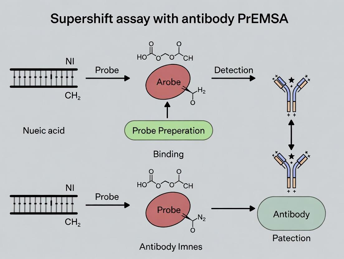
Protocol B: Supershift Assay with Antibody
- Follow Protocol A, Steps II.1-4 to set up the primary protein-DNA binding reaction.
- After the 20-30 minute incubation with the probe (Step II.6), add 1-2 µL of the specific antibody or isotype control antibody (0.5-2 µg) directly to the reaction.
- Incubate the mixture for an additional 30-60 minutes at 4°C or room temperature.
- Proceed with Protocol A, Part III (Gel Electrophoresis & Detection). Analyze for a further retarded band (supershift) above the original protein-DNA complex.
Diagrams
Title: EMSA Supershift Assay Workflow
Title: Expected EMSA/Supershift Gel Band Pattern
The Scientist's Toolkit: Key Research Reagent Solutions
Table 3: Essential Materials for EMSA & Supershift Assays
| Item | Function & Rationale |
|---|---|
| Purified DNA/RNA Probe | The labeled nucleic acid fragment containing the suspected protein-binding site. Must be of high purity and accurately quantified. |
| [γ-³²P] ATP or Non-radioactive Labeling Kit | Provides the tag for sensitive probe detection. Non-radioactive alternatives (e.g., chemiluminescent) improve safety and reagent stability. |
| T4 Polynucleotide Kinase (PNK) | Catalyzes the transfer of a phosphate group from ATP to the 5' end of the probe for radiolabeling. |
| Nuclear Extract Kit | Provides a method to obtain a protein fraction enriched for DNA-binding proteins like transcription factors from cells or tissues. |
| Poly(dI-dC) | A synthetic, non-specific competitor DNA used to bind and "absorb" proteins that interact weakly or non-specifically with nucleic acids. |
| High-Purity Specific Antibody | For supershift assays. Must recognize the native, DNA-bound conformation of the target protein. Monoclonal antibodies are preferred. |
| Non-denaturing Polyacrylamide Gel Kit | Provides reagents for casting gels that separate biomolecules based on size and charge without disrupting non-covalent protein-nucleic acid complexes. |
| Electrophoresis System (Cold Room Compatible) | Running the gel at 4°C is critical to maintain the stability of often labile protein-nucleic acid complexes during electrophoresis. |
| Phosphorimager or Chemiluminescence Imager | For high-sensitivity detection and quantification of the shifted bands, whether radioactively or non-radioactively labeled. |
The Electrophoretic Mobility Shift Assay (EMSA) is a cornerstone technique for studying protein-nucleic acid interactions. Within this framework, the antibody-mediated "supershift" assay is a critical method for specifically identifying individual protein components within a protein-DNA or protein-RNA complex. By incorporating a specific antibody into the binding reaction, a complex containing the target protein experiences a further reduction in electrophoretic mobility, resulting in a higher molecular weight "supershifted" band. This application note details the protocol and principles of the EMSA supershift assay, positioning it as an essential tool for validating protein specificity in transcriptional regulation and drug discovery research.
Core Principle and Quantitative Data
The supershift assay relies on the formation of a ternary complex: nucleic acid probe + DNA-binding protein + specific antibody. The key quantitative outcomes are the changes in migration distance and signal intensity, which confirm specific protein identification.
Table 1: Expected Gel Band Interpretations in a Supershift Assay
| Band Position | Description | Interpretation |
|---|---|---|
| Supershifted Band | Highest molecular weight, retarded migration. | Successful formation of a ternary complex (Probe + Protein + Antibody). Confirms presence of the specific target protein in the original complex. |
| Shifted Band (Complex) | Intermediate migration, above free probe. | Binary complex of the protein(s) bound to the nucleic acid probe. Intensity may decrease upon successful supershift. |
| Free Probe Band | Fastest migration at the gel front. | Unbound nucleic acid probe. Serves as an internal control for electrophoresis. |
Table 2: Common Antibody Types and Their Use in Supershift Assays
| Antibody Type | Target Epitope | Typical Use in Supershift | Notes on Efficacy |
|---|---|---|---|
| Monoclonal | Single, specific epitope. | High specificity; ideal when epitope is accessible in the protein-nucleic acid complex. | Most reliable for consistent supershift results. |
| Polyclonal | Multiple epitopes. | Higher chance of binding as multiple epitopes are targeted; can be more sensitive. | Potential for non-specific binding; requires careful control. |
| Phospho-specific | Phosphorylated amino acid. | Identifies specific active/phosphorylated form of the protein in the complex. | Confirms post-translational modification status of the bound protein. |
Detailed Supershift Assay Protocol
Materials and Reagents
- Nuclear Extract or Purified Protein: Source containing the DNA/RNA-binding protein of interest.
- Biotin- or Fluorophore-labeled DNA/RNA Probe: Typically 20-50 bp containing the known binding sequence.
- Unlabeled Competitor Probe: Identical to labeled probe (specific) or mutated (non-specific) for competition controls.
- Specific Antibody: Validated for use in EMSA/supershift assays (preferably monoclonal).
- Isotype Control Antibody: Same species and isotype as the specific antibody.
- EMSA Binding Buffer: 10 mM HEPES, 50 mM KCl, 1 mM DTT, 2.5% Glycerol, 0.05% NP-40, pH 7.9.
- Poly(dI-dC): Non-specific carrier DNA to reduce non-specific binding.
- Non-denaturing Polyacrylamide or Agarose Gel: Pre-cast in appropriate TBE or TAE buffer.
- Electrophoresis and Transfer Apparatus: For gel separation and blotting (if using chemiluminescent detection).
Procedure
Part A: Standard EMSA Binding Reaction
- Prepare Binding Reactions: On ice, assemble the following in a nuclease-free microcentrifuge tube:
- EMSA Binding Buffer: 10 µL
- Poly(dI-dC) (1 µg/µL): 1 µL
- Nuclear Extract (or purified protein): 5-10 µg (volume variable)
- Labeled Probe (10-20 fmol/µL): 1 µL
- Nuclease-free water to a final volume of 20 µL.
- Optional Competition Controls: In separate tubes, add a 100-fold molar excess of unlabeled specific or non-specific competitor probe before adding the labeled probe.
- Incubate: Mix gently and incubate at room temperature for 20-30 minutes.
Part B: Antibody Supershift
- Add Antibody: To the experimental tube, add 1-2 µg of the target-specific antibody. To the control tube, add an equivalent amount of isotype control antibody.
- Incubate: Mix gently and incubate the reaction at 4°C for 60 minutes or room temperature for 30 minutes. (Longer incubation at 4°C often enhances antibody binding to the pre-formed complex.)
- Load and Run: Add 5 µL of non-denaturing loading dye to each reaction. Load the entire volume onto a pre-run non-denaturing polyacrylamide gel (6-8%) in 0.5x TBE buffer. Run the gel at 100V at 4°C until the dye front migrates appropriately.
- Detection: Visualize bands according to your label (e.g., chemiluminescence for biotin, fluorescence imaging for fluorophores).
Visualization of Workflow and Molecular Interactions
Diagram 1: Supershift Assay Molecular Workflow
The Scientist's Toolkit: Key Research Reagent Solutions
Table 3: Essential Materials for Supershift Assays
| Reagent / Solution | Function & Importance in Supershift Assay |
|---|---|
| High-Affinity, Validated Antibodies | The cornerstone of the assay. Must recognize the native, often conformationally altered, protein within the nucleic acid complex. Antibodies validated for ChIP or EMSA are preferred. |
| Chemiluminescent Nucleic Acid Detection Kits | Provide sensitive, low-background detection of biotin- or digoxigenin-labeled probes, superior to traditional radioisotopes for most applications. |
| LightShift Chemiluminescent EMSA Kit (Thermo Fisher) | A commercial optimized system providing ready-to-use buffers, substrate, and positive controls for robust, reproducible supershift assays. |
| Pre-cast Non-denaturing Polyacrylamide Gels | Ensure consistent gel matrix and electrophoresis conditions, critical for clear resolution of supershifted complexes from standard shifted bands. |
| Poly(dI-dC) or Salmon Sperm DNA | Essential non-specific competitor DNA that binds and neutralizes non-sequence-specific nucleic acid-binding proteins, reducing background noise. |
| Protease and Phosphatase Inhibitor Cocktails | Added to extraction buffers to preserve the native state, post-translational modifications, and DNA-binding activity of proteins from cell lysates. |
| Recombinant Target Protein | Serves as an essential positive control to confirm antibody efficacy and optimize binding conditions before using complex cell extracts. |
Application Notes
Electrophoretic Mobility Shift Assay (EMSA), particularly the supershift variant, is a cornerstone technique in molecular biology for studying protein-nucleic acid interactions. Within the broader thesis on EMSA supershift assay optimization, its applications span from foundational discovery to intricate mechanistic studies. The core principle relies on the reduced electrophoretic mobility of a nucleic acid probe (often DNA) when bound by a protein. The addition of a specific antibody that recognizes the bound protein creates an even larger "supershifted" complex, providing unambiguous identification.
Key Applications:
- Transcription Factor (TF) Discovery and Validation: EMSA supershift is pivotal for confirming the identity of a protein binding to a specific DNA consensus sequence. When a putative TF is suspected, antibodies against it can confirm its presence in the DNA-protein complex.
- Analysis of Complex Composition: The assay can determine if multiple proteins (e.g., co-activators, dimeric partners) are present in a single DNA-bound complex. Sequential or combinatorial antibody additions can dissect these multiprotein assemblies.
- Studying Post-Translational Modifications (PTMs): Antibodies specific to phosphorylation, acetylation, or other PTMs can determine if such modifications are required or altered by DNA binding, linking signaling pathways to transcriptional activity.
- Drug and Inhibitor Screening: EMSA supershift can assess the efficacy of small-molecule inhibitors designed to disrupt specific protein-DNA interactions critical in disease (e.g., NF-κB in inflammation), providing a direct readout of target engagement.
Quantitative Data Summary: Table 1: Common Transcription Factors Analyzed by EMSA Supershift & Key Antibody Targets
| Transcription Factor | Common Consensus Sequence | Typical Antibody Target (for Supershift) | Associated Disease/Pathway |
|---|---|---|---|
| NF-κB | GGGRNNYYCC (R=purine, Y=pyrimidine) | p65 (RelA), p50, phospho-specific antibodies | Inflammation, Cancer |
| AP-1 | TGANTCA | c-Fos, c-Jun, phospho-c-Jun | Proliferation, Stress Response |
| STAT3 | TTCCCGGAA | STAT3, phospho-STAT3 (Tyr705) | Oncology, Autoimmunity |
| p53 | RRRCWWGYYY (R=purine, W=A/T, Y=pyrimidine) | p53, acetyl-p53 | Cancer, Genomic Stability |
| CREB | TGACGTCA | CREB, phospho-CREB (Ser133) | Metabolism, Neuronal Signaling |
| NFAT | GGAAAAT | NFATc1, NFATc2 | Immune Activation, Cardiac Hypertrophy |
Table 2: Comparison of EMSA Detection Methodologies
| Detection Method | Sensitivity | Required Equipment | Advantages | Best For |
|---|---|---|---|---|
| Radioactive (³²P) | Very High (zeptomole) | Geiger counter, Phosphorimager | Gold standard, quantitative | Low-abundance complexes, competition assays |
| Chemiluminescent | High (attomole) | Standard gel imager (CCD) | Safe, long shelf-life, good for publication | Most routine applications, labs without radioisotope permits |
| Fluorescent | Moderate | Fluorescence gel scanner | Multiplexing possible, safe | Pre-labeled probes, kinetic studies |
| Colorimetric | Lower | Visual inspection, standard imager | Inexpensive, no special equipment | High-abundance complexes, educational use |
Detailed Protocols
Protocol 1: Standard EMSA Supershift Assay for Nuclear Extract Analysis
Objective: To detect and confirm the identity of a transcription factor binding to a target DNA sequence using a supershift antibody.
Materials (Research Reagent Solutions):
- Biotech-Grade Non-Ionic Detergent: (e.g., NP-40 or Igepal CA-630). Function: Cell membrane lysis for nuclear isolation.
- Protease/Phosphatase Inhibitor Cocktail (100X): Function: Preserves native protein states by inhibiting degradation and maintaining PTMs.
- High-Binding DNA Probe Kit: Contains T4 Polynucleotide Kinase for end-labeling, and purification columns. Function: Generates high-specific-activity probe.
- Poly(dI•dC): Function: Non-specific competitor DNA to reduce background from non-specific protein binding.
- EMSA Grade 10% TBE-PAGE Gel: Pre-cast, non-denaturing polyacrylamide gel. Function: Matrix for separation of complexes based on size/charge.
- Chemiluminescent Nucleic Acid Detection Module: Contains streptavidin-HRP and stable peroxide/luminol reagents. Function: For sensitive, non-radioactive detection of biotinylated probes.
- Transcription Factor-Specific Monoclonal Antibody (Supershift Grade): Function: Binds specifically to target protein in the complex, inducing a mobility supershift. Critical: Must be validated for EMSA/supershift.
Method:
- Nuclear Extract Preparation: Harvest cells, wash in PBS, and resuspend in cold Hypotonic Lysis Buffer (10 mM HEPES pH 7.9, 1.5 mM MgCl₂, 10 mM KCl, 0.5% NP-40, 0.5 mM DTT, 1X inhibitor cocktail) for 10 min on ice. Centrifuge at 4°C. Pellet nuclei, resuspend in Nuclear Extraction Buffer (20 mM HEPES pH 7.9, 1.5 mM MgCl₂, 420 mM NaCl, 0.2 mM EDTA, 25% glycerol, 0.5 mM DTT, 1X inhibitor cocktail). Vortex, centrifuge, and aliquot supernatant (nuclear extract). Determine protein concentration.
- DNA Probe Labeling: Label 200 ng of complementary single-stranded oligonucleotides containing the consensus sequence with biotin using the 3’-end labeling kit. Anneal strands to form double-stranded probe.
- Binding Reaction: In a 20 µL total volume, combine: 4 µL 5X Binding Buffer (50 mM Tris, 250 mM NaCl, 5 mM MgCl₂, 2.5 mM EDTA, 20% glycerol, 5 mM DTT), 1 µL Poly(dI•dC) (1 µg/µL), 2-10 µg nuclear extract, and nuclease-free water. For supershift, add 1-2 µL of specific antibody or isotype control. Pre-incubate for 10 min at room temperature. Add 20 fmol of biotinylated probe. Incubate 20 min at room temperature.
- Gel Electrophoresis: Pre-run a 6% TBE-PAGE gel in 0.5X TBE buffer at 100V for 60 min. Load samples with 5X native loading dye. Run at 100V for 60-90 min at 4°C.
- Transfer & Detection: Electro-blot to positively charged nylon membrane at 380 mA for 30 min in 0.5X TBE. UV-crosslink the DNA to the membrane. Block membrane, incubate with streptavidin-HRP conjugate, wash, and develop with chemiluminescent substrate. Image.
Protocol 2: Competitive EMSA with Supershift for Affinity Assessment
Objective: To determine binding specificity and relative affinity using unlabeled competitor DNA alongside supershift confirmation.
Method:
- Follow Protocol 1 for probe labeling and extract preparation.
- Competition Setup: Set up binding reactions as in Protocol 1, but include a series of tubes with increasing molar excess (e.g., 10x, 50x, 100x, 200x) of unlabeled competitor DNA. Competitors should be: a) identical "specific" probe, b) probe with a mutated binding site, c) an unrelated sequence.
- Supershift Addition: In parallel, set up identical competition series but include the supershift antibody in the pre-incubation step.
- Analysis: Run gels and image. Specific binding is indicated by displacement only by the specific competitor. The supershift complex should also be competed away, confirming its identity.
Diagrams
Title: EMSA Supershift Assay Experimental Workflow
Title: EMSA Gel Lane Interpretation and Complex Composition
The Scientist's Toolkit: Essential Research Reagents
Table 3: Key Research Reagent Solutions for EMSA Supershift Assays
| Reagent Category | Specific Example | Function & Importance |
|---|---|---|
| Nuclear Extraction Kit | Commercial kits with optimized buffers/inhibitors. | Ensures high-quality, active TF yield from cells; saves optimization time. |
| Supershift-Validated Antibodies | Monoclonal antibodies to p65, c-Jun, STAT3, etc. | Critical for definitive complex identification. Must bind native, DNA-bound protein. |
| Biotin 3’-End DNA Labeling Kit | Contains terminal transferase & biotin-NTPs. | Safe, non-radioactive method for high-sensitivity probe generation. |
| Non-Radioactive Detection System | Chemiluminescent modules (HRP-based). | Provides publication-quality results without radiation safety concerns. |
| EMSA/Gel Shift Buffer Systems | 5X-10X concentrated binding buffers. | Ensures optimal ionic strength and pH for specific protein-DNA interactions. |
| High-Purity Competitor DNAs | Poly(dI•dC), specific & mutant cold probes. | Essential for demonstrating binding specificity in competition assays. |
| Pre-Cast Non-Denaturing Gels | 6% TBE-PAGE gels, multiple wells. | Provides consistent, reproducible separation matrix with minimal hands-on time. |
Application Notes
The Electrophoretic Mobility Shift Assay (EMSA) and its extension, the supershift assay, are cornerstone techniques for studying protein-nucleic acid interactions, particularly transcription factor binding. The choice between radioactive and non-radioactive detection, the quality of antibodies, and the preparation of nuclear extracts are critical determinants of success. These components are integral to thesis research focused on optimizing EMSA supershift protocols for drug discovery targeting transcription factors.
Probe Detection: Radioactive vs. Non-Radioactive
The core of EMSA is the labeled nucleic acid probe. The detection method impacts sensitivity, safety, cost, and workflow.
Radioactive Probes (³²P-labeled): Traditionally the gold standard due to unparalleled sensitivity, capable of detecting sub-femtomole quantities of protein. The direct incorporation of [γ-³²P]ATP via T4 Polynucleotide Kinase is common. However, stringent safety protocols, regulatory hurdles, and waste disposal issues are significant drawbacks.
Non-Radioactive Probes: Modern alternatives offer safer, more convenient workflows with comparable sensitivity for many applications.
- Biotinylated Probes: Detected by streptavidin-conjugated enzymes (HRP or AP) for chemiluminescent or colorimetric readouts. Sensitivity is high but can be influenced by non-specific binding.
- Fluorescently-labeled Probes: Probes tagged with fluorophores (e.g., Cy5, FAM) are detected directly by fluorescence imaging or scanning. They enable multiplexing and are ideal for quantitative analysis.
- Digoxigenin (DIG)-labeled Probes: Similar to biotin, detected by anti-DIG antibody conjugates, offering high specificity.
Quantitative Comparison:
Table 1: Comparison of Probe Detection Methods
| Parameter | ³²P Radioactive | Biotin/Chemiluminescence | Fluorescent |
|---|---|---|---|
| Sensitivity | Very High (0.1-1 fmol) | High (1-10 fmol) | Moderate to High (5-50 fmol) |
| Resolution | Excellent | Good | Good |
| Signal Stability | Short half-life (14.3 days for ³²P) | Stable, can be re-probed | Stable |
| Exposure Time | Minutes to Hours (film/phosphorimager) | Seconds to Minutes | Seconds (scanner) |
| Safety & Regulation | High; Requires licensing, special handling, disposal | Low; Standard lab safety | Low; Standard lab safety |
| Cost | Lower reagent cost, high waste/disposal cost | Higher reagent cost | Moderate reagent cost |
| Throughput & Speed | Slow (due to safety) | Medium | Fast |
| Multiplexing | No | Difficult | Yes (multiple colors) |
| Primary Best Use Case | Maximum sensitivity, low-abundance factors | Standard assays, regulated labs | Quantitative, high-throughput, multiplex assays |
Antibodies for Supershift Assays
The supershift assay employs specific antibodies to identify proteins in a protein-DNA complex. A "supershift" occurs when the antibody binds to the protein, causing a further retardation in the complex's mobility.
- Function: Confirms protein identity in the complex.
- Critical Quality: Must be specific for the native, DNA-bound conformation of the target protein. Polyclonal antibodies often perform better than monoclonals due to recognition of multiple epitopes.
- Controls: Include an isotype control antibody to rule out non-specific antibody-DNA or antibody-protein interactions.
Nuclear Extracts
Nuclear extracts are the primary protein source for studying transcription factors.
- Preparation: Involves cell lysis, isolation of nuclei, and high-salt extraction of nuclear proteins. Key steps must be performed at 4°C with protease and phosphatase inhibitors.
- Quality Assessment: Protein concentration (Bradford assay) and functional activity (positive control EMSA with a known probe) are essential.
- Storage: Aliquots at -80°C to prevent degradation and freeze-thaw cycles.
Protocols
Protocol 1: Preparation of Non-Radioactive Biotinylated DNA Probe
Materials: Oligonucleotides, Biotin 3'-End DNA Labeling Kit, Nuclease-free water, TE buffer.
- Anneal complementary oligonucleotides containing the target sequence.
- Use a biotinylation kit (e.g., using Terminal Deoxynucleotidyl Transferase, TdT, with Biotin-dUTP) to label the 3' ends of the duplex DNA.
- Purify the labeled probe using a spin column.
- Verify labeling efficiency via a dot-blot assay with streptavidin-HRP.
- Store at -20°C in aliquots.
Protocol 2: EMSA/Supershift Assay with Non-Radioactive Probe
Materials: Nuclear extract, biotinylated probe, poly(dI:dC), EMSA binding buffer, specific and control antibodies, non-denaturing polyacrylamide gel, 0.5X TBE buffer, vertical electrophoresis unit, nylon membrane, UV crosslinker, Chemiluminescent Nucleic Acid Detection Kit.
Binding Reaction (20 µL):
- Prepare master mix: 2 µL 10X binding buffer, 1 µL poly(dI:dC) (1 µg/µL), 1 µL 50% glycerol, x µL nuclear extract (2-10 µg protein), nuclease-free water to 18 µL.
- For supershift: Pre-incubate extract with 1-2 µg of specific or control antibody on ice for 20-30 minutes.
- Add 2 µL (20 fmol) of biotinylated probe. Incubate at room temperature for 20 minutes.
Electrophoresis:
- Load samples onto a pre-run 6% non-denaturing polyacrylamide gel in 0.5X TBE at 100V for 10 min, then run at 100V for 60-90 min at 4°C.
Transfer & Detection:
- Electro-blot to a positively charged nylon membrane at 380 mA for 30-45 min in 0.5X TBE.
- UV-crosslink the DNA to the membrane.
- Block membrane, incubate with Streptavidin-HRP conjugate, wash, and develop with chemiluminescent substrate. Image.
Protocol 3: Nuclear Extract Preparation (Mini-scale)
Materials: Cell culture, Hypotonic Lysis Buffer, Nuclear Extraction Buffer, protease inhibitors, DTT, centrifugation equipment.
- Harvest cells, pellet, and wash with PBS.
- Resuspend in cold Hypotonic Buffer. Incubate on ice 15 min. Centrifuge.
- Resuspend pellet in cold Nuclear Extraction Buffer. Vigorously vortex. Incubate on ice 30 min with mixing.
- Centrifuge at max speed, 4°C, for 10 min.
- Aliquot supernatant (nuclear extract), snap-freeze, store at -80°C.
Visualizations
Title: EMSA Supershift Assay Experimental Workflow
Title: Decision Tree for EMSA Probe Detection Method
The Scientist's Toolkit
Table 2: Essential Research Reagent Solutions for EMSA/Supershift Assays
| Reagent/Material | Function & Importance |
|---|---|
| Nuclear Extract Kit | Provides optimized buffers for efficient, high-quality extraction of active nuclear proteins. Critical for yield and activity. |
| Biotin 3'-End Labeling Kit | Enzymatically incorporates biotin into DNA probes for safe, sensitive non-radioactive detection. |
| Poly(dI:dC) | A non-specific competitor DNA that reduces interference from non-specific DNA-binding proteins in the binding reaction. |
| EMSAsafe Protease Inhibitor Cocktail | Prevents degradation of transcription factors during extract prep and binding reactions. |
| Non-Denaturing PAGE System | Pre-cast gels and buffers optimized for resolving protein-nucleic acid complexes without disrupting weak interactions. |
| Positively Charged Nylon Membrane | Essential for efficient transfer and retention of negatively charged DNA/protein complexes in non-radioactive blotting. |
| Chemiluminescent Detection Module | Streptavidin-HRP and stable substrate for high-sensitivity imaging of biotinylated probes. |
| Transcription Factor Specific Antibody | Validated for EMSA/supershift; recognizes native, DNA-bound protein. The key reagent for identification. |
| Gel Shift Binding Buffer (5X) | Optimized buffer with salts, glycerol, and carrier to promote specific binding in the reaction. |
Step-by-Step EMSA Supershift Protocol: From Probe Design to Gel Imaging
Within the framework of thesis research focused on optimizing the EMSA supershift assay with antibody protocol, the initial preparation phase is critical. This phase dictates the success of subsequent electrophoretic mobility and supershift experiments by ensuring the availability of high-quality, specific nucleic acid probes and functionally active protein extracts. This application note details current methodologies for the design and preparation of these core components.
Designing Optimal DNA/RNA Probes
The probe is a labeled, short, double-stranded DNA or RNA fragment containing the specific protein-binding sequence of interest.
Core Design Principles
- Length: Typically 20-40 base pairs. Longer probes (>50 bp) increase non-specific binding, while shorter probes (<15 bp) may lack necessary flanking sequences for optimal protein interaction.
- Sequence: Must contain the well-characterized consensus binding sequence for the target transcription factor (e.g., NF-κB, AP-1). Including 5-10 bp of flanking sequence on each side enhances stability and specificity.
- Labeling: Probes are commonly labeled at the 5' or 3' end with fluorophores (e.g., Cy5, FAM) for fluorescence-based detection or with biotin for chemiluminescent detection. Radioactive labeling (γ-32P ATP) remains a highly sensitive option.
- Specificity Control: A mutant probe with a scrambled or mutated core binding sequence is mandatory as a negative control to demonstrate binding specificity.
Table 1: Quantitative Parameters for Probe Design
| Parameter | Optimal Range | Purpose & Rationale |
|---|---|---|
| Probe Length | 20 - 40 bp | Balances sufficient binding site context with minimal non-specific interactions. |
| GC Content | 40 - 60% | Promotes probe duplex stability during annealing. |
| Melting Temp (Tm) | 60 - 75°C | Ensures probe is double-stranded under binding reaction conditions. |
| Labeling Efficiency | > 90% | Maximizes detection signal; measured by spectrophotometry or gel analysis. |
Protocol: Probe Annealing
Objective: To generate double-stranded, labeled probes from complementary single-stranded oligonucleotides. Materials: HPLC-purified sense and antisense oligonucleotides, Nuclease-Free Water, 10X Annealing Buffer (100 mM Tris, 1 M NaCl, 10 mM EDTA, pH 8.0). Method:
- Dilution: Resuspend oligonucleotides to 100 µM in nuclease-free water.
- Mixing: Combine equal volumes (e.g., 50 µL each) of the complementary oligonucleotides in a microcentrifuge tube.
- Addition of Buffer: Add 1/10 volume of 10X Annealing Buffer (e.g., 10 µL for a 100 µL total mixture).
- Annealing: Place the tube in a beaker containing 500 mL of water heated to 95°C. Allow the beaker to cool slowly to room temperature (~2-3 hours).
- Storage: Dilute the annealed probe to a working concentration (e.g., 1 µM), aliquot, and store at -20°C.
Preparing Nuclear and Cellular Extracts
The source of protein for EMSA can be whole cell extracts (for abundant proteins) or nuclear extracts (for transcription factors primarily localized to the nucleus).
Key Considerations for Extract Preparation
- Cell/ Tissue Type: Use relevant biological material. Cultured cells are common; tissue samples require homogenization.
- Lysis Method: Cytoplasmic lysis with a mild non-ionic detergent (e.g., NP-40) is used for nuclear extraction. Whole cell extracts use a more stringent RIPA-type buffer.
- Inhibition of Proteases & Phosphatases: Add fresh inhibitors (PMSF, aprotinin, leupeptin, sodium orthovanadate) to all buffers to preserve protein activity and modification states.
- Speed and Temperature: Perform steps quickly on ice or at 4°C to prevent protein degradation.
Table 2: Comparison of Extract Types for EMSA
| Extract Type | Target Proteins | Key Buffer Components | Typical Protein Yield (from 10⁷ cells) |
|---|---|---|---|
| Nuclear Extract | Nuclear transcription factors (e.g., p65, c-Jun) | Hypotonic buffer, NP-40, High-salt nuclear extraction buffer | 100 - 500 µg |
| Whole Cell Extract | Abundant cytoplasmic/nuclear proteins | RIPA Buffer (or variants with SDS, deoxycholate) | 500 - 2000 µg |
Protocol: Rapid Nuclear Extract Preparation from Cultured Cells
Objective: To isolate active nuclear proteins from adherent or suspension cell lines. Materials: PBS (ice-cold), Hypotonic Buffer (10 mM HEPES pH 7.9, 1.5 mM MgCl₂, 10 mM KCl, 0.5 mM DTT, protease inhibitors), Lysis Buffer (Hypotonic Buffer + 0.1% NP-40), Nuclear Extraction Buffer (20 mM HEPES pH 7.9, 1.5 mM MgCl₂, 420 mM NaCl, 0.2 mM EDTA, 25% glycerol, 0.5 mM DTT, protease inhibitors), Bradford Assay Reagent. Method:
- Harvest: Wash cells twice with ice-cold PBS. Scrape adherent cells into PBS and pellet (500 x g, 5 min, 4°C).
- Hypotonic Swell: Resuspend cell pellet in 5x pellet volume of Hypotonic Buffer. Incubate on ice for 10-15 min.
- Lysis: Add 0.1% NP-40 (final concentration). Vortex vigorously for 10 seconds. Centrifuge immediately (12,000 x g, 30 sec, 4°C). The supernatant (cytoplasmic fraction) can be discarded or saved.
- Nuclear Extraction: Resuspend the nuclear pellet in 1-2x pellet volume of Nuclear Extraction Buffer. Incubate on ice with vigorous shaking or vortexing every 5 min for 30 min total.
- Clarification: Centrifuge (12,000 x g, 10 min, 4°C). Carefully transfer the supernatant (nuclear extract) to a fresh pre-chilled tube.
- Quantification & Storage: Determine protein concentration using the Bradford assay. Aliquot extracts, snap-freeze in liquid nitrogen, and store at -80°C. Avoid repeated freeze-thaw cycles.
The Scientist's Toolkit: Research Reagent Solutions
Table 3: Essential Materials for Probe and Extract Preparation
| Item | Function & Application |
|---|---|
| HPLC-Purified Oligonucleotides | Ensures high-purity, contaminant-free single-stranded DNA for specific probe synthesis. |
| Fluorophore- or Biotin-Labeling Kits | Provides optimized enzymes and buffers for efficient, consistent 5' or 3' end-labeling of probes. |
| Nuclease-Free Water & Buffers | Prevents degradation of nucleic acid probes during resuspension and annealing. |
| Complete Protease Inhibitor Cocktail (Tablets/Liquid) | Broad-spectrum inhibition of serine, cysteine, aspartic proteases, and aminopeptidases in extracts. |
| Phosphatase Inhibitor Cocktail (Sodium Orthovanadate, etc.) | Preserves the phosphorylation state of transcription factors, critical for DNA-binding activity. |
| Non-ionic Detergent (NP-40/Igepal CA-630) | Selective lysis of the plasma membrane for nuclear isolation without disrupting the nuclear envelope. |
| High-Salt Nuclear Extraction Buffer | Disrupts nuclear envelope and solubilizes DNA-binding proteins via salt-dependent disruption of protein-DNA interactions. |
| Bradford or BCA Protein Assay Kit | Accurate quantification of total protein concentration in final extracts for normalizing EMSA reactions. |
Visualizing the Preparation Workflow and Pathway Context
Title: EMSA Phase 1 Workflow: Probe and Extract Preparation
Title: Signaling Pathway Context for EMSA Target Identification
Within the broader scope of thesis research on the Electrophoretic Mobility Shift Assay (EMSA) supershift protocol, Phase 2 is pivotal. This phase focuses on empirically determining the optimal binding conditions to facilitate stable and specific complex formation between the target nucleic acid (e.g., DNA probe) and the protein of interest (e.g., transcription factor). Suboptimal conditions are a primary source of false negatives or non-specific binding in subsequent EMSA and supershift steps. These Application Notes provide a detailed, current protocol for systematically optimizing the binding reaction.
Critical Parameters for Optimization
The stability and specificity of the protein-nucleic acid complex are influenced by several interdependent factors. The following table summarizes the key variables and their typical tested ranges.
Table 1: Key Parameters for Binding Reaction Optimization
| Parameter | Typical Range Tested | Purpose & Rationale |
|---|---|---|
| Protein (Lysate/Extract) Amount | 2 µg – 20 µg | Titrates active protein concentration; avoids probe exhaustion or non-specific binding from excess protein. |
| Labeled Probe Concentration | 0.1 nM – 1.0 nM (per reaction) | Ensures signal detection while maintaining conditions where protein is limiting. |
| Binding Buffer Ionic Strength (KCl/NaCl) | 0 mM – 150 mM | Modulates electrostatic interactions; high salt can disrupt weak specific complexes. |
| Mg²⁺ Concentration | 0 mM – 10 mM | Often required for DNA-protein interactions; stabilizes complex. |
| Non-Specific Competitor (poly(dI:dC)) | 0 µg – 5 µg per reaction | Blocks non-specific protein-probe interactions; type and amount are critical. |
| Carrier Protein (BSA) | 0 µg – 100 µg per reaction | Stabilizes dilute proteins and prevents adsorption to tubes. |
| Incubation Time | 10 min – 30 min | Allows equilibrium of complex formation. |
| Incubation Temperature | 4°C, 22°C (RT), 37°C | Affects reaction kinetics and protein stability. |
| Detergent (NP-40/Triton X-100) | 0% – 0.1% (v/v) | Reduces non-specific binding but can disrupt some complexes. |
Detailed Optimization Protocol
Experiment 1: Titration of Nuclear Extract and Non-Specific Competitor
This matrix experiment identifies the optimal balance between specific complex formation and suppression of non-specific shifts.
Materials:
- Purified, end-labeled DNA probe.
- Nuclear extract containing the target DNA-binding protein.
- 5X Binding Buffer (200 mM HEPES pH 7.9, 30 mM MgCl₂, 400 mM KCl, 50% Glycerol, 5 mM DTT - prepare fresh).
- poly(dI:dC) stock solution (1 µg/µL).
- Nuclease-free water.
- 0.5 mL thin-wall PCR tubes or similar.
Method:
- Prepare a master mix for the desired number of reactions (n+1) containing: 4 µL of 5X Binding Buffer, 1 µL of labeled probe (0.5 nM final concentration), and nuclease-free water to bring the volume to 18 µL per reaction after addition of extract and competitor.
- Aliquot 18 µL of the master mix into each reaction tube.
- Create a two-dimensional titration matrix by adding varying amounts of nuclear extract (e.g., 1, 2, 4, 8 µg) and poly(dI:dC) (e.g., 0, 0.5, 1, 2 µg) in a final reaction volume of 20 µL. Adjust water accordingly.
- Mix gently by pipetting. Centrifuge briefly.
- Incubate at room temperature (22°C) for 20 minutes.
- Immediately load 10 µL of each reaction on a pre-run (0.5X TBE, 100V, 30 min) 6% non-denaturing polyacrylamide gel.
- Run the gel at 100V in 0.5X TBE buffer at 4°C until the dye front is near the bottom.
- Image the gel using a phosphorimager or autoradiography. The optimal condition shows a strong, discrete shifted band with minimal smearing or higher-order complexes.
Experiment 2: Optimization of Monovalent and Divalent Cations
This experiment fine-tunes buffer composition for maximal complex stability.
Method:
- Prepare 5X Binding Buffer stocks varying only in KCl concentration (e.g., 0, 50, 100, 150, 200 mM final 1X concentration) and MgCl₂ concentration (e.g., 0, 2, 5, 10 mM final 1X concentration).
- Using the optimal extract and poly(dI:dC) amounts from Experiment 1, set up reactions with the different binding buffers.
- Follow steps 4-8 from Experiment 1. The optimal condition yields the most intense and clean shifted band.
The Scientist's Toolkit: Research Reagent Solutions
Table 2: Essential Materials for EMSA Binding Optimization
| Item | Function & Rationale |
|---|---|
| HEPES-based Binding Buffer | Maintains stable pH during the incubation. Superior to phosphate buffers for protein interactions. |
| High-Purity poly(dI:dC) | The standard non-specific competitor for DNA-binding proteins. Competes for non-sequence-specific electrostatic binding. |
| Carrier DNA (e.g., salmon sperm DNA) | An alternative competitor for some systems, often used in combination with poly(dI:dC). |
| Bovine Serum Albumin (BSA), Nuclease-Free | Stabilizes proteins, prevents loss on tube walls, and can reduce non-specific interactions. |
| Dithiothreitol (DTT) | Fresh reducing agent critical for maintaining cysteine-dependent DNA-binding domains in active state. |
| Glycerol | Added to binding buffer to increase viscosity, stabilize proteins, and facilitate gel loading. |
| Non-ionic Detergent (e.g., NP-40) | Used at low concentrations (≤0.1%) to minimize hydrophobic non-specific interactions. |
| Protease & Phosphatase Inhibitor Cocktails | Essential in extract preparation to preserve the native state and post-translational modifications of the DNA-binding protein. |
Visualizing the Optimization Workflow and Pathway
Title: EMSA Binding Optimization Workflow
Title: Specific vs. Non-Specific Binding in EMSA
A systematic approach to optimizing the binding reaction, as outlined here, is non-negotiable for generating reliable, interpretable data in EMSA and subsequent supershift assays. The identified optimal conditions form the foundation for Phase 3 (native gel electrophoresis) and Phase 4 (antibody-based supershift) of the thesis protocol, ensuring that observed shifts are attributable to specific protein-nucleic acid interactions.
Application Notes
Incorporating a specific antibody into an Electrophoretic Mobility Shift Assay (EMSA) to perform a "supershift" is a critical step for identifying protein components within a DNA-protein or RNA-protein complex. Phase 3 focuses on the precise conditions required for successful antibody addition, which, if optimized, can provide definitive evidence of a particular transcription factor's presence. The key variables are the timing of antibody addition (pre-incubation vs. post-incubation), the concentration of the antibody, and the implementation of rigorous specificity controls. Failure to optimize these parameters is a common source of false-negative or false-positive supershift results.
Optimal timing typically involves adding the antibody after the initial protein-nucleic acid complex has formed. This "post-incubation" or "supershift-only" approach minimizes the risk of the antibody sterically hindering the protein's ability to bind to its target probe. The effective concentration of the antibody must be determined empirically, as too little will not cause a visible shift, while too much can lead to nonspecific interactions or disruption of the primary complex. Specificity controls are non-negotiable and include the use of (1) an isotype-control antibody, (2) an antibody against an unrelated protein, and (3) a blocking peptide to pre-absorb the specific antibody.
The data below, compiled from recent literature and optimized protocols, summarizes the quantitative ranges for these critical parameters.
Table 1: Optimization Parameters for Antibody Incorporation in EMSA Supershift Assays
| Parameter | Recommended Range | Purpose & Rationale |
|---|---|---|
| Antibody Incubation Timing | 15-30 minutes post protein-probe binding | Allows complex formation before Ab addition, preventing interference with binding. |
| Incubation Temperature | 4°C (on ice) or Room Temperature (20-25°C) | 4°C favors complex stability; RT may improve Ab-antigen kinetics. Must be tested. |
| Polyclonal Antibody Concentration | 0.5 - 2 µg per 20 µL reaction | High-affinity polyclonals often work at lower concentrations. |
| Monoclonal Antibody Concentration | 1 - 4 µg per 20 µL reaction | May require higher amounts due to single epitope recognition. |
| Antibody:Protein Molar Ratio | 2:1 to 10:1 (Ab:Target Protein) | Ensures sufficient Ab for supershift without vast excess. |
| Key Specificity Controls | 1. Isotype-control IgG 2. Unrelated protein Ab 3. Antigen-blocking peptide | Verifies supershift is due to specific antigen-Ab interaction. |
Experimental Protocols
Protocol 3.1: Standard Supershift Assay with Post-Incubation
Objective: To identify a specific protein within a DNA-protein complex using a target-specific antibody.
Materials: Pre-formed protein-probe complex (from Phase 2 EMSA), target-specific antibody, control antibodies, EMSA gel shift buffer, ice.
Method:
- Perform Standard Binding Reaction: Set up your primary EMSA binding reaction (Nuclear extract/protein + labeled probe + buffer/poly dI:dC) as optimized in Phase 2. Incubate at appropriate temperature (e.g., 20-25°C) for 20 minutes to allow complex formation.
- Add Antibody: To the completed binding reaction, add the target-specific antibody. A typical starting amount is 1 µg of IgG per 20 µL reaction.
- Secondary Incubation: Incubate the reaction for an additional 30 minutes at the same temperature as step 1 (or on ice if complex stability is a concern).
- Load and Run: Add loading dye (without SDS, which may disrupt complexes) and immediately load the entire reaction onto a pre-run, native polyacrylamide gel. Run under previously optimized EMSA conditions (typically 4°C, 100-150 V).
- Analyze: Visualize shifted and "supershifted" complexes (further retarded bands) via autoradiography or phosphorimaging.
Protocol 3.2: Specificity Control Using Antigen Blocking Peptide
Objective: To confirm the specificity of the observed supershift by competitive inhibition.
Materials: Target-specific antibody, corresponding immunizing peptide (blocking peptide), control peptide.
Method:
- Pre-absorb the Antibody: In a separate tube, mix the target-specific antibody (1-2 µg) with a 5-10 fold molar excess of the blocking peptide. Incubate this mixture for 30-60 minutes on ice prior to the supershift assay.
- Perform Parallel Reactions: Set up two identical binding reactions (protein + probe). To one, add the pre-absorbed antibody mixture. To the other, add the untreated target-specific antibody (positive control for supershift).
- Complete the Assay: Follow Protocol 3.1 from step 3 onwards. The supershift should be abolished or significantly diminished in the reaction with the pre-absorbed antibody, confirming specificity.
Protocol 3.3: Titration of Antibody Concentration
Objective: To determine the minimal effective antibody concentration for a clear supershift while avoiding nonspecific effects.
Method:
- Prepare Reaction Master Mix: Create a master mix containing all components for the binding reaction (protein, probe, buffer) for n+1 reactions, where n is the number of antibody concentrations to be tested.
- Aliquot and Add Antibody: Aliquot equal volumes of the master mix into separate tubes. Add a serial dilution of the target-specific antibody to each tube (e.g., 0.1, 0.5, 1.0, 2.0, 4.0 µg per reaction). Include one tube with no antibody and one with an isotype-control antibody.
- Incubate and Run: Incubate all tubes (post-addition) for 30 minutes, then load and run on a native gel.
- Analyze: Identify the concentration that yields a clear supershift without smearing or loss of the original protein-probe complex.
Diagrams
Title: Antibody Addition Timing in Supershift Assay
Title: Supershift Assay Feasibility and Validation Path
The Scientist's Toolkit
Table 2: Key Reagents for EMSA Supershift Assays
| Reagent | Function & Importance in Phase 3 |
|---|---|
| Target-Specific Antibody | Primary reagent for supershift. Must recognize the native, non-denatured protein epitope within the complex. Polyclonals often have higher success rates. |
| Isotype-Control IgG | An antibody of the same species and isotype (e.g., IgG) but without specificity for the target. Essential negative control to rule out nonspecific band shifts. |
| Blocking Peptide | The specific antigenic peptide used to generate the antibody. Used in pre-absorption experiments to competitively inhibit the supershift, confirming antibody specificity (Protocol 3.2). |
| Antibody against Unrelated Protein | An additional negative control antibody targeting a protein not present in the extract or complex. Further validates the specificity of the observed supershift. |
| Native Gel Electrophoresis System | Includes gel casting apparatus, running buffers, and a cooling unit (4°C). Critical for maintaining the integrity of antibody-protein-DNA complexes during separation. |
| High-Sensitivity Detection Reagents | Such as phosphor screens or high-performance film. Supershifted complexes may be of lower abundance and require sensitive detection for visualization. |
| Non-denaturing Loading Dye | Glycerol-based dye without SDS or β-mercaptoethanol, which would disrupt non-covalent protein-antibody interactions before gel entry. |
This Application Note details the critical Phase 4 of the Electrophoretic Mobility Shift Assay (EMSA) supershift protocol, as framed within a broader thesis investigating protein-DNA interactions and complex supershifting with specific antibodies. Following the formation of protein-nucleic acid and antibody-supershift complexes (Phases 1-3), Native Polyacrylamide Gel Electrophoresis (Native PAGE) is employed to separate complexes based on charge and size without denaturation. Subsequent signal visualization enables the detection and analysis of shifted bands, confirming specific interactions and supershift phenomena.
Key Principles of Native PAGE for EMSA/Supershift
Native PAGE preserves the native conformation and biological activity of protein-DNA complexes. The electrophoresis running buffer (typically Tris-Glycine or Tris-Borate) maintains a pH (~8.3-8.8) that keeps proteins negatively charged. The polyacrylamide gel matrix (typically 4-10%) acts as a molecular sieve. Key parameters influencing separation:
- Gel Percentage: Lower % (e.g., 4-6%) for larger complexes (>200 kDa); higher % (e.g., 8-10%) for smaller complexes.
- Buffer System: Absence of SDS (Sodium Dodecyl Sulfate) and reducing agents is mandatory.
- Temperature: Electrophoresis is typically performed at 4°C to stabilize complexes and prevent gel overheating.
- Voltage/Current: Constant current (e.g., 25-35 mA) is recommended to minimize heat generation.
Detailed Protocol: Native PAGE Setup and Electrophoresis
A. Gel Casting
- Prepare Gel Solutions: For a 6% resolving gel (10 ml volume):
- 2.0 ml 30% acrylamide/bis-acrylamide (29:1)
- 2.5 ml 5x Tris-Glycine buffer (or Tris-Borate)
- 5.4 ml nuclease-free water
- 100 µl 10% Ammonium Persulfate (APS)
- 10 µl Tetramethylethylenediamine (TEMED)
- Mix swiftly and pour immediately between assembled glass plates.
- Prepare Stacking Gel (Optional but Recommended): A 4% stacking gel (3 ml) can be poured on top of the polymerized resolving gel to sharpen bands.
- Polymerization: Allow gels to polymerize completely (20-30 min at RT).
B. Sample and Electrophoresis Setup
- Pre-electrophoresis: Assemble gel apparatus in tank filled with pre-chilled 1x running buffer. Pre-run the gel for 30-60 min at 70-100 V, 4°C, to remove residual APS and equilibrate pH.
- Sample Loading: Mix binding reaction samples (from supershift assay) with 6x Native Loading Dye (non-denaturing, contains Ficoll or glycerol and tracking dyes). Do not heat. Load samples into wells.
- Electrophoresis Run: Run gel at constant current (e.g., 25-35 mA per gel) in cold room (4°C) until the bromophenol blue dye front migrates to the bottom 1/4 of the gel. Time varies with gel size and percentage.
Table 1: Recommended Native PAGE Conditions for EMSA Complexes
| Complex Size Range | Recommended Gel % | Suggested Running Buffer | Typical Run Time (Mini-gel, 25mA) | Key Consideration |
|---|---|---|---|---|
| >250 kDa | 4-5% | 0.5x TBE or Tris-Glycine | 1.5 - 2 hours | Low % gel fragile; use high-strength glass plates. |
| 100-250 kDa | 6% | 0.5x TBE or Tris-Glycine | 1 - 1.5 hours | Standard condition for most nuclear extract DNA-binding complexes. |
| 50-100 kDa | 8% | 0.5x TBE or Tris-Glycine | 45 min - 1 hour | Provides better resolution for smaller complexes. |
| Supershift Assays | 4-6% | 0.25x TBE (low ionic strength) | 1.5 - 2 hours | Low ionic strength helps preserve large antibody-antigen-DNA complexes. |
Detailed Protocol: Signal Visualization
A. Post-Electrophoresis Transfer (For Blot-Based Detection)
- Electroblotting: For sensitive detection or probe recovery, transfer separated complexes from gel to a positively charged nylon membrane via wet or semi-dry electroblotting in 0.5x TBE buffer at 4°C (e.g., 380 mA for 1 hour).
- Crosslinking: If using a labeled DNA probe, covalently link DNA to membrane via UV crosslinking (e.g., 120 mJ/cm²).
B. Detection Methods Method selection depends on the label used on the nucleic acid probe (radioactive or non-radioactive).
1. Radioactive Detection (³²P-labeled probe):
- Direct Autoradiography: Dry gel or membrane, expose to storage phosphor screen or X-ray film at -80°C (film) or RT (screen).
- Quantification: Scan phosphor screen with a phosphorimager. Data can be quantified using ImageJ or AIDA software.
- Typical Exposure Times: Phosphor screen: 30 min - 2 hours; X-ray film with intensifying screen: 2-16 hours.
2. Non-Radiochemical Detection (Biotin/Digoxigenin/Fluorescent-labeled probe):
- Chemiluminescence (Biotin/DIG):
- Blocking: Incubate membrane in blocking buffer (e.g., 5% BSA in TBST) for 1 hour.
- Conjugate Incubation: Incubate with Streptavidin-HRP (for biotin) or Anti-DIG-HRP (for digoxigenin) in blocking buffer for 30-60 min.
- Washing: Wash membrane 3x for 10 min in TBST.
- Substrate Incubation: Incubate with enhanced chemiluminescent (ECL) substrate per manufacturer's instructions.
- Imaging: Capture signal using a CCD-based chemiluminescence imager. Exposure times range from 10 seconds to 10 minutes.
- Fluorescence Imaging: For IRDye or Cy-labeled probes, scan gel/membrane directly using an appropriate fluorescence scanner (e.g., LI-COR Odyssey) at the relevant excitation/emission wavelengths.
Table 2: Comparison of Primary Detection Methodologies
| Method | Typical Sensitivity (Moles of DNA) | Dynamic Range | Required Equipment | Time to Result (Post-Electrophoresis) | Key Advantage |
|---|---|---|---|---|---|
| ³²P Autoradiography (Phosphorimager) | 0.1-1 fmol | >10⁵ | Phosphorimager | 1-3 hours (screening) | Highest sensitivity; gold standard for quantitation. |
| ³²P Autoradiography (X-ray Film) | 1-10 fmol | ~10³ | Film Developer | 12-24 hours | Widely accessible; permanent record. |
| Chemiluminescence (ECL) | 1-10 fmol | ~10⁴ | Chemiluminescence Imager | 2-3 hours | No radioactivity; good sensitivity. |
| Fluorescence (Direct) | 10-100 fmol | ~10⁴ | Fluorescence Scanner | 1-2 hours | Fast; multiplexing possible; no additional steps. |
| Colorimetric (BCIP/NBT) | 100 fmol - 1 pmol | ~10² | Benchtop Scanner | 4-24 hours | Inexpensive; no special imager needed. |
The Scientist's Toolkit: Research Reagent Solutions
Table 3: Essential Materials for Native PAGE and Detection in EMSA Supershift Assays
| Item | Function & Critical Specification |
|---|---|
| Acrylamide/Bis-acrylamide (29:1 or 37.5:1) | Forms the porous polyacrylamide gel matrix. Ratio determines crosslinking density. Must be high-purity, electrophoresis grade. |
| TEMED & APS | Catalyzer (TEMED) and initiator (APS) for acrylamide polymerization. Prepare APS fresh or store aliquots at -20°C. |
| Native Running Buffer (10x TBE or Tris-Glycine) | Provides conductive ions and maintains pH during electrophoresis. For EMSA, 0.25x-0.5x working concentration is common to reduce complex dissociation. |
| Non-Denaturing Loading Dye (6x) | Increases sample density for well loading; contains inert polymers (e.g., Ficoll, glycerol) and visible tracking dyes (bromophenol blue, xylene cyanol). Contains no SDS or β-mercaptoethanol. |
| Pre-cast Native PAGE Gels | Commercial gels offering consistency, convenience, and time savings. Ensure they are specified for Native protein analysis. |
| Positively Charged Nylon Membrane | For blotting nucleic acid probes; strong positive charge ensures efficient retention of negatively charged DNA. |
| Phosphor Screen & Imager | For quantitative detection of radioisotopes (³²P, ³³P). Offers wide dynamic range and faster results than film. |
| HRP-Conjugated Streptavidin/Anti-DIG | Secondary detection conjugate for biotin- or digoxigenin-labeled probes. High affinity and specific activity are crucial for sensitivity. |
| Enhanced Chemiluminescence (ECL) Substrate | HRP enzyme catalyzes luminescent reaction. "Enhanced" kits provide higher signal intensity and duration. |
| Fluorescent Scanner (e.g., LI-COR Odyssey) | Enables direct, in-gel detection of fluorescently labeled probes (Cy3, Cy5, IRDye). Allows multiplexing. |
Diagrams
Title: EMSA Native PAGE & Detection Workflow
Title: EMSA Band Identity and Characteristics
Solving Common EMSA Supershift Problems: A Troubleshooting Handbook
No Shift or Supershift? Diagnosing Issues with Probes, Proteins, and Antibodies.
Application Notes
Electrophoretic Mobility Shift Assays (EMSAs), particularly supershift assays incorporating antibodies, are critical for studying protein-nucleic acid interactions in transcription factor research and drug discovery. Recent data (2022-2024) indicates a high failure rate (~30-40%) in achieving a successful supershift, often due to suboptimal reagent quality or protocol execution. Key quantitative findings from current literature are summarized below.
Table 1: Common Failure Points and Success Rates in EMSA/Supershift Assays
| Failure Point | Estimated Prevalence in Failed Assays | Key Impact | Typical Resolution Success Rate* |
|---|---|---|---|
| Probe Issues (Labeling efficiency, purity, degradation) | 35% | No shift observed; high background. | 95% with HPLC-purified, fresh probe. |
| Protein Issues (Activity, concentration, buffer) | 30% | Weak or absent primary shift. | 90% with validated recombinant protein or nuclear extract. |
| Antibody Issues (Specificity, affinity, epitope occlusion) | 25% | Supershift absent or primary shift disrupted. | 85% with monoclonal or validated supershift-grade antibodies. |
| Assay Conditions (Ion strength, carrier protein, time) | 10% | Non-specific shifts or smearing. | 80% with systematic optimization. |
*Success rate after implementing the recommended diagnostic and corrective protocol.
A successful supershift requires that the antibody binds to the protein-DNA complex without disrupting the core interaction. Epitope accessibility is paramount; antibodies targeting the DNA-binding domain often disrupt the primary shift. Current best practice emphasizes monoclonal antibodies or polyclonals raised against a full-length protein for a higher supershift success probability.
Experimental Protocols
Protocol 1: Diagnostic EMSA for Probe and Protein Viability
Objective: To establish a functional primary protein-probe interaction as a baseline. Research Reagent Solutions:
- Biotin-End Labeled DNA Probe: Chemically synthesized, HPLC-purified duplex. Function: High-specific-activity target.
- Recombinant Transcription Factor: Purified, activity-validated protein. Function: Binding agent.
- EMSA/Gel-Shift Binding Buffer (5X): Contains glycerol, Nonidet P-40, MgCl2, EDTA, DTT. Function: Provides optimal binding conditions.
- Poly(dI·dC): Non-specific competitor DNA. Function: Reduces non-specific protein-probe binding.
- 6% DNA Retardation Gel: Non-denaturing polyacrylamide gel. Function: Resolves bound vs. unbound probe.
- Chemiluminescent Nucleic Acid Detection Module: Streptavidin-HRP and stable peroxide/luminal reagents. Function: Visualizes probe.
Methodology:
- Prepare Reaction Mix: For a 20 µL binding reaction, combine:
- 4 µL 5X Binding Buffer
- 1 µL Poly(dI·dC) (1 µg/µL)
- 1 µL Biotin-labeled Probe (20 fmol)
- Nuclease-free water to 17 µL.
- 2-3 µg of recombinant protein or nuclear extract (titrate for optimal signal).
- Incubate: Mix gently and incubate at room temperature for 20-30 minutes.
- Load and Run: Add loading dye, load onto pre-chilled 6% retardation gel in 0.5X TBE. Run at 100V for 60-90 minutes at 4°C.
- Transfer and Detect: Electroblot to positively charged nylon membrane. Crosslink, block, and detect using the chemiluminescent module.
- Analysis: A clear mobility shift confirms viable probe and protein. Proceed to supershift only if this primary shift is robust.
Protocol 2: Antibody-Mediated Supershift Assay
Objective: To confirm protein identity in the primary complex using a specific antibody. Research Reagent Solutions:
- Supershift-Grade Antibody: Monoclonal antibody or polyclonal serum validated for EMSA. Function: Specifically binds to protein in complex.
- Isotype Control Antibody: Same host species and isotype as supershift antibody. Function: Controls for non-specific antibody effects.
- Modified Binding Buffer (5X): As in Protocol 1, but may require optimization of salt concentration.
Methodology:
- Establish Primary Complex: Set up the binding reaction as in Protocol 1, step 1. Incubate for 20 minutes at room temperature.
- Add Antibody: Add 1-2 µg of the supershift-grade antibody or isotype control to separate reaction tubes. Do not increase total volume significantly.
- Secondary Incubation: Incubate for an additional 30-60 minutes at 4°C (reduces complex disruption; optimal temperature requires testing).
- Load and Run: Proceed with gel electrophoresis, transfer, and detection as in Protocol 1.
- Analysis: A successful supershift is indicated by a further reduction in mobility (higher molecular weight complex) or a diminution of the primary shift band with a corresponding appearance of a higher band. Disappearance of the primary shift without a supershift suggests antibody disruption.
Title: EMSA Supershift Assay Diagnostic Troubleshooting Flowchart
Title: Stepwise Workflow for Antibody Supershift EMSA Protocol
Research Reagent Solutions Table
| Reagent | Function & Importance | Specification Notes |
|---|---|---|
| HPLC-Purified Biotin-DNA Probe | Provides high-specific-activity target for binding; purity is critical for low background and clear shifts. | Double-stranded, 20-35 bp, end-labeled. Verify concentration and labeling efficiency spectrophotometrically. |
| Active Target Protein | The core binding agent; activity is more critical than absolute concentration. | Use recombinant protein with verified DNA-binding activity or high-quality, concentrated nuclear extracts. |
| Supershift-Grade Antibody | Binds specifically to the protein in the DNA-protein complex, causing a further mobility shift. | Monoclonal antibodies are preferred. Must be validated for EMSA/supershift. Isotype control is mandatory. |
| Non-specific Competitor DNA | Suppresses non-specific protein-probe interactions, sharpening the specific shift band. | Poly(dI·dC) or sheared salmon sperm DNA. Requires titration for each new protein/extract source. |
| Optimized Binding Buffer | Provides the ionic strength, pH, and cofactors necessary for specific interaction stability. | Often contains Mg²⁺, DTT, glycerol, and non-ionic detergent. May require optimization for each system. |
| Non-denaturing Polyacrylamide Gel | Matrix that resolves complexes based on size/charge; low ionic strength preserves complexes during run. | Typically 4-6% acrylamide:bis (29:1 or 37.5:1). Must be pre-run and run at 4°C for optimal resolution. |
This application note is framed within a broader thesis investigating transcription factor complexes using the Electrophoretic Mobility Shift Assay (EMSA) with supershift protocols. A recurring technical challenge in this research is the appearance of high background signals and non-specific bands, which obscure the interpretation of specific protein-nucleic acid interactions and subsequent antibody-mediated supershifts. This document details systematic optimization strategies focusing on competitor DNA and buffer composition to enhance assay specificity and signal-to-noise ratio, thereby strengthening the validity of conclusions drawn in the thesis regarding specific transcription factor-DNA interactions.
Core Principles & Optimization Targets
Non-specific bands and high background primarily result from the binding of non-target nuclear proteins to the labeled probe or the solid support (e.g., membrane in chemiluminescent detection). Key optimization levers are:
- Competitor DNA: To sequester non-specific DNA-binding proteins.
- Binding & Wash Buffer Composition: To modulate ionic strength and detergent concentration for optimal stringency.
- Blocking Agents: To reduce non-specific adsorption during detection.
Table 1: Optimization of Poly(dI•dC) Competitor DNA Concentration
Data from internal thesis experiments using a 32P-labeled NF-κB consensus oligonucleotide and HeLa nuclear extract.
| Competitor [Poly(dI•dC)] (ng/μL) | Specific Band Intensity (Arbitrary Units) | Background Intensity (Arbitrary Units) | Signal-to-Background Ratio | Non-Specific Bands Observed |
|---|---|---|---|---|
| 0 | 85 | 95 | 0.89 | High |
| 0.1 | 92 | 65 | 1.42 | Medium |
| 0.5 | 88 | 35 | 2.51 | Low |
| 1.0 | 75 | 25 | 3.00 | Very Low |
| 2.0 | 45 | 20 | 2.25 | None |
Conclusion: 0.5 ng/μL provided the optimal balance between suppressing non-specific binding and retaining specific complex formation for this system.
Table 2: Effect of Buffer Stringency on Band Specificity
Systematic testing of binding/wash buffers (pH 7.5) with a constant 0.5 ng/μL Poly(dI•dC).
| Buffer Variant | KCl (mM) | NP-40 (%) | Glycerol (%) | Specific Band Clarity | Background | Recommended Use Case |
|---|---|---|---|---|---|---|
| Low Stringency | 50 | 0.1 | 5 | Poor (smearing) | Very High | Initial binding step |
| Moderate Stringency (Optimal) | 100 | 0.25 | 5 | Excellent | Low | Standard binding & wash |
| High Stringency | 200 | 0.5 | 2.5 | Reduced Intensity | Very Low | Final wash to reduce background |
| Very High Stringency | 300 | 0.5 | 0 | Lost | None | Not recommended for standard EMSA |
Detailed Experimental Protocols
Protocol 1: Systematic Competitor DNA Titration
Objective: To determine the optimal concentration of non-specific competitor DNA (e.g., Poly(dI•dC), salmon sperm DNA) for minimizing non-specific bands without disrupting the specific protein-DNA complex.
Materials:
- Labeled specific DNA probe
- Nuclear protein extract
- Poly(dI•dC) stock solution (1 mg/mL)
- 5X Binding Buffer (see Protocol 3)
- Nuclease-free water
- Pre-cast 6% non-denaturing polyacrylamide gel
Methodology:
- Prepare a master mix for N binding reactions (N = number of competitor concentrations + controls). Per reaction, combine:
- 4 μL of 5X Binding Buffer
- 1 μL of 10 μg/mL BSA
- Protein extract (e.g., 5-10 μg)
- Nuclease-free water to 18 μL (before adding competitor/probe).
- Aliquot 18 μL of master mix into each tube.
- Add 1 μL of serially diluted Poly(dI•dC) to each tube to achieve final concentrations (e.g., 0, 0.1, 0.5, 1.0, 2.0 ng/μL). Include a no-competitor control.
- Pre-incubate for 10 minutes at room temperature.
- Add 1 μL of labeled probe (≈ 20,000 cpm) to each tube. Incubate 20 minutes at room temperature.
- Load onto gel and run under appropriate conditions. Visualize using autoradiography or phosphorimaging.
- Quantify bands as shown in Table 1.
Protocol 2: Buffer Stringency Optimization
Objective: To optimize ionic strength and detergent concentration in binding and wash buffers to minimize non-specific interactions.
Materials:
- Optimized competitor DNA concentration (from Protocol 1)
- Nuclear protein extract and labeled probe
- Stock solutions: 1M KCl, 10% NP-40, 100% glycerol, 1M Tris-HCl (pH 7.5), 0.5M EDTA, 1M DTT.
- Gel shift assay kit components (optional).
Methodology:
- Prepare 5X Binding Buffers with varying stringency (see Table 2 for final 1X concentrations). Example for "Moderate Stringency" 5X stock: 500 mM KCl, 12.5% Glycerol, 25 mM Tris-HCl, 2.5 mM EDTA, 2.5 mM DTT, 1.25% NP-40.
- Set up binding reactions as in Protocol 1, using the optimized competitor concentration and the different 5X binding buffers.
- After binding, load and run the gel.
- For subsequent detection (if using a non-radioactive method), prepare corresponding wash buffers matching the ionic strength of the binding buffer (e.g., same KCl and NP-40 concentration as the 1X binding buffer).
- Compare specific band sharpness and background levels.
Protocol 3: Supershift Assay with Optimized Conditions
Objective: To perform an antibody-mediated supershift assay using the optimized competitor and buffer conditions established above.
Materials:
- All materials from Protocols 1 & 2 (at optimized conditions).
- Specific antibody against the target transcription factor.
- Isotype control antibody.
Methodology:
- Set up the standard binding reaction (with optimized competitor and buffer) as described in Protocol 1, but scale up volume by 25%.
- After the 20-minute incubation with the labeled probe, divide the reaction mixture into three aliquots:
- Aliquot 1: No antibody (control).
- Aliquot 2: Add 1-2 μg of specific antibody.
- Aliquot 3: Add 1-2 μg of isotype control antibody.
- Incubate further for 1-2 hours at 4°C (or as recommended for the antibody).
- Load all samples onto the gel. A successful supershift will appear as a further retardation (higher molecular weight complex) of the specific band only in Aliquot 2.
Visualization of Optimization Strategy
Diagram Title: EMSA Optimization Workflow for Thesis Research
The Scientist's Toolkit: Key Research Reagent Solutions
Table 3: Essential Materials for EMSA Optimization
| Reagent/Material | Function & Rationale | Example/Note |
|---|---|---|
| Non-Specific Competitor DNA (Poly(dI•dC), Salmon Sperm DNA) | Sequesters non-specific DNA-binding proteins in the extract, reducing background and non-specific bands. Poly(dI•dC) is preferred for many transcription factors. | Critical titration required. Use high-quality, sonicated, and denatured carrier DNA. |
| Labeled Specific DNA Probe | The target sequence for the transcription factor of interest. Enables detection of the specific complex. | Can be radioactively (32P) or non-radioactively (biotin, digoxigenin) labeled. Must be gel-purified. |
| High-Quality Nuclear Extract | Source of the transcription factor protein. Purity and activity are paramount. | Prepare fresh or use validated commercial extracts. Avoid repeated freeze-thaw cycles. |
| Antibody for Supershift | Binds to the protein in the DNA-protein complex, causing a further mobility shift, confirming protein identity. | Must be capable of recognizing native protein. Check for EMSA/supershift validation. |
| Optimized Binding/Wash Buffers | Provides the ionic environment for specific interaction. Components (KCl, Mg2+, DTT, detergents) dramatically affect specificity. | See Table 2. Include protease/phosphatase inhibitors as needed. |
| Non-Denaturing Polyacrylamide Gel | Matrix for separation of protein-DNA complexes from free probe based on size and charge. | Typically 4-10% acrylamide. Pre-run and run at 4°C for best resolution. |
| Blocking Agent (e.g., Non-Fat Dry Milk, BSA) | For non-radioactive detection, blocks non-specific binding sites on the membrane after transfer. | Must be compatible with your detection system (e.g., milk is not suitable for phospho-specific probes). |
| Chemiluminescent Substrate | For visualization of non-radioactive probes. Sensitivity and low background are key. | Use high-sensitivity substrates for low-abundance factors. |
The Electrophoretic Mobility Shift Assay (EMSA) supershift assay is a cornerstone technique for validating specific protein-DNA interactions and identifying components of DNA-binding complexes. However, the successful application of antibody-mediated supershifts is fraught with technical pitfalls. Within the broader thesis of optimizing EMSA antibody protocols, this Application Note addresses three critical, often overlooked variables: verifying antibody supershift-compatibility, employing rigorous isotype controls, and confirming epitope accessibility in the context of the native nucleoprotein complex.
Table 1: Common Antibody Pitfalls in EMSA Supershift Assays
| Pitfall Category | Consequence | Estimated Frequency* | Key Mitigation Strategy |
|---|---|---|---|
| Non-Supershift-Compatible Antibody | No shift, false negative result. | 40-50% | Use antibodies validated for EMSA/bandshift. |
| Missing/Inadequate Isotype Control | False positive supershift from non-specific binding. | ~30% | Include same-host species, same isotype IgG. |
| Epitope Masking in Complex | No shift despite target presence (false negative). | 20-35% | Pre-incubate Ab with protein before adding probe. |
| Antibody Excess | Complete gel retardation (smear, no clear band). | 15-25% | Perform antibody titration (0.2-2 µg per reaction). |
| Target Protein Denaturation | Disruption of native protein-DNA interaction. | 10-20% Use gentle binding buffers; avoid detergents like SDS. |
*Frequency estimates based on literature analysis of troubleshooting forums and methodological reviews.
Table 2: Antibody Titration Optimization Results
| Antibody per Reaction (µg) | Supershift Band Intensity | Free Probe Intensity | Specificity (vs. Isotype Control) | Recommended |
|---|---|---|---|---|
| 0.2 | Faint but detectable | Strong | High | For high-affinity antibodies |
| 0.5 | Clear, strong | Strong | High | Optimal starting point |
| 1.0 | Very strong | Reduced | Moderate | May see non-specific retardation |
| 2.0 | Smear / complete shift | Very weak | Low | Not recommended |
Detailed Experimental Protocols
Protocol 1: Primary EMSA Supershift Assay
Objective: To identify a transcription factor in a DNA-protein complex using a supershift-compatible antibody.
Materials (see Toolkit below): Nuclear extract, biotinylated DNA probe, binding buffer, antibody, isotype control, non-denaturing loading dye, 6% native PAGE gel, transfer membrane, chemiluminescent detection kit.
Procedure:
- Prepare Binding Reactions: On ice, assemble in order:
- Nuclease-free water to 20 µL final volume.
- 2 µL 10X Binding Buffer.
- 1 µg Poly(dI·dC) or other non-specific competitor DNA.
- 2-5 µg nuclear extract protein.
- Experimental: 0.5-1 µg specific antibody. Control 1: 0.5-1 µg isotype control IgG. Control 2: No antibody.
- Incubate at 4°C for 30 minutes.
- Add Probe: Add 20-50 fmol biotinylated DNA probe. Incubate at room temperature for 20 minutes.
- Electrophoresis: Add loading dye. Load samples onto pre-run 6% native PAGE gel in 0.5X TBE at 100V for 60-90 minutes at 4°C.
- Transfer & Detect: Electroblot to positively charged nylon membrane. Crosslink DNA. Perform chemiluminescent detection per kit instructions.
Protocol 2: Epitope Accessibility Pre-incubation Test
Objective: To determine if antibody epitope is accessible when the target protein is bound to DNA.
Procedure:
- Set up two parallel binding reactions for each antibody.
- Condition A (Standard): Follow Protocol 1 (add antibody after extract, before probe).
- Condition B (Pre-incubation): Incubate nuclear extract with the antibody at 4°C for 30 minutes first. Then, add the DNA probe and incubate at room temperature for 20 minutes.
- Run both conditions on the same gel. A supershift in Condition B but not A indicates epitope masking in the formed complex. A supershift in both confirms accessibility.
Protocol 3: Supershift-Compatibility Verification
Objective: To test a novel antibody for supershift utility.
Procedure:
- Use a known positive control system (e.g., NF-κB p65 subunit with a consensus κB probe).
- Test the candidate antibody alongside a commercially validated "supershift-grade" anti-p65 antibody.
- Compare the ability of each to produce a clear, retarded supershift band without causing smearing or complete loss of the protein-DNA complex band.
Visualizations
Diagram 1: EMSA Supershift Assay Workflow & Pitfalls
Diagram 2: Antibody Epitope Accessibility Scenarios
The Scientist's Toolkit
Table 3: Essential Research Reagent Solutions
| Reagent / Material | Function in Supershift Assay | Critical Consideration |
|---|---|---|
| Supershift-Compatible Antibody | Binds native, DNA-bound protein without disrupting the interaction. | Must be validated for EMSA; check supplier datasheets. |
| Matched Isotype Control IgG | Distinguishes specific supershift from non-specific antibody-complex interaction. | Must be same host species, same immunoglobulin class/subclass. |
| Native Nuclear Extract | Source of transcription factors and DNA-binding proteins. | Quality is paramount; avoid repeated freeze-thaw cycles. |
| Biotinylated DNA Probe | High-sensitivity, non-radioactive detection of DNA-protein complexes. | Contains the specific protein-binding consensus sequence. |
| Poly(dI·dC) | Non-specific competitor DNA to reduce non-specific protein-probe binding. | Concentration requires optimization for each protein extract. |
| Non-Denaturing Loading Dye | Facilitates gel loading without disrupting weak protein-DNA interactions. | Must contain no SDS; often includes glycerol and tracking dyes. |
| Chemiluminescent Nucleic Acid Detection Module | Sensitive detection of biotinylated probes on membranes. | More sensitive than colorimetric methods for low-abundance complexes. |
Application Notes
This protocol details advanced optimization strategies for the Electrophoretic Mobility Shift Assay (EMSA) supershift assay, specifically within the context of a broader thesis investigating protein-DNA interactions and complex composition. The primary challenges in EMSA are non-specific binding, poor complex resolution, and high background, which obscure the specific "supershifted" band indicating antibody-protein-DNA complex formation. Systematic optimization of salt gradients and detergent inclusion in binding and electrophoresis buffers is critical for enhancing the signal-to-noise ratio (SNR), leading to more definitive and reproducible results.
Core Principles:
- Salt Gradients (Cation Modulation): Potassium chloride (KCl) concentration directly influences binding affinity. Lower ionic strength (e.g., 20-50 mM KCl) promotes tighter protein-DNA binding but can increase non-specific interactions. Higher ionic strength (e.g., 80-150 mM) weakens electrostatic interactions, favoring specific, high-affinity binding sites. An optimized gradient can selectively destabilize non-specific complexes.
- Detergent Selection: Non-ionic detergents (NP-40, Tween-20) reduce hydrophobic interactions that cause aggregation and lane smearing. They solubilize components without denaturing proteins, leading to cleaner, sharper bands. Inclusion in the gel and running buffer minimizes re-association during electrophoresis.
- SNR Enhancement: The combined effect is a dramatic reduction in background smear and stabilization of the specific ternary complex (DNA-Probe + Target Protein + Antibody), making the supershift band prominent and quantifiable.
Quantitative Optimization Data Summary: Table 1: Effect of KCl Concentration on EMSA Signal-to-Noise Ratio (SNR)
| KCl Concentration (mM) | Specific Complex Intensity (AU) | Background Intensity (AU) | Calculated SNR | Supershift Clarity |
|---|---|---|---|---|
| 25 | 8500 | 4200 | 2.0 | Poor, high background |
| 50 | 8200 | 2100 | 3.9 | Moderate |
| 75 | 7800 | 950 | 8.2 | Good |
| 100 | 6500 | 550 | 11.8 | Optimal |
| 125 | 4500 | 400 | 11.3 | Good, but signal loss |
Table 2: Impact of Detergent Addition on Band Resolution
| Detergent (0.1% v/v) | Specific Band Sharpness | Lane Smearing Index (Lower is better) | Supershift Band Recovery | Recommended Use |
|---|---|---|---|---|
| None | Low | 85 | 100% | Not Recommended |
| Tween-20 | High | 25 | 98% | General Use |
| NP-40 | Very High | 15 | 95% | For problematic aggregates |
| Triton X-100 | Moderate | 45 | 90% | Less common |
Protocols
Protocol 1: Optimized EMSA Supershift Assay with Salt & Detergent Titration
I. Reagent Preparation
- Binding Buffer (10X Stock): 200 mM HEPES (pH 7.9), 500 mM KCl, 50 mM MgCl₂, 10 mM DTT, 10 mM EDTA. Add detergents fresh from 10% stock.
- Poly(dI-dC) Competitor: 1 mg/mL stock in TE buffer.
- Non-ionic Detergent Stocks: 10% (v/v) Tween-20 or NP-40 in water.
- Antibody: High-quality, validated antibody against the target transcription factor (preferably affinity-purified).
- Native PAGE Gel: 6% acrylamide:bis (29:1) in 0.5X TBE. Add 0.1% Tween-20 to both gel and running buffer (0.5X TBE).
II. Binding Reaction Setup (Salt Gradient)
- Prepare a master mix for n+1 reactions containing: 2 μL of 10X Binding Buffer (without KCl), 1 μL of 1 mg/mL poly(dI-dC), 1 μL of 10 mM DTT (if not in buffer), 1 μL of 100% glycerol, and nuclease-free water to 18 μL per reaction.
- Aliquot 18 μL of master mix into each tube.
- Add KCl from a 1M stock to each tube to create the desired final concentration gradient (e.g., 50, 75, 100, 125 mM) in a 20 μL final volume.
- Add 1 μL of labeled DNA probe (~20 fmol) to each tube.
- Add 1 μL of nuclear extract or purified protein (2-5 μg). Vortex gently and incubate at room temperature for 20 min.
- For supershift: Add 1-2 μg of specific antibody (or control IgG) to the completed protein-DNA complex. Mix and incubate at 4°C for 60-90 minutes (or as optimized for the antibody).
- Load entire reaction onto the pre-run native PAGE gel.
III. Electrophoresis and Detection
- Run gel in 0.5X TBE (+ 0.1% Tween-20) at 100V at 4°C until dye front migrates appropriately.
- Transfer to imaging system (e.g., phosphorimager for radioisotopes or gel doc for chemiluminescence/fluorescence).
Protocol 2: Systematic SNR Optimization Workflow
- Establish Base Conditions: Perform a standard EMSA with 100 mM KCl and no detergent.
- Titrate Salt: Perform parallel reactions with KCl from 25 mM to 150 mM (Protocol 1, steps II.1-II.5). Identify concentration yielding strongest specific complex with minimal background.
- Titrate Detergent: Using the optimal KCl concentration, add Tween-20 or NP-40 to binding reactions (0.01%, 0.05%, 0.1%) and to the gel/running buffer.
- Integrate Supershift: Perform antibody incubation at the determined optimal salt/detergent conditions. Include controls: no antibody, isotype control antibody, and antibody + excess unlabeled competitor probe.
- Quantify: Use image analysis software to measure mean pixel intensity of specific complex, supershift, and background regions. Calculate SNR as (Specific Band Intensity - Background Intensity) / Background Intensity.
Visualizations
Title: EMSA Optimization Pathway for Enhanced SNR
Title: Supershift Complex Formation vs. Non-Specific Background
The Scientist's Toolkit: Key Research Reagent Solutions
Table 3: Essential Materials for Optimized EMSA Supershift Assays
| Reagent/Material | Function & Role in Optimization | Example Product/Note |
|---|---|---|
| High-Purity KCl | Modulates ionic strength to discriminate specific vs. non-specific protein-DNA binding. Critical for establishing the optimal salt gradient. | Molecular biology grade, RNase/DNase-free. |
| Non-Ionic Detergent (Tween-20 or NP-40) | Reduces hydrophobic protein aggregation and adherence to tubes, minimizing lane smearing and background for clearer band resolution. | 10% stock solution, prepared in nuclease-free water. |
| Carrier DNA (Poly(dI-dC)) | Competes for and sequesters non-specifically binding proteins, essential for reducing background. Amount must be titrated for each protein extract. | Pharmacia or equivalent high-quality synthetic polymer. |
| Native Gel Acrylamide Mix (29:1) | Matrix for separation of protein-DNA complexes based on size/shift. Must be of high purity for consistent polymerization and low background fluorescence. | Commercially prepared 40% stock, avoid old batches. |
| Specific & Validated Antibody | Binds to the target protein in the DNA-protein complex, causing a further mobility shift ("supershift") to confirm protein identity. Must be tested for EMSA compatibility (non-denaturing). | Use affinity-purified polyclonal or monoclonal antibodies; check for lot-to-lot consistency. |
| Chemiluminescent Nucleic Acid Detection Module | Enables sensitive, non-radioactive detection of biotin- or digoxigenin-labeled probes. Critical for achieving a high SNR in imaging. | Examples: Thermo Fisher LightShift or Roche DIG systems. |
| Magnetic Shift Assay Kits (Optional Alternative) | Bead-based, non-gel alternative. Solution-phase kinetics can be less prone to some artifacts. Useful for validation or high-throughput screening. | Examples: Pierce Magnetic EMSA Kit. |
Validating Your Supershift Data and Comparative Analysis with Other Techniques
Within the broader thesis on EMSA supershift assay methodologies, this document outlines the critical experimental controls required to validate specificity, interpret supershift results accurately, and prevent common artifacts. A supershift assay, which employs specific antibodies to identify proteins in a nucleic acid-protein complex, is powerful but prone to misinterpretation without stringent controls.
Core Validation Controls: Purpose and Implementation
The following controls are non-negotiable for a robust supershift experiment. Their outcomes must be interpreted collectively.
Table 1: Essential Supershift Assay Controls and Their Interpretation
| Control Type | Purpose | Expected Result | Problem Indicated if Not Met |
|---|---|---|---|
| "No Protein" / Probe Only | Baseline for unbound probe migration. | Single band of free probe. | Probe degradation or gel issues. |
| "Protein + Probe" (No Antibody) | Confirms formation of primary complex(es). | Clear band(s) for specific complex(es). | Binding conditions are suboptimal. |
| *Isotype Control Antibody* | Distinguishes specific from non-specific antibody effects. | Migration identical to "Protein + Probe" lane. | Non-specific antibody interference with binding. |
| "Antibody Only" + Probe | Rules out antibody-probe direct interaction. | Migration identical to "No Protein" lane. | Antibody binds probe artifactually. |
| Cold Competition (Unlabeled Probe) | Confirms sequence-specific binding. | Disappearance of specific complex(es). | Complexes are non-specific. |
| Mutant Cold Competition | Further verifies binding specificity. | No reduction in complex intensity. | Validates true sequence specificity. |
| "Pre-immune" or Non-Relevant Antibody | Confirms supershift specificity. | No supershift; may show non-specific depletion. | Supershift is not target-specific. |
| Protein/Component Depletion (e.g., siRNA, KO extract) | Ultimate validation of target protein's role. | Loss of original complex & supershift. | Antibody may cross-react or bind indirectly. |
Detailed Protocol: A Controlled Supershift Assay
I. Materials & Reagent Preparation
- Nuclear Extract: Prepare from relevant cell/tissue using standard methods (e.g., NE-PER kit). Determine optimal protein concentration (typically 2-10 µg/ reaction).
- DNA/RNA Probe: End-label 20-50 fmol of dsDNA or RNA oligonucleotide with [γ-³²P]ATP or a fluorophore using T4 Polynucleotide Kinase. Purify using spin column.
- Antibodies: High-quality, supershift-validated monoclonal or polyclonal antibodies against target epitope. Reconstitute and store as recommended.
- Binding Buffer (10X): 100 mM Tris, 500 mM KCl, 10 mM DTT, 10 mM EDTA, 50% Glycerol, pH 7.5. Add 1 µg/µL Poly(dI•dC) as non-specific competitor from a fresh 1X working solution.
- Cold Competitors: Identical unlabeled probe (specific) and probe with mutated binding site (non-specific).
II. Step-by-Step Procedure
- Set Up Binding Reactions (20 µL final volume):
- Prepare reactions on ice in the order listed in Table 2. Add antibody last to the relevant tubes after the initial binding incubation (Step 2).
- Critical: Include all control lanes from Table 1.
Initial Binding Incubation:
- For Tubes A, B, and the master mix for C/D: Mix gently, centrifuge briefly. Incubate at room temperature for 20 minutes.
Antibody Addition & Supershift Incubation:
- Add the specific or control antibody to Tubes C and D. Mix gently.
- Incubate at room temperature for 30-60 minutes OR at 4°C overnight for higher affinity antibodies.
Gel Electrophoresis:
- Pre-run a 4-6% non-denaturing polyacrylamide gel (29:1 acrylamide:bis) in 0.5X TBE at 100V for 30-60 min in a cold room (4-10°C).
- Load samples (do not add loading dye with bromophenol blue, as it can interfere; use dye-only lane). Run at 80-100V until free probe is near the bottom (1.5-2 hrs).
Visualization:
- For radioactive probes: Dry gel and expose to a phosphorimager screen or X-ray film.
- For fluorescent probes: Image using appropriate gel scanner.
The Scientist's Toolkit: Key Reagent Solutions
| Reagent / Solution | Critical Function & Rationale |
|---|---|
| High-Affinity, Supershift-Validated Antibody | Must recognize native, DNA-bound protein epitope. The single most critical reagent. |
| Poly(dI•dC) or tRNA | Non-specific nucleic acid competitor to suppress protein interactions with probe backbone. |
| DTT or β-Mercaptoethanol | Reducing agent to maintain protein integrity and prevent oxidation of DNA-binding domains. |
| Glycerol (in binding buffer) | Stabilizes proteins and aids in gel loading. |
| Non-denaturing Polyacrylamide Gel | Maintains protein-nucleic acid interactions during separation. Low percentage (4-6%) resolves large complexes. |
| Cold Room/Circulating Chiller | Maintains gel temperature during run to prevent complex dissociation and gel "smiling." |
| Phosphorimager / Fluorescent Scanner | Enables sensitive, quantitative detection of complex bands beyond traditional X-ray film. |
Experimental Workflow and Data Interpretation Logic
Title: Supershift Assay Control Workflow & Logic
Title: Molecular Basis of a Supershift
Within the broader thesis on EMSA supershift assay with antibody protocol research, this analysis evaluates the incremental value of the antibody-based supershift assay over the standard Electrophoretic Mobility Shift Assay (EMSA). The core thesis posits that while standard EMSA identifies protein-nucleic acid interactions, the supershift variant provides critical, high-specificity data on the identity of individual protein components within complexes, thereby resolving ambiguities in transcriptional regulatory studies and drug mechanism-of-action investigations.
Comparative Data Analysis: Standard EMSA vs. Supershift EMSA
The added value of the supershift assay is quantifiable across several experimental parameters, as summarized below.
Table 1: Comparative Output and Performance Metrics
| Parameter | Standard EMSA | Supershift EMSA | Informational Gain |
|---|---|---|---|
| Primary Output | Detection of protein-nucleic acid complex formation. | Identification of specific protein(s) within a detected complex. | Confirmation of protein identity; resolves complexes with similar mobility. |
| Specificity | Moderate. Confirms interaction but not participant identity. | High. Antibody specificity confirms the presence of a particular protein. | Eliminates ambiguity from related protein family members (e.g., NF-κB p50 vs. p65). |
| Complex Resolution | Based on size/charge shift. Multiple proteins may cause similar shift. | Causes a further, specific "supershift" or complex ablation. | Distinguishes between components in multi-protein complexes. |
| False Positive Risk | Higher. Non-specific protein or contaminant binding possible. | Lower. Antibody binding adds a layer of specificity verification. | Increases confidence in the biological relevance of the observed shift. |
| Sample Requirement | ~5-20 µg nuclear extract. | ~10-30 µg nuclear extract (higher due to antibody addition). | Marginal increase for substantial informational gain. |
| Key Application | Screening for binding activity to a target DNA/RNA sequence. | Defining the transcription factor composition in a regulatory complex. | Essential for mechanistic studies and drug target validation. |
Table 2: Common Outcomes and Interpretations in Supershift Assays
| Observed Gel Result | Interpretation | Informational Value |
|---|---|---|
| Complete Supershift | The antibody binds to the protein-DNA complex, causing a further mobility retardation. | The target protein is a core component of the DNA-binding complex. |
| Ablation/Disruption | The antibody epitope is occluded or binding disrupts the complex, leading to loss of the shifted band. | The target protein is essential for complex stability or DNA binding. |
| No Effect | The shifted band remains unchanged. | The target protein is not present in the specific complex detected. |
| Partial Supershift | A portion of the shifted band moves higher, a portion remains. | Indicates heterogeneous complexes; only a subset contains the target protein. |
Detailed Experimental Protocols
Protocol 1: Standard EMSA (Core Binding Assay)
Objective: To detect the formation of a protein complex on a labeled nucleic acid probe.
Key Reagent Solutions:
- Binding Buffer (10X): 100 mM Tris, 500 mM KCl, 10 mM DTT, pH 7.5. Store at -20°C.
- Poly(dI-dC) (1 µg/µL): Non-specific competitor DNA. Aliquot and store at -20°C.
- Labeled Probe: 5'-end biotinylated or ³²P-labeled double-stranded DNA oligonucleotide containing the protein binding consensus sequence.
- Nuclear Extract: Prepared from treated/untreated cells using a hypotonic lysis/NP-40 method or commercial kits. Typical working concentration: 2-5 µg/µL.
Methodology:
- Prepare Reaction Mix (per 20 µL reaction):
- Nuclease-free water to 20 µL
- 2 µL 10X Binding Buffer
- 1 µL Poly(dI-dC) (1 µg/µL)
- 1 µL 50% Glycerol
- 1 µL 1% NP-40
- 1 µL 100 mM MgCl₂
- 1 µL 50 mM DTT (if not in buffer)
- X µL Nuclear Extract (4-10 µg total protein)
- Negative Control: Replace extract with water or storage buffer.
- Pre-incubate: Mix gently and incubate at room temperature for 10 minutes.
- Add Probe: Add 1 µL of labeled probe (20-50 fmol) to each reaction. Mix gently.
- Incubate: Incubate at room temperature for 20-30 minutes.
- Load and Run: Add 5 µL of 5X native loading dye. Load entire sample onto a pre-run 6% non-denaturing polyacrylamide gel in 0.5X TBE buffer. Run at 100 V at 4°C until dye front is near bottom.
- Detection: For biotin probes, transfer to nylon membrane, UV crosslink, and detect with streptavidin-HRP/chemiluminescence. For radioisotopes, dry gel and expose to film/phosphorimager.
Protocol 2: Supershift EMSA (Antibody-Based Identification)
Objective: To identify a specific protein component within a DNA-protein complex detected in the standard EMSA.
Key Reagent Solutions:
- All reagents from Protocol 1.
- Specific Antibody: High-quality, EMSA-validated antibody against the target transcription factor. Isotype control antibody is essential.
Methodology:
- Perform Standard Binding Reaction: Follow Protocol 1, steps 1-3, using the optimal amount of nuclear extract determined from the standard assay.
- Antibody Addition: After the 20-30 minute incubation with the probe, add 1-2 µL of the specific antibody (typically 1-2 µg) to the appropriate reaction. For the control, add an equal amount of isotype control antibody or antibody storage buffer.
- Critical: Some protocols add the antibody before the probe for 10-30 minutes. This must be empirically determined, as it can affect supershift vs. ablation outcomes.
- Secondary Incubation: Incubate the reaction at the optimal temperature (4°C, room temperature, or 37°C) for 30-60 minutes. Longer, colder incubations often favor supershift formation.
- Load and Run: Follow Protocol 1, steps 5-6. The supershifted complex will migrate higher (slower) than the original protein-DNA complex. Ablation will result in a diminished or absent original band.
Visualizations
Title: EMSA vs Supershift Experimental Decision Workflow
Title: Molecular Complex Formation in EMSA vs Supershift
The Scientist's Toolkit: Key Research Reagent Solutions
Table 3: Essential Materials for EMSA/Supershift Assays
| Reagent / Solution | Function & Importance | Notes for Optimal Results |
|---|---|---|
| High-Affinity DNA/RNA Probe | Contains the consensus binding sequence for the target protein. Must be labeled (Biotin, ³²P, Fluorescence) for detection. | HPLC-purified oligos recommended. Verify annealing for double-stranded probes. |
| Nuclear Extract Kit | Provides purified nuclear proteins from cultured cells or tissues. Maintains native protein interactions and post-translational modifications. | Use fresh extracts or aliquots stored at -80°C. Avoid repeated freeze-thaw cycles. |
| Non-Specific Competitor DNA (poly dI-dC) | Blocks non-specific binding of proteins to the probe, reducing background and sharpening specific bands. | Titration is critical; too much can compete away weak specific interactions. |
| EMSA-Validated Antibody | Binds specifically to a protein in the complex, causing a supershift or ablation. Must recognize native, non-denatured protein. | Crucial: Not all antibodies work in EMSA. Use antibodies cited in supershift literature. |
| Native Gel Electrophoresis System | Separates protein-nucleic acid complexes based on size/charge without denaturing the complex. | Pre-running and running at 4°C minimizes heat-induced complex dissociation. |
| Chemiluminescent Detection Kit (Biotin) | Sensitive, non-radioactive detection of biotin-labeled probes via streptavidin-HRP and substrate. | Offers safety and stability advantages over radioisotopes with good sensitivity. |
| Positive Control Extract & Probe | Extract from cells known to express the target TF and a probe with a validated binding site. | Essential for troubleshooting and validating assay performance. |
| Isotype Control Antibody | Control for non-specific effects of antibody addition (e.g., salt, glycerol). | Rules out artifacts from the antibody addition step itself. |
The EMSA supershift assay is a cornerstone technique for identifying specific proteins within a protein-nucleic acid complex by introducing specific antibodies. Within the broader thesis of EMSA supershift protocol research, it is critical to understand its relative position among other methods for studying biomolecular interactions. The following table compares key quantitative and qualitative parameters.
Table 1: Comparative Analysis of Protein-Nucleic Acid Interaction Techniques
| Parameter | EMSA Supershift | Chromatin Immunoprecipitation (ChIP) | Microscale Thermophoresis (MST) | Surface Plasmon Resonance (SPR) |
|---|---|---|---|---|
| Primary Measurement | Complex mobility shift (gel electrophoresis) | DNA enrichment (qPCR/seq) | Thermophoretic movement (fluorescence) | Resonance angle shift (RU) |
| Key Output | Identity of bound protein(s) | In vivo DNA binding sites | Binding affinity (KD), stoichiometry | Real-time kinetics (kon, koff), affinity (KD) |
| Affinity Range (Typical KD) | Qualitative / Semi-quantitative (nM-µM) | Not directly measured | pM - mM | pM - µM |
| Sample Consumption | Moderate (µg of protein) | High (106-107 cells) | Very Low (< 1 µL, nM-pM) | Low (~µg of ligand) |
| Throughput | Low | Low to Medium (with ChIP-seq) | Medium (capillary-based) | Medium (multi-channel) |
| Native / In Vivo Context | In vitro (cell lysate) | In vivo | In vitro (purified components) | In vitro (immobilized component) |
| Real-Time Kinetics | No | No | Yes (equilibrium) | Yes (direct) |
| Key Advantage | Specific protein identification in complexes | Genomic binding site mapping | Label-free or fluorescent, minimal sample | Label-free, detailed kinetic profiling |
Detailed Methodologies & Protocols
Protocol A: EMSA Supershift Assay (Core Reference)
Research Reagent Solutions:
- Binding Buffer (10X): 100 mM Tris, 500 mM KCl, 10 mM DTT; pH 7.5. Provides ionic conditions for specific binding.
- Poly(dI·dC) (1 µg/µL): Non-specific competitor DNA to reduce non-specific protein binding.
- Biotin- or Radioisotope-End-Labeled Probe: Double-stranded DNA/RNA containing the target sequence for detection.
- Specific & Control Antibodies: For supershift (specific) and verifying assay specificity (isotype control).
- Non-Denaturing Polyacrylamide Gel (4-6%): Matrix for separating complexes based on size/shape.
- Native Gel Running Buffer (0.5X TBE or TAE): Maintains pH and conductivity without disrupting complexes.
Procedure:
- Prepare Binding Reactions: Combine on ice: 2 µL 10X binding buffer, 1 µL poly(dI·dC) (1 µg), 1-2 µg nuclear extract or purified protein, 1 µL antibody (for supershift condition), nuclease-free water to 18 µL. Include control reactions without protein and without antibody.
- Pre-incubate: Incubate for 10-15 minutes at room temperature to allow antibody-protein binding.
- Add Probe: Add 2 µL of labeled probe (20-50 fmol) to each reaction. Final volume = 20 µL.
- Binding Reaction: Incubate for 20-30 minutes at room temperature.
- Gel Electrophoresis: Load samples onto pre-run non-denaturing polyacrylamide gel. Run in 0.5X TBE at 100V, 4°C, until dye front migrates appropriately.
- Detection: Transfer to membrane if using biotin probe (detect via chemiluminescence) or expose gel directly for radioisotopic probes.
Protocol B: Chromatin Immunoprecipitation (ChIP-qPCR)
Procedure:
- Cross-linking: Treat cells (~106) with 1% formaldehyde for 10 min at RT to fix protein-DNA interactions.
- Cell Lysis & Sonication: Lyse cells and shear chromatin via sonication to yield 200-500 bp DNA fragments.
- Immunoprecipitation: Incubate chromatin with protein-specific or isotype control antibody overnight at 4°C. Capture complexes with Protein A/G beads.
- Washing & Elution: Wash beads stringently. Elute protein-DNA complexes and reverse cross-links (65°C, overnight).
- DNA Purification: Treat with RNase/Proteinase K, purify DNA.
- Analysis: Quantify target DNA enrichment via quantitative PCR (qPCR) compared to input and control IgG samples.
Protocol C: Affinity Measurement via Microscale Thermophoresis (MST)
Research Reagent Solutions:
- Capillaries: Coated glass capillaries for sample loading.
- MST-Optimized Buffer: e.g., PBS + 0.05% Tween-20 to prevent adhesion.
- Fluorescently-Labeled Target Molecule: Protein or nucleic acid labeled with compatible fluorophore (e.g., Cy5, Alexa Fluor 647).
- Serial Dilutions of Ligand: 16 1:1 dilutions of the binding partner in the same buffer.
Procedure:
- Prepare Titration Series: Keep fluorescent target at constant concentration (e.g., 10 nM). Prepare serial dilutions of the unlabeled ligand.
- Mix & Load: Mix equal volumes of target and each ligand concentration. Load into individual capillaries.
- MST Measurement: Place capillaries in instrument. Measure fluorescence as an IR-laser creates a microscale temperature gradient. Monitor thermophoretic movement (change in fluorescence distribution).
- Data Analysis: Plot normalized fluorescence (Fnorm) or thermophoresis (ΔFnorm) vs. ligand concentration. Fit curve to determine KD.
Protocol D: Kinetic Analysis via Surface Plasmon Resonance (SPR)
Procedure:
- Surface Preparation: Immobilize one interactant (ligand) onto a sensor chip surface (e.g., via amine coupling).
- Binding Kinetics: Flow the other interactant (analyte) in a series of concentrations over the chip in running buffer.
- Real-Time Monitoring: The SPR response (Resonance Units, RU) is monitored in real-time during association (analyte on) and dissociation (buffer on) phases.
- Regeneration: Remove bound analyte with a mild regeneration solution (e.g., low pH or high salt) to reset the surface.
- Data Fitting: Fit the resulting sensorgrams globally to a 1:1 binding model to extract association (kon) and dissociation (koff) rate constants. KD = koff/kon.
Visualizing Methodological Relationships
Title: Technique Selection Based on Research Question
Title: EMSA Supershift Assay Workflow
The Electrophoretic Mobility Shift Assay (EMSA) supershift, augmented with specific antibodies, has been a cornerstone in fundamental transcription factor research, validating protein-DNA interactions. Within a thesis on EMSA supershift methodology, this protocol explores its translational extension into clinical and therapeutic discovery. The core principle—using specific antibodies to identify and "supershift" protein complexes—is directly analogous to techniques used for validating clinically relevant protein-biomarker interactions, such as transcription factors driving oncogenic pathways. This document provides Application Notes and Protocols for leveraging this foundational knowledge towards identifying predictive biomarkers and developing targeted therapies.
Application Notes: From Transcription Factor Detection to Clinical Biomarker Validation
Note 1: Transcription Factors as Theranostic Biomarkers Dysregulated transcription factors (TFs) are prime candidates for clinical biomarkers and therapeutic targets. EMSA supershift assays can confirm the presence and activity of specific TFs (e.g., NF-κB, STAT3, p53) in patient-derived nuclear extracts. Quantification of these DNA-binding activities can correlate with disease stage, prognosis, or treatment response.
Note 2: Protocol Translation for Patient Samples Standard EMSA protocols using cell lines must be adapted for complex clinical samples (e.g., tumor biopsies, peripheral blood mononuclear cells). Key modifications include optimized nuclear extraction protocols for limited sample quantities and the inclusion of comprehensive protease/phosphatase inhibitors to preserve post-translational modifications critical for TF activity.
Note 3: Integration with Omics Data EMSA supershift data on TF activity should be integrated with genomic (DNA-seq), transcriptomic (RNA-seq), and proteomic data from the same patient sample. This multi-parametric validation strengthens biomarker identification, moving from correlation to mechanistic causation.
Table 1: Quantitative Correlation of TF Activity with Clinical Outcomes in Glioblastoma (Representative Data)
| Transcription Factor | EMSA Band Intensity (Relative Units) | Correlation with Overall Survival (Hazard Ratio) | p-value | Potential Targeted Therapy Class |
|---|---|---|---|---|
| NF-κB (p65 subunit) | High (>2.5) | 2.8 | <0.001 | IKK inhibitors |
| STAT3 | High (>3.0) | 2.1 | 0.005 | JAK/STAT inhibitors |
| p53 (wild-type) | Detectable | 0.7 | 0.03 | MDM2 antagonists |
Detailed Protocol: EMSA Supershift for TF Biomarker Validation from Tumor Biopsies
A. Sample Preparation: Nuclear Extract from Core Needle Biopsy
- Homogenize: Mechanically homogenize 10-20 mg of fresh-frozen tumor tissue in 500 µL of cold Hypotonic Buffer (10 mM HEPES pH 7.9, 1.5 mM MgCl₂, 10 mM KCl, 0.5 mM DTT, protease inhibitors).
- Incubate: Incubate on ice for 15 min. Add 25 µL of 10% IGEPAL CA-630. Vortex for 10 sec.
- Pellet Nuclei: Centrifuge at 12,000 x g for 30 sec at 4°C. Discard supernatant (cytoplasmic fraction).
- Extract Nuclei: Resuspend nuclear pellet in 50 µL of cold High-Salt Extraction Buffer (20 mM HEPES pH 7.9, 25% glycerol, 0.42 M NaCl, 1.5 mM MgCl₂, 0.2 mM EDTA, 0.5 mM DTT, protease/phosphatase inhibitors).
- Agitate: Rock at 4°C for 30 min. Centrifuge at 20,000 x g for 5 min at 4°C.
- Aliquot: Collect supernatant (nuclear extract). Quantify protein (Bradford assay). Aliquot and store at -80°C.
B. EMSA Supershift for Clinical Biomarker Identification
- Probe Labeling: Label 2 pmol of dsDNA oligonucleotide containing the consensus binding site for your target TF (e.g., NF-κB site) with [γ-³²P] ATP using T4 Polynucleotide Kinase. Purify using a spin column.
- Binding Reaction (10 µL total):
- 2 µL 5X Binding Buffer (50 mM Tris pH 7.5, 250 mM NaCl, 5 mM EDTA, 25% glycerol, 5 mM DTT)
- 2 µg Poly(dI-dC) as non-specific competitor
- 5-10 µg of patient nuclear extract protein
- For Supershift: Add 1-2 µg of specific anti-TF antibody (e.g., anti-p65) or isotype control antibody.
- Incubate at room temperature for 20 min.
- Add 0.5 µL of labeled probe (~50,000 cpm). Incubate 20 min at room temperature.
- Electrophoresis: Load reaction onto a pre-run 6% non-denaturing polyacrylamide gel in 0.5X TBE buffer. Run at 100V for 60-90 min at 4°C.
- Detection: Dry gel and expose to phosphorimager screen or X-ray film. Quantify band intensity.
C. Data Analysis for Clinical Correlation
- Normalize TF-DNA complex band intensity to a positive control lane run on every gel.
- Correlate normalized intensity values with patient clinical data (e.g., survival, tumor grade) using statistical software (e.g., GraphPad Prism, R).
Visualizing the Translational Workflow and Signaling Pathways
Diagram Title: Translational Workflow from Biopsy to Therapy
Diagram Title: NF-κB Pathway as a Therapeutic Target
The Scientist's Toolkit: Key Research Reagent Solutions
Table 2: Essential Reagents for Translational EMSA Supershift Research
| Item | Function in Protocol | Key Consideration for Clinical Research |
|---|---|---|
| Anti-Transcription Factor Antibodies (e.g., anti-p65, anti-STAT3) | For specific supershift and identification of TF in complex. | Must be validated for supershift (not just WB/IP); monoclonal preferred for consistency. |
| Phospho-Specific Antibodies | To detect activated, phosphorylated TFs in the supershift assay. | Critical for linking pathway activation to TF DNA-binding activity. |
| Patient-Derived Nuclear Extracts | The source of the TF biomarker. | Extraction kits optimized for low-yield, fibrous, or necrotic tissues are essential. |
| ³²P-labeled or Chemiluminescent EMSA Probes | To visualize TF-DNA interaction. | Chemiluminescent systems (e.g., LightShift Kit) reduce radioisotope use in clinical labs. |
| Protease & Phosphatase Inhibitor Cocktails | To preserve the native state and post-translational modifications of TFs. | Comprehensive, broad-spectrum cocktails are non-negotiable for clinical samples. |
| Validated Consensus & Mutant Oligonucleotides | As specific probes and cold competitors for binding validation. | Mutant probes control for specificity; biotinylated probes allow non-radioactive detection. |
| High-Sensitivity Chemiluminescence Imager | For quantification of EMSA/supershift bands. | Required for accurate, quantitative data suitable for statistical clinical correlation. |
Conclusion
The EMSA supershift assay remains an indispensable, cost-effective tool for directly identifying specific proteins within nucleic acid-protein complexes. Mastering this technique requires a solid grasp of foundational EMSA principles, a meticulous and optimized protocol, systematic troubleshooting, and rigorous validation through appropriate controls. While newer high-throughput methods exist, the supershift assay offers unique advantages in specificity and direct visualization of complexes. Its successful application continues to drive discoveries in gene regulation, mechanisms of disease, and the identification of novel therapeutic targets. Future integration with quantitative methods and single-cell approaches promises to further expand its utility in translational biomedical research.