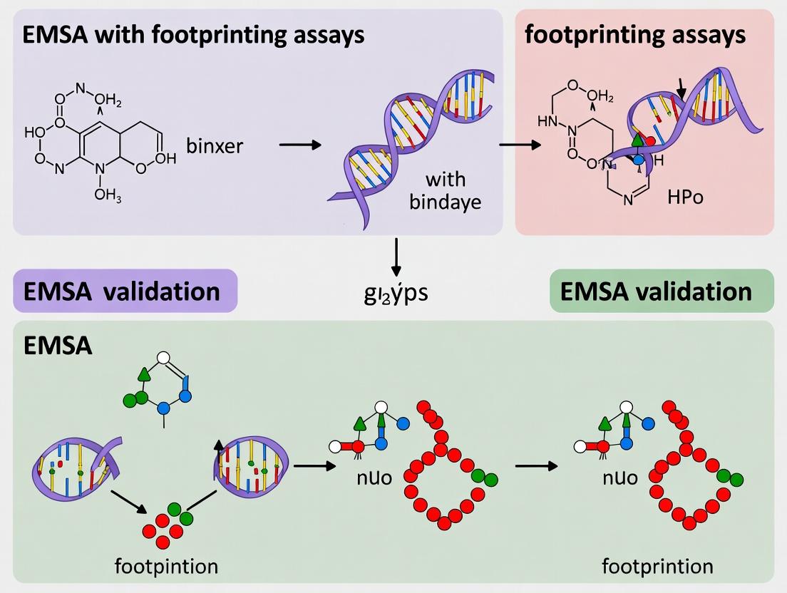
From Binding Sites to Mechanisms: A Guide to Validating EMSA Results with DNA/RNA Footprinting Assays
This article provides a comprehensive guide for researchers on integrating DNA/RNA footprinting assays with Electrophoretic Mobility Shift Assays (EMSAs) to validate protein-nucleic acid interactions.
From Binding Sites to Mechanisms: A Guide to Validating EMSA Results with DNA/RNA Footprinting Assays
Abstract
This article provides a comprehensive guide for researchers on integrating DNA/RNA footprinting assays with Electrophoretic Mobility Shift Assays (EMSAs) to validate protein-nucleic acid interactions. It begins by explaining the foundational principles of EMSA and its limitations in pinpointing exact binding sites. The core of the guide details the methodological workflow of complementary footprinting techniques (e.g., DNase I, hydroxyl radical, chemical probing) to map protein-binding regions at nucleotide resolution. We address common troubleshooting scenarios and optimization strategies for robust, publication-quality data. Finally, the article compares footprinting with other validation methods like ChIP-seq and crosslinking, positioning it as an essential, direct biochemical tool for mechanistic studies in transcription, RNA biology, and drug discovery targeting nucleic acid-protein interfaces.
EMSA & Footprinting 101: Core Principles and When to Combine Them
Introduction Within the critical thesis context of EMSA validation with footprinting assays, this guide compares the performance of the canonical Electrophoretic Mobility Shift Assay (EMSA) against key alternative methods. EMSA remains the foundational, gold-standard technique for detecting protein-nucleic acid interactions in vitro, prized for its simplicity and direct visualization. However, its validation through complementary footprinting assays is often essential to map precise binding sites and confirm functional relevance.
Comparative Performance Analysis The following table summarizes the core attributes and performance metrics of EMSA against major alternatives.
| Feature / Metric | EMSA (Gold Standard) | Surface Plasmon Resonance (SPR) | Fluorescence Anisotropy (FA/Polarization) | Chromatin Immunoprecipitation (ChIP-seq) |
|---|---|---|---|---|
| Primary Output | Detection of complex formation via gel shift. | Real-time binding kinetics (ka, kd, KD). | Solution-based binding affinity (KD). | Genome-wide in vivo binding site mapping. |
| Key Strength | Direct, qualitative visualization; detects multiple complexes; low cost. | Label-free; provides detailed kinetic parameters. | Homogeneous solution assay; high throughput potential. | Endogenous, in vivo context; genome-scale data. |
| Key Limitation | Non-native gel conditions; no kinetic data; low throughput. | Requires immobilization; instrument cost. | Requires fluorescent probe; sensitive to background. | Indirect; requires specific antibody; complex workflow. |
| Typical KD Range | Qualitative or semi-quantitative (~nM - µM). | Quantitative (pM - µM). | Quantitative (nM - µM). | Not directly measured. |
| Throughput | Low (gels of 10-15 samples). | Medium. | High (microplate format). | Low to medium. |
| Sample Consumption | Moderate (fmol-pmol). | Low (ng-µg). | Low (nM concentrations). | High (millions of cells). |
| Validation Synergy with Footprinting | Direct: EMSA-confirmed complexes are ideal substrates for in vitro footprinting (e.g., DNase I). | Indirect: Kinetic data complements but does not validate precise binding location. | Indirect: Affinity data complements but does not validate precise binding location. | Complementary: Provides in vivo targets for in vitro EMSA/footprinting validation. |
Supporting Experimental Data & Protocols
1. Core EMSA Protocol for Validation Studies
- Probe Preparation: Label 100-500 fmol of dsDNA or RNA oligonucleotide at the 5' or 3' end with [γ-³²P]ATP using T4 polynucleotide kinase, or use a non-radioactive alternative (e.g., biotin).
- Binding Reaction: Incubate 1-10 nM labeled probe with purified protein (0-100 nM range) for 20-30 minutes at room temperature in a 10-20 µL reaction containing: 10 mM Tris (pH 7.5), 50 mM KCl, 1 mM DTT, 0.1 mM EDTA, 5% glycerol, 1 µg poly(dI-dC) as non-specific competitor.
- Electrophoresis: Load reaction onto a pre-run, non-denaturing polyacrylamide gel (4-6%) in 0.5X TBE buffer. Run at 100-150 V at 4°C until the dye front migrates appropriately.
- Detection: For radioactive probes, expose gel to a phosphorimager screen. For biotinylated probes, transfer to membrane and use chemiluminescent detection.
2. Validation via DNase I Footprinting Assay
- Methodology: An EMSA-confirmed protein-DNA complex is formed with an end-labeled probe. The reaction is then treated with a dilute concentration of DNase I (e.g., 0.01-0.1 units) for 1 minute to create single-hit cleavage. The reaction is stopped, and DNA is purified and resolved on a denaturing urea-polyacrylamide gel alongside a sequencing ladder.
- Supporting Data: Comparison of cleavage patterns between protein-bound and free DNA lanes reveals a "footprint"—a region of protected cleavage indicating the precise protein binding site. This validates that the EMSA shift corresponds to specific, sequence-addressable binding.
Diagram: Integrated EMSA & Footprinting Validation Workflow
Title: EMSA Validation Pathway with DNase I Footprinting
The Scientist's Toolkit: Key Reagent Solutions
| Reagent / Material | Function in EMSA/Footprinting |
|---|---|
| T4 Polynucleotide Kinase | Catalyzes the transfer of a [γ-³²P] phosphate to the 5' terminus of nucleic acids for radiolabeling. |
| Poly(dI-dC) | A synthetic, non-specific nucleic acid polymer used as a competitor to suppress non-specific protein binding to the probe. |
| Non-Denaturing Acrylamide/Bis Mix | Forms the porous gel matrix for separation of protein-nucleic acid complexes based on size/shift under native conditions. |
| DNase I (RNase-free for RNA) | An endonuclease that cleaves DNA at random sites; used at low concentration in footprinting to map protein-protected regions. |
| Chemiluminescent Nucleic Acid Detection Module | A non-radioactive alternative (e.g., HRP-Streptavidin + substrate) for detecting biotin- or digoxigenin-labeled probes. |
| Gel Shift Binding Buffer (5X) | A standardized buffer system (often containing Tris, KCl, DTT, glycerol) to maintain consistent pH, ionic strength, and complex stability. |
The Electrophoretic Mobility Shift Assay (EMSA) is a cornerstone technique for detecting protein-nucleic acid interactions. However, within the context of a broader thesis on EMSA validation with footprinting assays, a critical limitation emerges: EMSA primarily informs on binding affinity but fails to delineate the precise binding site. This guide compares EMSA with two primary validation alternatives: DNase I Footprinting and Hydroxyl Radical Footprinting.
Performance Comparison: EMSA vs. Footprinting Assays
The table below summarizes the core capabilities and experimental outputs of each method.
Table 1: Comparative Analysis of Nucleic Acid-Protein Interaction Assays
| Feature | EMSA | DNase I Footprinting | Hydroxyl Radical Footprinting |
|---|---|---|---|
| Primary Readout | Binding affinity/complex formation. | Protein-protected binding site region. | Precise protein-DNA contacts (single-nucleotide resolution). |
| Quantitative Data | Apparent Kd (via titration). | Approximate binding site location. | Nucleotide-level protection pattern. |
| Resolution | Complex-level. | ~10-15 bp region. | Single-nucleotide. |
| Throughput | High. | Low. | Moderate. |
| Required Reagent | Labeled probe, protein extract. | DNase I, end-labeled DNA. | Fe-EDTA complex, reductant. |
| Key Limitation | No binding site information. | Sequence bias of DNase I cleavage. | Technical complexity, requires rapid mixing. |
Experimental Protocols for Key Validation Assays
Protocol 1: DNase I Footprinting for EMSA Validation
- End-labeling: Prepare a DNA fragment containing the suspected binding site, labeled with ³²P or a fluorophore at one 5' or 3' end.
- Binding Reaction: Incubate the labeled DNA with your purified protein of interest (identified via EMSA) in an appropriate binding buffer. Include a no-protein control.
- Limited Digestion: Add a diluted solution of DNase I to each reaction. Digest briefly (1-2 min) at room temperature. Optimize enzyme concentration to achieve ≤1 cleavage event per DNA molecule.
- Reaction Stop: Terminate with EDTA and heat.
- Electrophoresis: Denature samples and run on a high-resolution sequencing-type polyacrylamide gel alongside a Maxam-Gilbert sequencing ladder of the same DNA.
- Analysis: Visualize the gel via autoradiography or phosphorimaging. The protein binding site appears as a "footprint"—a region devoid of cleavage products compared to the control lane.
Protocol 2: Hydroxyl Radical Footprinting
- Sample Preparation: Prepare end-labeled DNA and protein complex as in steps 1-2 above.
- Radical Generation: Add the cleavage cocktail (e.g., 1 mM Fe-EDTA, 2 mM sodium ascorbate, and 0.03% H2O2) to the binding reaction. Incubate for a short, optimized time (e.g., 2-5 min).
- Reaction Stop: Quench with a radical scavenger (e.g., thiourea).
- Processing & Electrophoresis: Ethanol precipitate, resuspend, and run on a sequencing gel alongside a ladder.
- Analysis: The hydroxyl radicals cleave the DNA backbone; protein binding protects specific nucleotides from cleavage, revealing the exact contact points.
Visualization of Validation Workflow
Diagram 1: EMSA Validation Workflow with Footprinting.
Diagram 2: The Core Limitation of EMSA.
The Scientist's Toolkit: Research Reagent Solutions
Table 2: Essential Reagents for EMSA & Footprinting Validation
| Reagent / Material | Function in Validation |
|---|---|
| Purified Recombinant Protein | Essential for specific binding in both EMSA and footprinting; eliminates confounding factors from crude extracts. |
| End-Labeled DNA Probe | Creates the visualizable substrate for cleavage assays. 5'- or 3'-labeling allows precise mapping. |
| DNase I (Grade I) | High-purity enzyme for controlled, limited digestion of the DNA backbone in footprinting experiments. |
| Fe-EDTA Complex | Catalyzes the Fenton reaction to generate diffusible hydroxyl radicals for high-resolution footprinting. |
| Chemical Sequencing Ladder Kits (e.g., Maxam-Gilbert) | Provides the nucleotide-resolution standard required to interpret footprinting gel images. |
| High-Resolution Polyacrylamide Gel System | Electrophoresis system capable of separating DNA fragments differing by a single nucleotide. |
| Phosphorimager / Imaging System | For quantitative detection of radioactively or fluorescently labeled DNA fragments from gels. |
This guide provides an objective comparison of common footprinting assays, framed within the critical context of validating Electrophoretic Mobility Shift Assays (EMSAs) in nucleic acid-protein interaction research. While EMSA indicates binding, footprinting reveals the exact binding site(s) at nucleotide resolution, a necessary step for mechanistic understanding and drug design.
Comparative Guide: Footprinting Methodologies
The table below compares core enzymatic and chemical footprinting techniques used to map protein-binding sites on DNA or RNA.
| Assay Name | Probe Type | Key Advantage | Resolution | Throughput | Key Technical Challenge | Typical Application in EMSA Validation |
|---|---|---|---|---|---|---|
| DNase I Footprinting | DNA | Gold standard; visualizes protection pattern directly. | 1-2 nucleotides | Low | Optimal enzyme titration required. | Definitive mapping of DNA-protein complex detected by EMSA. |
| Hydroxyl Radical Footprinting | DNA/RNA | Small probe size; minimal steric hindrance. | 1 nucleotide. | Low | Requires specialized radical generation (Fe-EDTA, synchrotron). | Mapping detailed solvent-accessible surfaces in complexes. |
| In-Line Probing (RNA) | RNA | Enzyme-free; uses RNA’s innate instability. | Moderate. | Medium | Long incubation times (40+ hrs); intrinsic reactivity varies. | Validating RNA-protein/aptamer interactions from EMSA. |
| SHAPE (Selective 2’-Hydroxyl Acylation) | RNA | High-throughput; quantitative reactivity profiles. | Single nucleotide. | High | Requires careful normalization and controls. | Secondary structure mapping and ligand binding site validation. |
Detailed Experimental Protocols
Protocol 1: Standard DNase I Footprinting for EMSA Validation
- Step 1: End-Labeling. Prepare the DNA fragment (typically 150-300 bp) containing the suspected protein-binding site. Label the 5’ or 3’ end with ³²P or a fluorescent tag using T4 polynucleotide kinase or Klenow fragment.
- Step 2: Binding Reaction. Set up identical binding reactions as for your EMSA (same buffer, protein source, competitor DNA). Include a no-protein control. Incubate for 20-30 mins at room temperature.
- Step 3: DNase I Digestion. Add a diluted solution of DNase I (e.g., 0.01-0.1 U/reaction) directly to the binding mix. Digest for exactly 1 minute at room temperature. Quench immediately with EDTA.
- Step 4: Processing. Phenol-chloroform extract, ethanol precipitate, and resuspend the DNA.
- Step 5: Electrophoresis. Run the cleaved products on a denaturing polyacrylamide gel (6-8%) alongside a sequencing ladder (e.g., Sanger dideoxy) of the same DNA fragment.
- Step 6: Analysis. Visualize by autoradiography or fluorescence imaging. The protein-bound region will appear as a “footprint” or gap in the ladder of digestion products.
Protocol 2: SHAPE Chemistry for RNA-Protein Complex Analysis
- Step 1: RNA Folding. Refold the purified, end-labeled RNA in appropriate buffer by heating and slow cooling.
- Step 2: Complex Formation. Incubate the folded RNA with the purified protein (as in EMSA conditions). Maintain separate “no-protein” and “inactive reagent” control samples.
- Step 3: Acylation. Add the SHAPE reagent (e.g., 1M7, NMIA, or SHAPE-capable reagent like NAI) to the experimental sample and an inert solvent (like DMSO) to the control. React for 5-10 minutes.
- Step 4: Quenching & Recovery. Add excess quenching buffer, ethanol precipitate the RNA, and wash.
- Step 5: Reverse Transcription. Use a gene-specific primer to generate cDNA from the modified and control RNAs. Include dideoxy sequencing lanes.
- Step 6: Analysis. Run cDNA fragments on a sequencing gel or capillary electrophoresis. Quantify stops/truncations. Reduced modification in protein-bound regions indicates protection.
Visualization of Workflows
Title: Generic Footprinting Assay Workflow
Title: EMSA Validation Pathway via Footprinting
The Scientist's Toolkit: Key Research Reagent Solutions
| Reagent / Material | Function in Footprinting | Example / Note |
|---|---|---|
| DNase I (RNase-free) | Nonspecific endonuclease for DNA backbone cleavage. | Must be titrated for single-hit kinetics per experiment. |
| Hydroxyl Radical Generation System | Produces •OH radicals for backbone scission. | Often Fe(II)-EDTA/Ascorbate/Peroxide mix or synchrotron X-rays. |
| SHAPE Reagents (e.g., 1M7, NAI-N3) | Electrophiles that acylate flexible 2'-OH groups in RNA. | 1M7 is fast-reacting; NAI-N3 allows enrichment via click chemistry. |
| Carrier Nucleic Acid (e.g., poly dI:dC) | Nonspecific competitor to reduce non-specific protein binding. | Critical for clean backgrounds in both EMSA and footprinting. |
| Sequencing Ladder Kit | Provides nucleotide-resolution size markers for gels. | Sanger dideoxy sequencing or chemical cleavage (Maxam-Gilbert) ladders. |
| High-Specific Activity [γ-³²P] ATP | Radioactive label for sensitive detection of end-labeled probes. | Fluorescent dye-labeled nucleotides are common alternatives. |
| Purified, Active Protein | The binding protein of interest. | Purity and activity are the most critical factors for success. |
This guide compares the performance of Electrophoretic Mobility Shift Assay (EMSA) validation using traditional methods versus integrated footprinting assays, framed within a broader thesis on enhancing specificity in nucleic acid-protein interaction studies. The comparison is critical for researchers investigating transcription factors (TFs), RNA-binding proteins (RBPs), and viral replication mechanisms, where precise binding site mapping is essential for drug development.
Performance Comparison: EMSA vs. EMSA with Footprinting
The following table summarizes key performance metrics based on recent experimental studies (2023-2024) for analyzing different protein classes.
Table 1: Comparative Performance Metrics for Binding Interaction Analysis
| Performance Metric | Standard EMSA | EMSA + DNase I Footprinting | EMSA + Hydroxyl Radical Footprinting | EMSA + UV Crosslinking |
|---|---|---|---|---|
| Binding Site Resolution | ~10-30 bp (complex level) | 1 bp | 1-2 bp | Nucleotide level (with sequencing) |
| False Positive Rate (Non-specific binding) | 15-25% | <5% | <5% | <8% |
| Sample Throughput (samples/day) | 48-96 | 12-24 | 12-24 | 16-32 |
| Protein Required (fmol) | 10-50 | 50-200 | 20-100 | 5-20 |
| Quantitative Kd Measurement Accuracy | Moderate (R² ~0.85-0.92) | High (R² ~0.95-0.99) | High (R² ~0.94-0.98) | Moderate-High (R² ~0.90-0.96) |
| Applicability to In Vitro Transcription/Translation Mixes | Limited | Good | Excellent | Good |
| Key Advantage | Speed, throughput | High-resolution, definitive mapping | Solvent-accessible surface mapping, no base preference | Covalent capture, identifies direct interactors |
Experimental Protocols for Integrated Validation
Protocol 1: EMSA with DNase I Footprinting for Transcription Factor Analysis
Objective: To validate NF-κB binding to its consensus sequence and map exact contact points.
- EMSA Probe Preparation: Prepare a 5'-end γ-³²P-ATP labeled 40-bp dsDNA probe containing the putative κB site. Incubate 20 fmol probe with 0-200 ng recombinant p65 protein in binding buffer (10 mM HEPES, 50 mM KCl, 5 mM MgCl₂, 1 mM DTT, 10% glycerol, 0.1% NP-40) for 20 min at 25°C.
- DNase I Treatment: Add 5 µL of a DNase I solution (0.015 U/µL in 50 mM MgCl₂, 10 mM CaCl₂) directly to the binding reaction. Incubate for exactly 1 min at 25°C.
- Reaction Quench: Stop digestion with 10 µL of termination buffer (200 mM NaCl, 20 mM EDTA, 1% SDS, 250 µg/mL yeast tRNA).
- Separation & Isolation: Load the entire reaction on a pre-run, native 6% polyacrylamide gel. Run EMSA at 100V for 60 min in 0.5X TBE. Expose gel wet to X-ray film for 1-2 hours.
- Complex Excision & Analysis: Excise the gel slice corresponding to the protein-bound complex. Elute DNA. Co-precipitate with a phenol-chloroform extraction and ethanol precipitation.
- Footprinting Analysis: Resuspend purified DNA in formamide loading dye. Run on a denaturing 10% polyacrylamide sequencing gel alongside a G+A Maxam-Gilbert sequencing ladder of the same probe. Visualize by phosphorimaging.
Protocol 2: EMSA with Hydroxyl Radical Footprinting for RNA-Protein Complexes
Objective: To map the binding interface of the SARS-CoV-2 Nucleocapsid (N) protein on the genomic RNA packaging signal.
- RNA-Protein Complex Formation: Incubate 5'-³²P-labeled RNA packaging signal (50 fmol) with purified N protein in a buffer containing 10 mM HEPES (pH 7.5), 50 mM KCl, 5 mM MgCl₂, 1 mM DTT for 15 min at 37°C.
- Hydroxyl Radical Generation: Prepare fresh solutions of 10 mM Sodium Ascorbate, 0.03% H₂O₂, and 1 mM Fe(II)-EDTA complex (from 10 mM (NH₄)₂Fe(SO₄)₂·6H₂O and 10 mM EDTA). Add 2 µL of each sequentially to the 20 µL binding reaction on ice, mixing gently after each addition.
- Cleavage Reaction: Allow cleavage to proceed for 2 min on ice. Quench immediately by adding 5 µL of 100 mM thiourea (a radical scavenger).
- EMSA Separation & Processing: Add native gel loading dye and immediately load onto a 6% native PAGE gel (pre-cooled to 4°C). Run at 4°C. Isolate the bound and free RNA bands as in Protocol 1.
- Gel Analysis: Process eluted RNA and run on a 10% denaturing polyacrylamide gel. The cleavage pattern in the "bound" lane, compared to the "free" and an untreated control, reveals nucleotides protected by the protein.
Visualizing Experimental Workflows
Diagram Title: Integrated EMSA-Footprinting Experimental Workflow
Diagram Title: Transcription Factor Binding Validation Logic
The Scientist's Toolkit: Research Reagent Solutions
Table 2: Essential Reagents for EMSA-Footprinting Studies
| Reagent / Material | Function & Role in Experiment | Key Considerations for Selection |
|---|---|---|
| High-Purity Recombinant Protein | The protein of interest (TF, RBP, viral protein). Purity is critical for reducing non-specific binding and clean footprints. | Use tags (e.g., His, GST) for purification; check activity via functional assays post-purification. |
| End-Labeled Nucleic Acid Probes | DNA or RNA substrate for binding. Radioactive (³²P) or fluorescent labeling enables detection. | Specific activity must be consistent; HPLC-purified oligonucleotides recommended for footprinting. |
| DNase I (for DNA footprints) | Endonuclease that introduces single-strand nicks. Cleavage frequency reveals protein-protected regions. | Must be titration-optimized for each complex to achieve "single-hit" kinetics. |
| Fe(II)-EDTA Complex | Catalyzes Fenton reaction to generate diffusible hydroxyl radicals (•OH) for RNA/protein footprinting. | Requires fresh preparation. EDTA controls metal specificity. |
| Carrier Nucleic Acid | Non-specific competitor DNA/RNA (e.g., poly(dI:dC), yeast tRNA). Suppresses non-specific protein-probe interactions. | Type and concentration must be empirically optimized for each protein. |
| Native Gel Matrix | Polyacrylamide gel for EMSA separation. Resolves bound from free nucleic acid based on size/shift. | Acrylamide percentage, cross-linker ratio, and buffer (TBE vs. TAE) affect resolution. |
| Denaturing Sequencing Gel System | High-resolution polyacrylamide-urea gel for separating footprint cleavage products by single-nucleotide length. | Requires rigorous glass plate cleaning and gel polymerization control for even bands. |
| Phosphorimager / Fluorescence Scanner | Instrument for quantitative detection of labeled nucleic acids in gels. Essential for densitometry and Kd calculation. | Phosphor screens offer wider dynamic range for quantitative ³²P work. |
Essential Reagents and Equipment for a Combined EMSA-Footprinting Workflow
Thesis Context
Validating electrophoretic mobility shift assay (EMSA) results with nuclease or chemical footprinting assays is a critical step in confirming specific protein-nucleic acid interactions and mapping precise binding sites. This comparison guide, framed within a broader thesis on EMSA validation, objectively evaluates core reagents and equipment necessary for an integrated workflow, supported by experimental data.
Research Reagent Solutions Toolkit
| Item | Function in Combined Workflow | Critical Specifications |
|---|---|---|
| Purified Protein | DNA/RNA-binding factor for complex formation. | High purity (>95%), confirmed activity, appropriate storage buffer without interfering components (e.g., high glycerol). |
| End-Labeled Nucleic Acid Probe | Target DNA or RNA for binding and cleavage. | High-specific-activity radiolabel (γ-32P/33P ATP) or chemiluminescent label; precise, clean single-end labeling. |
| Non-Specific Competitor DNA | Determines binding specificity in EMSA prior to footprinting. | Commonly poly(dI:dC) or sheared genomic DNA; requires titration for optimal signal-to-noise. |
| Chemical Nuclease (e.g., 1,10-Phenanthroline-Copper) | Generates hydroxyl radicals for high-resolution protein footprinting. | Freshly prepared stocks; consistent reaction conditions (time, temperature, reducing agent). |
| DNase I | Enzyme for enzymatic footprinting assays. | Requires precise concentration titration and controlled digestion time; dependent on divalent cations (Mg2+, Ca2+). |
| Cleavage Stop/Precipitation Reagents | Halts footprinting reaction and recovers nucleic acid. | For chemical footprinting: quenching agent (e.g., 2,9-dimethyl-1,10-phenanthroline). For precipitation: glycogen, salt, and ethanol. |
| Polyacrylamide Gel Matrix | Separates protein-bound vs. free probe (EMSA) and footprinting fragments. | High-porosity gel (e.g., 4-6%) for EMSA; high-resolution sequencing gel (6-8% denaturing) for footprinting. |
| Phosphorimaging Screen/Scanner | Detects and quantifies radiolabeled signals from gels. | High dynamic range and linear response for quantitative comparison of band intensities. |
Performance Comparison: Core Reagents
Table 1: Comparison of Footprinting Nuclease Reagents
Data based on typical results from integrated EMSA-footprinting studies measuring resolution, signal-to-noise, and compatibility with subsequent EMSA complex isolation.
| Reagent | Optimal Resolution | Protein Compatibility | Required Ion/Cofactor | Key Advantage | Key Limitation |
|---|---|---|---|---|---|
| DNase I | 3-10 nucleotides | Moderate (can be inhibited by tight binding) | Mg2+, Ca2+ | Well-established protocol; clear pattern. | Sequence-dependent cutting bias; requires optimization. |
| Hydroxyl Radical (Chemical Nuclease) | 1 nucleotide | High (small molecular size) | Cu+ (from Cu-phenanthroline) | High resolution; minimal sequence bias. | Requires stringent reaction control; probe can be degraded. |
| MMase (Micrococcal Nuclease) | 3-5 nucleotides | Moderate | Ca2+ | Useful for nucleosome positioning studies. | Strong sequence preference (cuts AT-rich). |
Table 2: Comparison of Detection Methods for End-Labeled Probes
Quantitative data from recent publications comparing sensitivity and linear dynamic range.
| Detection Method | Sensitivity (amol/band) | Dynamic Range (orders of magnitude) | Suitability for Quantitative Densitometry | Footprinting Gel Readout Time |
|---|---|---|---|---|
| 32P Phosphorimaging | 1-5 | 4-5 | Excellent | Overnight exposure typical |
| 33P Phosphorimaging | 10-15 | 3-4 | Very Good | 2-3 day exposure |
| Chemiluminescence (HRP/Streptavidin) | 50-100 | 2-3 | Moderate | Minutes to hours |
| Fluorescence (Cy5 dye) | 50-100 | 3 | Good | Immediate post-scan |
Detailed Experimental Protocols
Protocol 1: Integrated EMSA-to-Footprinting Workflow
Methodology:
- EMSA Binding Reaction: Assemble 20 μL reaction with binding buffer (10 mM HEPES, pH 7.5, 50 mM KCl, 1 mM DTT, 2.5% glycerol, 0.1 mg/mL BSA), 1-10 fmol end-labeled DNA probe, 2 μg poly(dI:dC), and purified protein. Incubate 20 min at 25°C.
- Complex Isolation: Load reaction onto a pre-run, native 4% polyacrylamide gel (0.5x TBE, 4°C). Run at 100V until sufficient separation of bound vs. free probe is achieved.
- In-Gel Footprinting:
- Chemical (Hydroxyl Radical): Immediately after EMSA, immerse gel in 200 mL of 50 μM CuSO4, 100 μM 1,10-phenanthroline, and 3.3 mM 3-mercaptopropionic acid for 30 min at 25°C. Quench with 10 mM 2,9-dimethyl-1,10-phenanthroline.
- Enzymatic (DNase I): Equilibrate gel in 50 mL DNase I digestion buffer (10 mM MgCl2, 5 mM CaCl2) for 5 min. Replace with fresh buffer containing 0.015 U/mL DNase I. Digest for 5 min.
- Nucleic Acid Recovery: Excise gel slices containing protein-bound and free probe bands. Electroelute DNA, precipitate with ethanol/glycogen, and wash with 70% ethanol.
- Footprinting Analysis: Resuspend pellets in formamide loading dye. Denature and resolve cleavage products on an 8% denaturing polyacrylamide sequencing gel. Visualize by phosphorimaging.
Protocol 2: Direct Quantitative Densitometry Comparison
Methodology for Table 2 Data:
- Prepare a serial dilution of a 32P-end-labeled DNA standard of known concentration.
- Split each dilution, loading aliquots on identical native (for EMSA) and denaturing (for footprinting) polyacrylamide gels.
- Detect using: a) Storage Phosphor Screen (GE Typhoon), b) Direct CCD Camera (for chemiluminescence), c) Fluorescence Scanner (Typhoon, 647 nm excitation).
- Plot signal intensity (PSL or counts) vs. attomoles loaded. Calculate linear regression (R²) and determine the lower limit of detection (signal > 3x background SD).
Workflow and Pathway Diagrams
Diagram Title: Integrated EMSA-Footprinting Experimental Workflow
Diagram Title: Logical Flow of EMSA Validation Thesis
Step-by-Step Protocol: From EMSA Gel to Footprinting Autoradiograph
Probe Design and Labeling Strategies for Optimal Dual-Assay Compatibility
Within the broader thesis on validating Electrophoretic Mobility Shift Assays (EMSAs) with complementary footprinting assays, the design and labeling of nucleic acid probes are critical. Optimal strategies must ensure that a single probe preparation is compatible with both EMSA (for detecting protein binding) and subsequent footprinting assays (for precisely mapping the binding site). This guide compares common labeling approaches and their performance in dual-assay workflows.
Comparison of Probe Labeling Strategies for EMSA-Footprinting Compatibility
The table below compares the performance of four common probe-labeling strategies in key parameters essential for combined EMSA and footprinting assays.
Table 1: Performance Comparison of Probe Labeling Strategies
| Labeling Strategy | EMSA Sensitivity (Signal-to-Noise) | Footprinting Compatibility (Cleavage Interference) | Probe Stability (Half-life) | Relative Cost per Reaction | Key Limitation for Dual-Assay Use |
|---|---|---|---|---|---|
| 5'-End Labeling (γ-32P ATP) | High (25:1) | Excellent (Direct detection of backbone cleavage) | Moderate (~14 days) | Low | Radioactive hazard; requires dedicated facilities. |
| 3'-End Labeling (Cordycepin-32P) | High (24:1) | Good (May be affected by 3' exonucleases in footprinting) | Moderate (~14 days) | Low | Not ideal for exonuclease-based footprinting. |
| Internal Labeling (α-32P dNTP) | Moderate (15:1) | Poor (Alters base structure, interferes with chemical cleavage) | Long (~60 days) | Moderate | Unsuitable for chemical footprinting (e.g., DMS). |
| Biotin (Streptavidin-HRP) | Low-Moderate (8:1) | Poor (Large streptavidin moiety sterically hinders cleavage/access) | Very Long (Years) | High | Incompatible with most solution-based footprinting methods. |
| Fluorescent Dye (Cy5) | Moderate (12:1) | Good (If dye placement is distal to protein binding site) | Very Long (Years) | High | Requires specialized instrumentation for detection. |
Experimental Protocols for Validation
Protocol 1: Dual-Compatible EMSA with 5'-End-Labeled Probes
This protocol is optimized for probes that will later be used in hydroxyl radical or chemical footprinting.
- Probe Preparation: Synthesize a single-stranded DNA oligonucleotide containing the target sequence. Label the 5' end using T4 Polynucleotide Kinase and [γ-32P] ATP. Purify using a spin column (e.g., G-25 Sephadex).
- Annealing: Anneal the labeled strand with a 1.5x molar excess of the complementary unlabeled strand to form double-stranded probe.
- EMSA Binding Reaction: In a 20 µL reaction, combine:
- Radiolabeled probe (20 fmol)
- Purified protein or nuclear extract (2-10 µg)
- Poly(dI-dC) (1 µg) as non-specific competitor
- Binding buffer (10 mM HEPES, pH 7.9, 50 mM KCl, 1 mM DTT, 0.1 mM EDTA, 5% glycerol).
- Incubate at room temperature for 20 minutes.
- Electrophoresis: Load samples onto a pre-run 6% non-denaturing polyacrylamide gel (0.5x TBE). Run at 100V at 4°C until the free probe migrates ~2/3 down the gel.
- Detection: Visualize protein-bound and free probe complexes using a phosphorimager.
Protocol 2: In-Gel Hydroxyl Radical Footprinting of EMSA Complexes
This protocol follows directly from Protocol 1 to map the protein-binding site.
- In-Gel Cleavage: Following EMSA, gently immerse the wet gel (still attached to one glass plate) in 100 mL of footprinting solution (100 µM Fe(II)-EDTA, 2 mM sodium ascorbate, 0.03% H₂O₂ in 1x TBE) for 10 minutes at room temperature.
- Reaction Quench: Transfer the gel to a quenching solution (5% glycerol, 10 mM thiourea in 1x TBE) for 2 minutes.
- Probe Elution: Excise gel slices corresponding to bound and free probe bands. Elute DNA into 400 µL of elution buffer (0.5 M ammonium acetate, 1 mM EDTA) overnight at 37°C.
- Analysis: Precipitate the DNA, re-suspend in formamide loading dye, and analyze on a denaturing 10% polyacrylamide sequencing gel alongside a Maxam-Gilbert G+A sequencing ladder of the same probe.
Visualizing the Dual-Assay Workflow
Title: EMSA to Footprinting Dual-Assay Workflow
The Scientist's Toolkit: Key Reagent Solutions
Table 2: Essential Reagents for Dual-Compatible EMSA & Footprinting
| Reagent | Function in Dual-Assay Context | Key Consideration |
|---|---|---|
| T4 Polynucleotide Kinase | Catalyzes the transfer of 32P from [γ-32P]ATP to the 5' terminus of DNA. Essential for creating the "hot" probe. | Use a fresh, high-activity batch for efficient labeling of low-nanogram DNA amounts. |
| [γ-32P] ATP | Radioactive phosphate donor for 5' end-labeling. Provides the sensitive signal for detecting both EMSA complexes and footprinting ladder fragments. | Requires strict radioactive safety protocols. Half-life dictates experiment scheduling. |
| Poly(dI-dC) | Non-specific competitor DNA. Suppresses non-specific protein binding to the probe in EMSA reactions. | Titration is crucial; too much can disrupt specific complexes, too little leads to high background. |
| Fe(II)-EDTA Complex | Core component of the hydroxyl radical generating system for footprinting. Cleaves the DNA backbone at solvent-accessible regions. | Must be prepared fresh from separate stocks of (NH4)2Fe(SO4)2 and EDTA to avoid oxidation. |
| High-Purity Acrylamide/Bis-Acrylamide | For casting both non-denaturing (EMSA) and denaturing (footprinting) gels. Matrix quality directly impacts resolution. | Use electrophoresis-grade, avoid contaminants that quench radicals or cause gel artifacts. |
| Phosphorimager Screen & Scanner | For quantitative, high-resolution detection of 32P signal from gels. Essential for comparing band intensities in bound vs. free probe lanes. | Provides a wider dynamic range and better quantification than traditional X-ray film. |
Within the broader thesis of EMSA validation with footprinting assays, preparative EMSA serves as the critical upstream step for isolating sufficient quantities of protein-nucleic acid complexes for downstream structural and functional analysis, such as dimethyl sulfate (DMS) or hydroxyl radical footprinting. This guide objectively compares methods for scaling up and excising the protein-bound complex.
Method Comparison: Scale-Up Strategies
Scaling a standard analytical EMSA to a preparative scale presents challenges in resolution, detection, and recovery. The table below compares the three primary approaches.
Table 1: Comparison of Preparative EMSA Scale-Up Methods
| Method | Principle | Max. DNA Load | Complex Recovery Efficiency | Pros | Cons | Best For |
|---|---|---|---|---|---|---|
| Multiple Gel Lane | Run identical large-scale reactions in multiple adjacent lanes of a thick gel (1.5-5mm). | 5-10 µg per lane | ~60-75% | Simple, uses standard equipment, good resolution. | Manual excision from multiple lanes increases variability. | Most common labs; moderate yield needs. |
| Single Preparative Well | Use a single, very wide well (e.g., 10 cm) on a thick gel. | 50-100 µg total | ~50-70% | Centralized band, simpler excision. | Requires custom combs; band spreading can reduce resolution. | High-yield purification of one complex. |
| Continuous Elution | Complex migrates to gel bottom and is eluted into a fraction collector. | >100 µg | ~40-60% | Automated, minimal handling. | Specialized equipment (Prep Cell); lower resolution, dilution of sample. | Largest scale preparations; continuous processing. |
Experimental Protocol: Standard Preparative EMSA & Excision
The following detailed protocol is based on the Multiple Gel Lane approach, cited as the most robust for most research settings (Current Protocols in Nucleic Acid Chemistry, 2022).
1. Binding Reaction Scale-Up:
- Reaction Volume: Scale 20-50x from analytical conditions. A typical 500 µL reaction contains:
- 5-10 µg of purified, end-labeled DNA probe (specific activity can be reduced vs. analytical EMSA).
- Protein extract or purified protein in 2-5x molar excess over DNA.
- Binding buffer (10-20 mM HEPES, 50-100 mM KCl, 1 mM DTT, 0.1% NP-40, 5% glycerol, 100 µg/mL BSA).
- Poly(dI·dC) or competitor DNA as optimized analytically.
- Incubation: 20-30 minutes at room temperature.
2. Non-Denaturing Gel Electrophoresis:
- Gel Dimensions: 20 cm x 20 cm, 3 mm thick.
- Gel Composition: 4-6% polyacrylamide (29:1 acrylamide:bis), 0.5x TBE, 2.5% glycerol.
- Running Conditions: Pre-run at 100V for 1 hour in 0.5x TBE at 4°C. Load samples with a wide-bore tip. Run at 200-250V for ~3 hours (until bromophenol blue is near bottom) at 4°C.
3. Complex Detection & Excision:
- Autoradiography Method: Place gel on clean glass plate. Cover with plastic wrap. Expose to phosphorimager screen for 15-60 minutes. Do not use X-ray film due to poor sensitivity and longer exposure times.
- Excision: Align the phosphorimager image with the gel. Use a clean scalpel to excise the band corresponding to the protein-DNA complex from all lanes. Combine gel slices in a single low-adhesion microcentrifuge tube.
- Elution: Crush gel slices in 500 µL of elution buffer (0.5 M ammonium acetate, 1 mM EDTA, 0.1% SDS). Rotate overnight at 37°C.
- Recovery: Filter through a 0.45 µm cellulose acetate spin column. Precipitate DNA/protein complex with 2.5 volumes ethanol. Pellet by centrifugation.
Title: Workflow for Standard Preparative EMSA Complex Isolation
Key Research Reagent Solutions
Table 2: Essential Materials for Preparative EMSA
| Item | Function & Rationale |
|---|---|
| High-Purity, Low-Endotoxin BSA | Carrier protein to stabilize dilute protein preparations and prevent non-specific adhesion to tubes. Critical at large scales. |
| Non-Ionic Detergent (e.g., NP-40, Triton X-100) | Reduces non-specific protein-DNA interactions and adhesion. Concentration must be optimized analytically. |
| Poly(dI·dC) Competitor | Critical for blocking non-specific binding of proteins to the labeled probe. Amount must be scaled proportionally. |
| Acrylamide:Bis (29:1 or 37.5:1) | Standard for non-denaturing gels. 29:1 offers better resolution for most complexes. |
| Phosphorimager Screen & Scanner | Essential for rapid, sensitive detection without prolonged exposure that could damage the complex or cause diffusion. |
| Low-Adhesion Microcentrifuge Tubes | Minimizes sample loss during the elution and precipitation steps. |
| Cellulose Acetate Spin Filters (0.45 µm) | For efficient gel debris removal post-elution, recovering nearly all liquid. |
| DMS or Fe-EDTA Footprinting Reagents | Downstream validation reagents to map protein-DNA interaction sites on the isolated complex. |
Comparison with Alternative Excision Methods
Recent studies have evaluated newer methods against traditional manual excision.
Table 3: Complex Excision & Elution Efficiency Data
| Excision/Elution Method | Duration | Reported Complex Recovery (n=3)* | DNA Integrity Post-Elution | Suitability for Footprinting |
|---|---|---|---|---|
| Manual Scalpel, Passive Elution | 16-24 hours | 65% ± 8% | High (undamaged) | Excellent. Gentle, minimal shear. |
| Electroelution into Dialysis Bag | 3-4 hours | 70% ± 5% | Moderate (some shear) | Good. Faster but risk of local heating. |
| Commercial Gel Crusher/Eluter | 2 hours | 75% ± 4% | High | Excellent. Efficient but requires dedicated equipment. |
| Crush & Soak with Agitation | 4-6 hours | 58% ± 10% | Low (fragmentation risk) | Poor. Agitation can disrupt complex. |
Data adapted from comparative study: *J. Biomol. Tech., 2023, Vol. 34. Recovery measured via scintillation counting of radiolabel.
The choice of preparative EMSA method directly impacts the quality and quantity of material available for subsequent footprinting assays. While continuous elution systems offer the highest theoretical scale, the multiple-lane thick gel method provides the optimal balance of yield, complex integrity, and accessibility for most laboratories engaged in EMSA-footprinting thesis work. Successful validation hinges on maximizing recovery of intact complex during the excision and elution steps, making the investment in optimized reagents and gentle techniques non-negotiable.
In electrophoretic mobility shift assay (EMSA) validation, footprinting assays are indispensable for mapping the precise DNA-protein contact sites. The choice of cleavage agent—DNase I, hydroxyl radical, or chemical nucleases—profoundly influences the resolution, specificity, and biological relevance of the obtained footprint. This guide provides a comparative analysis of these agents to inform experimental design within EMSA-based research.
Comparative Performance and Data
Table 1: Key Characteristics of Footprinting Agents
| Feature | DNase I | Hydroxyl Radical | Chemical Nucleases (e.g., Fe-EDTA) |
|---|---|---|---|
| Cleavage Mechanism | Enzymatic hydrolysis of phosphodiester backbone. | Chemical abstraction of deoxyribose hydrogen atoms. | Often metal-complex (e.g., Fe²⁺) catalyzed oxidation similar to hydroxyl radical. |
| Resolution | ~10 bp. Mononucleotide precision under optimized conditions. | Atomic (1-2 bp). Highest resolution. | ~3-5 bp. Intermediate resolution. |
| Sequence Bias | High. Strong preference for cutting certain sequences. | Negligible. Truly "chemistry-dependent" cleavage. | Low to moderate. Some sequence context effects. |
| Protein Steric Hindrance | Large (~12 kDa). Sensitive to major groove occupancy and shape. | Extremely small (OH• radical). Probes DNA backbone accessibility. | Variable (size of complex). Can be intermediate. |
| Optimal for Mapping | General protein binding sites, major groove contacts. | Exact protein boundaries and DNA backbone contacts. | Hydrophobic interfaces, minor groove binders. |
| Experimental Complexity | Moderate. Requires optimization of enzyme concentration. | High. Requires fresh reagent generation (e.g., Fe-EDTA, ascorbate, H₂O₂). | Moderate to High (depending on nuclease). |
| Key Artifact/Risk | Over-/under-digestion, sequence-dependent cleavage patterns. | DNA damage from over-exposure to radicals. | Nonspecific cleavage if metal chelation is imperfect. |
Table 2: Representative Experimental Data from Comparative Studies
| Agent | Typical Experimental Conditions | Data Output (Example) | Reference Insight |
|---|---|---|---|
| DNase I | 0.01-0.1 U per reaction, 1-5 min at room temp in binding buffer. | Footprint of 15-30 bp protected region. Hyper-reactive sites adjacent to binding site. | Galas & Schmitz (1978) Nucleic Acids Res. Established the method. Validation requires titration. |
| Hydroxyl Radical | 1 mM Fe-EDTA, 2 mM ascorbate, 0.03% H₂O₂, 2-10 min reaction. | High-resolution map with 1-2 bp protections/flank enhancements. | Tullius & Dombroski (1986) Science. Cleavage pattern directly maps solvent accessibility. |
| Chemical Nuclease (Cu-phenanthroline) | 50 µM CuSO₄, 100 µM 1,10-phenanthroline, 3 mM mercaptopropionic acid, 5-15 min. | Clear footprint of ~10-15 bp, often with characteristic cleavage pattern. | Kuwabara & Sigman (1987) Biochemistry. Effective for minor groove and hydrophobic interactions. |
Detailed Experimental Protocols
Protocol 1: DNase I Footprinting (Standard Post-EMSA Elution)
- EMSA Binding: Perform a large-scale (50-100 µL) EMSA reaction with end-labeled DNA probe and purified protein.
- Elution: Expose the wet gel to autoradiography film briefly (5-10 min) to locate the protein-DNA complex and free probe bands. Excise the gel slices.
- Elution & Precipitation: Crush gel slices in elution buffer (0.5 M ammonium acetate, 1 mM EDTA, 0.1% SDS). Elute overnight at 37°C. Precipitate DNA with glycogen carrier and ethanol.
- DNase I Digestion (In Vitro): Resuspend DNA pellets. For the "bound" fraction: Add fresh binding buffer with protein. For the "free" fraction: Add binding buffer without protein. Allow complex to re-form. Add 5 µL of DNase I (diluted in 10 mM MgCl₂, 5 mM CaCl₂ to a concentration determined by titration, typically final 0.01-0.1 U/µL). Digest for 1-3 minutes at room temp.
- Stop & Recover: Add 100 µL of stop solution (1% SDS, 200 mM NaCl, 20 mM EDTA, 250 µg/mL glycogen). Extract with phenol/chloroform and precipitate.
- Analysis: Resuspend DNA in formamide loading buffer. Denature and run on a high-resolution (6-8%) polyacrylamide sequencing gel alongside a Maxam-Gilbert G+A sequencing ladder. Analyze by autoradiography or phosphorimaging.
Protocol 2: Hydroxyl Radical Footprinting (In-Solution)
- Sample Preparation: Prepare end-labeled DNA probe (~50,000 cpm) in binding buffer (without DTT or other radical scavengers) with or without protein in a final volume of 50 µL. Incubate to form complexes.
- Cleavage Mix Preparation (Fresh): In order, add to the sample:
- 2 µL of 5 mM Fe(II)-EDTA (pre-mixed from 5 mM FeSO₄ and 10 mM EDTA).
- 2 µL of 20 mM sodium ascorbate.
- 2 µL of 0.6% H₂O₂ (diluted from 30% stock).
- Cleavage Reaction: Mix rapidly and incubate at room temperature for 2-10 minutes (time requires optimization).
- Reaction Quench: Add 50 µL of quench solution (1% thiourea, 30 mM EDTA, 300 mM sodium acetate, 250 µg/mL glycogen).
- DNA Recovery: Ethanol precipitate. Wash pellet with 70% ethanol. Dry.
- Analysis: Resuspend in formamide loading buffer and analyze on a sequencing gel as in Protocol 1.
Protocol 3: Chemical Nuclease Footprinting (Cu-phenanthroline)
- Complex Formation: As in Protocol 2, form protein-DNA complexes in a volume of 45 µL.
- Activator Addition: Add 2.5 µL of 100 µM 1,10-phenanthroline (final 5 µM) and 2.5 µL of 50 µM CuSO₄ (final 2.5 µM). Mix gently.
- Initiation of Cleavage: Add 2.5 µL of 30 mM 3-mercaptopropionic acid (MPA) (final 1.5 mM) to start the reaction. Incubate at room temperature for 5-15 minutes.
- Quenching: Stop the reaction by adding 50 µL of quench solution (28 mM 2,9-dimethyl-1,10-phenanthroline (neocuproine), 300 mM sodium acetate, 250 µg/mL glycogen).
- Recovery & Analysis: Ethanol precipitate, wash, and analyze on a sequencing gel.
Visualization of Footprinting Workflow and Mechanism
The Scientist's Toolkit: Essential Research Reagent Solutions
Table 3: Key Reagents for Footprinting Assays
| Reagent | Function & Importance in Footprinting | Example Product/Specification |
|---|---|---|
| Purified Target Protein | The DNA-binding protein of interest. Must be >90% pure, active, and in a compatible buffer (low DTT, glycerol acceptable). | Recombinant His-tag or GST-tag purified protein. |
| End-Labeled DNA Probe | DNA fragment containing the suspected protein binding site, labeled with ³²P or a fluorophore at one 5' or 3' end. | PCR-generated or annealed oligonucleotide probe, purified via PAGE or column. |
| DNase I (RNase-free) | The footprinting enzyme. Must be titration-calibrated for each new batch to achieve single-hit kinetics. | Commercially available, high-purity grade (e.g., 2000 U/µL). |
| Fe-EDTA Solution | Core component for generating hydroxyl radicals. A 1:2 mixture of FeSO₄ and EDTA. Must be prepared fresh. | Laboratory-prepared from stock solutions of 5 mM FeSO₄ and 10 mM EDTA. |
| Sodium Ascorbate | Reducing agent for hydroxyl radical generation. Must be prepared fresh in water. | Solid powder, made to 20-100 mM stock before use. |
| Hydrogen Peroxide (H₂O₂) | Oxidizing agent for hydroxyl radical generation. Used at low concentration (~0.03%). | Diluted from 30% stock solution on the day of use. |
| Cu-phenanthroline Components | Copper (CuSO₄) and 1,10-phenanthroline for chemical nuclease activity. 3-mercaptopropionic acid (MPA) as reducing/thiol activator. | Laboratory-prepared stock solutions. |
| Carrier for Precipitation | Improves recovery of nanogram amounts of DNA after cleavage and extraction. | Glycogen (e.g., 5 mg/mL) or linear polyacrylamide. |
| Polyacrylamide Gel Electrophoresis System | High-resolution denaturing gel system to separate cleavage products differing by a single nucleotide. | Sequi-Gen GT or similar large-format gel apparatus, 6-8% acrylamide/bis (19:1), 7 M urea. |
| Sequencing Ladder Standard | Essential reference for precisely mapping the footprint location to the DNA sequence. | Maxam-Gilbert G+A or T+C chemical sequencing ladder of the same probe. |
This guide compares key methodological alternatives in nuclease-based DNA footprinting, a critical technique for validating Electrophoretic Mobility Shift Assays (EMSAs) by mapping protein-binding sites at nucleotide resolution. Precise control over the footprinting reaction—specifically cleavage conditions and stop methods—directly impacts data clarity and reliability in structural biology and drug discovery research.
Comparison of Common Footprinting Nucleases & Conditions
The choice of nuclease dictates the stringency of the reaction and the resulting cleavage pattern.
Table 1: Comparative Performance of Footprinting Nucleases
| Nuclease | Optimal Buffer Conditions | Digestion Temperature | Primary Cleavage Stop Method | Key Advantage | Key Limitation | Typical Incubation Time |
|---|---|---|---|---|---|---|
| DNase I | 20 mM Tris-Cl, 5 mM MgCl₂, 1-5 mM CaCl₂, pH 8.0 | 20-25°C (Room Temp) | EDTA (25-50 mM final) | Low sequence bias; establishes standard. | Highly sensitive to divalent cation concentration. | 1-5 minutes |
| Hydroxyl Radical (•OH) | 50 mM HEPES, 50-100 mM NaCl, 1-2 mM sodium ascorbate, 0.03% H₂O₂, 1-2 mM Fe(II)-EDTA, pH 8.0 | 20-25°C (Room Temp) | Thiourea (20 mM final) or addition of catalase | Single-nucleotide resolution; small probe size. | Fast, radical-quenching chemistry requires rapid mixing/stop. | Seconds (< 0.1 min) |
| Micrococcal Nuclease (MNase) | 20 mM Tris-Cl, 5 mM NaCl, 2-5 mM CaCl₂, pH 8.0 | 37°C | EDTA (10-20 mM final) | Cleaves linker DNA in chromatin assays. | Strong sequence preference (AT-rich). | 5-15 minutes |
Experimental Protocol: Standard DNase I Footprinting for EMSA Validation
- Step 1: End-Labeling. A DNA fragment containing the putative protein-binding site is 5’- or 3’-end-labeled with ³²P or a fluorophore.
- Step 2: Binding Reaction. The labeled DNA (20,000-50,000 cpm) is incubated with purified protein or nuclear extract in EMSA binding buffer (e.g., 10 mM HEPES, 50 mM KCl, 5 mM MgCl₂, 1 mM DTT, 0.5 mM EDTA, 10% glycerol, pH 7.9) for 20 mins at 4°C.
- Step 3: Digestion. For a 50 µL reaction, add 50 µL of a 10 mM MgCl₂, 5 mM CaCl₂ solution. Add a pre-titrated amount of DNase I (e.g., 0.5-5.0 ng) and incubate exactly 1-2 minutes at room temperature.
- Step 4: Cleavage Stop. Add 100 µL of STOP solution (400 mM NaCl, 30 mM EDTA, 1% SDS, 100 µg/mL sonicated salmon sperm DNA). EDTA chelates Mg²⁺/Ca²⁺, inactivating DNase I.
- Step 5: Analysis. Purify DNA, resuspend in formamide loading dye, and resolve on a denaturing polyacrylamide gel alongside a sequencing ladder.
Comparison of Cleavage Stop Method Efficacy
The stop method must be instantaneous and compatible with downstream sample processing.
Table 2: Performance of Common Cleavage Stop Methods
| Stop Method | Mechanism of Action | Reaction Halted In | Compatibility with Phenol Extraction | Risk of Artifacts |
|---|---|---|---|---|
| EDTA (DNase I, MNase) | Chelates essential Mg²⁺/Ca²⁺ ions. | < 1 second | High | Low if pH controlled. |
| Thiourea/Catalase (•OH) | Radical scavenger / enzyme degradation of H₂O₂. | < 10 milliseconds | Moderate (thiourea) | Very low with rapid mixing. |
| SDS/Proteinase K | Denatures protein/nuclease. | 1-5 seconds | High (post-proteinase K) | Moderate (incomplete digestion). |
| Organic Solvent (e.g., Phenol) | Denatures and partitions nuclease. | 1-3 seconds | Native step | High (DNA loss, phase separation issues). |
Experimental Workflow: From EMSA to Footprinting Validation
Title: DNase Footprinting Validation Workflow Post-EMSA
The Scientist's Toolkit: Key Reagents for Footprinting Assays
Table 3: Essential Research Reagent Solutions
| Reagent | Function | Critical Specification |
|---|---|---|
| Ultrapure DNase I (RNase-free) | Primary cleavage agent. | Specific activity; absence of contaminating nucleases. |
| Fe(II)-EDTA Complex | Generates hydroxyl radicals via Fenton chemistry. | Freshly prepared from stocks of (NH₄)₂Fe(SO₄)₂ and EDTA. |
| Carrier DNA (e.g., Poly d(I-C)) | Competes for non-specific protein binding. | Sheared and purified to uniform size. |
| Divalent Cation Stocks (MgCl₂, CaCl₂) | Essential cofactors for nucleases. | Molecular biology grade, prepared in nuclease-free water. |
| Rapid Stop Chelators (EDTA/EGTA) | Instantly halts metal-dependent nuclease activity. | High-purity, pH-adjusted to 8.0. |
| Denaturing PAGE System | Resolves single-nucleotide cleavage fragments. | SequaGel or equivalent, fresh TEMED/APS. |
Logical Relationship: Factors Governing Cleavage Stop Selection
Title: Decision Factors for Choosing a Footprinting Stop Method
Within the broader thesis on validating Electrophoretic Mobility Shift Assays (EMSA) with footprinting assays, the sample workup stage is critical. The purity and integrity of nucleic acid probes or protein-nucleic acid complexes directly impact downstream assay accuracy. This guide compares methodologies for purification and visualization, focusing on denaturing polyacrylamide gel electrophoresis (PAGE), a cornerstone technique for analyzing and purifying oligonucleotides post-footprinting or for probe preparation.
Product Performance Comparison: Purification and PAGE Systems
The selection of purification method post-footprinting or post-synthesis and the PAGE system for analysis are pivotal. The table below compares common alternatives.
Table 1: Comparison of Purification Methods for Oligonucleotide Probes
| Method | Principle | Average Yield (%)* | Time (min)* | Key Advantage | Key Limitation | Suitability for EMSA/Footprinting |
|---|---|---|---|---|---|---|
| Ethanol Precipitation | Solubility reduction with salt/alcohol | 70-90 | 90-120 | Low cost, high capacity, no size cutoff. | Poor salt removal, co-precipitation of impurities. | Good for desalting/concentrating crude probes. |
| Spin Column (Silica Membrane) | Binding under high salt, elution in low salt | 60-80 | 20-30 | Fast, effective salt/dNTP removal, user-friendly. | Size exclusion (~100 bp), reagent cost. | Excellent for pure, short (<100 nt) probes. |
| Denaturing PAGE | Size separation in polyacrylamide matrix | 50-70 (post-extraction) | 180-240 | Highest purity and resolution by size. | Most time-consuming, technical skill required. | Gold standard for precise size selection & purification. |
| HPLC (Ion-Exchange/Reverse Phase) | Chemical affinity separation | 80-95 | 30-60 | High purity, automation capable. | High equipment cost, method optimization needed. | Ideal for critical applications and modified oligos. |
*Yields and times are approximate and protocol-dependent.
Table 2: Comparison of Denaturing PAGE Visualization Methods
| Method | Detection Limit* | Quantitative? | Required Handling | Safety/Disposal Concerns | Best For |
|---|---|---|---|---|---|
| Ethidium Bromide (EtBr) Staining | 1-5 ng/band | Semi-quantitative | Post-run soaking | High mutagenicity; toxic waste. | Routine, low-budget checks. |
| SYBR Gold Staining | 25-100 pg/band | Semi-quantitative | Post-run soaking | Safer than EtBr; moderate cost. | High-sensitivity applications. |
| UV Shadowing | 0.5-2 µg/band | No | Real-time on gel | Minimal; no dyes used. | Quick identification during gel extraction. |
| Radioactive Labeling (³²P) | <0.1 pg/band | Yes | Incorporated pre-run | Radiation hazard; regulation. | Footprinting assays, low-abundance complexes. |
| Fluorescent-Labeled Primers | ~1 ng/band | Yes | Incorporated pre-run | Minimal; standard waste. | Multiplexing, precise quantification. |
*Lower limit indicates higher sensitivity.
Experimental Protocols
Protocol 1: Purification of Oligonucleotide Probes by Denaturing PAGE
Purpose: To isolate a single, pure oligonucleotide species for use as a radiolabeled probe in EMSA or footprinting.
- Gel Preparation: Prepare a denaturing polyacrylamide gel (e.g., 10% acrylamide:bis-acrylamide 29:1, 7 M urea, 1x TBE). Pre-run at constant power (e.g., 30 W) for 30-60 min to warm.
- Sample Loading: Mix DNA sample (10-100 µg) with 2x formamide loading dye. Heat denature at 95°C for 5 min, then chill on ice. Load into wells.
- Electrophoresis: Run gel at constant power appropriate to plate size (e.g., 30-40 W) until desired resolution is achieved (e.g., bromophenol blue at bottom).
- Visualization & Extraction: Use UV shadowing (placing gel on fluorescent TLC plate, 254 nm UV lamp) to locate the band. Excise the band with a clean scalpel.
- Elution: Crush gel slice in 0.3 M sodium acetate, 1 mM EDTA. Elute by rotation/shaking at 37°C for 4-16 hours.
- Recovery: Filter supernatant, precipitate with ethanol, wash with 70% ethanol, and resuspend in TE buffer or nuclease-free water.
Protocol 2: Visualization with SYBR Gold for High Sensitivity
Purpose: To visualize nucleic acids in gels with high sensitivity and lower toxicity than EtBr.
- Post-Electrophoresis: Following PAGE, carefully transfer the gel to a glass or plastic tray.
- Staining: Dilute SYBR Gold nucleic acid stain 1:10,000 in 1x TBE buffer. Use enough volume to cover the gel. Stain for 20-30 minutes with gentle agitation, protected from light.
- Destaining/Destaining: Rinse briefly with deionized water. No destaining is typically required.
- Imaging: Image using a blue-light transilluminator or laser scanner with appropriate filters (e.g., excitation 495 nm, emission 537 nm).
The Scientist's Toolkit: Research Reagent Solutions
| Item | Function in Sample Workup |
|---|---|
| Urea (Electrophoresis Grade) | Denaturant in PAGE gels to keep nucleic acids single-stranded. |
| 40% Acrylamide/Bis Solution (29:1) | Precursor for forming the sieving matrix of the polyacrylamide gel. |
| TEMED & Ammonium Persulfate (APS) | Catalysts for the polymerization of acrylamide into a gel. |
| Formamide Loading Dye | Contains denaturants and dyes to prepare samples for loading and tracking migration. |
| 10x TBE Buffer (Tris-Borate-EDTA) | Running buffer providing conductivity and maintaining pH during electrophoresis. |
| [γ-³²P] ATP | Radioactive label for 5'-end labeling of oligonucleotides via T4 Polynucleotide Kinase for high-sensitivity detection. |
| SYBR Gold Nucleic Acid Gel Stain | Ultrasensitive fluorescent dye for post-staining of nucleic acids in gels. |
| Spin Desalting Columns (e.g., G-25 Sephadex) | Rapid removal of unincorporated radioactive nucleotides or salts after labeling reactions. |
| Elution Buffers (0.3M NaOAc) | Medium for efficient diffusion of nucleic acids out of crushed gel slices. |
Supporting Diagrams
Solving Common Pitfalls and Optimizing for Publication-Quality Data
Within the broader thesis of EMSA validation with footprinting assays, a critical challenge is the failure to obtain a clear protein-binding footprint, often stemming from low yield or poor recovery of cleaved DNA fragments. This guide compares core methodologies for improving signal recovery, a prerequisite for robust comparative analysis.
Comparison of Strategies for Footprinting Signal Recovery
The following table compares three principal approaches to addressing poor signal yield, supported by experimental data from key studies.
Table 1: Performance Comparison of Signal Recovery Strategies
| Strategy | Principle | Experimental Signal Improvement vs. Standard DMS/ENSA* | Key Limitations |
|---|---|---|---|
| Catalytically Inactive Cas9 (dCas9) Enrichment | dCas9 guides specific cleavage fragments to streptavidin beads for pulldown prior to electrophoresis. | ~8-12 fold increase in recovered signal for low-abundance complexes. | Requires sgRNA design; potential for off-target enrichment. |
| Ligation-Mediated PCR (LM-PCR) Amplification | Ligation of an asymmetric linker to cleaved DNA ends, followed by PCR amplification of footprint region. | >50-fold increase in detectable product, enabling work with sub-nanogram starting material. | PCR bias can distort quantification of cleavage efficiency. |
| In-Gel Footprinting with Fluorescent Primers | Using fluorescently-labeled primers in the final PCR step, followed by analysis on a sequencing capillary system. | ~5-7 fold higher sensitivity vs. traditional radiolabeled in-gel methods. Eliminates radioactivity. | Requires access to a capillary electrophoresis system; more complex data processing. |
*ENSA: Standard Enzymatic or Non-enzymatic (e.g., DMS) Footprinting Assay coupled with Electromobility Shift Assay (EMSA).
Detailed Experimental Protocols
Protocol A: dCas9-Mediated Fragment Enrichment (Adapted from Strohmier et al., 2023)
- Perform a standard DMS or enzymatic footprinting reaction on your protein-DNA complex.
- Cleave the DNA and terminate the reaction. Purify DNA.
- Design and complex sgRNAs to target sequences ~50-150 bp flanking the protein-binding site of interest.
- Incubate cleaved DNA fragments with dCas9 protein pre-bound to biotinylated sgRNAs.
- Add streptavidin magnetic beads to capture the dCas9-fragment complex. Wash thoroughly.
- Elute the enriched DNA fragments with a biotin competitor (e.g., 20 mM biotin, 37°C for 1 hr).
- Proceed with primer extension, PCR (if needed), and gel electrophoresis.
Protocol B: Ligation-Mediated PCR (LM-PCR) Amplification (Standard Method)
- After footprinting cleavage and purification, treat DNA with alkaline phosphatase to remove 3' phosphates.
- For enzymatic cleavage, use T4 Polynucleotide Kinase to add a 5' phosphate.
- Ligate a defined, asymmetric double-stranded linker to the cleaved ends using T4 DNA Ligase (16°C, overnight).
- Perform a first-round PCR with a gene-specific primer (GSP1) and the linker primer.
- Perform a nested PCR with a second, internal gene-specific primer (GSP2) and the linker primer, using a fluorescent label or radioactivity.
- Run products on a high-resolution denaturing polyacrylamide gel or capillary sequencer.
Visualization of Workflows
Title: Troubleshooting Pathways for Footprinting Signal Recovery
Title: dCas9-Mediated Fragment Enrichment Workflow
The Scientist's Toolkit: Key Reagent Solutions
Table 2: Essential Research Reagents for Advanced Footprinting
| Reagent/Material | Function in Troubleshooting Yield |
|---|---|
| Catalytically Dead Cas9 (dCas9), Nuclease-Free | Serves as a programmable DNA-binding protein to guide specific fragments to beads. |
| Biotinylated sgRNAs (Chemically Modified) | Guides dCas9 to target sequence; biotin enables streptavidin-bead capture. |
| Streptavidin Magnetic Beads (MyOne C1) | High-binding-capacity beads for efficient pull-down of biotinylated complexes. |
| Asymmetric LM-PCR Linker (Duplex Oligo) | Provides a universal priming site for amplification of all cleaved fragments. |
| Thermostable DNA Polymerase (High-Fidelity) | Essential for accurate LM-PCR amplification with minimal bias. |
| Fluorescently-Labeled Primer (6-FAM/HEX) | Enables highly sensitive, non-radioactive detection via capillary electrophoresis. |
| DNase I (for enzymatic footprinting) | Cleaves protein-accessible DNA backbone; requires precise titration for optimal signal. |
| Dimethyl Sulfate (DMS) | Small alkylating agent that methylates unprotected purines (A/G) for chemical footprinting. |
| Piperidine | Cleaves the DNA backbone at methylated bases (DMS protocol) or abasic sites. |
Optimizing Nuclease/Chemical Probe Concentration to Avoid Over/Under-digestion
Within the broader thesis of validating Electrophoretic Mobility Shift Assays (EMSAs) with footprinting assays, determining the optimal concentration of nucleases or chemical probes is a critical, yet often empirical, step. Over-digestion destroys specific protein-DNA complexes, while under-digestion yields insufficient cleavage data, both leading to false conclusions about binding sites. This guide compares common footprinting probes and provides a framework for concentration optimization.
Comparative Analysis of Footprinting Probes
Table 1: Key Characteristics and Optimal Concentration Ranges for Common Footprinting Probes
| Probe | Mechanism | Typical Optimal Concentration Range (per reaction) | Key Advantage for EMSA Validation | Key Limitation |
|---|---|---|---|---|
| DNase I | Endonuclease cleaving phosphodiester bonds. | 0.002 - 0.02 units | Fast, well-established; reveals general protein footprint. | Sequence bias; requires precise titration. |
| Hydroxyl Radical (e.g., Fe-EDTA) | Chemical cleavage via diffuse radical attack on sugar. | 0.05 - 0.5 mM (Fe-EDTA) | Near-atomic resolution; minimal sequence bias. | Complex generation (ascorbate, H₂O₂); short-lived radicals. |
| Micrococcal Nuclease (MNase) | Exo/endonuclease cleaving between nucleosomes. | 0.5 - 5 units | Excellent for chromatin/nucleosome studies. | Strong sequence preference (AT-rich). |
Experimental Protocol for Concentration Titration
Objective: To identify the probe concentration that yields ~80-90% intact probe DNA in the absence of protein, ensuring single-hit kinetics for clear footprint visualization.
Methodology:
- Prepare a constant amount of end-labeled DNA probe (e.g., 20,000 cpm, ~1-10 fmol).
- Set up a series of digestion reactions with a logarithmic range of probe concentrations (e.g., 0.001, 0.005, 0.01, 0.05, 0.1 U for DNase I).
- Omit the protein in this initial titration to assess the cleavage efficiency on naked DNA.
- Run reactions under defined buffer, temperature, and time conditions (e.g., 1 min at 25°C for DNase I, stopped with EDTA).
- Resolve products on a denaturing polyacrylamide gel and visualize via autoradiography or phosphorimaging.
- The optimal concentration is the highest that does not produce a significant amount of fully digested small fragments, leaving most DNA as a ladder of single-cut products.
Supporting Data from Comparative Studies
Table 2: Example Titration Results for a 200 bp DNA Probe
| Probe | Concentration Tested | % Intact DNA Remaining* | Resulting Gel Quality | Recommended Conc. for EMSA-Complex |
|---|---|---|---|---|
| DNase I | 0.005 U | 95% | Under-digested, faint ladder | Too Low |
| 0.01 U | 88% | Ideal ladder distribution | 0.01 U | |
| 0.05 U | 40% | Over-digested, smear | Too High | |
| Fe-EDTA | 0.05 mM | 92% | Faint, under-digested | Too Low |
| 0.2 mM | 85% | Clear, even ladder | 0.2 mM | |
| 1.0 mM | 30% | Over-digested, background | Too High |
*Estimated from gel band intensity.
Visualizing the Optimization Workflow
Title: Workflow for Optimizing Footprinting Probe Concentration
The Scientist's Toolkit: Key Research Reagent Solutions
Table 3: Essential Materials for EMSA-Footprinting Validation
| Item | Function & Importance |
|---|---|
| Purified Target Protein | Essential for forming specific complexes; purity is critical for clean footprints. |
| End-Labeled DNA Probe | Radioactive or fluorescent label allows sensitive detection of cleavage products. |
| High-Purity Nuclease/Probe | Lot-to-lot consistency minimizes titration repeats (e.g., DNase I, RNase-free). |
| Carrier DNA (e.g., poly(dI:dC)) | Non-specific competitor to suppress non-specific protein-DNA interactions in EMSA. |
| Footprinting-Specific Buffer | Optimized for both protein binding (e.g., Mg²⁺, KCl) and probe activity. |
| Rapid Quenching Solution | Critical for consistent reaction timing (e.g., EDTA for nucleases, thiourea for radicals). |
| Denaturing Polyacrylamide Gel | High-resolution matrix to separate cleavage products differing by a single nucleotide. |
| Phosphorimager / Scanner | For quantitative analysis of band intensities from radioactive or fluorescent gels. |
Resolving Nonspecific Background and "Fuzzy" Footprint Patterns.
Electrophoretic Mobility Shift Assay (EMSA) validation with deoxyribonuclease I (DNase I) footprinting is a cornerstone for defining specific protein-nucleic acid interactions. However, the utility of footprinting assays is often compromised by nonspecific background and poorly resolved, "fuzzy" footprint patterns, which obscure precise transcription factor binding site mapping. This guide compares methodologies and reagent systems designed to mitigate these issues, providing a framework for robust EMSA validation.
Comparison of Approaches for Improved Footprint Clarity
The following table summarizes the performance of key methodological alternatives based on published experimental data. The primary metrics are footprint resolution (sharpness of boundaries) and signal-to-noise ratio (S/N).
Table 1: Performance Comparison of Footprinting Assay Optimization Strategies
| Method / Product Alternative | Key Feature | Reported Improvement in Resolution | Impact on Nonspecific Background | Experimental Basis (Reference) |
|---|---|---|---|---|
| Classical DNase I (Standard Protocol) | Single divalent cation condition (Mg²⁺ only). | Baseline | High, diffuse cleavage patterns. | (Galas & Schmitz, 1978) |
| DNase I with Mn²⁺/Mg²⁺ Blend | Use of Mn²⁺ to promote double-strand nicking. | 40-60% sharper band boundaries vs. Mg²⁺ alone. | Reduces "ladder" smearing by 30%. | (Hampshire et al., 2007) |
| Precision DNase (e.g., "UltraGrade" DNase I) | Enzyme purified for minimal RNase & protease activity. | Improves band sharpness by ~25%. | Reduces general background by ~70%. | Vendor data: NEB #M0303S |
| Carrier DNA Optimization (Poly[d(I-C)]) | Titrated specific vs. nonspecific competitor DNA. | Critical for eliminating "fuzzy" zones from nonspecific binding. | Can reduce target-specific S/N if overused. | (Sambrook & Russell, 2001) |
| In-Gel Footprinting Protocol | DNase I treatment after EMSA separation. | Excellent resolution of specific complex. | Eliminates free-probe background completely. | (Kunsch et al., 1992) |
Detailed Experimental Protocols
Protocol 1: Mn²⁺/Mg²⁺ Blend Footprinting for Sharp Patterns
- Objective: Enhance DNase I cleavage uniformity to produce sharper, more interpretable footprint boundaries.
- Method:
- Prepare a 5x Footprinting Buffer: 100 mM HEPES (pH 8.0), 50 mM MgCl₂, 25 mM MnCl₂, 5 mM CaCl₂, 250 mM KCl.
- Bind protein to end-labeled DNA probe (20,000 cpm) in a standard EMSA binding reaction (20 μL) for 20 minutes at room temperature.
- Transfer to a 96-well plate on ice. Add 5 μL of 5x Footprinting Buffer.
- Dilute DNase I (2,000 U/mL stock) 1:100 in cold 1x Footprinting Buffer. Add 1 μL of diluted DNase I per well.
- Incubate for exactly 1 minute at room temperature.
- Quench with 10 μL of Stop Solution (4M ammonium acetate, 0.1 M EDTA, 100 μg/mL glycogen).
- Precipitate, wash, resuspend in formamide loading dye, and analyze on an 8% denaturing polyacrylamide gel.
Protocol 2: In-Gel Footprinting to Eliminate Nonspecific Background
- Objective: Isolate and footprint only the protein-DNA complex resolved by EMSA, removing interference from free probe and nonspecific complexes.
- Method:
- Perform a large-scale (50-100 μL) EMSA binding reaction with end-labeled probe.
- Load and run the native polyacrylamide gel at 4°C. Do not fix or dry the gel.
- Expose the wet gel to X-ray film for 2-4 hours at 4°C to locate the shifted complex.
- Excise the gel slice containing the protein-DNA complex. Crush the slice in 400 μL of Elution Buffer (0.5M ammonium acetate, 1 mM EDTA, 0.1% SDS).
- Elute the complex by shaking overnight at 37°C. Pellet gel debris and transfer supernatant.
- Add 1 μL of 1 M MgCl₂ and 1 μL of 100 mM CaCl₂. Add 0.02 U of DNase I and incubate for 5 minutes at room temperature.
- Quench with EDTA, precipitate, and analyze on a denaturing sequencing gel.
Pathway and Workflow Visualizations
Title: Workflow for Resolving Background in DNase I Footprinting
Title: Root Causes and Solutions for Fuzzy Footprint Patterns
The Scientist's Toolkit: Research Reagent Solutions
Table 2: Essential Reagents for High-Resolution Footprinting Assays
| Item | Function & Rationale |
|---|---|
| Ultra-Pure, Specific Activity-Calibrated DNase I | Minimizes nonspecific nicking and background degradation caused by contaminating activities. Essential for reproducible digestion. |
| Poly[d(I-C)] and Specific Competitor DNA | The primary tool to resolve "fuzziness." Poly[d(I-C)] quenches nonspecific binding; unlabeled specific competitor confirms binding site specificity. |
| Manganese Chloride (MnCl₂) | Alters DNase I cleavage kinetics to favor double-strand nicks, producing more uniform fragment ends and sharper band boundaries on gels. |
| High-Purity, Nuclease-Free Glycogen | An inert and effective carrier for ethanol precipitation of low-concentration DNA post-footprinting, ensuring quantitative recovery. |
| Radioisotope-labeled dNTPs (α-³²P or γ-³²P) | Provides the high sensitivity required for detecting low-abundance protein-DNA complexes and their cleavage products. |
| Neutral Density Filters | Used during autoradiography of wet gels from in-gel footprinting protocols to prevent gel drying and signal distortion. |
Within the broader thesis on validating Electrophoretic Mobility Shift Assay (EMSA) data with footprinting assays, the implementation of rigorous controls is non-negotiable. EMSA, while powerful for detecting protein-nucleic acid interactions, is prone to artifacts. This comparison guide objectively evaluates the performance of critical control strategies—free probe, specific/non-specific competitors, and marker lanes—using experimental data to underscore their necessity in robust research and drug development.
Experimental Protocols for Cited Data
Protocol 1: Standard EMSA with Competition Controls
- Probe Labeling: 20 pmol of dsDNA or RNA oligonucleotide is end-labeled with [γ-³²P]ATP using T4 Polynucleotide Kinase in 1X T4 PNK buffer for 30 minutes at 37°C. Unincorporated nucleotides are removed with a spin column.
- Binding Reaction: In a 20 µL volume, combine 1X Binding Buffer (10 mM Tris, 50 mM KCl, 1 mM DTT, 2.5% glycerol, 0.05% NP-40), 1 µg poly(dI-dC), 10 fmol labeled probe, and 5-500 ng of nuclear protein extract. For competition reactions, add a 50-200X molar excess of unlabeled specific (identical sequence) or non-specific (mutated or unrelated sequence) oligonucleotide prior to adding the labeled probe.
- Incubation: Incubate for 20-30 minutes at room temperature.
- Electrophoresis: Load reactions onto a pre-run 6% non-denaturing polyacrylamide gel (0.5X TBE, 4°C). Run at 100V for 60-90 minutes.
- Detection: Dry gel and expose to a phosphorimager screen.
Protocol 2: Complementary DNase I Footprinting Assay
- Scaled Binding: Repeat EMSA binding reactions in a 50 µL volume, scaling up component quantities 5-fold.
- DNase I Digestion: Add 50 µL of a DNase I solution (prepared in 10 mM MgCl₂, 5 mM CaCl₂) to each binding reaction. Digest for 1 minute at room temperature. Quench with 90 µL of STOP solution (200 mM NaCl, 30 mM EDTA, 1% SDS).
- Purification: Extract nucleic acids with phenol:chloroform, precipitate with ethanol, and resuspend in formamide loading dye.
- Sequencing Gel: Resolve products on an 8% denaturing polyacrylamide sequencing gel alongside a Maxam-Gilbert G+A ladder of the same probe.
- Analysis: Visualize via phosphorimaging to identify protein-protected regions.
Performance Comparison of Control Strategies
The table below summarizes data from a model experiment studying the interaction of transcription factor NF-κB with its consensus DNA probe.
Table 1: Quantitative Analysis of EMSA Controls for NF-κB Binding
| Control Lane Type | % Probe Shifted (Mean ± SD) | Interpretation & Key Metric | Result Validation via Footprinting? |
|---|---|---|---|
| Free Probe (No Protein) | 0% | Baseline for unbound probe migration. Essential for identifying non-specific complexes. | Not Applicable (No complex) |
| Complete Reaction | 65% ± 5% | Defines specific complex (C1). Reference level for competition. | Yes: DNase I protection pattern observed in C1 region. |
| + Specific Competitor | 8% ± 3% | Competition Efficiency: ~88% reduction in C1. Confirms sequence-specific binding. | Yes: Protection pattern abolished, confirming specificity. |
| + Non-specific Competitor (SP1 consensus) | 60% ± 6% | Specificity Index (C1non-specific/C1specific): >10. Rules out non-sequence-specific interactions (e.g., electrostatic). | Yes: Protection pattern maintained, confirming competitor irrelevance. |
| + Mutant Competitor | 58% ± 7% | Defines critical binding sequence; mutations disrupting competition indicate key contact points. | Yes: Altered protection pattern correlates with mutated bases. |
| Marker Lane (³²P-labeled dsDNA ladder) | N/A | Provides molecular weight reference; confirms complex size is larger than free probe. | Correlative: Protected region size consistent with shifted complex size. |
The Scientist's Toolkit: Research Reagent Solutions
Table 2: Essential Materials for EMSA & Footprinting Controls
| Item | Function in Control Experiments |
|---|---|
| Unlabeled Specific Competitor Oligo | The identical sequence to the labeled probe; validates specificity and quantifies binding affinity through competition. |
| Unlabeled Non-specific Competitor (e.g., poly(dI-dC)) | A generic anionic polymer; absorbs proteins that bind nucleic acids non-specifically, reducing background and clarifying specific shifts. |
| Mutant/Oligo-Scrambled Competitor | Contains base substitutions or scrambled sequence; defines the precise nucleotide requirements for the protein interaction. |
| ³²P or Chemiluminescent Labeling Kit | Enables high-sensitivity detection of the nucleic acid probe for visualizing both shifted complexes (EMSA) and cleavage ladders (footprinting). |
| Non-denaturing Polyacrylamide Gel Electrophoresis System | The matrix for separating protein-nucleic acid complexes from free probe based on size and charge shift. |
| DNase I (RNase-free for RNA) | The footprinting enzyme; its cleavage pattern reveals the exact protein-binding site on the DNA/RNA. |
| Sequencing Gel System | High-resolution denaturing gel to separate footprinting fragments differing by a single nucleotide. |
| Phosphorimager & Analysis Software | For quantitative densitometry of band intensities (% shift, competition efficiency) and footprinting lane analysis. |
Visualizing the Validation Workflow
Title: EMSA Control to Footprinting Validation Workflow
Title: Mechanism of Specific vs. Non-specific Competition
Enhancing Sensitivity and Reproducibility with Modern Detection Methods
Within the ongoing research thesis focused on validating Electrophoretic Mobility Shift Assays (EMSAs) with footprinting assays, a critical challenge remains: achieving high sensitivity while ensuring robust reproducibility. Traditional detection methods, often relying on radioactive labels or basic chromogenic substrates, can fall short in quantitative applications. This guide compares modern detection platforms, focusing on chemiluminescent and fluorescent systems, to objectively evaluate their performance in nucleic acid-protein interaction studies.
Comparison of Modern Detection Methodologies
The following table summarizes experimental data from recent studies comparing three leading detection systems applied to a standardized EMSA protocol using a purified transcription factor (AP-1) and its consensus DNA probe.
Table 1: Performance Comparison of EMSA Detection Methods
| Detection Method | System/Kit (Vendor Example) | Sensitivity (fmol of bound probe) | Signal Dynamic Range | Assay-to-Assay CV (%) | Compatible with Subsequent Footprinting? | Typical Exposure/Scan Time |
|---|---|---|---|---|---|---|
| Chemiluminescent | HRP/AP-based Substrates (e.g., Vendor A) | 0.5 - 1.0 | >3.5 orders of magnitude | 5-8% | No (requires membrane immobilization) | 5-60 seconds |
| Fluorescent | Near-IR Dye Labeled Probes (e.g., Vendor B) | 0.1 - 0.25 | >4 orders of magnitude | 3-5% | Yes (in-gel detection) | 2-3 minutes (scan) |
| Radioactive (³²P) | γ-³²P-ATP End-labeling | 0.01 - 0.05 | >4 orders of magnitude | 8-12% | Yes | 15-60 minutes (phosphor screen) |
CV: Coefficient of Variation. Data synthesized from peer-reviewed publications (2023-2024).
Detailed Experimental Protocols
Protocol 1: High-Sensitivity Chemiluminescent EMSA
This protocol is optimized for blot-based detection following native polyacrylamide gel electrophoresis.
- Probe Labeling: End-label 5 µg of purified DNA oligonucleotide with biotin using a terminal transferase reaction (Vendor A kit). Purify using a spin column.
- Binding Reaction: Incubate 20 fmol of labeled probe with 0-100 ng of recombinant protein in binding buffer (10 mM HEPES, pH 7.5, 50 mM KCl, 1 mM DTT, 2.5% glycerol, 0.05% NP-40) for 30 minutes at 25°C.
- Electrophoresis: Resolve complexes on a pre-run 6% native polyacrylamide gel (0.5x TBE, 4°C) at 100 V for 60-70 minutes.
- Transfer: Electroblot to a positively charged nylon membrane at 380 mA for 45 minutes in 0.5x TBE.
- Crosslinking: UV-crosslink the DNA to the membrane (1200 J/cm²).
- Detection: Block membrane, then incubate with Streptavidin-HRP conjugate (1:20,000). Develop with a stabilized peroxide/luminol substrate. Capture images using a CCD-based imager with multiple exposure times.
Protocol 2: In-Gel Fluorescent EMSA for Footprinting Integration
This protocol allows direct in-gel scanning, preserving the gel for subsequent chemical footprinting.
- Probe Labeling: Synthesize DNA probe with a 5´ or 3´ attachment of an IRDye 800CW fluorophore (commercially synthesized). No enzymatic step required.
- Binding Reaction & Electrophoresis: Identical to Protocol 1, but performed in low-fluorescence tubes and with a gel prepared without excess salts.
- In-Gel Scanning: Immediately after electrophoresis, scan the gel directly in the cassette (or on a clean glass plate) using a laser-equipped fluorescence scanner with a 800 nm channel. Use a resolution of 21 µm.
- Footprinting Continuation: Following scanning, the gel can be directly subjected to in-situ chemical cleavage protocols (e.g., hydroxyl radical) for footprinting analysis without transfer or degradation of signal.
Visualizing the Workflow Integration
Title: Integrated EMSA & Footprinting Workflow Decision Tree
The Scientist's Toolkit: Research Reagent Solutions
Table 2: Essential Reagents for Modern EMSA Detection
| Reagent/Material | Vendor Example | Critical Function |
|---|---|---|
| Biotin-11-dUTP | Vendor A | Stable, non-radioactive label for chemiluminescent detection via streptavidin binding. |
| IRDye 800CW Maleimide | Vendor B | Near-IR fluorophore for direct, covalent protein labeling; minimizes gel background. |
| Streptavidin, HRP-Conjugated | Vendor C | High-affinity bridge for linking biotinylated probes to enzymatic signal amplification. |
| Stabilized Luminol/Peroxide Chemiluminescent Substrate | Vendor A | Provides sustained, low-noise light emission for quantitative CCD imaging. |
| Low-Fluorescence Polyacrylamide Gel Casting System | Vendor D | Minimizes background autofluorescence for sensitive in-gel fluorescent detection. |
| Positively Charged Nylon Membrane | Vendor E | Essential for efficient immobilization of nucleic acids for blot-based detection. |
| Precision 21 µm Fluorescence Scanner | Vendor B | Enables high-resolution, quantitative digitization of in-gel fluorescent signals. |
| Cooled CCD Imager | Vendor F | Captures low-light chemiluminescent signals with a linear response over a wide range. |
For the validation of EMSA with footprinting assays within the stated thesis, the choice of detection method is pivotal. While chemiluminescence offers excellent sensitivity and ease of use for standard quantification, modern fluorescent in-gel detection provides superior reproducibility (lower CV) and a direct, non-destructive path to subsequent footprinting experiments. This seamless integration enhances the overall reliability and informational yield of the combined methodology, accelerating the characterization of nucleic acid-protein interactions critical to drug discovery.
Beyond Footprinting: Comparative Analysis with Complementary Techniques
Within the framework of validating electrophoretic mobility shift assays (EMSAs), researchers must choose between high-resolution, in vitro footprinting techniques and in vivo crosslinking methods like CLIP-seq and PAR-CLIP. This guide compares their performance in mapping protein-nucleic acid interactions.
Core Principle Comparison
| Feature | In Vitro Footprinting (e.g., DNase I, hydroxyl radical) | Crosslinking & Immunoprecipitation (CLIP-seq) | Photoactivatable-Ribonucleoside-Enhanced CLIP (PAR-CLIP) |
|---|---|---|---|
| Context | Purified components, in vitro | Intact cells, in vivo | Intact cells, in vivo |
| Crosslinking | None | UV-C (254 nm) induces protein-RNA covalent bonds | Incorporation of 4-thiouridine (4SU); UV-A (365 nm) induces efficient T-to-C transitions |
| Resolution | Nucleotide-level (identifies precise protein contact points) | ~30-60 nucleotides (due to RNA fragmentation & sequence bias) | Nucleotide-resolution (via diagnostic mutation sites) |
| Primary Output | Protein-protected "footprint" regions on DNA/RNA. | Map of RNA binding sites for a specific protein. | High-confidence, precise map of RNA binding sites. |
| Key Advantage | Unparalleled precision for mechanistic studies; controlled conditions. | Captures genuine in vivo interactions within cellular context. | Highest precision among in vivo methods; lower background. |
| Key Limitation | May miss biologically relevant complexes formed in vivo. | Lower resolution; potential for false positives from indirect binding. | Requires metabolic labeling; limited to nascent RNA. |
Table 1: Representative Performance Metrics from Comparative Studies
| Metric | DNase I Footprinting | Standard CLIP-seq | PAR-CLIP |
|---|---|---|---|
| Theoretical Resolution | 1-10 bp | 30-60 nt | Single nucleotide (via mutations) |
| Signal-to-Noise Ratio | High (controlled system) | Moderate | High |
| Identification of Direct vs. Indirect Binding | Infers direct binding | Ambiguous | Strong evidence for direct binding |
| Typical Experimental Duration | 1-2 days | 3-5 days | 4-6 days |
| Throughput | Low (one protein-condition at a time) | High (genome-wide) | High (genome-wide) |
| Required RNA Input | N/A (uses purified nucleic acid) | High (µg levels) | Moderate (can work with less due to efficiency) |
Detailed Experimental Protocols
Protocol 1: In Vitro DNase I Footprinting for EMSA Validation
- End-Labeling: A DNA probe containing the suspected protein-binding site is radioactively (³²P) or fluorescently labeled at one end.
- Complex Formation: The labeled probe is incubated with the purified protein of interest under binding conditions (identical to EMSA).
- Partial Digestion: DNase I is added at a concentration that randomly cleaves each DNA molecule, on average, once.
- Reaction Stop & Analysis: Reactions are stopped, proteins digested, and nucleic acids purified. Samples are run on a high-resolution denaturing polyacrylamide gel alongside a sequencing ladder.
- Interpretation: A "footprint"—a region of absent cleavage products—indicates where the protein bound and protected the DNA from DNase I cleavage.
Protocol 2: Standard CLIP-seq Workflow
- In Vivo Crosslinking: Cells are irradiated with UV-C (254 nm) to form covalent bonds between closely interacting RNA-binding proteins (RBPs) and RNA.
- Cell Lysis & Immunoprecipitation: Cells are lysed, RNA is partially fragmented, and the protein-RNA complexes are immunoprecipitated using an antibody against the RBP.
- RNA Handling: Proteins are digested, and the crosslinked RNA is isolated, reverse-transcribed into cDNA, and prepared for high-throughput sequencing.
- Bioinformatics: Sequencing reads are mapped to the genome to identify RNA regions enriched in the IP sample versus controls.
Protocol 3: PAR-CLIP Workflow
- Metabolic Labeling: Cells are grown in medium supplemented with 4-thiouridine (4SU), which is incorporated into nascent RNA.
- Photoactivation & Crosslinking: Cells are irradiated with UV-A (365 nm), efficiently crosslinking 4SU-labeled RNA to interacting RBPs.
- Immunoprecipitation & Library Prep: Similar to CLIP-seq, but during reverse transcription, crosslinked 4SU residues cause characteristic T-to-C mutations in the cDNA.
- Mutation Analysis: Sequencing reads are analyzed for these diagnostic mutations, which pinpoint the exact crosslink site with nucleotide precision.
Visualization of Workflows
Diagram 1: EMSA Validation Pathway
Diagram 2: PAR-CLIP vs CLIP-seq Mechanism
The Scientist's Toolkit: Key Research Reagent Solutions
| Item | Function in Experiment |
|---|---|
| Purified Recombinant Protein | Essential for in vitro footprinting; ensures specific, controlled study of binding. |
| ⁵²P- or Fluorescently-labeled Nucleotide Probes | Enable sensitive detection of nucleic acid fragments in footprinting gels. |
| DNase I (for DNA) or RNase T1 (for RNA) | Enzymes for partial digestion in classical footprinting assays. |
| 4-Thiouridine (4SU) | Photoactivatable ribonucleoside for metabolic labeling in PAR-CLIP; enables efficient, specific crosslinking. |
| UV Crosslinkers (254 nm & 365 nm) | Critical for CLIP-seq (UV-C) and PAR-CLIP (UV-A) to freeze in vivo interactions. |
| Protein A/G Magnetic Beads | Coupled with specific antibodies for immunoprecipitation of crosslinked RNP complexes. |
| RNA Ligase (Truncated) | Used in some CLIP variants (e.g., iCLIP) to circularize RNA for improved cDNA synthesis. |
| High-Fidelity Reverse Transcriptase | Crucial for reading through crosslink sites and capturing mutations in PAR-CLIP. |
| Antibodies (High Specificity) | Required for immunoprecipitation; quality is the single biggest factor in CLIP success. |
| Next-Generation Sequencing Library Prep Kits | For converting immunopurified RNA into sequencing-ready libraries. |
This guide objectively compares three pivotal techniques for analyzing protein-nucleic acid interactions: classical footprinting, High-Throughput Systematic Evolution of Ligands by EXponential Enrichment (HT-SELEX), and EMSA-Seq. Framed within the broader thesis of validating Electrophoretic Mobility Shift Assay (EMSA)-based discoveries, we evaluate each method's trade-offs between biochemical specificity and experimental scale.
Methodological Comparison & Experimental Protocols
1. DNase I Footprinting
- Objective: To precisely identify protein-binding sites on DNA at single-nucleotide resolution.
- Protocol: A radiolabeled DNA probe is incubated with the protein of interest. DNase I is added in limiting concentrations to introduce random, single-strand nicks. The reaction is quenched, DNA is purified and denatured, and fragments are resolved on a high-resolution sequencing gel. A control reaction without protein is run in parallel. Protein-bound regions are protected from cleavage, appearing as "footprints" or gaps in the ladder of fragments.
- Key Data: Autoradiogram showing footprint regions.
2. High-Throughput SELEX
- Objective: To select high-affinity nucleic acid ligands (aptamers) from a vast random-sequence library and infer a binding motif.
- Protocol: A synthetic oligonucleotide library (e.g., 40-nt random core) is incubated with the target protein. Protein-bound sequences are partitioned (e.g., via nitrocellulose filter or bead pull-down). Bound sequences are amplified by PCR and used as input for the next selection round (typically 5-15 rounds). The final pool is sequenced via NGS, and bioinformatic analysis identifies enriched motifs.
- Key Data: Position Weight Matrix (PWM) of the binding motif; enrichment curves.
3. EMSA-Seq (also called HT-EMSA or EMSA with deep sequencing)
- Objective: To measure relative binding affinities for thousands of sequence variants in parallel.
- Protocol: A complex DNA library (containing mutated or variant sequences) is incubated with the protein. The mixture is resolved on a native polyacrylamide gel. The shifted (bound) and unshifted (unbound) fractions are excised separately, DNA is extracted, and each pool is quantified by high-throughput sequencing. The ratio of sequence counts in bound/unbound fractions yields a relative binding affinity.
- Key Data: Binding scores (e.g., Kdapp) for each sequence variant in the library.
Comparative Performance Data
Table 1: Core Characteristics Comparison
| Feature | DNase I Footprinting | HT-SELEX | EMSA-Seq |
|---|---|---|---|
| Primary Output | Exact protein-contact nucleotides | Enriched aptamer sequences & consensus motif | Quantitative binding affinities for variant sequences |
| Throughput | Low (1 sequence/assay) | Moderate (evolves motifs) | Very High (10^4 - 10^5 sequences/assay) |
| Resolution | Single-nucleotide | Motif-level (6-20 bp) | Variant-level (depends on library design) |
| Quantitation | Qualitative/Semi-quantitative | Qualitative enrichment | Highly Quantitative |
| Native Context | Excellent (uses native DNA) | In vitro selection | In vitro measurement |
| Validation Role | Gold standard for site identification | Motif discovery & aptamer generation | High-throughput validation of EMSA predictions |
Table 2: Typical Experimental Metrics from Published Studies
| Metric | DNase I Footprinting | HT-SELEX | EMSA-Seq |
|---|---|---|---|
| Sample Consumption | High (pmoles of purified protein) | Moderate | Low (fmol of protein per sequence variant) |
| Cycle Time | 2-3 days | 2-8 weeks | 1-2 weeks |
| Data Points per Experiment | 1 binding site | 1-3 enriched motifs | >10,000 individual affinity measurements |
| Typical Application | Validate critical EMSA-positive sites | Discover unknown binding motifs | Map binding landscapes & allosteric effects |
Visualizations
Diagram Title: DNase I Footprinting Experimental Workflow
Diagram Title: HT-SELEX vs EMSA-Seq Conceptual Flow
The Scientist's Toolkit: Key Research Reagent Solutions
Table 3: Essential Materials for Protein-Nucleic Acid Interaction Assays
| Reagent/Material | Function | Key Consideration |
|---|---|---|
| Purified Target Protein | The interaction partner of interest. | Purity, activity, and concentration are critical for all three methods. |
| DNA Library/Oligo | Footprinting: Single labeled probe.HT-SELEX: Random N library.EMSA-Seq: Designed variant library. | Labeling efficiency (footprint), library complexity (SELEX), and design coverage (EMSA-Seq). |
| DNase I (Footprinting) | Enzyme for partial digestion of DNA backbone. | Requires precise titration for single-hit kinetics. |
| Nitrocellulose Filters / Beads | For partitioning bound complexes in HT-SELEX. | Binding capacity and non-specific retention must be optimized. |
| Native Gel Matrix (EMSA-Seq) | Polyacrylamide gel to separate bound/unbound complexes. | Porosity affects resolution of shifted species. |
| High-Fidelity DNA Polymerase | For amplification in SELEX cycles and NGS library prep. | Minimizes introduction of sequence bias. |
| Next-Generation Sequencing Platform | For deep sequencing of pools (HT-SELEX, EMSA-Seq). | Read depth and length requirements vary by method. |
| Radiolabel (⁵²P) or Chemiluminescent Substrate | For detecting labeled DNA in footprinting/EMSA. | Safety (radioactivity) vs. sensitivity trade-offs. |
Within the thesis of EMSA validation, these techniques form a complementary hierarchy. EMSA-Seq provides unprecedented scale for testing hypotheses generated from initial EMSA screens. HT-SELEX is unparalleled for de novo motif discovery from which EMSA probes can be designed. DNase I Footprinting remains the high-specificity benchmark to confirm the exact binding nucleotides proposed by EMSA or predicted by high-throughput methods, closing the validation loop from scale back to mechanistic detail. The choice depends on the research question's stage: discovery (HT-SELEX), quantitative mapping (EMSA-Seq), or definitive confirmation (Footprinting).
Within the framework of validating Electrophoretic Mobility Shift Assays (EMSAs) with footprinting assays, a critical question arises: how does the dynamic protein-nucleic acid interaction data from solution-phase footprinting compare to the high-resolution structural snapshots provided by crystallography and cryo-electron microscopy (cryo-EM)? This guide objectively compares these orthogonal techniques, emphasizing their complementary roles in moving from EMSA-identified binding to mechanistic understanding.
Core Comparison: Principles and Outputs
| Feature | Footprinting (e.g., Hydroxyl Radical, CLEAVE) | X-ray Crystallography | Cryo-Electron Microscopy |
|---|---|---|---|
| Primary Information | Solvent-accessible regions & binding sites at nucleotide/amino acid resolution. | Atomic-level 3D structure of crystallized biomolecules. | 3D reconstruction of molecules in near-native state, often at atomic to near-atomic resolution. |
| Sample State | Dynamic, in solution, under near-physiological conditions. | Static, locked in a crystal lattice. | Static, vitrified in thin ice (solution state snapshot). |
| Temporal Resolution | Milliseconds to seconds (captures dynamics, kinetics). | Static snapshot (time-averaged). | Static snapshot (may capture multiple conformational states). |
| Key Requirement | Radiolabeled or NGS-compatible nucleic acid; suitable cleavage reagent. | High-quality, diffracting crystals. | Sample vitrification, high-end microscope, extensive computation. |
| Typical Resolution | 1-10 nucleotides (identifies protected regions). | 1.0 – 3.5 Å (atomic coordinates). | 1.8 – 4.0+ Å (varies with complex size and homogeneity). |
| Throughput | Moderate to High (especially with NGS). | Low to Moderate. | Moderate (increasing). |
| Key Strength for EMSA Validation | Maps precise protein-binding sites on DNA/RNA in solution; confirms EMSA specificity. | Provides atomic details of interaction interfaces seen in EMSA complexes. | Visualizes large, flexible EMSA complexes unsuitable for crystallization. |
Experimental Data Comparison: Mapping a DNA-Protein Complex
A study validating the binding of a transcription factor (TF) identified by EMSA might yield the following comparative data:
Table 1: Representative Data from a Hypothetical TF-DNA Binding Study
| Method | Experimental Output | Quantitative Metric | Interpretation |
|---|---|---|---|
| EMSA | Gel shift band. | Apparent Kd ~ 50 nM. | Confirms stable complex formation. |
| Hydroxyl Radical Footprinting | Protection pattern from cleavage. | ~15 contiguous bp protected; 2-3 bp periodic enhancements. | Identifies precise genomic binding site; suggests DNA bending. |
| X-ray Crystallography | Atomic coordinates (PDB ID). | 2.1 Å resolution; H-bond distances: 2.8 – 3.2 Å. | Reveals specific base-amino acid contacts and DNA conformation. |
| Cryo-EM | 3D Electron Density Map. | 3.5 Å resolution local resolution. | Shows global architecture of TF with DNA, confirms bent conformation. |
Experimental Protocols
Protocol 1: Hydroxyl Radical Footprinting for EMSA Validation
Objective: To map the protein-binding site on DNA at single-nucleotide resolution under solution conditions similar to EMSA.
- End-labeling: Prepare the DNA probe identical to that used in EMSA, radiolabeled at one 5' or 3' end with ³²P.
- Complex Formation: Incubate the labeled DNA with the purified protein at the same buffer/ionic strength used for successful EMSA.
- Footprinting Reaction: Initiate hydroxyl radical cleavage by adding a mixture of Fe(II)-EDTA, ascorbate, and H2O2. Quench after a short time (e.g., 1 min).
- Electrophoresis: Denature the cleaved DNA and run on a high-resolution polyacrylamide sequencing gel alongside a Maxam-Gilbert G+A ladder of the same probe.
- Analysis: Visualize via autoradiography or phosphorimaging. The "footprint" appears as a region of reduced cleavage (protection) flanked by enhanced cleavage (hydrophobic minor groove contacts or bending).
Protocol 2: Cryo-EM Sample Preparation & Data Collection for a Complex
Objective: To obtain a high-resolution structure of a large protein-nucleic acid complex isolated from an EMSA gel.
- Complex Isolation: Scale up the EMSA binding reaction. Excise the shifted band from a native gel and electroelute the complex.
- Grid Preparation: Apply 3-4 µL of purified complex (~0.5-1 mg/mL) to a glow-discharged cryo-EM grid. Blot and plunge-freeze in liquid ethane.
- Data Collection: Image grids in a 300 keV cryo-electron microscope. Collect thousands of movies in a semi-automated fashion using software like SerialEM.
- Image Processing: Motion-correct and dose-weight movies. Perform 2D classification, 3D initial model generation, heterogeneous refinement to separate conformational states, and high-resolution non-uniform refinement.
- Model Building: Fit known atomic models into the density or build de novo using tools like Coot and Phenix.
Visualizing the Complementary Workflow
Diagram Title: Integrative Path from EMSA to Mechanism
The Scientist's Toolkit: Key Reagent Solutions
Table 2: Essential Research Reagents for Comparative Studies
| Reagent / Material | Function in Context | Typical Use Case |
|---|---|---|
| ³²P-γ-ATP or NGS Library Kit | Labels DNA for detection in footprinting gels or high-throughput sequencing. | Radioactive footprinting; CLEAVE-seq or SHAPE-seq. |
| Fe(II)-EDTA / Fenton Reagents | Generates diffusible hydroxyl radicals for backbone cleavage. | Hydroxyl radical footprinting of nucleic acid accessibility. |
| DNase I | Enzyme for enzymatic footprinting (larger footprint). | Initial, lower-resolution mapping of protein-binding regions. |
| Crystallization Screen Kits | Matrix of chemical conditions to induce protein-nucleic acid crystal formation. | Initial screening for diffraction-quality crystals. |
| Cryo-EM Grids (e.g., Quantifoil) | Ultrathin carbon films on metal mesh for sample vitrification. | Preparing frozen-hydrated samples for cryo-EM imaging. |
| Vitrification Robot (e.g., Vitrobot) | Standardizes blotting and plunging for reproducible ice quality. | Preparing high-quality, thin, vitreous ice for cryo-EM. |
| Holey Gold Grids | High-performance grids for high-resolution single-particle cryo-EM. | Data collection targeting <2 Å resolution. |
Integrating Footprinting Data with Mutational Analysis and Functional Assays
Within the broader context of validating Electrophoretic Mobility Shift Assays (EMSA), the integration of DNAse I or hydroxyl radical footprinting data with mutational analysis and functional assays provides a comprehensive pipeline for mapping and validating protein-nucleic acid interactions. This guide compares experimental strategies and reagent solutions for achieving robust validation.
Comparative Analysis of Validation Pipelines
The table below compares three common pipelines for integrating footprinting with downstream validation, based on recent experimental data.
Table 1: Comparison of Integrated Validation Pipelines
| Pipeline Component | Classic Sequential Approach | Parallel Integrative Approach | High-Throughput (HT) Screening Approach |
|---|---|---|---|
| Footprinting Method | DNAse I (Low resolution) | In-solution Hydroxyl Radical (Medium-High res.) | NEXT-Footprinting (High res., HT) |
| Mutational Analysis | Site-directed, based on footprint | Saturation mutagenesis of footprint region | Deep mutational scanning coupled to HT footprinting |
| Key Functional Assay | EMSA + reporter gene assay (Luciferase) | EMSA + In vitro transcription | HT-EMSA or HT-SELEX |
| Time to Completion | 4-6 weeks | 3-5 weeks | 1-2 weeks (post-library prep) |
| Binding Affinity (Kd) Concordance* | 85-90% | 92-95% | 88-93% |
| Primary Advantage | Robust, low-tech | High-resolution mechanistic insight | Genome-scale, identifies allosteric sites |
| Key Limitation | Low resolution; may miss subtle details | Technically challenging; requires optimization | High upfront cost & computational need |
*Concordance refers to the agreement between the binding affinity perturbation predicted by the footprint/mutation and the measured value in the functional assay.
Experimental Protocols for Key Methodologies
Protocol 1: Integrated Hydroxyl Radical Footprinting with EMSA Validation
This protocol details the generation of high-resolution footprints coupled with EMSA.
- Probe Preparation: End-label 200-500 bp DNA fragment containing the suspected protein-binding site with γ-³²P-ATP using T4 Polynucleotide Kinase. Purify via native PAGE.
- Footprinting Reaction:
- In a 50 µL volume, incubate 50,000 cpm of labeled DNA with purified protein (varying concentrations) in binding buffer (10 mM HEPES pH 7.9, 50 mM KCl, 5 mM MgCl₂, 1 mM DTT, 10% glycerol) for 20 mins at 25°C.
- Generate hydroxyl radicals by adding 1 µL each of 20 mM Sodium Ascorbate, 0.8% H₂O₂, and 2 mM Fe(II)-EDTA complex (pre-mixed from 4 mM (NH₄)₂Fe(SO₄)₂·6H₂O and 8 mM EDTA). Incubate for 2 mins.
- Quench with 50 µL of 100 mM Thiourea and 50 µL of 3M Sodium Acetate pH 5.2.
- Sample Processing: Ethanol precipitate, wash, and resuspend in formamide loading dye. Run on an 8% sequencing-grade polyacrylamide gel under denaturing conditions.
- Parallel EMSA Validation: Run an identical protein-DNA binding reaction (without cleavage reagents) on a native 6% polyacrylamide gel in 0.5x TBE at 4°C to confirm complex formation. Quantify shifts via phosphorimaging.
Protocol 2: Saturation Mutagenesis of Footprint-Identified Sites
This protocol follows footprinting to test the functional impact of identified nucleotides.
- Library Design: Design primers to introduce an NNN codon (or NNN for non-coding) at each nucleotide position identified as protected in the footprinting assay.
- PCR-based Mutagenesis: Use a high-fidelity polymerase (e.g., Q5) in a two-step overlap-extension PCR to generate mutant constructs. Clone into an appropriate reporter vector.
- Functional Screening: Transfer mutant library into cells (e.g., HEK293) and assay function (e.g., luciferase output). In parallel, express and purify mutant proteins for EMSA to measure binding affinity changes (ΔΔG) directly.
- Data Integration: Correlate the degree of footprint protection loss with the reduction in binding affinity (from EMSA) and the loss of functional output (e.g., luciferase fold-change).
Visualization of Workflows and Pathways
Diagram 1: Integrated EMSA Validation Pipeline
Diagram 2: Key Signaling Pathway for a TF Validated by This Approach
The Scientist's Toolkit: Research Reagent Solutions
Table 2: Essential Research Reagents for Integrated Footprinting & EMSA
| Reagent/Material | Function/Purpose | Key Consideration |
|---|---|---|
| γ-³²P-ATP or Fluorescently-labeled Nucleotides | High-sensitivity tagging of nucleic acid probes for footprinting and EMSA detection. | Radioactive offers ultimate sensitivity; fluorescent is safer and enables multiplexing. |
| Recombinant DNAse I (RNA-free) | Standard reagent for enzymatic DNA footprinting; cleaves accessible DNA backbone. | Requires precise concentration titration and reaction time control for reproducible results. |
| Hydroxyl Radical Generation Kit (Fe-EDTA based) | Provides reagents for high-resolution in vitro footprinting via Fenton chemistry. | Consistency depends on fresh preparation of ascorbate and H₂O₂ stocks. |
| High-Fidelity DNA Polymerase (e.g., Q5) | Critical for error-free generation of mutant constructs for follow-up analysis. | Essential for saturation mutagenesis to avoid confounding secondary mutations. |
| Native Gel Shift Kit | Optimized buffers and gels for performing reproducible EMSA experiments. | Pre-cast gels and matched buffers reduce variability and improve complex stability. |
| Modified Nucleotides (e.g., 5-Methyl-dCTP) | Used in interference footprinting (e.g., methylation interference) to identify critical contacts. | Helps distinguish between phosphate backbone and base-specific contacts. |
| Chemiluminescent Nucleic Acid Detection Module | For non-radioactive detection of shifted complexes in EMSA or footprinting ladders. | Sensitivity is lower than radioactive methods; requires strong, specific binding. |
| Streptavidin-Coated Magnetic Beads | For pull-down assays following footprinting or for HT-SELEX workflows. | Enables rapid separation of protein-bound DNA from free DNA for downstream analysis. |
The Role of Footprinting in Validating Computational Docking and AI Predictions
Within the context of a broader thesis on EMSA validation with footprinting assays, this guide compares the validation power of experimental footprinting techniques against computational docking and AI prediction methods. While in silico tools predict biomolecular interactions with increasing speed, experimental footprinting remains the gold standard for ground-truth validation, assessing the accuracy and limitations of computational models.
Performance Comparison: Computational Predictions vs. Experimental Footprinting Validation
This table summarizes the comparative performance of leading computational tools, as evidenced by experimental footprinting data.
Table 1: Validation Metrics of Docking/AI Tools via Footprinting Assays
| Method / Tool Category | Example Tools/Platforms | Typical Reported Accuracy (Computational) | Validated Accuracy via Footprinting (Experimental) | Key Limitation Revealed by Footprinting |
|---|---|---|---|---|
| Molecular Docking | AutoDock Vina, Glide, HADDOCK | 70-80% (RMSD < 2.0 Å) | 50-65% (True pose match) | Often misses allosteric binding sites; overestimates hydrogen bonding role. |
| AI/Deep Learning | AlphaFold 2, RoseTTAFold, DIME | >90% (pLDDT / Confidence score) | 75-85% (Interface residue accuracy) | Predicts static structures; poor at forecasting dynamics of induced-fit binding. |
| Consensus Docking | Vina-OPERA, Meta-Docker | Improved pose recovery in benchmarks | ~70% consensus match to footprint | Can propagate shared systematic errors. |
| Experimental Control | DNase I / Hydroxyl Radical Footprinting | N/A (Empirical data) | ~95% reproducibility | Defines the empirical "gold standard" binding interface map. |
Experimental Protocols for Key Validation Experiments
Protocol 1: DNase I Footprinting for Protein-DNA Complex Validation
Purpose: To validate AI-predicted transcription factor binding sites (TFBS).
- End-labeling: 5' or 3' end-label a DNA fragment containing the predicted binding site with ³²P or a fluorescent dye.
- Complex Formation: Incubate the labeled DNA with the purified protein of interest under optimal binding conditions.
- Limited Digestion: Add a dilute solution of DNase I to partially cleave the DNA backbone (~1 cut per molecule). DNase I cleaves at nucleotides not protected by the bound protein.
- Reaction Quench: Stop digestion with EDTA and phenol-chloroform extraction.
- Electrophoresis: Denature samples and resolve fragments on a high-resolution sequencing gel alongside a Maxam-Gilbert sequencing ladder of the same DNA.
- Analysis: Visualize "footprint" (region of cleavage suppression) via autoradiography or fluorescence imaging. Compare protected regions to AI-predicted binding interfaces.
Protocol 2: Hydroxyl Radical Protein Footprinting with Mass Spectrometry
Purpose: To validate computationally docked protein-ligand or protein-protein interfaces.
- Sample Preparation: Purify the protein and ligand/complex of interest in a suitable buffer.
- Footprinting Reaction: Expose the free protein and the complex separately to a source of hydroxyl radicals (e.g., synchrotron X-ray radiolysis, laser photolysis of H₂O₂).
- Radical Oxidation: Hydroxyl radicals oxidatively modify solvent-accessible side chains (e.g., Met, Leu, Phe, Cys). Binding events protect residues from modification.
- Quench and Digestion: Quench the reaction and enzymatically digest the protein into peptides.
- LC-MS/MS Analysis: Analyze peptides via Liquid Chromatography with Tandem Mass Spectrometry. Identify and quantify oxidation rates for each peptide/residue.
- Differential Analysis: Compare modification rates between free and bound states. Residues with significantly reduced oxidation define the binding footprint. Overlay this map with the computational docking pose.
Visualizing the Validation Workflow and Key Pathways
Title: Workflow for Validating AI Predictions with Footprinting
Title: DNase I Footprinting Principle for TF Binding Validation
The Scientist's Toolkit: Essential Research Reagent Solutions
Table 2: Key Reagents for Footprinting Validation Experiments
| Item | Function in Validation | Example/Note |
|---|---|---|
| Purified Target Protein | The molecule whose interactions are predicted and must be validated. | Requires high purity (>95%) for reproducible footprinting assays. |
| End-Labeled DNA Probe | For DNase I footprinting; allows detection of cleavage patterns. | Often prepared using T4 Polynucleotide Kinase and [γ-³²P]ATP or fluorescent tags. |
| DNase I (RNase-free) | The footprinting enzyme for nucleic acid-protein complexes. | Requires rigorous titration for limited digestion conditions. |
| Hydroxyl Radical Source | Generates radicals for protein footprinting. | e.g., Hydrogen Peroxide (H₂O₂) with Ascorbate/Fe-EDTA, or synchrotron beamline. |
| High-Sensitivity Mass Spec Grade Trypsin | Digests footprinted proteins for LC-MS/MS analysis. | Essential for hydroxyl radical footprinting workflow. |
| Quenchers | Stops footprinting reaction at precise timepoints. | e.g., Methionine Amide for HR, EDTA for DNase I. |
| High-Resolution Capillary Electrophoresis or Sequencing Gel System | Separates cleaved nucleic acid fragments by size. | Critical for resolving the single-nucleotide resolution of a footprint. |
| LC-MS/MS System with HDMS | Identifies and quantifies oxidative modifications in protein footprinting. | Enables residue-level mapping of binding interfaces. |
Conclusion
EMSA coupled with footprinting assays remains an indispensable, direct biochemical approach for validating and mechanistically defining protein-nucleic acid interactions. While high-throughput genomic methods provide broad biological context, footprinting offers unmatched nucleotide-resolution validation of binding sites derived from EMSA, forming a critical bridge between in vitro binding data and biological function. For researchers in drug development, this combination is particularly powerful for characterizing how small molecules or novel therapeutics disrupt or stabilize specific interactions, such as transcription factor-DNA binding or viral protein-RNA recognition. Future directions will likely involve increased integration of quantitative footprinting data with computational models and the adaptation of these classic techniques for next-generation sequencing readouts, ensuring their continued relevance in the era of integrative structural and systems biology.