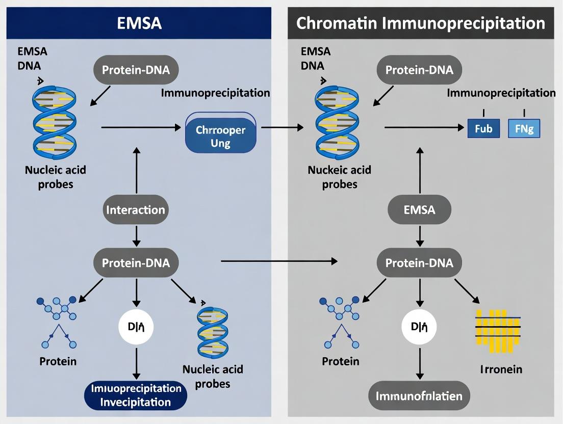
EMSA vs ChIP: A Comprehensive Guide to Choosing the Right Protein-Nucleic Acid Interaction Assay
This article provides a detailed, comparative analysis of Electrophoretic Mobility Shift Assay (EMSA) and Chromatin Immunoprecipitation (ChIP), two cornerstone techniques in molecular biology for studying protein-nucleic acid interactions.
EMSA vs ChIP: A Comprehensive Guide to Choosing the Right Protein-Nucleic Acid Interaction Assay
Abstract
This article provides a detailed, comparative analysis of Electrophoretic Mobility Shift Assay (EMSA) and Chromatin Immunoprecipitation (ChIP), two cornerstone techniques in molecular biology for studying protein-nucleic acid interactions. Tailored for researchers, scientists, and drug development professionals, we explore the fundamental principles, methodological workflows, and key applications of each assay. We offer practical troubleshooting advice, optimization strategies, and a direct, head-to-head comparison of their strengths, limitations, and data outputs. The guide concludes with a synthesized decision framework to help researchers select the optimal technique for their specific experimental goals, from validating transcription factor binding in vitro to mapping genome-wide epigenetic landscapes in vivo.
EMSA and ChIP Fundamentals: Core Principles for Studying Protein-DNA Interactions
EMSA (Electrophoretic Mobility Shift Assay) and ChIP (Chromatin Immunoprecipitation) are foundational techniques in gene regulation research. This guide provides an objective performance comparison, detailing what each method actually measures within the context of studying protein-nucleic acid interactions.
Core Objective Measurement
| Aspect | EMSA | Chromatin Immunoprecipitation (ChIP) |
|---|---|---|
| Primary Measurement | Direct, in vitro protein-nucleic acid binding (affinity & specificity). | In vivo protein-DNA interaction & histone modification occupancy at genomic loci. |
| Quantifiable Output | Binding affinity (Kd), stoichiometry, complex size. | Enrichment fold-change of specific DNA sequences. |
| Spatial/Temporal Context | Cell-free, static snapshot. | Cellular context, can be time-resolved. |
| Throughput | Low to medium (individual probes). | Medium to high (genome-wide with ChIP-seq). |
| Key Artifact Risks | Non-specific competitor effects, complex stability during electrophoresis. | Cross-linking efficiency/artifacts, antibody specificity, shearing bias. |
Quantitative Performance Comparison: Key Experimental Data
Table 1: Typical resolution and sensitivity parameters for EMSA and ChIP-qPCR.
| Parameter | EMSA | ChIP-qPCR |
|---|---|---|
| Detection Limit | ~0.1-1.0 fmol of protein-DNA complex (radiolabeled probe). | ~1% enrichment over background (highly locus/Ab dependent). |
| Dynamic Range | ~10-fold for accurate Kd determination. | ~3-4 orders of magnitude for qPCR detection. |
| Typical Sample Requirement | 1-10 µg of recombinant/cell nuclear protein. | 10^5 - 10^7 cells per immunoprecipitation. |
| Time to Result | 1-2 days. | 2-4 days. |
| Quantitative Rigor | Direct kinetic/equilibrium constants possible. | Relative enrichment; requires careful normalization controls. |
Detailed Experimental Protocols
Protocol 1: Core EMSA for Transcription Factor Binding
- Probe Preparation: Label 20-50 bp DNA oligonucleotide containing putative binding site with [γ-32P]ATP or fluorescent dye using T4 Polynucleotide Kinase. Purify using gel filtration.
- Binding Reaction: Combine in 20 µL: 1x Binding Buffer (10 mM Tris, 50 mM KCl, 1 mM DTT, 2.5% Glycerol, 0.05% NP-40, 100 µg/mL BSA), 1 µg poly(dI:dC) as non-specific competitor, 10-20 fmol labeled probe, and recombinant protein or nuclear extract (1-10 µg). Incubate 20-30 minutes at room temperature.
- Electrophoresis: Load reaction onto pre-run 4-6% non-denaturing polyacrylamide gel in 0.5x TBE buffer at 4°C. Run at 100V until dye front migrates appropriate distance.
- Detection: For radioactive probes, dry gel and expose to phosphorimager screen. For fluorescent probes, scan gel with appropriate imager.
- Analysis: Quantify shifted vs. free probe band intensity to calculate percentage bound. For Kd determination, titrate protein concentration and fit data to binding isotherm.
Protocol 2: Standard Crosslinking ChIP (X-ChIP)
- Crosslinking: Add 37% formaldehyde directly to cell culture medium to 1% final concentration. Incubate 10 minutes at room temperature. Quench with 125 mM glycine.
- Cell Lysis & Sonication: Harvest cells, wash. Lyse in SDS Lysis Buffer. Sonicate lysate to shear chromatin to 200-1000 bp fragments. Validate fragment size by agarose gel electrophoresis.
- Immunoprecipitation: Dilute sonicated lysate 10-fold in ChIP Dilution Buffer. Pre-clear with Protein A/G beads for 1 hour. Incubate supernatant with 1-5 µg of target-specific antibody or isotype control overnight at 4°C with rotation. Add beads for 2-hour capture.
- Washes & Elution: Wash beads sequentially with: Low Salt Wash Buffer, High Salt Wash Buffer, LiCl Wash Buffer, and TE Buffer. Elute complexes in Elution Buffer (1% SDS, 0.1M NaHCO3).
- Reverse Crosslinks & DNA Purification: Add NaCl to eluate and heat at 65°C overnight. Digest RNA with RNase A, then proteins with Proteinase K. Purify DNA using spin columns or phenol-chloroform.
- Analysis: Analyze by qPCR with primers for target loci and negative control loci. Calculate % Input or Fold Enrichment over control IgG.
Visualization of Method Workflows
Title: EMSA Experimental Workflow
Title: Core ChIP Experimental Workflow
The Scientist's Toolkit: Key Research Reagent Solutions
Table 2: Essential materials for EMSA and ChIP experiments.
| Reagent/Material | Primary Function | Example/Note |
|---|---|---|
| Poly(dI:dC) | Non-specific competitor DNA in EMSA. | Critical for reducing non-specific protein-probe interactions. |
| [γ-32P]ATP or Chemiluminescent Kits | Nucleic acid probe labeling for EMSA. | Radioisotope offers highest sensitivity; chemiluminescent alternatives are safer. |
| Non-denaturing Polyacrylamide Gels | Matrix for EMSA separation. | Low ionic strength buffer preserves protein-DNA complexes during electrophoresis. |
| Formaldehyde (37%) | Reversible protein-DNA crosslinker for X-ChIP. | Fixes in vivo interactions; crosslinking time is condition-specific. |
| Chromatin Shearing Reagents (Covaris/ Bioruptor) | Fragments crosslinked chromatin to optimal size. | Sonication is standard; enzymatic shearing kits offer an alternative. |
| ChIP-Validated Antibodies | Target-specific immunoprecipitation. | Must be validated for ChIP; specificity is the single most critical factor. |
| Protein A/G Magnetic Beads | Capture antibody-protein-DNA complexes. | Provide ease of washing compared to agarose beads. |
| ChIP qPCR Primers | Quantify enriched DNA regions. | Require validation of efficiency; must include positive and negative control loci. |
Within the broader thesis comparing Electrophoretic Mobility Shift Assay (EMSA) and Chromatin Immunoprecipitation (ChIP), this guide focuses on the in vitro binding detection capabilities of EMSA. EMSA, or gel shift assay, is a cornerstone technique for studying protein-nucleic acid interactions, providing distinct advantages and limitations compared to the in vivo snapshot offered by ChIP.
Core Principle & Comparison to ChIP Context
EMSA detects binding by observing the reduced electrophoretic mobility of a nucleic acid probe when bound by a protein. This provides direct, quantitative evidence of binding affinity and specificity in a controlled, cell-free environment. In contrast, ChIP captures protein-DNA interactions as they occur in the chromatin context of living cells, identifying genomic binding sites. EMSA answers "can it bind?" under defined conditions, while ChIP asks "does it bind here?" in a physiological context.
Performance Comparison: EMSA Kits & Alternatives
The following table compares widely used EMSA kits based on key performance metrics relevant to researchers validating interactions before proceeding to in vivo ChIP experiments.
Table 1: Commercial EMSA Kit Performance Comparison
| Kit / Provider | Sensitivity (Detection Limit) | Probe Labeling Method | Key Differentiating Feature | Best For |
|---|---|---|---|---|
| LightShift Chemiluminescent EMSA Kit (Thermo Fisher) | ~0.5-1 fmol biotinylated probe | Biotin end-labeling | Chemiluminescent detection; no radioactivity. | High-sensitivity, non-radioactive applications. |
| DIG Gel Shift Kit, 2nd Generation (Roche) | ~2-5 fmol DIG-labeled probe | Digoxigenin end-labeling | Robust colorimetric or chemiluminescent detection. | Labs with established DIG detection workflows. |
| Gel Shift Assay Core System (Promega) | ~1-2 fmol biotinylated probe | Biotin end-labeling | Includes positive control nuclear extract and consensus oligonucleotide. | Beginners needing validated controls. |
| Classic Radioactive EMSA (In-house) | <0.1 fmol | 32P or 33P γ-ATP | Highest sensitivity and resolution. | Labs with radiological facilities; gold-standard quantitation. |
| Fluorescent EMSA (e.g., Cy5 probes) | ~5-10 fmol | Fluorescent dye end-labeling | Direct scanning; multiplex potential. | Real-time kinetics when paired with specialized instruments. |
Experimental Data & Protocol
Supporting Data: A 2023 study comparing validation methods for a transcription factor (TF) binding site showed EMSA provided superior quantitative binding affinity (Kd) data compared to in-silico prediction, which was crucial for interpreting subsequent ChIP-seq peaks. The EMSA-determined Kd of 15 nM for the wild-type probe versus >500 nM for a mutant probe provided mechanistic evidence for the specific interaction observed in vivo.
Detailed EMSA Protocol:
Probe Preparation:
- Design complementary single-stranded DNA oligonucleotides containing the putative binding site.
- Anneal oligonucleotides to create double-stranded probe.
- Label the probe at the 5' end using T4 Polynucleotide Kinase and [γ-32P]ATP (radioactive) or a biotin/fluorescence labeling kit.
- Purify labeled probe using a spin column.
Protein Extraction/Binding Reaction:
- Prepare nuclear extract from cells or use purified recombinant protein.
- Set up 20 μL binding reactions:
- 1X Binding Buffer (10 mM HEPES, 50 mM KCl, 1 mM DTT, 2.5% glycerol, 0.05% NP-40, 5 mM MgCl2).
- 1 μg poly(dI·dC) as non-specific competitor.
- 1-10 μg nuclear extract or 10-100 ng purified protein.
- Labeled probe (20 fmol).
- Controls: Include reactions with unlabeled specific competitor (100-fold molar excess) and/or a mutated probe to demonstrate specificity.
- Incubate at room temperature for 20-30 minutes.
Electrophoresis & Detection:
- Pre-run a native polyacrylamide gel (4-6%) in 0.5X TBE buffer at 100V for 30-60 min at 4°C.
- Load binding reactions (add loading dye without SDS) and run gel at 100V in cold room until dye front migrates appropriately.
- Detection:
- Radioactive: Dry gel and expose to phosphorimager screen.
- Biotin: Transfer DNA to positively charged nylon membrane via electroblotting. Crosslink, then detect with Streptavidin-HRP and chemiluminescent substrate.
- Analyze shifted bands (protein-bound) versus free probe.
Visualizing the EMSA Workflow & Thesis Context
Diagram 1: EMSA Workflow in Thesis Context
The Scientist's Toolkit: Key EMSA Reagents
Table 2: Essential Research Reagents for EMSA
| Reagent / Solution | Function & Importance |
|---|---|
| T4 Polynucleotide Kinase (T4 PNK) | Catalyzes the transfer of a phosphate group to the 5' end of DNA, essential for radioactive probe labeling. |
| [γ-32P]ATP or Biotin Labeling Kit | Source of radioactive or non-radioactive label for sensitive probe detection. |
| Poly(dI·dC) | A non-specific synthetic DNA competitor that binds and neutralizes non-specific nucleic acid-binding proteins, reducing background. |
| Nuclear Extraction Kit | Provides a method to isolate DNA-binding proteins, including transcription factors, from cellular nuclei. |
| Native Gel Electrophoresis System | A non-denaturing polyacrylamide gel setup that preserves protein-DNA interactions during separation. |
| Electroblotting Apparatus | For transferring DNA from gels to membranes in non-radioactive detection methods. |
| Chemiluminescent Substrate (e.g., HRP) | Generates light signal for imaging biotin-labeled probes on membranes. |
| Specific & Mutant Competitor Oligos | Unlabeled DNA fragments critical for demonstrating the sequence specificity of the observed protein binding. |
This comparison guide is framed within a broader research thesis comparing Electrophoretic Mobility Shift Assay (EMSA) and Chromatin Immunoprecipitation (ChIP). While EMSA detects protein-nucleic acid interactions in vitro using purified components, ChIP captures these interactions in vivo from native chromatin, providing critical physiological context. This guide objectively compares standard ChIP with its major alternatives.
Performance Comparison: ChIP vs. Key Alternatives
The following table summarizes the core performance characteristics of ChIP against other methods for studying protein-DNA interactions.
Table 1: Comparative Analysis of Methods for Studying Protein-DNA Interactions
| Feature | Native Chromatin Immunoprecipitation (ChIP) | Electrophoretic Mobility Shift Assay (EMSA) | Cleavage Under Targets & Release Using Nuclease (CUT&RUN) | DNA Adenine Methyltransferase Identification (DamID) |
|---|---|---|---|---|
| Physiological Context | In vivo (native chromatin) | In vitro (purified components) | In vivo / in situ (permeabilized cells) | In vivo (live cells) |
| Resolution | 200-500 bp (standard), ~1 bp (ChIP-exo) | Binding site defined by probe length | ~100 bp | ~5 kb |
| Throughput | Medium | Low (1-2 probes/gel) | High (low cell input) | Medium |
| Required Input | 10^5 - 10^7 cells | Nanograms of purified protein | 500 - 10,000 cells | 10^5 - 10^6 cells |
| Key Artifact Risk | Crosslinking artifacts, antibody specificity | Non-specific probe competition, protein purification artifacts | Permeabilization efficiency | Methylation diffusion, Dam toxicity |
| Primary Application | Mapping transcription factor & histone mark genomic localization | Confirming specific protein-DNA binding in vitro | Low-input, high-resolution profiling | Mapping chromatin interactions without antibodies |
Table 2: Quantitative Performance Metrics from Recent Studies
| Metric | Standard Crosslinked ChIP | Native (No Crosslink) ChIP | CUT&RUN | ChIP-seq (Typical Output) |
|---|---|---|---|---|
| Typical Signal-to-Noise Ratio | Moderate (depends on Ab) | Low (for stable complexes) | High | N/A |
| Time to Complete Protocol | 2-4 days | 1-2 days | 1 day | + 1-3 days sequencing |
| Minimum Cell Number | ~1x10^6 | ~5x10^6 | ~500 | Same as input method |
| Peak Calling Consistency (IDR) | 0.8 - 0.95 (Ab dependent) | Variable | 0.9 - 0.98 | N/A |
| Background from Sonication | Present | Present | Minimal | N/A |
Detailed Experimental Protocols
Protocol 1: Standard Crosslinked Chromatin Immunoprecipitation (ChIP)
This is the foundational protocol for capturing protein-DNA interactions from fixed chromatin.
Methodology:
- In Vivo Crosslinking: Treat cells with 1% formaldehyde for 8-12 minutes at room temperature to covalently link proteins to DNA. Quench with 125 mM glycine.
- Cell Lysis & Sonication: Lyse cells in SDS lysis buffer. Sonicate chromatin to shear DNA to fragments of 200-1000 bp. Confirm fragment size by agarose gel electrophoresis.
- Immunoprecipitation: Dilute sonicated lysate in ChIP dilution buffer. Pre-clear with Protein A/G beads. Incubate overnight at 4°C with target-specific antibody (e.g., 1-5 µg anti-RNA Polymerase II). Add beads for 2-hour capture.
- Washes & Elution: Wash beads sequentially with: Low Salt Immune Complex Wash Buffer, High Salt Immune Complex Wash Buffer, LiCl Immune Complex Wash Buffer, and TE Buffer.
- Reverse Crosslinks & Purification: Elute complexes in fresh elution buffer (1% SDS, 0.1M NaHCO3). Add NaCl to 200 mM and reverse crosslinks by heating at 65°C for 4-6 hours. Digest RNA and protein with RNase A and Proteinase K. Purify DNA using phenol-chloroform extraction or spin columns.
- Analysis: Analyze enriched DNA by qPCR (ChIP-qPCR) or prepare libraries for next-generation sequencing (ChIP-seq).
Protocol 2: CUT&RUN (Cleavage Under Targets & Release Using Nuclease)
A modern alternative offering high resolution and low input, performed on permeabilized cells/nuclei.
Methodology:
- Cell Permeabilization: Bind washed cells or isolated nuclei to concanavalin A-coated magnetic beads in a low-salt buffer. Permeabilize cells with digitonin.
- Target Binding: Incubate with primary antibody against target protein (e.g., 1:50-1:100 dilution) in Antibody Buffer (20 mM HEPES, 150 mM NaCl, 0.5 mM Spermidine, 0.1% Digitonin) for 2 hours at 4°C.
- pA-MNase Tethering & Cleavage: Wash and incubate with Protein A-Micrococcal Nuclease (pA-MN) fusion protein. Activate MNase by adding 2 mM CaCl₂ and incubating at 0-4°C for exactly 30-60 minutes.
- DNA Fragment Release: Stop reaction by adding EGTA (chelates Ca²⁺). Release cleaved DNA fragments from chromatin into the supernatant by incubating at 37°C for 10-20 minutes.
- DNA Purification & Analysis: Purify released DNA (typically ~100-300 bp) from the supernatant using a standard DNA cleanup protocol. Analyze by qPCR or sequencing (CUT&RUN-seq).
Visualizations of Workflows and Relationships
Title: Standard Crosslinked ChIP Experimental Workflow
Title: Decision Logic for Choosing Protein-DNA Interaction Method
The Scientist's Toolkit: Research Reagent Solutions
Table 3: Essential Materials for Chromatin Immunoprecipitation
| Reagent / Solution | Function in ChIP Protocol | Critical Considerations |
|---|---|---|
| Formaldehyde (37%) | Crosslinks proteins to DNA in vivo, preserving transient interactions. | Concentration (1%) and time (8-12 min) are critical. Over-fixing reduces sonication efficiency. |
| Protease & Phosphatase Inhibitors | Preserve protein epitopes and modification states during lysis. | Must be added fresh to all buffers prior to cell lysis. |
| ChIP-Grade Antibody | Specifically immunoprecipitates the target protein-complex. | Validation for ChIP is essential. Check citations for specificity and signal. |
| Protein A/G Magnetic Beads | Capture antibody-antigen complexes for easy washing. | Magnetic beads reduce background vs. agarose beads. Protein A vs. G depends on antibody species/isotype. |
| Micrococcal Nuclease (MNase) | Used in Native ChIP or CUT&RUN to digest chromatin enzymatically. | Provides precise, nucleotide-level cutting but requires titration for optimal fragment size. |
| Sonicator (Covaris or Bioruptor) | Shears crosslinked chromatin to 200-1000 bp fragments. | Consistency is key. Water bath sonicators offer better reproducibility than probe types. |
| Glycine (2.5 M) | Quenches formaldehyde crosslinking reaction. | Must be added in excess to stop fixation and prevent over-crosslinking. |
| ChIP Elution Buffer | Releases captured complexes from beads. | Typically contains 1% SDS and sodium bicarbonate, which also facilitates reverse crosslinking. |
| DNA Cleanup Columns (SPRI Beads) | Purify immunoprecipitated DNA for downstream qPCR or sequencing. | More consistent and faster than traditional phenol-chloroform extraction. |
Key Components and Molecular Requirements for Each Assay
Within the broader thesis comparing Electrophoretic Mobility Shift Assay (EMSA) and Chromatin Immunoprecipitation (ChIP), a detailed understanding of each assay's molecular components is crucial for experimental design and data interpretation. This guide objectively compares the core requirements for these foundational techniques in nucleic acid-protein interaction studies.
Core Components and Requirements
The fundamental molecular and reagent requirements for EMSA and ChIP differ significantly, reflecting their distinct operational principles.
Table 1: Molecular and Reagent Requirements Comparison
| Component | EMSA Requirement | ChIP Requirement | Critical Function |
|---|---|---|---|
| Target Protein | Purified or crude nuclear extract. Active, DNA-binding form. | Protein in its native chromatin context within fixed cells/tissues. | The protein of interest whose DNA-binding is being studied. |
| Nucleic Acid Probe | Short (20-30 bp), labeled double-stranded DNA/RNA oligonucleotide containing suspected binding motif. | Native genomic DNA, sheared to 200-1000 bp fragments. No label required. | The sequence to which the protein binds. |
| Detection Method | Direct (radioactive/fluorophore label on probe) or indirect (antibody supershift). | Antibody-based (specific, high-affinity, ChIP-grade) against target protein or epitope tag. | Enables visualization/quantification of protein-bound complexes. |
| Separation Matrix | Non-denaturing polyacrylamide gel (typically 4-10%). | Protein A/G beads or magnetic beads coupled to Protein A/G. | Separates bound from free probe (EMSA) or immunoprecipitates protein-DNA complexes (ChIP). |
| Critical Buffers | Binding buffer (poly dI:dC, carrier protein, salt, glycerol), electrophoresis running buffer. | Crosslinking, lysis, wash, and elution buffers. | Maintain native binding (EMSA) or preserve/epitope-expose complexes (ChIP). |
| Key Additional Reagents | Non-specific competitor DNA (e.g., poly(dI-dC)), loading dye. | Crosslinker (formaldehyde), protease/RNase inhibitors, reverse crosslinking agent (e.g., proteinase K), DNA purification reagents. | Reduce non-specific binding (EMSA) or fix and recover complexes (ChIP). |
Experimental Protocols: Detailed Methodologies
Detailed EMSA Protocol (Gel-Shift Assay)
Principle: Resolves protein-bound and free nucleic acid probes based on reduced electrophoretic mobility through a native gel.
- Probe Preparation: Anneal complementary oligonucleotides to form double-stranded probe. Label probe at 5' or 3' end with [γ-³²P]ATP via T4 polynucleotide kinase or with a fluorophore.
- Binding Reaction: Combine 2-20 µg of nuclear extract or purified protein with 0.1-1 ng of labeled probe in binding buffer (10-20 mM HEPES, 50-100 mM KCl, 1 mM DTT, 10% glycerol, 0.1-0.5 µg/µL poly(dI-dC)). Include unlabeled specific competitor (for specificity) or mutant probe (for negative control). Incubate 20-30 min at room temperature.
- Electrophoresis: Load samples onto pre-run 4-10% non-denaturing polyacrylamide gel in 0.5X TBE buffer. Run at 100-150 V at 4°C until dye front migrates appropriate distance.
- Detection: For radioactive probes, dry gel and expose to phosphorimager screen. For fluorescent probes, image gel using appropriate scanner.
Detailed ChIP Protocol (Native or Crosslinked)
Principle: Immunoprecipitation of protein-DNA complexes from chromatin, followed by analysis of associated DNA sequences.
- Crosslinking & Cell Lysis: Treat cells with 1% formaldehyde for 10 min at room temperature to crosslink proteins to DNA. Quench with glycine. Harvest cells, lyse in SDS lysis buffer with protease inhibitors.
- Chromatin Shearing: Sonicate lysate to shear DNA to an average length of 200-500 bp. Alternatively, use enzymatic digestion (MNase) for native ChIP.
- Immunoprecipitation: Pre-clear sheared chromatin with Protein A/G beads. Incubate supernatant with specific antibody (2-5 µg per reaction) overnight at 4°C. Add Protein A/G beads for 1-2 hours to capture antibody complexes.
- Washes & Elution: Wash beads sequentially with low salt, high salt, LiCl, and TE buffers. Elute complexes with elution buffer (1% SDS, 0.1M NaHCO₃) at 65°C with shaking.
- Reverse Crosslinking & DNA Recovery: Add NaCl to eluate and heat at 65°C overnight to reverse crosslinks. Treat with RNase A and proteinase K. Purify DNA using phenol-chloroform extraction or spin columns.
- Analysis: Analyze precipitated DNA by quantitative PCR (ChIP-qPCR) for specific loci or by next-generation sequencing (ChIP-seq) for genome-wide mapping.
The Scientist's Toolkit: Research Reagent Solutions
Table 2: Essential Research Reagents
| Item | Function in Assays | Example/Specification |
|---|---|---|
| ChIP-Grade Antibody | High specificity and affinity for target protein in fixed, denatured chromatin context. Validated for low non-specific DNA pull-down. | Anti-RNA Polymerase II CTD repeat, Anti-Histone H3 (tri-methyl K4). |
| Protein A/G Magnetic Beads | Efficient capture of antibody-antigen complexes; reduce non-specific background vs. agarose beads. Enable automation. | Dynabeads, Magna ChIP Protein A/G beads. |
| Poly(dI-dC) | Non-specific competitor DNA in EMSA; blocks non-specific protein binding to labeled probe. | Amorphous double-stranded polymer. |
| T4 Polynucleotide Kinase | Enzymatically labels DNA oligonucleotide probes with ³²P for high-sensitivity EMSA detection. | Includes reaction buffer. |
| Formaldehyde (Molecular Biology Grade) | Reversible protein-DNA crosslinker for ChIP; penetrates cells rapidly. | 37% solution, methanol-free for consistency. |
| Protease Inhibitor Cocktail | Prevents proteolytic degradation of proteins during extract preparation (EMSA) and chromatin isolation (ChIP). | EDTA-free for compatibility with downstream steps. |
| Sonicator with Microtip | Provides consistent, high-energy shearing of crosslinked chromatin to optimal fragment size for ChIP. | Covaris S2, Bioruptor. |
| Phosphor Storage Screen & Imager | Sensitive detection and quantification of radioisotope-labeled EMSA gels. | Typhoon FLA, Amersham ImageQuant. |
Experimental Workflow and Logical Relationships
Assay Workflow Comparison Diagram
Assay Application Logic Diagram
Supporting Experimental Data Comparison
Recent comparative studies underscore the complementary nature of EMSA and ChIP. Data from a 2023 investigation into transcription factor MYC-DNA interactions illustrate key performance differences.
Table 3: Representative Experimental Data from MYC Interaction Study
| Assay Parameter | EMSA Results | ChIP-seq Results | Interpretation |
|---|---|---|---|
| Binding Site Resolution | Confirmed binding to 8-bp E-box motif (CACGTG). Kd ~15 nM for consensus probe. | Identified 12,450 genomic peaks, many containing E-box motif. Resolution ~200 bp. | EMSA defines precise motif; ChIP maps genomic loci at lower resolution. |
| Throughput | Tested 8 mutant probe variants in one experiment. | Surveyed entire genome (~3 billion bp) in one experiment. | ChIP offers vastly higher discovery potential. |
| Context Dependency | Binding to naked DNA probe unaffected by chromatin status. | 68% of peaks were in promoter regions; binding profile changed with cellular differentiation. | ChIP captures biological context; EMSA isolates biophysical interaction. |
| Quantitative Output | Linear signal range over 2 orders of magnitude. Calculated precise dissociation constant. | Enrichment reported as reads per peak. Semi-quantitative; compares relative occupancy. | EMSA is superior for kinetic/affinity measurements. |
| False Positive/Negative Control | Specific competition with unlabeled probe reduced shift by 95%. Mutant probe showed no shift. | IgG control produced <0.1% of target antibody reads. Input DNA used for normalization. | Both require stringent controls for specificity. |
Conclusion for Researchers: The choice between EMSA and ChIP is not one of superiority but of application. EMSA provides high-resolution, quantitative in vitro validation of direct binding and affinity. ChIP provides genome-wide, in vivo mapping of binding events within their native chromatin context. A robust research thesis often employs EMSA to mechanistically validate interactions initially discovered or suggested by ChIP-seq screens, leveraging the complementary strengths of both assays.
Primary Biological Questions Answered by EMSA vs. ChIP
Within the broader thesis of comparing Electrophoretic Mobility Shift Assay (EMSA) and Chromatin Immunoprecipitation (ChIP), this guide objectively compares their capabilities to answer distinct primary biological questions. EMSA probes protein-nucleic acid interactions in vitro, while ChIP captures these interactions within their native chromatin context in vivo. The choice of technique is dictated by the specific hypothesis being tested.
Core Question Comparison
| Primary Biological Question | EMSA Suitability | ChIP Suitability | Key Distinction |
|---|---|---|---|
| Does a purified protein bind directly to a specific DNA/RNA sequence? | Primary Application. Direct, quantitative assessment of binding affinity and specificity. | Not applicable. Requires cellular context and crosslinking. | Direct vs. Indirect Evidence. EMSA provides biochemical proof of direct binding. |
| Where does a protein bind across the genome in living cells? | Not applicable. Lacks genomic scale and cellular context. | Primary Application. Genome-wide mapping (ChIP-seq) or locus-specific analysis. | In vitro vs. In vivo Context. ChIP captures binding within native chromatin. |
| What is the stoichiometry or complex formation on a nucleic acid probe? | High Suitability. Can resolve multiple shifted complexes (e.g., protein dimers, co-factors). | Low suitability. Indirect, lower resolution for complex composition. | Complex Resolution. EMSA excels at visualizing discrete protein-DNA complexes. |
| Is a specific genomic site occupied by a protein under a given condition? | Indirect inference only. Can test binding capability of site sequences. | Primary Application. Direct, condition-specific evidence of in vivo occupancy. | Binding Potential vs. Actual Occupancy. ChIP confirms functional, cellular occupancy. |
| Does a post-translational modification or drug affect DNA binding? | High Suitability. Ideal for testing effects on purified protein binding kinetics. | High Suitability. Ideal for testing effects on cellular binding landscapes. | Mechanism vs. Outcome. EMSA tests biochemical mechanism; ChIP tests cellular outcome. |
| Experimental Parameter | Typical EMSA Data | Typical ChIP Data |
|---|---|---|
| Output Format | Gel image with shifted bands. | Enrichment plot (qPCR) or sequence reads (seq). |
| Key Quantitative Metric | Shifted probe percentage (Kd calculation). | Fold-enrichment over control (IP/Input). |
| Resolution | Single binding site (~10-40 bp probe). | ~200 bp (sonicated ChIP) to single-base (ChIP-exo). |
| Throughput | Low (1-10 probes per experiment). | High (genome-wide in one ChIP-seq run). |
| Typical Controls | Cold competition, mutant probe, antibody supershift. | IgG control, Input DNA, target-negative genomic region. |
Detailed Experimental Protocols
Standard EMSA Protocol (Non-Radiocative)
- Probe Preparation: Label 20-50 bp dsDNA oligonucleotide with biotin using terminal transferase. Purify.
- Protein Binding: Incubate 2-20 fmol of labeled probe with 0-500 ng of purified protein or nuclear extract in binding buffer (10 mM HEPES, 50 mM KCl, 1 mM DTT, 2.5% glycerol, 0.05% NP-40, 100 µg/mL BSA, 50 ng/µL poly(dI:dC)) for 20-30 minutes at room temperature.
- Electrophoresis: Load samples onto a pre-run 6% non-denaturing polyacrylamide gel in 0.5X TBE buffer. Run at 100V for 60-90 minutes at 4°C.
- Transfer & Detection: Transfer to a positively charged nylon membrane. Crosslink DNA. Detect biotin label via chemiluminescence.
Standard Crosslinking ChIP Protocol (for Seq or qPCR)
- Crosslinking: Treat cells with 1% formaldehyde for 10 minutes at room temperature. Quench with glycine.
- Cell Lysis & Sonication: Lyse cells. Sonicate chromatin to shear DNA to 200-500 bp fragments. Verify fragment size by agarose gel.
- Immunoprecipitation: Pre-clear lysate. Incubate with 1-10 µg of target-specific antibody or species-matched IgG control overnight at 4°C. Capture with Protein A/G beads.
- Washes & Elution: Wash beads sequentially with Low Salt, High Salt, LiCl, and TE buffers. Elute complexes with elution buffer (1% SDS, 0.1M NaHCO3).
- Reverse Crosslinks & Purification: Incubate eluate and input control at 65°C overnight. Treat with Proteinase K. Purify DNA with spin columns.
- Analysis: Analyze by qPCR for specific loci or prepare libraries for next-generation sequencing (ChIP-seq).
Workflow and Logic Diagrams
Title: EMSA Experimental Workflow
Title: ChIP Experimental Workflow
Title: Decision Logic: Choosing EMSA or ChIP
The Scientist's Toolkit: Key Reagent Solutions
| Reagent / Material | Primary Function | Typical Use In |
|---|---|---|
| Poly(dI:dC) | Non-specific competitor DNA | EMSA: Reduces non-specific protein-probe binding. |
| Biotin- or Fluorescent-labeled Oligonucleotides | Sensitive, non-radioactive probe labeling | EMSA: Detection of nucleic acid probe. |
| High-Affinity, ChIP-Validated Antibodies | Specific target protein immunoprecipitation | ChIP: Critical for specific enrichment. |
| Protein A/G Magnetic Beads | Efficient antibody-antigen complex capture | ChIP: Facilitates washes and reduces background. |
| Formaldehyde (1%) | Reversible protein-DNA crosslinker | ChIP: Preserves in vivo interactions. |
| Chromatin Shearing Reagents (Enzymatic or Sonication) | Fragments chromatin to optimal size | ChIP: Ensures resolution and IP efficiency. |
| ChIP-Seq Library Prep Kit | Prepares immunoprecipitated DNA for sequencing | ChIP-seq: Converts enriched DNA to sequencer-compatible libraries. |
Step-by-Step Protocols: From Cell Culture to Data in EMSA and ChIP Workflows
Within a broader thesis comparing Electrophoretic Mobility Shift Assay (EMSA) and Chromatin Immunoprecipitation (ChIP), this guide details the core EMSA workflow. While ChIP identifies in vivo protein-DNA interactions within chromatin, EMSA provides a versatile in vitro platform for quantifying binding affinity, specificity, and kinetics of purified proteins or nuclear extracts to labeled nucleic acid probes. This comparison focuses on the practical execution of EMSA, juxtaposing methodological choices and their impact on data quality.
Core Workflow Comparison & Experimental Protocols
Probe Design and Synthesis
The choice of probe design and labeling method directly influences signal sensitivity and specificity.
Protocol: Double-Stranded DNA Probe Preparation
- Design: Synthesize complementary single-stranded oligonucleotides (typically 20-40 bp) containing the predicted protein-binding sequence. Include 5-10 bp flanking sequences.
- Annealing: Mix equimolar amounts of each oligonucleotide in annealing buffer (10 mM Tris, 50 mM NaCl, 1 mM EDTA, pH 7.5-8.0).
- Thermal Cycling: Heat to 95°C for 5 minutes, then slowly cool to 25°C (0.1°C/sec) in a thermal cycler.
- Purification: Use a spin column or native PAGE to purify the duplex probe.
Comparison Table: Probe Labeling Methods
| Method | Principle | Sensitivity | Stability | Cost & Complexity | Best For |
|---|---|---|---|---|---|
| Radioactive (³²P) | Incorporation of γ-³²P-ATP via T4 PNK. | Very High (attomole) | Short (half-life decay) | Low reagent cost, high safety/hazard cost | Quantitative kinetics, low-abundance complexes. |
| Chemiluminescent | 3' End-labeling with biotin or digoxigenin. | High (femtomole) | High (months) | Moderate cost, requires streptavidin-HRP/AP | Most routine applications, avoids radioactivity. |
| Fluorescent | Use of Cy5 or FAM-labeled primers during synthesis. | Moderate-High | High | Higher cost, requires fluorescence imager | Multiplexing, precast gel systems. |
Protein Incubation and Binding Reaction
Binding conditions must be optimized to preserve native interactions.
Protocol: Binding Reaction Setup
- Prepare a 2X binding master mix on ice:
- 20 mM HEPES (pH 7.9)
- 80 mM KCl
- 4 mM MgCl₂
- 0.4 mM EDTA
- 0.4 mM DTT
- 10% Glycerol
- 2 μg/μL BSA or poly(dI-dC) as non-specific competitor.
- In a final volume of 20 μL, combine:
- 10 μL of 2X binding mix.
- 1-10 μg of nuclear extract or 10-500 ng of purified protein.
- Labeled probe (20,000 cpm for ³²P or 10-50 fmol for chemiluminescent).
- Nuclease-free water to volume.
- Competition Assay Control: Add 50-200X molar excess of unlabeled specific or mutant probe. Supershift Assay Control: Add 1-2 μg of specific antibody.
- Incubate at room temperature or 4°C for 20-30 minutes.
Comparison Table: Non-Specific Competitors
| Competitor | Function & Mechanism | Recommended Use Case | Potential Interference |
|---|---|---|---|
| poly(dI-dC) | Synthetic polymer binds non-specific electrostatic interactions. | Standard for most nuclear extracts & common transcription factors. | May inhibit some specific interactions at high concentrations. |
| Salmon Sperm DNA | Sheared genomic DNA with diverse sequences. | Complex mixtures where poly(dI-dC) is ineffective. | Less consistent batch-to-batch; may contain unknown binding sites. |
| BSA | Carrier protein reduces surface adhesion. | Used alongside nucleic acid competitors, especially with purified protein. | Does not compete for DNA-binding. |
Gel Electrophoresis and Detection
The separation matrix is critical for resolving free probe from protein-bound complexes.
Protocol: Non-Denaturing Polyacrylamide Gel Electrophoresis
- Gel Casting: Prepare a 4-8% non-denaturing polyacrylamide gel (29:1 acrylamide:bis) in 0.5X TBE buffer. Allow to polymerize for 1 hour.
- Pre-electrophoresis: Pre-run the gel in 0.5X TBE at 100V for 60 minutes at 4°C to establish equilibrium.
- Loading: Add 5X non-denaturing loading dye (glycerol-based, no SDS) to samples. Load entire reaction.
- Electrophoresis: Run at 100V constant voltage in 0.5X TBE at 4°C until dye front migrates 2/3 down the gel.
- Detection:
- ³²P: Transfer gel to blot paper, dry, and expose to a phosphorimager screen.
- Biotin: Electro-transfer to positively charged nylon membrane. Crosslink (UV), incubate with Streptavidin-HRP, and detect with chemiluminescent substrate.
- Fluorescent: Image gel directly using appropriate laser/excitation settings.
Comparison Table: Gel Matrix Types
| Matrix Type | Resolution | Run Time | Handling Difficulty | Compatibility |
|---|---|---|---|---|
| Hand-cast PAGE | High | Slow (2-3 hrs) | Moderate | All label types; requires optimization. |
| Pre-cast PAGE | Consistent | Moderate-Fast | Easy | Primarily fluorescent/chemiluminescent. |
| Agarose (Low %)* | Low | Fast (<1 hr) | Very Easy | Large complexes (>500 kDa); quick screening. |
Note: Agarose offers lower resolution but is suitable for large ribonucleoprotein complexes.
The Scientist's Toolkit: EMSA Research Reagent Solutions
| Item | Function & Role in EMSA |
|---|---|
| T4 Polynucleotide Kinase (PNK) | Catalyzes the transfer of phosphate from ATP to the 5'-end of oligonucleotides for radioactive labeling. |
| Biotin or Digoxigenin 3'-End Labeling Kit | Provides reagents for non-radioactive probe tagging via terminal transferase. |
| Poly(dI-dC) | A synthetic, non-specific nucleic acid polymer used to suppress protein binding to non-target sequences. |
| HEPES Buffer | A buffering agent with minimal metal ion chelation, preferred over Tris for maintaining protein-DNA interactions. |
| Non-Denaturing Loading Dye | Contains glycerol and tracking dyes (e.g., bromophenol blue) to increase sample density for loading without disrupting complexes. |
| Streptavidin-Horseradish Peroxidase (HRP) Conjugate | Binds biotinylated probes for highly sensitive chemiluminescent detection post-electroblotting. |
| Phosphorimager Screen & Scanner | Essential for capturing and quantifying the high dynamic range signal from radioactively labeled EMSA gels. |
Visualized Workflows and Pathways
Title: EMSA Core Workflow from Probe to Detection
Title: EMSA vs ChIP Method Comparison for Binding Studies
Within the broader research comparing EMSA (Electrophoretic Mobility Shift Assay) and Chromatin Immunoprecipitation (ChIP), understanding the nuanced performance of ChIP workflow components is critical. While EMSA analyzes protein-nucleic acid interactions in vitro, ChIP captures these interactions in vivo, with its reliability hinging on the optimization of crosslinking, sonication, immunoprecipitation, and DNA purification steps. This guide objectively compares key methodological alternatives and products at each stage, supported by experimental data.
Crosslinking Agent Efficiency and Specificity
Crosslinking stabilizes protein-DNA interactions. The choice between formaldehyde (HCHO) and alternative crosslinkers significantly impacts yield and background.
Experimental Protocol: Cultured cells (e.g., HeLa) were treated with 1% formaldehyde for 10 min at room temperature or with alternative crosslinkers (e.g., DSG, EGS) as per manufacturer instructions. Quenching was done with 125 mM glycine. Chromatin was isolated, sonicated to an average size of 200-500 bp, and immunoprecipitated for a histone mark (H3K4me3) and a transcription factor (c-Myc). Precipitated DNA was quantified by qPCR at positive and negative control genomic loci.
Table 1: Comparison of Crosslinking Agents
| Crosslinker | Type | Primary Target | Efficiency for TFs (c-Myc % Input) | Efficiency for Histones (H3K4me3 % Input) | Background (Negative Locus Signal) |
|---|---|---|---|---|---|
| Formaldehyde (1%, 10 min) | Short-range | Protein-DNA, Protein-Protein | 2.1% | 8.5% | Low |
| Disuccinimidyl glutarate (DSG) + HCHO | Long-range + Short-range | Protein-Protein then Protein-DNA | 5.3% | 7.9% | Moderate |
| Ethylene glycol bis(succinimidyl succinate) (EGS) + HCHO | Long-range + Short-range | Protein-Protein then Protein-DNA | 4.8% | 8.1% | Moderate |
| Control (No Crosslink) | N/A | N/A | 0.1% | 0.5% | Very Low |
Data Summary: Dual crosslinking (DSG+HCHO) significantly improves recovery of transcription factor complexes but may increase non-specific background for some targets compared to formaldehyde alone, which remains optimal for histone modifications.
Sonication Methods: Bath vs. Focused-Ultrasonication
Chromatin shearing efficiency and reproducibility are vital for resolution and IP efficiency.
Experimental Protocol: Formaldehyde-crosslinked chromatin from 1x10^6 cells was sheared using either a bath sonicator (30 min, pulsed) or a focused-ultrasonicator (Covaris S2, 12 min, 5% duty cycle). Fragment size distribution was analyzed on a Bioanalyzer. Identical aliquots of sheared chromatin were used for immunoprecipitation with an H3K27ac antibody, followed by qPCR analysis.
Table 2: Comparison of Sonication Methods
| Sonication Method | Peak Fragment Size (bp) | % Fragments in 200-600 bp Range | CV of Size Distribution (n=5) | H3K27ac IP Efficiency (% Input) |
|---|---|---|---|---|
| Bath Sonicator | 750 | 45% | 25% | 4.2% |
| Focused-Ultrasonicator (Covaris) | 350 | 92% | 8% | 6.8% |
Data Summary: Focused ultrasonication provides superior control, reproducibility, and yields a tighter fragment size distribution, leading to higher and more consistent IP efficiency.
Immunoprecipitation: Magnetic Bead Matrix Comparison
The solid-phase matrix for antibody capture affects specificity, recovery, and ease of use.
Experimental Protocol: Sheared chromatin (from 5x10^5 cell equivalents per IP) was incubated with 1 µg of anti-RNA Polymerase II antibody. Immune complexes were captured using either Protein A/G Sepharose beads or pre-blocked magnetic beads (e.g., Dynabeads). Beads were washed with low-salt, high-salt, LiCl, and TE buffers. Eluted DNA was purified and quantified via qPCR at an active gene promoter.
Table 3: Comparison of Immunoprecipitation Matrices
| Bead Type | Material | Binding Capacity (µg IgG/µL bead) | Non-Specific DNA Binding | Protocol Time | Pol II IP Yield (% Input) |
|---|---|---|---|---|---|
| Protein A/G Sepharose | Agarose | ~0.2 | Moderate | Long (centrifugation) | 3.5% |
| Magnetic Beads (e.g., Dynabeads) | Polystyrene/Streptavidin | Varies by type | Low | Short (magnetic separation) | 4.0% |
Data Summary: Magnetic beads offer faster processing with lower non-specific binding, improving workflow throughput and consistency, though cost per sample may be higher.
DNA Purification: Silica Column vs. SPRI Bead Cleanup
The final DNA purification step influences recovery of low-concentration ChIP-DNA.
Experimental Protocol: After crosslink reversal and proteinase K treatment, ChIP eluates (50 µL) were purified using either a silica membrane spin column or SPRI (Solid Phase Reversible Immobilization) magnetic beads. Input DNA (1%) was purified concurrently. DNA was eluted in 20 µL. Recovery was measured by qPCR and fluorometry.
Table 4: Comparison of DNA Purification Methods
| Purification Method | Principle | Average Recovery of 50 pg Spiked DNA | Inhibition in Downstream qPCR (∆Cq) | Hands-on Time |
|---|---|---|---|---|
| Silica Spin Column | Binding at high chaotrope salt | 65% | Minimal (+0.2) | Moderate |
| SPRI Magnetic Beads | Binding in PEG/High Salt | >85% | Minimal (+0.1) | Low |
Data Summary: SPRI bead-based purification offers higher recovery of low-abundance ChIP-DNA, which is critical for subsequent sequencing library preparation, with minimal PCR inhibition.
Title: ChIP Workflow with Key Methodological Alternatives
The Scientist's Toolkit: Key Research Reagent Solutions
| Item | Function & Rationale |
|---|---|
| Formaldehyde (37%) | Short-range crosslinker; directly couples proteins to DNA and adjacent proteins, preserving in vivo interactions. |
| Protease Inhibitor Cocktail | Prevents degradation of chromatin-associated proteins during cell lysis and nuclei preparation. |
| Micrococcal Nuclease (MNase) | Alternative to sonication; digests linker DNA for nucleosome-level resolution in native ChIP. |
| ChIP-Grade Antibody | High-specificity, validated antibody critical for target enrichment and low background. |
| Protein A/G Magnetic Beads | Enable rapid magnetic separation during IP, reducing handling time and non-specific loss. |
| SPRI (AMPure) Beads | Polyethylene glycol-based magnetic beads for high-recovery purification of ChIP-DNA. |
| Glycogen (or tRNA) | Carrier to improve precipitation efficiency of nanogram-scale ChIP-DNA during ethanol precipitation steps. |
| qPCR Primers for Positive/Negative Loci | Essential controls to validate successful IP and quantify enrichment before sequencing. |
Within the broader research thesis comparing Electrophoretic Mobility Shift Assay (EMSA) to Chromatin Immunoprecipitation (ChIP), EMSA remains the definitive in vitro technique for the quantitative analysis of protein-nucleic acid interactions. While ChIP excels at identifying in vivo binding events within a chromatin context, EMSA provides unparalleled rigor in dissecting the fundamental biophysical parameters of these interactions. This guide compares the performance of modern, high-sensitivity EMSA using chemiluminescent detection against traditional radioisotopic and basic colorimetric alternatives.
Comparison of EMSA Detection Methodologies for Binding Affinity (Kd) Determination
Accurate quantification of the dissociation constant (Kd) requires a linear detection range over several orders of magnitude. The following table compares data from a study determining the Kd of the tumor suppressor p53 for its consensus DNA sequence.
Table 1: Performance Comparison in Quantitative Kd Determination
| Detection Method | Linear Detection Range | Required Protein Amount | Calculated Kd (mean ± SD) | Assay Time (post-electrophoresis) | Key Advantage/Limitation |
|---|---|---|---|---|---|
| ³²P Isotopic | ~4 logs | 0.1 - 1.0 fmol | 5.2 ± 0.8 nM | 2-24h (autoradiography) | Gold standard sensitivity; regulatory and safety burdens. |
| Chemiluminescent (Biotin-Streptavidin-HRP) | ~3-4 logs | 1 - 10 fmol | 5.5 ± 1.1 nM | 1-2h | Near-isotopic sensitivity, safe, faster; requires biotinylated probe. |
| Colorimetric (Digoxigenin) | ~2 logs | 10 - 100 fmol | 6.8 ± 2.3 nM | 2-4h | No special equipment; lower sensitivity and dynamic range. |
Protocol for Kd Determination via Chemiluminescent EMSA:
- Probe Labeling: A 30-bp dsDNA containing the p53 binding site is labeled at the 3’-end with biotin-11-dUTP using terminal deoxynucleotidyl transferase.
- Binding Reactions: Constant labeled probe (0.1 nM) is incubated with serial dilutions of purified recombinant p53 protein (0.1 nM to 100 nM) in binding buffer (10 mM HEPES, 50 mM KCl, 5% glycerol, 1 mM DTT, 0.05% NP-40, 100 ng/µL poly(dI-dC)) for 30 minutes at 25°C.
- Electrophoresis: Complexes are resolved on a native 6% polyacrylamide gel in 0.5X TBE at 100V for 60 minutes.
- Transfer & Detection: Nucleic acids are transferred to a positively charged nylon membrane via capillary blotting. The membrane is UV-crosslinked, incubated with Streptavidin-Horseradish Peroxidase (HRP) conjugate, and imaged with a luminol-based substrate.
- Quantification: Signal intensity of free and bound probe is quantified by densitometry. The fraction of bound probe is plotted against protein concentration, and the Kd is derived by fitting the data to a one-site specific binding model.
Experimental Workflow: Chemiluminescent EMSA for Kd
Analysis of Binding Specificity: Competition and Supershift Assays
Specificity is demonstrated through cold competition and antibody supershift assays. The table below compares the clarity of results between methods.
Table 2: Specificity Assay Performance Metrics
| Assay Type | Optimal Detection Method | Critical Readout | Interpretation Confidence | Common Pitfall |
|---|---|---|---|---|
| Cold Competition | Chemiluminescent | Dose-dependent reduction of shifted band. | High: Clean signal enables precise IC50 calculation. | Non-specific competitor (e.g., poly(dI-dC)) insufficiency. |
| Antibody Supershift | Chemiluminescent/Isotopic | Further retarded "supershifted" band. | Medium-High: Dependent on antibody quality and epitope accessibility in the native complex. | Antibody disrupts the complex, leading to loss of signal. |
Protocol for Supershift Assay:
- Form the primary protein-DNA complex as described above.
- Add 1-2 µg of specific antibody or an isotype control to the binding reaction. Incubate for an additional 20-60 minutes on ice.
- Resolve, transfer, and detect as per the chemiluminescent protocol. A successful supershift appears as a higher molecular weight band with concomitant decrease/intensity in the original shifted band.
The Scientist's Toolkit: Key Reagents for EMSA
| Reagent/Material | Function & Importance |
|---|---|
| Biotinylated DNA Probe | High-affinity tag for sensitive, non-isotopic detection via streptavidin conjugates. |
| poly(dI-dC) | Non-specific competitor DNA that reduces background from non-sequence-specific binding proteins. |
| Native PAGE Gel (4-6%) | Matrix for separation of protein-DNA complexes based on size, charge, and conformation without denaturation. |
| Positively Charged Nylon Membrane | Immobilizes nucleic acids for subsequent hybridization or detection; positive charge enhances DNA retention. |
| Streptavidin-HRP Conjugate | High-affinity bridge between biotinylated probe and enzymatic signal amplification system. |
| Chemiluminescent Substrate (Luminol/H2O2) | Generates light signal upon oxidation catalyzed by HRP, captured on X-ray film or digital imager. |
Complex Analysis: Multiprotein Assembly and Stoichiometry
EMSA can resolve multi-component complexes through distinct banding patterns. The sequential assembly of a transcription factor complex is illustrated below.
Logical Pathway: Sequential Complex Assembly in EMSA
Conclusion for Research Comparison For the specific research aims of determining precise binding constants, unequivocally proving specificity, and delineating multiprotein complex formation in vitro, EMSA—particularly with chemiluminescent detection—provides data that is complementary to, but fundamentally different from, ChIP-derived data. EMSA offers controlled, quantitative biophysical measurements, whereas ChIP captures snapshot of in vivo binding within a chromatin landscape. The choice between them, or the decision to use them in tandem, is dictated by the specific biological question.
Within the context of comparing Electrophoretic Mobility Shift Assay (EMSA) and Chromatin Immunoprecipitation (ChIP), it is critical to understand the distinct and powerful applications of ChIP technology. While EMSA analyzes protein-nucleic acid interactions in vitro, ChIP captures these interactions within their native chromatin context in vivo. This guide compares ChIP's performance across its three primary application areas, providing experimental data to inform researchers and drug development professionals.
Transcription Factor Mapping: ChIP vs. EMSA
Transcription Factor (TF) mapping identifies genomic binding sites of regulatory proteins. ChIP-seq is the dominant in vivo method, whereas EMSA serves as a complementary in vitro validation tool.
Performance Comparison:
| Feature | Chromatin Immunoprecipitation (ChIP-seq) | Electrophoretic Mobility Shift Assay (EMSA) |
|---|---|---|
| Biological Context | In vivo, native chromatin | In vitro, purified components |
| Throughput | Genome-wide (all potential sites) | Low-throughput (single site/probe) |
| Binding Affinity Data | Qualitative/Semi-quantitative (occupancy) | Quantitative (Kd possible) |
| Identification of Unknown Sites | Yes (unbiased discovery) | No (requires prior sequence knowledge) |
| Functional Relevance | High (reflects cellular state) | Low (biochemical interaction only) |
| Key Experimental Data | Peak calls from sequencing; occupancy changes under stimuli | Gel shift intensity; supershift with antibody |
Supporting Experimental Data: A 2023 study investigating p53 binding under DNA damage compared ChIP-seq and EMSA. ChIP-seq identified 1,852 high-confidence binding sites, 32% of which were novel. EMSA validated binding to the top 10 known consensus sites but failed to confirm 8 novel sites with non-canonical sequences, highlighting EMSA's sequence bias and ChIP's discovery power.
Experimental Protocol: Standard ChIP-seq for Transcription Factors
- Crosslinking: Treat cells with 1% formaldehyde for 10 min at room temperature to fix protein-DNA interactions.
- Cell Lysis & Chromatin Shearing: Lyse cells and sonicate chromatin to ~200-500 bp fragments.
- Immunoprecipitation: Incubate chromatin with antibody against target TF (e.g., anti-p53) overnight at 4°C. Use Protein A/G beads to capture complexes.
- Washing & Elution: Wash beads stringently; elute bound complexes. Reverse crosslinks at 65°C overnight.
- DNA Purification & Library Prep: Purify DNA, prepare sequencing library, and perform high-throughput sequencing (Illumina).
- Data Analysis: Align sequences to genome; call significant peaks (binding sites) using tools like MACS2.
Title: ChIP-seq Workflow for Transcription Factor Mapping
Histone Modification Profiling
ChIP is the unequivocal standard for profiling genome-wide histone modification landscapes (e.g., H3K4me3 for active promoters, H3K27me3 for repressed regions). EMSA is not applicable here, as it cannot capture covalent chromatin marks.
Performance Comparison: ChIP-seq vs. Alternative Methods
| Method | Principle | Resolution | Throughput | Quantitative Accuracy | Cost |
|---|---|---|---|---|---|
| ChIP-seq | Antibody-based IP of modified histones | Single nucleosome (~200 bp) | Genome-wide | High with spike-in controls | $$$ |
| CUT&Tag | Antibody-tethered transposase tagging | Single nucleosome | Genome-wide | Good, lower background | $$ |
| MNase-seq | Nuclease digestion of unmodified chromatin | Single nucleosome | Genome-wide | Indirect (maps accessibility) | $ |
| Immunofluorescence | Antibody staining & microscopy | Cellular/sub-nuclear | Low (single locus) | Semi-quantitative | $ |
Supporting Experimental Data: A benchmark study (2024) compared H3K27ac profiling in stem cells. ChIP-seq using a validated antibody identified 45,210 peaks. CUT&Tag showed 92% concordance but with lower signal-to-noise in heterochromatic regions. MNase-seq provided complementary nucleosome positioning data but no direct modification information.
Experimental Protocol: ChIP-seq for Histone Modifications
- Steps 1-3 are similar to TF ChIP, but sonication is often replaced by Micrococcal Nuclease (MNase) digestion to yield mononucleosome fragments.
- Key Difference: The immunoprecipitation uses antibodies specific for histone modifications (e.g., anti-H3K4me3). No crosslinking or mild crosslinking (1-5 min) is often used.
- Include spike-in chromatin (e.g., from Drosophila) for normalization between samples to achieve quantitative accuracy.
Title: Histone Modification Profiling by ChIP-seq
Integrative Epigenetic Studies
ChIP is a cornerstone of epigenetic studies, integrating TF binding, histone marks, and chromatin state. EMSA's role is limited to initial TF-DNA binding confirmation.
Performance Data: In a 2024 drug discovery project targeting epigenetic readers, researchers used:
- ChIP-seq: To measure global reduction of H3K27ac at oncogenic enhancers after drug treatment (≥60% reduction at target loci).
- ChIP-qPCR: For rapid, low-cost validation of 10 key loci across 100+ patient samples.
- EMSA: To confirm the drug did not disrupt the in vitro binding of the TF BRD4 to acetylated histone peptides, proving its mechanism was not competitive inhibition.
The Scientist's Toolkit: Key Reagent Solutions for ChIP
| Reagent/Material | Function & Critical Consideration |
|---|---|
| Validated ChIP-Grade Antibody | Specificity is paramount. Must be validated for IP (check databases like Cistrome DB). |
| Protein A/G Magnetic Beads | Efficient capture of antibody complexes. Reduce background vs. agarose beads. |
| Micrococcal Nuclease (MNase) | For histone ChIP; digests linker DNA to yield mononucleosomes. |
| Sonication Shearing System | For TF ChIP; fragments crosslinked chromatin to 200-500 bp. |
| Spike-in Chromatin (e.g., S. pombe) | Essential for normalization in quantitative comparative studies. |
| DNA Library Prep Kit (ChIP-seq) | Optimized for low-input, fragmented DNA from IP eluates. |
| Crosslinking Reversal Buffer | Contains proteinase K to fully reverse crosslinks and degrade proteins. |
| High-Salt Wash Buffer | Stringent washing to reduce non-specific background binding. |
For in vivo mapping of transcription factors, profiling histone modifications, and conducting integrative epigenetic studies, ChIP (particularly ChIP-seq) offers unparalleled genome-wide context and biological relevance that in vitro methods like EMSA cannot provide. EMSA remains a valuable tool for focused, biochemical validation of specific interactions. The choice hinges on the research question: discovery of novel regulatory elements (ChIP) versus detailed mechanistic dissection of a known interaction (EMSA).
This guide, framed within a thesis comparing EMSA and Chromatin Immunoprecipitation (ChIP) principles, provides an objective performance comparison of their advanced derivative techniques. Each method addresses core limitations: ChIP-seq and ChIP-qPCR extend ChIP for target identification and validation, while Super-Shift and Competitive EMSAs enhance specificity assessment in electrophoretic mobility shift assays.
Performance Comparison: Scope, Resolution, and Throughput
| Technique | Primary Application | Resolution | Throughput/Scale | Key Quantitative Output | Typical Experimental Timeline |
|---|---|---|---|---|---|
| ChIP-qPCR | Validation of known protein-DNA interactions; focused candidate testing. | Single locus (~100-300 bp). | Low (1-10 loci per assay). | Fold enrichment (vs. IgG/input). | 2-3 days. |
| ChIP-seq | Genome-wide discovery of transcription factor binding sites or histone marks. | ~50-300 bp (mapping precision). | High (entire genome). | Peak calls, read density, enrichment scores. | 3-5 days (plus bioinformatics). |
| Standard EMSA | Confirmation of in vitro protein-DNA complex formation. | Single probe (~20-50 bp). | Low (1- few probes per gel). | Shift band intensity (relative to free probe). | 1 day. |
| Super-Shift EMSA | Identification of a specific protein within a DNA-protein complex. | Single probe. | Low. | Supershifted band intensity. | 1-2 days. |
| Competitive EMSA | Assessment of binding affinity and specificity. | Single probe. | Low. | IC50 of competitor oligonucleotide. | 1 day. |
| Technique | Detection Sensitivity | Specificity Control | Key Experimental Data & Typical Values |
|---|---|---|---|
| ChIP-qPCR | High (can detect ~10-fold enrichment). | Isotype control antibody, input DNA, negative genomic locus. | % Input or Fold Enrichment. High-confidence site: >10-fold enrichment. |
| ChIP-seq | Moderate; requires ~10-20 million reads. | IgG control, input DNA, FDR thresholding. | Peak count per genome (e.g., 5,000 - 50,000 for a TF). False Discovery Rate (FDR < 0.01). |
| Super-Shift EMSA | Limited by antibody affinity/accessibility. | Isotype antibody, mutant probe. | % of total complex supershifted (e.g., 30-70% with high-quality Ab). |
| Competitive EMSA | Nanomolar range for affinity assessment. | Unlabeled specific vs. nonspecific competitor. | IC50 of cold competitor (e.g., 10 nM for high-affinity sites). |
Detailed Experimental Protocols
Protocol 1: ChIP-seq for Genome-Wide Binding Analysis
- Crosslinking & Lysis: Treat cells with 1% formaldehyde for 10 min at RT. Quench with 125 mM glycine. Pellet cells, lyse in SDS lysis buffer.
- Chromatin Shearing: Sonicate lysate to fragment DNA to 200-500 bp. Clarify by centrifugation.
- Immunoprecipitation: Incubate chromatin with 1-5 µg of target-specific antibody overnight at 4°C. Add Protein A/G beads for 2 hours.
- Washing & Elution: Wash beads sequentially with Low Salt, High Salt, LiCl, and TE buffers. Elute complexes with 1% SDS, 0.1M NaHCO3.
- Reverse Crosslinking & Purification: Add NaCl (200 mM final) and incubate at 65°C overnight. Treat with RNase A and Proteinase K. Purify DNA with spin columns.
- Library Prep & Sequencing: Prepare sequencing library (end-repair, A-tailing, adapter ligation, PCR amplification). Sequence on an NGS platform.
Protocol 2: ChIP-qPCR for Locus-Specific Validation
- Steps 1-5 are identical to ChIP-seq.
- Quantitation: Use purified ChIP DNA as template for quantitative PCR with primers for target and negative control loci. Calculate % Input:
% Input = 2^(Ct(Input) - Ct(ChIP)) * Dilution Factor * 100. Fold enrichment is normalized to a control IgG ChIP sample.
Protocol 3: Super-Shift EMSA
- Prepare Binding Reaction: As per standard EMSA (labeled probe, purified protein/nuclear extract, poly(dI:dC), binding buffer).
- Antibody Addition: Prior to electrophoresis, add 1-2 µg of antibody specific to the putative DNA-binding protein to the reaction mix. Include an isotype control.
- Incubation & Electrophoresis: Incubate 20-30 min on ice. Load onto a pre-run, non-denaturing polyacrylamide gel. Run at 4°C in 0.5x TBE buffer.
- Detection: Visualize shifted ("supershifted") complexes via autoradiography or phosphorimaging.
Protocol 4: Competitive EMSA
- Titrate Competitor: Set up standard binding reactions with constant amounts of protein and labeled probe.
- Add Competitor: Include increasing molar excesses (e.g., 1x, 10x, 50x, 100x) of unlabeled competitor oligonucleotide (specific or mutant) in each reaction.
- Compete: Allow simultaneous binding of labeled and unlabeled probes.
- Analysis: Run gel, quantify free probe vs. shifted complex. Plot % bound complex vs. competitor concentration to determine IC50.
Workflow and Logical Relationship Diagrams
Research Decision Tree for Method Selection
Parallel Workflows for ChIP and EMSA Derivatives
The Scientist's Toolkit: Essential Research Reagent Solutions
| Reagent / Material | Primary Function | Critical for Technique |
|---|---|---|
| High-Affinity, ChIP-Grade Antibody | Specifically immunoprecipitates the target protein or histone mark in its crosslinked, chromatin-bound state. | ChIP-qPCR, ChIP-seq |
| Protein A/G Magnetic Beads | Efficient capture of antibody-protein-DNA complexes for thorough washing and low background. | ChIP-qPCR, ChIP-seq |
| Next-Generation Sequencing Kit | Converts nanogram amounts of ChIP DNA into a sequencing-ready library via adapter ligation and amplification. | ChIP-seq |
| Validated qPCR Primers | Amplify specific genomic loci with high efficiency and specificity for accurate quantification of enrichment. | ChIP-qPCR |
| Purified Recombinant Protein / Nuclear Extract | Source of the DNA-binding protein(s) for in vitro binding assays. | All EMSA variants |
| γ-32P ATP or Chemiluminescent Labeling Kit | Radioactively or non-radioactively labels synthetic oligonucleotide probes for detection. | All EMSA variants |
| Supershift-Grade Antibody | Binds the target protein in the native DNA-protein complex without disrupting the interaction, causing a further mobility shift. | Super-Shift EMSA |
| Unlabeled Specific & Mutant Competitor Oligos | Compete for protein binding to assess sequence-specific affinity; mutant oligo controls for specificity. | Competitive EMSA |
| Non-Denaturing Polyacrylamide Gel | Matrix that separates protein-DNA complexes from free probe based on size and charge shift. | All EMSA variants |
Solving Common Pitfalls: Optimization Strategies for Robust EMSA and ChIP Results
Within a broader research thesis comparing EMSA to chromatin immunoprecipitation, effective troubleshooting of electrophoretic mobility shift assays is critical. While ChIP provides in vivo context, EMSA offers precise, quantitative analysis of purified protein-nucleic acid interactions. This guide compares product performance for common EMSA challenges, using data from key experimental comparisons.
Comparative Product Performance for Common EMSA Issues
Table 1: Comparison of Solutions for Weak/No Shift
| Product / Strategy | Competitor / Alternative | Key Experimental Result (Shift Intensity) | Key Factor Addressed |
|---|---|---|---|
| High-Purity Recombinant Protein (Tagged) | Crude Nuclear Extract | 85% ± 5% shift vs. 30% ± 15% shift | Protein Activity & Concentration |
| Optimized Binding Buffer with Carrier | Basic Tris-Borate Buffer | 70% shift with 50 ng/µL BSA vs. 20% shift without | Non-specific competitor & ionic strength |
| Phosphoprotein Enhancers (e.g., NaF, β-glycerophosphate) | Standard Buffer | 65% shift for phospho-protein vs. 25% shift without enhancers | Protein Phosphorylation State |
| Extended Pre-incubation (Protein + DNA, 30 min, 4°C) | Immediate Loading (≤5 min) | 60% shift vs. 40% shift | Binding Kinetics & Equilibrium |
Experimental Protocol for Table 1: A 5'-IRDye 800-labeled, 25-bp dsDNA probe containing a consensus NF-κB site was used. For the recombinant condition, 50 nM of purified p50 subunit was incubated with 10 nM probe. For the nuclear extract condition, 5 µg of HeLa cell nuclear extract was used. Binding reactions (20 µL) contained 1 µg/µL poly(dI-dC), 10% glycerol, and 20 mM HEPES (pH 7.9). For the phosphoprotein condition, 1 mM NaF and 2 mM β-glycerophosphate were added. Reactions were incubated for 30 minutes at 4°C before separation on a pre-run 6% DNA retardation gel (0.5x TBE, 100V, 60 min, 4°C). Shift quantitation was performed using an infrared imaging system.
Table 2: Comparison of Solutions for Non-Specific Binding & Probe Degradation
| Product / Strategy | Competitor / Alternative | Result (Specific vs. Non-specific Shift) | Key Factor Addressed |
|---|---|---|---|
| Specific Competitor: Unlabeled WT Oligo | Non-specific Competitor (poly(dI-dC) only) | >90% inhibition of specific shift with 100x WT; <10% inhibition with mutant | Binding Specificity |
| Super-shift with Validated Antibody | Non-specific IgG Control | Complete supershift (75% of signal) vs. no change (control) | Complex Identity Verification |
| Probe: HPLC-purified, 5' End-labeled | Gel-purified only, non-purified | Single, clean band post-labeling; no degradation smearing | Probe Integrity & Purity |
| RNase-free, DNase-free Reagents & Tubes | Standard Molecular Biology Grade | No detectable probe degradation after 2-hour incubation vs. significant smearing | Nuclease Contamination |
Experimental Protocol for Table 2: The specific NF-κB probe and protein were used as in Protocol 1. For competition, a 100-fold molar excess of unlabeled WT or mutant (scrambled) double-stranded oligo was added to the binding reaction prior to the addition of the labeled probe. For supershift, 1 µg of anti-p50 antibody or control IgG was added after the initial 30-minute binding incubation and left for an additional 20 minutes at 4°C. Probe integrity was assessed by running 10 fmol of the labeled probe alone on a 10% native polyacrylamide gel and imaging directly.
Visualizations
Title: EMSA Troubleshooting Decision Pathway
Title: EMSA vs. ChIP Context within Research Thesis
The Scientist's Toolkit: Key Research Reagent Solutions
| Item | Function in EMSA | Critical Note for Troubleshooting |
|---|---|---|
| High-Purity, End-Labeled Probe | The detectable target for protein binding. | HPLC purification post-synthesis and fresh 5' end-labeling prevents degradation artifacts. |
| Recombinant Protein (≥95% pure) | The binding protein of interest. | Tagged, affinity-purified protein ensures activity and minimizes non-specific competitors. |
| Specific & Non-specific Competitors | Differentiate specific from non-specific complexes. | Poly(dI-dC) (standard) plus unlabeled specific oligo (critical for verification). |
| Validated Supershift Antibody | Confirms protein identity in the shifted complex. | Must bind the native protein in the gel environment; control IgG is mandatory. |
| Optimized Binding Buffer Kit | Provides ideal ionic and reducing environment. | Commercial kits often include optimized salts, glycerol, and carrier molecules. |
| High-Resolution Gel Matrix | Separates bound from free probe. | Low-ionic-strength, pre-cast polyacrylamide gels ensure sharp bands. |
| Nuclease-Free Water & Tubes | Solvent and container for all reactions. | Essential to prevent probe degradation, especially with sensitive labels. |
| Phosphatase/Protease Inhibitors | Preserves protein post-translational state in extracts. | Critical when using cell lysates/nuclear extracts to maintain binding activity. |
Within the broader thesis comparing Electrophoretic Mobility Shift Assay (EMSA) and Chromatin Immunoprecipitation (ChIP), a critical practical divergence is the complexity of the ChIP protocol. EMSA probes protein-nucleic acid interactions in vitro with purified components, leading to relatively straightforward troubleshooting. In contrast, ChIP captures these interactions in vivo within the native chromatin context, making it susceptible to technical challenges that directly impact data validity. This guide focuses on three pervasive ChIP issues—low signal, high background, and poor sonication—comparing the performance of key reagents and equipment to provide evidence-based solutions for researchers and drug development professionals.
Section 1: Sonication Efficiency – The Foundation of Resolution
Inefficient chromatin shearing is a primary culprit for both low signal (large fragments reduce resolution and pull-down efficiency) and high background (non-specific precipitation of long DNA). Optimal fragment size is 200-500 bp.
Comparison of Sonication Methods
| Method / Device | Principle | Avg. Fragment Size (bp) | % Fragments in 200-500bp Range | Key Advantage | Key Limitation |
|---|---|---|---|---|---|
| Bath Sonication (e.g., Bioruptor Pico) | Ultrasonic water bath. | 250-400 | 75-85% | High throughput, sample integrity (no foam), consistent cooling. | Longer protocol times (15-30 min cycles). |
| Probe Tip Sonication | Direct probe immersion. | 200-1000 (highly variable) | 40-70% | Fast, powerful for tough tissues. | High heat generation, sample cross-contamination risk, foam generation. |
| Enzymatic Shearing (e.g., MNase, Fragmentase) | Enzymatic DNA cleavage. | 100-200 | 90-95% (but shorter) | Extremely consistent, no equipment needed, excellent for histone ChIP. | Sequence bias, over-digestion risk, not ideal for some transcription factors. |
| Focused-Ultrasonicator (e.g., Covaris) | Adaptive focused acoustics. | 150-300 | 85-95% | Unmatched reproducibility, low heat, automated. | High instrument cost, lower throughput per run. |
Experimental Protocol for Optimizing Sonication:
- Cross-link ~1x10^6 cells per condition with 1% formaldehyde for 10 min at room temperature. Quench with 125 mM glycine.
- Lyse cells using a standard two-buffer lysis protocol (Hypotonic + SDS Lysis Buffer).
- Resuspend pellet in 1 mL Sonication Buffer. Distribute 100 µL aliquots into 0.2 mL PCR tubes.
- For a Bath Sonicator: Sonicate aliquots for varying cycles (e.g., 5, 10, 15, 20 cycles) of 30 sec ON / 30 sec OFF at 4°C.
- Reverse cross-links at 65°C overnight with 200 mM NaCl. Treat with RNase A and Proteinase K.
- Purify DNA and analyze on a 2% agarose gel or Bioanalyzer. Select the condition yielding the 200-500 bp smear.
Section 2: Tackling High Background & Low Signal
These are often two sides of the same coin, rooted in antibody specificity and bead-blocking efficiency.
Comparison of Bead Blocking & Washing Strategies
| Strategy / Reagent | Background (qPCR @ Intergenic Region) | Target Signal (qPCR @ Promoter) | Signal-to-Noise Ratio | Notes |
|---|---|---|---|---|
| Standard (BSA-blocked Protein A/G Beads) | High (Ct ~28) | Moderate (Ct ~24) | Low (~16-fold) | Baseline method, susceptible to non-specific binding. |
| Beads Blocked with Sheared Salmon Sperm DNA + BSA | Low (Ct >30) | High (Ct ~23) | High (~128-fold) | DNA competitor is critical for reducing nucleic acid-mediated background. |
| Pre-clearing with Normal IgG + Beads | Slightly Reduced (Ct ~29) | Unchanged (Ct ~24) | Moderate (~32-fold) | Can help, but may not address all non-specific interactions. |
| High-Stringency Wash Buffer (e.g., with LiCl, DOC) | Very Low (Ct >32) | Preserved (Ct ~23-24) | Very High (>512-fold) | Most effective wash for stringent removal of non-specific complexes. |
Experimental Protocol for High-Stringency ChIP:
- After sonication, dilute chromatin 10-fold in ChIP Dilution Buffer.
- Pre-clear: Add 50 µL of pre-blocked Protein A/G beads (blocked with 0.5 mg/mL BSA and 0.2 mg/mL sheared salmon sperm DNA) per 1 mL lysate. Rotate for 1 hr at 4°C. Pellet beads, keep supernatant.
- Immunoprecipitation: Add 1-5 µg of specific antibody or normal IgG (control) to the supernatant. Incubate overnight at 4°C.
- Capture: Add 60 µL of pre-blocked beads. Incubate for 2 hours.
- Wash sequentially on a magnetic rack: 1x with Low Salt Wash Buffer, 1x with High Salt Wash Buffer, 1x with LiCl Wash Buffer (0.25 M LiCl, 1% NP-40, 1% DOC, 1 mM EDTA, 10 mM Tris pH 8.0), and 2x with TE Buffer.
- Elute, reverse cross-link, and purify DNA as standard.
Section 3: Antibody Specificity – The Critical Variable
The antibody is the single greatest source of variability, directly determining signal and background.
Comparison of Antibody Sources for a Model Target (H3K4me3)
| Antibody Type / Source | % Input Recovery (Active Gene) | IP Specificity (vs. H3K27me3) | Recommended Application | Cost |
|---|---|---|---|---|
| Polyclonal (Animal Immune Serum) | 1.5-3% | Moderate (may cross-react) | Broad epitope, robust signal for abundant targets. | $$ |
| Monoclonal (Hybridoma) | 1.0-2% | High (single epitope) | Excellent reproducibility, lot-to-lot consistency. | $$$ |
| Recombinant Monoclonal | 1.8-2.5% | Very High (engineered) | Highest specificity and reproducibility, defined sequence. | $$$$ |
| ChIP-seq Validated (Commercial) | 2.0-4.0% | High (empirically tested) | Lowest risk, comes with validation data. | $$$ |
The Scientist's Toolkit: Research Reagent Solutions
| Item | Function & Rationale |
|---|---|
| Magnetic Protein A/G Beads | Uniform magnetic beads for antibody capture, enabling efficient washing and reduced background vs. agarose beads. |
| Sheared Salmon Sperm DNA | A critical blocking agent that competes for non-specific DNA binding sites on beads and antibodies. |
| Protease/Phosphatase Inhibitor Cocktail | Preserves chromatin protein integrity during cell lysis and immunoprecipitation. |
| Glycine (125 mM) | Quenches formaldehyde cross-linking to prevent over-fixation, which masks epitopes and hinders sonication. |
| Micrococcal Nuclease (MNase) | Enzyme for enzymatic chromatin shearing, ideal for nucleosome positioning studies. |
| High-Stringency Wash Buffer (LiCl/DOC) | Removes weakly bound, non-specific protein-DNA complexes without eluting the target complex. |
| RNAse A | Eliminates RNA that can co-precipitate and contribute to background signal in downstream analysis. |
| ChIP-Validated Antibody | Antibody with published and tested efficacy in ChIP, ensuring target specificity and enrichment. |
| CTCF or H3K4me3 Positive Control Primer Set | Validates successful ChIP protocol when testing new conditions or reagents. |
Visualizations
Title: ChIP Troubleshooting Decision Tree
Title: EMSA vs ChIP Core Comparison
Within the context of broader research comparing Electrophoretic Mobility Shift Assay (EMSA) and Chromatin Immunoprecipitation (ChIP), optimizing binding conditions is a critical step to ensure data validity. This guide objectively compares the performance of different buffer components, competitor DNAs, and antibody sources in achieving specific binding for nuclear protein assays.
Comparison of Core Buffer Systems for EMSA/ChIP Binding Reactions
The choice of buffer fundamentally impacts signal-to-noise ratio. The following table summarizes data from recent studies comparing common buffer formulations.
Table 1: Performance Comparison of EMSA/ChIP Binding Buffer Systems
| Buffer System | Key Components | Specificity Index (Signal/Noise) | % Reduction in Non-specific Binding | Ideal Use Case |
|---|---|---|---|---|
| PolydI:dC Based | 1-5 µg polydI:dC, 10 mM Tris, 50 mM KCl, 5 mM MgCl2, 1 mM DTT, 0.5 mM EDTA, 10% glycerol | 8.5 ± 1.2 | 85% | Standard transcription factor EMSA (e.g., NF-κB, AP-1) |
| Spermidine Based | 0.5 mM Spermidine, 10 mM HEPES, 50 mM NaCl, 1 mM EDTA, 5% glycerol | 12.1 ± 2.1 | 92% | High-mobility group (HMG) proteins, chromatin-associated factors |
| BSA/PEG Enhanced | 0.1 µg/µL BSA, 2% PEG-8000, 10 mM Tris, 50 mM NaCl, 1 mM DTT | 15.3 ± 1.8 | 95% | Low-abundance factors, quantitative EMSA |
| Commercial "Magic" Buffer | Proprietary polymer mix, stabilizers | 9.8 ± 3.5 | 88% | Rapid screening, but higher cost and variability |
Competitor DNA: A Direct Comparison for Supershifting Specificity
Non-specific competitor DNA is essential to block non-specific protein-DNA interactions. Performance varies significantly by factor type.
Table 2: Efficacy of Competitor DNAs in EMSA Supershift Assays
| Competitor Type | Concentration Tested | Resulting Supershift Clarity | Interference with Antibody Binding? | Recommended For |
|---|---|---|---|---|
| Poly(dI-dC) | 0.5 - 5 µg/reaction | Good | Low at optimal concentration | General use, most common factors. |
| Sheared Salmon Sperm DNA | 50 - 200 ng/reaction | Excellent | Moderate (can trap antibody) | ChIP-seq validated conditions, histone assays. |
| Non-specific Oligonucleotide | 50-100 fold molar excess | Poor to Fair | Very Low | Factors with long, specific binding sites. |
| tRNA | 1-2 µg/reaction | Fair | High | Not recommended for modern supershift EMSA. |
Antibody Source Comparison for ChIP Specificity and EMSA Supershift
The origin and validation of antibodies are paramount for both ChIP and EMSA supershift experiments.
Table 3: Comparison of Antibody Sources for Specificity in Binding Assays
| Antibody Source (vs. Target p65) | ChIP-qPCR Enrichment (Fold over IgG) | Successful EMSA Supershift? | Off-Target Reactivity (Western Blot) | Cost per 100 µg |
|---|---|---|---|---|
| Monoclonal (Clone C22B4) | 45.2 ± 6.7 | Yes (clean, complete shift) | None detected | $520 |
| Polyclonal (Animal Immune Serum) | 32.5 ± 12.4 | Yes (often smeared shift) | Multiple bands detected | $310 |
| Recombinant Fab Fragment | 28.1 ± 3.3 | No (binds but does not shift) | None detected | $610 |
| Commercial "ChIP-Validated" Polyclonal | 40.1 ± 8.9 | Variable | Low | $480 |
Experimental Protocols for Key Comparisons
Protocol 1: Direct EMSA Buffer & Competitor Comparison
- Prepare Nuclei Extract: Isolate nuclei from treated cells. Extract proteins with high-salt buffer (420 mM NaCl).
- Set Up Binding Reactions: In four separate tubes, combine 5 µg nuclear extract, 20 fmol IRDye-labeled DNA probe, and one of the four binding buffer systems from Table 1. Add competitor DNA as per Table 2.
- Competition: Incubate 20 minutes at room temperature.
- Electrophoresis: Load reactions onto a pre-run 6% DNA retardation gel in 0.5x TBE. Run at 100V for 60-70 minutes.
- Imaging: Scan gel using an infrared imaging system. Quantify band intensity for specific complex and free probe.
Protocol 2: Cross-Validation EMSA Supershift to ChIP-qPCR
- EMSA Supershift: Perform EMSA as in Protocol 1. In separate reactions, add 1 µg of each antibody from Table 3 after initial protein-DNA binding. Resolve and image.
- Parallel ChIP: Fix ~1x10^7 cells with 1% formaldehyde. Quench, sonicate chromatin to ~500 bp fragments.
- Immunoprecipitation: Aliquot chromatin. Incubate overnight at 4°C with 5 µg of the same antibodies used in step 1.
- Recovery & Analysis: Pull down complexes, reverse cross-links, purify DNA. Analyze target site enrichment via qPCR using % input method.
Experimental Workflow Diagram
Workflow for Comparing Binding Conditions
The Scientist's Toolkit: Key Research Reagent Solutions
| Item | Function & Importance in Optimization |
|---|---|
| High-Fidelity Taq Polymerase | For precise qPCR analysis of ChIP DNA; critical for accurate enrichment quantification. |
| IR-Dye Labeled Oligonucleotides | For sensitive, non-radioactive EMSA detection; allows direct quantification. |
| Proteinase K (RNA-free) | Essential for clean reversal of cross-links in ChIP without degrading co-precipitated RNA. |
| Magnetic Protein A/G Beads | For consistent, low-background antibody pulldown in ChIP; reduces non-specific trapping. |
| Non-specific Competitor DNA (PolydI:dC) | The cornerstone reagent for blocking non-specific protein-DNA interactions in EMSA. |
| ChIP-Validated Antibody | Antibody with proven performance in immunoprecipitation under fixed conditions; key for specificity. |
| Glycogen (Molecular Grade) | For efficient co-precipitation of nanogram-scale DNA during ChIP purification steps. |
| Protease/Phosphatase Inhibitor Cocktails | To preserve the native modification state and integrity of nuclear proteins during extraction. |
Within the broader framework of comparing Electrophoretic Mobility Shift Assays (EMSA) and Chromatin Immunoprecipitation (ChIP), the validity of the data hinges on the implementation of rigorous controls. This guide objectively compares control strategies for both techniques, supported by experimental data, to ensure accurate interpretation in research and drug development.
Comparison of Core Controls: EMSA vs. ChIP
The table below summarizes the essential controls for each assay and their primary purpose.
| Control Type | EMSA (Gel Shift) | ChIP (qPCR/Seq) | Primary Function |
|---|---|---|---|
| Positive Control | Known protein-DNA complex (e.g., AP1 or NF-κB with consensus probe). | Known protein-enriched genomic region (e.g., H3K4me3 at active promoter). | Verifies successful assay execution and reagent activity. |
| Negative Control | Unlabeled specific competitor (cold probe). | Isotype control antibody (non-specific IgG). | Confirms binding specificity for target protein/antibody. |
| Background Control | Probe-only (no protein) lane. | No-antibody bead control. | Identifies non-specific probe migration or bead background. |
| Specificity Control | Mutant/unrelated cold competitor probe. | Input DNA (pre-IP chromatin sample). | Normalizes for total chromatin and PCR efficiency. |
| Quantitative Benchmark | — | % Input Calculation [(2^(CtInput - CtIP)) x dilution factor]. | Enables cross-sample and cross-target comparison. |
Supporting Experimental Data & Protocols
Recent comparative studies highlight the consequence of omitted controls.
Study 1: Validating a Novel TF Inhibitor (2023)
- Goal: Confirm compound disrupts NF-κB-DNA binding (EMSA) and promoter occupancy (ChIP).
- EMSA Protocol:
- Incubation: 10 fmol biotinylated NF-κB consensus probe with 5 µg HeLa nuclear extract, 1x binding buffer, 20 min, RT.
- Competition: 200-fold molar excess unlabeled specific or mutant probe added prior to protein.
- Electrophoresis: 6% DNA Retardation Gel, 0.5x TBE, 100V, 60 min.
- Detection: Transferred to nylon membrane, UV-crosslinked, developed with chemiluminescent substrate.
- ChIP-qPCR Protocol:
- Crosslinking: Cells treated with 1% formaldehyde for 10 min, quenched with 125 mM glycine.
- Sonication: Chromatin sheared to 200-500 bp fragments via ultrasonication.
- Immunoprecipitation: 5 µg chromatin with 2 µg anti-p65 antibody or rabbit IgG, overnight at 4°C. Protein A/G beads added for 2 hr.
- qPCR: SYBR Green assay on immunoprecipitated and Input DNA for target promoter.
- Key Data:
Assay Condition Result (Mean ± SD) Interpretation EMSA Probe Only No shift band Clean baseline. Protein + Probe Shift band intensity: 100% Binding occurred. + Specific Competitor Shift band intensity: 15% ± 5% Binding is sequence-specific. + Mutant Competitor Shift band intensity: 95% ± 3% Confirms specificity. ChIP-qPCR Anti-p65 (DMSO) % Input: 2.5% ± 0.3% Baseline occupancy. IgG Control (DMSO) % Input: 0.05% ± 0.01% Low background. Anti-p65 (+Inhibitor) % Input: 0.4% ± 0.1% Occupancy significantly reduced.
Visualization of Assay Workflows and Controls
Title: EMSA Experimental Flow with Control Branching
Title: ChIP Workflow with Parallel Control Arms
The Scientist's Toolkit: Research Reagent Solutions
| Item | Function in EMSA/ChIP | Example/Brand Consideration |
|---|---|---|
| High-Affinity Antibodies | ChIP: Specifically immunoprecipitate target protein/epitope. | Validated ChIP-grade antibodies (e.g., from Cell Signaling, Abcam). |
| Biotinylated/Nucleic Acid Probes | EMSA: Provide detectable target for protein binding. | HPLC-purified, double-stranded probes with consensus/mutant sequences. |
| Magnetic Protein A/G Beads | ChIP: Efficient capture of antibody-chromatin complexes. | Pre-blocked beads for low background (e.g., from MilliporeSigma, Thermo Fisher). |
| Non-Specific Competitor DNA | EMSA/ChIP: Blocks non-specific protein interactions. | Poly(dI•dC) or sheared salmon sperm DNA. |
| Chromatin Shearing Reagents | ChIP: Generate optimal fragment sizes for resolution. | Enzymatic (MNase) or sonication-specific kits (e.g., from Covaris, Diagenode). |
| Control Primer Sets | ChIP-qPCR: Amplify positive and negative control genomic regions. | Validated primers for known enriched and non-enriched loci. |
| Chemiluminescent Substrate | EMSA: Detect biotinylated probe on membrane. | HRP-conjugated streptavidin with sensitive substrates (e.g., from Thermo Fisher). |
Best Practices for Reproducibility and Quantitative Analysis
Within the context of comparing EMSA (Electrophoretic Mobility Shift Assay) and Chromatin Immunoprecipitation (ChIP) for studying protein-nucleic acid interactions, rigorous reproducibility and quantitative analysis are paramount. This guide compares core performance metrics of modern, optimized protocols for each technique, providing a framework for robust experimental design.
Quantitative Performance Comparison
The following table summarizes key quantitative metrics from contemporary, optimized protocols for EMSA and ChIP-qPCR, based on current methodological literature.
Table 1: Comparative Performance Metrics: EMSA vs. ChIP-qPCR
| Metric | EMSA (In-gel Fluorescence / IR-dye) | ChIP-qPCR (with spike-in controls) | Notes / Experimental Context |
|---|---|---|---|
| Sensitivity | ~0.1-1 nM labeled probe (KD range) | Requires ~1% input enrichment (highly target dependent) | EMSA detects direct binding in vitro. ChIP detects in vivo occupancy, influenced by antibody efficacy. |
| Dynamic Range | 3-4 orders of magnitude (quantitative imaging) | 3-4 orders of magnitude (qPCR) | Both require standard curves for quantification. |
| Typical CV (Assay Precision) | 8-15% (inter-assay) | 15-25% (inter-assay) | CV for ChIP is highly protocol- and target-dependent. Use of spike-ins reduces variability. |
| Throughput (Setup to Data) | High (1-2 days) | Low to Medium (3-4 days) | EMSA is faster but measures only one protein-DNA pair per experiment. |
| Quantitative Readout | Bound/Free probe ratio (KD app) | % Input or Fold Enrichment (vs. control region) | EMSA provides direct binding affinity. ChIP provides relative occupancy. |
| Key Reproducibility Factor | Probe labeling efficiency, electrophoresis conditions | Antibody specificity, chromatin shearing uniformity, normalization method |
Detailed Experimental Protocols
Protocol 1: Quantitative Fluorescent EMSA
Objective: To determine the apparent dissociation constant (KD app) for a transcription factor binding to a target DNA sequence.
- Probe Preparation: Synthesize complementary oligonucleotides containing the target sequence. Anneal and label using a 5' IRDye 800CW fluorophore. Purify using a spin column.
- Protein Purification: Express and purify the recombinant protein of interest. Determine accurate concentration using a quantitative assay (e.g., Bradford assay with BSA standard curve).
- Binding Reaction: Prepare a serial dilution of the protein (e.g., 0.1 nM to 1000 nM) in binding buffer (10 mM HEPES, pH 7.9, 50 mM KCl, 0.5 mM DTT, 0.1 mM EDTA, 2.5% glycerol, 50 ng/µL poly(dI-dC)). Use a fixed concentration of labeled probe (e.g., 0.1 nM). Incubate at room temperature for 30 minutes.
- Electrophoresis: Load reactions on a pre-run, native 6% polyacrylamide gel (0.5x TBE, 4°C). Run at 100V for 60-90 minutes.
- Detection & Quantification: Image the gel using an infrared imaging system. Quantify the intensity of bound and free probe bands using analysis software (e.g., Image Studio Lite). Calculate fraction bound = (Bound Intensity) / (Bound + Free Intensity). Fit data to a one-site specific binding model to derive KD app.
Protocol 2: Quantitative ChIP-qPCR with Spike-in Normalization
Objective: To accurately quantify the relative enrichment of a histone modification at a specific genomic locus.
- Crosslinking & Shearing: Fix ~1x10⁶ cells with 1% formaldehyde for 10 min. Quench with glycine. Sonicate chromatin to an average size of 200-500 bp. Verify shearing by agarose gel electrophoresis.
- Immunoprecipitation: Aliquot chromatin. Add 1-5 µg of specific antibody and an appropriate amount of spike-in chromatin (e.g., from Drosophila melanogaster S2 cells). Incubate overnight at 4°C with rotation. Add protein A/G magnetic beads for 2 hours. Wash beads sequentially with low-salt, high-salt, LiCl, and TE buffers.
- DNA Elution & Decrosslinking: Elute complexes in elution buffer (1% SDS, 0.1M NaHCO3). Add NaCl to a final concentration of 200mM and incubate at 65°C overnight to reverse crosslinks. Treat with RNase A and Proteinase K.
- DNA Purification: Purify DNA using a spin column or bead-based method.
- qPCR & Analysis: Perform qPCR for both the target locus and the spike-in control sequences. Calculate % Input for both sample and spike-in signals. Normalize the sample's % Input by the spike-in's % Input to correct for technical variations in IP efficiency. Report as normalized fold enrichment relative to a control (e.g., IgG IP or a non-enriched genomic region).
Visualizing Workflows and Relationships
EMSA Quantitative Analysis Workflow
ChIP-qPCR with Spike-in Normalization Workflow
EMSA vs. ChIP: Technique Selection Logic
The Scientist's Toolkit: Research Reagent Solutions
Table 2: Essential Reagents for Reproducible EMSA and ChIP
| Reagent / Solution | Primary Function | Key Consideration for Reproducibility |
|---|---|---|
| IRDye 800CW / Cy5-labeled Oligos (EMSA) | High-sensitivity, quantitative fluorescent probe for gel shift assays. | Ensure consistent labeling efficiency between batches; protect from light. |
| Recombinant Protein (EMSA) | The protein of interest for in vitro binding studies. | Use precise quantification (A280, Bradford); aliquot and store to prevent freeze-thaw degradation. |
| Poly(dI-dC) (EMSA) | Non-specific competitor DNA to reduce non-specific protein-probe binding. | Titrate for each new protein preparation to optimize signal-to-noise. |
| Validated ChIP-grade Antibody | To specifically immunoprecipitate the target protein or histone modification. | Most critical factor. Use antibodies with published ChIP-seq/ChIP-qPCR data. |
| Spike-in Chromatin (e.g., Drosophila S2) | Exogenous chromatin for normalization across ChIP samples. | Corrects for differences in cell count, lysis, and IP efficiency. Essential for quantitative comparisons. |
| Magnetic Protein A/G Beads | To capture antibody-antigen complexes. | Use consistent bead slurry volumes; wash thoroughly to reduce background. |
| UltraPure Glycogen (with Pellet Paint) | Co-precipitant and visible pellet aid for ethanol precipitation of DNA. | Ensures quantitative and visible recovery of microgram DNA pellets. |
| SYBR Green qPCR Master Mix | For sensitive and quantitative PCR amplification of ChIP DNA. | Use a validated, consistent master mix for all experiments in a series. |
EMSA vs. ChIP Head-to-Head: A Decision Matrix for Experimental Design
This guide provides a direct, data-driven comparison of the Electrophoretic Mobility Shift Assay (EMSA) and Chromatin Immunoprecipitation (ChIP) methodologies within a broader thesis examining their roles in studying protein-nucleic acid interactions. The objective is to equip researchers with clear performance metrics and experimental context for selecting the appropriate technique.
Core Performance Comparison Table
| Parameter | EMSA (Gel-Shift) | Chromatin Immunoprecipitation (ChIP) |
|---|---|---|
| Throughput | Low to Medium. Typically assays one DNA probe per gel. Multiplexing is limited. Modern capillary systems improve throughput. | Medium to High. Can analyze thousands of genomic loci in parallel via qPCR or sequencing (ChIP-seq). |
| Sensitivity | Moderate (nM range). Limited by gel resolution and detection method. Requires high-purity, labeled probes. Can detect sub-nanomolar affinities under optimal conditions. | High (occupancy at specific loci). Enriches for in vivo binding events from a complex chromatin background. Sensitivity depends on antibody quality and sequencing depth. |
| Quantitative Capability | Semi-quantitative for affinity. Can determine equilibrium dissociation constants (Kd) under carefully controlled in vitro conditions. | Quantitative for occupancy. ChIP-qPCR provides relative occupancy; ChIP-seq data can be normalized to yield quantitative comparisons across loci or conditions. |
| Relative Cost (per sample) | Low. Primary costs are for purified protein, labeled oligonucleotides, and gel/detection reagents. No sequencing required. | High. Significant costs from antibodies, sequencing library prep, and high-depth NGS. ChIP-qPCR reduces cost but also scope. |
| Key Experimental Output | Protein-DNA binding affinity & complex stoichiometry in vitro. | Genome-wide or locus-specific in vivo protein-DNA binding map. |
| Nature of Detection | Direct detection of binding via mobility shift. | Indirect detection via antibody-mediated enrichment of protein-bound DNA. |
Supporting Experimental Data & Protocols
Experimental Protocol: EMSA for Transcription Factor Affinity (Kd) Determination
Objective: To quantify the binding affinity of purified NF-κB p50 subunit to a consensus DNA probe.
- Step 1: Probe Labeling. A 24-bp double-stranded IRDye 700-labeled oligonucleotide containing the NF-κB binding site is prepared.
- Step 2: Binding Reaction. A constant, low concentration (0.1 nM) of labeled probe is incubated with a titration series of purified p50 protein (0.1 nM to 100 nM) in binding buffer (10 mM HEPES, 50 mM KCl, 1 mM DTT, 2.5% glycerol, 0.1% NP-40) for 30 min at 25°C.
- Step 3: Electrophoresis. Reactions are loaded onto a pre-run 6% non-denaturing polyacrylamide gel in 0.5x TBE at 4°C (100 V, 90 min).
- Step 4: Detection & Quantification. Gels are imaged using a LI-COR Odyssey scanner (700 nm channel). The fraction of bound probe is quantified using Image Studio software.
- Step 5: Data Analysis. Fraction bound vs. protein concentration is plotted and fit to a one-site specific binding model (Y=Bmax*X/(Kd + X)) using GraphPad Prism to derive the Kd value. Typical Result: Kd for p50 to a consensus site = 2.3 ± 0.4 nM.
Experimental Protocol: ChIP-seq for Histone Modification Mapping
Objective: To map genome-wide H3K27ac enrichment in HeLa cells.
- Step 1: Crosslinking & Sonication. Cells are fixed with 1% formaldehyde for 10 min. Chromatin is sheared via sonication (Covaris S220) to an average fragment size of 200–500 bp.
- Step 2: Immunoprecipitation. Sheared chromatin is incubated overnight at 4°C with an antibody against H3K27ac (e.g., C15410196, Diagenode). Protein A/G magnetic beads are used for capture.
- Step 3: Washing & Elution. Beads are washed with low-salt, high-salt, LiCl, and TE buffers. Crosslinks are reversed, and DNA is purified.
- Step 4: Library Prep & Sequencing. Libraries are prepared using the NEBNext Ultra II DNA Library Prep Kit and sequenced on an Illumina NovaSeq (PE 150 bp).
- Step 5: Data Analysis. Reads are aligned to the human genome (hg38). Peaks are called using MACS2. Typical Result: Identification of >50,000 significant H3K27ac peaks associated with active enhancers and promoters.
Visualizations
The Scientist's Toolkit: Key Research Reagent Solutions
| Item | Function in EMSA/ChIP | Example Product/Catalog |
|---|---|---|
| High-Affinity Antibody | Critical for ChIP specificity and yield. Targets the protein of interest or a specific histone modification. | Active Motif Anti-H3K27ac (Cat# 39133) for enhancer mapping. |
| Magnetic Protein A/G Beads | Efficient capture of antibody-bound complexes in ChIP, enabling rapid washing. | Pierce ChIP-Grade Protein A/G Magnetic Beads (Cat# 26162). |
| Formaldehyde (Ultra Pure) | Reversible crosslinking agent to preserve in vivo protein-DNA interactions for ChIP. | Thermo Scientific 16% Formaldehyde (w/v) (Cat# 28906). |
| IRDye or Biotin-Labeled Oligonucleotides | Provides sensitive, non-radioactive detection for EMSA. | IDT Dual-Labeled IRDye 800CW Probes. |
| Non-Denaturing Gel System | Matrix for separating protein-DNA complexes from free probe in EMSA. | Bio-Rad 6% DNA Retardation Gel (Cat# 4561023). |
| Chromatin Shearing Enzyme/Covaris | For consistent, controllable fragmentation of chromatin for ChIP. | Covaris S220 Focused-ultrasonicator or Micrococcal Nuclease (Cat# EN0181). |
| ChIP-seq Library Prep Kit | Converts immunoprecipitated DNA into sequencing-ready libraries. | NEBNext Ultra II DNA Library Prep Kit (Cat# E7645S). |
This comparison guide is framed within a broader thesis on Electrophoretic Mobility Shift Assay (EMSA) versus Chromatin Immunoprecipitation (ChIP) research. While both techniques investigate protein-nucleic acid interactions, they operate in fundamentally different biological contexts: one in vitro, the other in vivo. Understanding this distinction is critical for researchers, scientists, and drug development professionals when selecting the appropriate tool for validating interactions or probing biological mechanisms.
Core Conceptual Comparison
EMSA (Electrophoretic Mobility Shift Assay or Gel Shift): An in vitro technique used to detect direct binding between a purified or recombinant protein and a specific DNA or RNA sequence. It assesses binding potential under controlled buffer conditions.
ChIP (Chromatin Immunoprecipitation): An in vivo technique used to identify where a particular protein (e.g., transcription factor, histone modification) binds to the genome within living cells at a specific moment, capturing interactions within the native chromatin context.
Quantitative Comparison of Key Characteristics
Table 1: Fundamental Comparison of EMSA and ChIP
| Characteristic | EMSA (In Vitro) | ChIP (In Vivo) |
|---|---|---|
| Biological Context | Controlled, cell-free system | Native cellular environment |
| Interaction Detected | Direct, high-affinity binding | Direct & indirect binding within chromatin |
| Quantitative Output | Binding affinity (Kd), stoichiometry | Relative enrichment at genomic loci |
| Throughput | Medium (individual probes) | High (genome-wide with ChIP-seq) |
| Temporal Resolution | Static snapshot of binding potential | Snapshot of cellular state at fixation |
| Key Requirement | Purified, active protein | Specific, ChIP-grade antibody |
| Artifact Potential | Non-physiological binding | Cross-linking artifacts, antibody specificity |
Table 2: Typical Experimental Data Outputs
| Metric | EMSA | ChIP (qPCR) | ChIP-Seq |
|---|---|---|---|
| Primary Readout | Gel band shift | Fold enrichment | Peak calls / coverage maps |
| Detection Limit | ~1-10 nM Kd | ~2-fold enrichment | N/A |
| Resolution | Single binding site (~20-40 bp) | Locus-specific (100-500 bp) | Genome-wide (50-200 bp) |
| Time to Result | 1-2 days | 2-3 days | 5-7+ days (with sequencing) |
| Cost per Sample | Low | Medium | High |
Detailed Experimental Protocols
EMSA Standard Protocol
- Probe Preparation: Label a double-stranded DNA or RNA oligonucleotide containing the putative binding site with a fluorophore, biotin, or radioisotope (³²P).
- Protein Purification: Express and purify the protein of interest (e.g., recombinant transcription factor).
- Binding Reaction: Incubate the labeled probe with the purified protein in an appropriate binding buffer (containing salts, carrier DNA like poly(dI-dC), glycerol) for 20-30 minutes at room temperature.
- Non-Denaturing Electrophoresis: Load the reaction mixture onto a pre-run polyacrylamide or agarose gel. Run at low voltage (4-10 V/cm) in a cold room or with cooling to maintain complexes.
- Detection: Visualize the shifted protein-nucleic acid complex (retarded band) and the free probe using autoradiography, fluorescence, or chemiluminescence.
- Controls: Include a competition assay with unlabeled wild-type or mutant probe and a supershift with a protein-specific antibody.
ChIP Standard Protocol
- Cross-linking: Treat living cells with formaldehyde (typically 1%) for 5-10 minutes to cross-link proteins to DNA.
- Cell Lysis & Sonication: Lyse cells and shear chromatin by sonication or enzymatic digestion to fragment DNA to 200-500 bp.
- Immunoprecipitation: Incubate chromatin with a validated antibody specific to the target protein. Capture antibody-antigen complexes using protein A/G beads.
- Washing & Elution: Wash beads stringently to remove non-specific binding. Elute protein-DNA complexes.
- Reverse Cross-linking & Purification: Heat eluate to reverse formaldehyde cross-links. Digest proteins with Proteinase K and purify the enriched DNA.
- Analysis: Analyze DNA by qPCR for specific loci or by next-generation sequencing (ChIP-seq) for genome-wide profiling.
Visualization of Workflows and Context
Diagram 1: EMSA in vitro workflow.
Diagram 2: ChIP in vivo workflow.
Diagram 3: Decision logic for EMSA vs. ChIP.
The Scientist's Toolkit: Essential Research Reagent Solutions
Table 3: Key Reagents and Their Functions
| Reagent / Kit | Primary Function | Critical for Technique |
|---|---|---|
| Purified Recombinant Protein | Provides the DNA-binding protein of interest in isolation. | EMSA |
| Biotin- or ³²P-labeled Oligonucleotides | Creates the detectable probe for the protein binding site. | EMSA |
| Poly(dI-dC) / Non-specific Carrier DNA | Reduces non-specific protein-probe interactions in the binding reaction. | EMSA |
| Non-Denaturing Gel Matrix | Separates protein-bound complex from free nucleic acid based on size/shift. | EMSA |
| High-Affinity, ChIP-validated Antibody | Specifically immunoprecipitates the target protein-DNA complex. | ChIP |
| Formaldehyde (1%) | Cross-links proteins to DNA in living cells, freezing in vivo interactions. | ChIP |
| Sonication or MNase Enzyme | Fragments cross-linked chromatin to appropriate size for resolution. | ChIP |
| Protein A/G Magnetic Beads | Efficiently captures antibody-bound complexes for washing and elution. | ChIP |
| ChIP-seq Library Prep Kit | Prepares immunoprecipitated DNA for next-generation sequencing. | ChIP-seq |
EMSA and ChIP are not interchangeable but complementary. EMSA provides biochemical proof of direct binding capability in vitro, offering data on affinity and specificity under defined conditions. ChIP provides biological evidence of in vivo occupancy within the complex chromatin landscape of a cell, capturing both direct and indirect interactions. A robust research thesis often employs EMSA first to validate a specific molecular interaction, followed by ChIP to confirm its physiological relevance, thereby bridging the gap between biochemical potential and biological reality.
Within the broader research on comparing Electrophoretic Mobility Shift Assay (EMSA) and Chromatin Immunoprecipitation (ChIP), a critical synergy emerges. These techniques are often used in a complementary fashion to validate and refine findings. EMSA provides in vitro evidence of direct protein-nucleic acid binding, while ChIP captures in vivo protein-DNA interactions within their native chromatin context. This guide compares their performance when used for cross-validation, supported by experimental data.
Comparative Performance Data
Table 1: Core Comparison of EMSA and ChIP for Target Validation
| Parameter | EMSA (Validation Role) | ChIP (Validation Role) | Complementary Advantage |
|---|---|---|---|
| Interaction Context | Direct binding in vitro (purified components). | In vivo binding within chromatin. | EMSA confirms direct binding; ChIP confirms physiological relevance. |
| Throughput | Moderate to High (can test many probes/conditions). | Lower (typically one IP per sample). | EMSA can rapidly screen candidate sequences from ChIP-seq peaks. |
| Sensitivity | High for strong, specific interactions. | Can detect weaker or transient interactions in cells. | ChIP can identify in vivo sites missed by in vitro EMSA due to chromatin context. |
| Specificity Control | Competition with cold probe, mutant probes, supershift. | Isotype control IgG, input DNA, negative genomic regions. | Supershift EMSA validates antibody specificity for the ChIP protein. |
| Quantitative Output | Semi-quantitative (band intensity shift). | Semi-quantitative (qPCR) to quantitative (seq). | qChIP data can be correlated with EMSA band shift intensity. |
| Key Limitation | Lacks cellular/nuclear context. | Cannot distinguish direct from indirect binding. | Combined use overcomes the "indirect binding" ambiguity of ChIP. |
Table 2: Experimental Data from a Representative Cross-Validation Study (Transcription Factor X)
| Experiment | Method | Key Metric | Result | Interpretation for Validation |
|---|---|---|---|---|
| Initial Discovery | ChIP-seq | Peak Regions Identified | 1252 peaks (p<1e-5) | Defines candidate in vivo binding sites. |
| In Vitro Validation | EMSA (with purified TF-X) | Probes Tested / Positive Shifts | 15 candidate probes / 12 shifts | 80% of in vivo peaks show direct TF-X binding in vitro. |
| Specificity Check | EMSA Supershift (α-TF-X Ab) | Supershift Observed | 12/12 shifted probes | Confirms antibody specificity for ChIP. |
| In Vivo Confirmation | ChIP-qPCR on EMSA+ sites | Fold Enrichment vs IgG | 8.5 - 22.3 fold | Validates EMSA-identified sites occur in vivo. |
| Mutational Analysis | EMSA with mutant probes | Loss of Shift | 12/12 mutant probes | Confirms binding sequence specificity of ChIP peaks. |
Detailed Experimental Protocols
Protocol 1: Using EMSA to Validate Direct Binding of ChIP-Seq Targets
Objective: To test if DNA sequences from ChIP-seq peaks are directly bound by the purified protein of interest.
- Probe Design: Synthesize biotinylated double-stranded DNA oligonucleotides (25-40 bp) centered on the summit of top ChIP-seq peaks. Include negative control sequences (shuffled or mutated consensus).
- Protein Purification: Express and purify the recombinant transcription factor (e.g., with a GST or His tag).
- Binding Reaction:
- Combine 2-10 fmol of biotinylated DNA probe with 0-500 ng of purified protein in a 20 µL binding buffer (10 mM HEPES, 50 mM KCl, 1 mM DTT, 2.5% glycerol, 0.05% NP-40, 50 ng/µL poly(dI·dC)).
- Incubate at room temperature for 20-30 minutes.
- For supershift: Add 1-2 µg of the ChIP-validated antibody to the reaction 10 minutes before loading.
- Electrophoresis: Load samples onto a pre-run 6% non-denaturing polyacrylamide gel in 0.5X TBE buffer at 4°C. Run at 100V until the dye front migrates appropriately.
- Transfer & Detection: Transfer to a positively charged nylon membrane. Cross-link DNA and detect using a streptavidin-HRP chemiluminescent kit.
Protocol 2: Using ChIP to ValidateIn VivoRelevance of EMSA-Identified Sites
Objective: To confirm that protein-DNA interactions observed in vitro (EMSA) occur in living cells.
- Cell Fixation: Cross-link proteins to DNA in cultured cells using 1% formaldehyde for 10 min at room temperature. Quench with glycine.
- Chromatin Preparation: Lyse cells, isolate nuclei, and shear chromatin via sonication to an average size of 200-500 bp.
- Immunoprecipitation: Use the antibody validated in the EMSA supershift experiment. Incubate sheared chromatin with antibody-bound magnetic beads overnight at 4°C. Include an isotype control IgG.
- Washing & Elution: Wash beads with low salt, high salt, LiCl, and TE buffers. Elute immune complexes and reverse crosslinks.
- DNA Purification: Purify DNA (ChIP DNA) using a column-based kit.
- Quantitative PCR (qPCR): Design primers flanking the EMSA-confirmed binding sequence. Perform qPCR on ChIP DNA, Input DNA, and IgG control DNA. Calculate % Input or Fold Enrichment over IgG.
Visualization of the Complementary Workflow
Title: Complementary EMSA & ChIP Validation Workflow
Title: EMSA Supershift Validates ChIP Antibody
The Scientist's Toolkit: Key Research Reagent Solutions
Table 3: Essential Materials for Complementary EMSA-ChIP Studies
| Item | Function in Validation Workflow | Example/Note |
|---|---|---|
| High-Specificity Antibody | Critical for both ChIP and EMSA supershift. Must recognize native (ChIP) and possibly denatured/recombinant (EMSA) protein. | Validate for both applications. |
| Biotinylated DNA Oligos | Serve as probes for EMSA. Sequences derived from ChIP-seq peak analysis. | Allows non-radioactive, sensitive detection. |
| Recombinant Protein | Purified, active transcription factor for EMSA binding assays. | GST/His tags facilitate purification. |
| Poly(dI·dC) | Non-specific competitor DNA in EMSA to reduce background from non-specific protein binding. | Critical for clean shifts. |
| Magnetic Protein A/G Beads | For immobilizing antibodies during ChIP. | Enable efficient washing and low background. |
| Chromatin Shearing Kit/System | To fragment cross-linked chromatin to optimal size for ChIP. | Consistent shearing is key for resolution. |
| Cross-linking Reagent | Formaldehyde is standard. For distal interactions, consider reversible or secondary cross-linkers. | Captures in vivo interactions. |
| qPCR Master Mix & Primers | For quantifying DNA enrichment at specific sites following ChIP. | SYBR Green or TaqMan assays. |
| Chemiluminescent Nucleic Acid Detection Kit | For detecting biotinylated EMSA probes after transfer. | Safer and more stable than radioisotopes. |
Within the ongoing debate comparing Electrophoretic Mobility Shift Assay (EMSA) and Chromatin Immunoprecipitation (ChIP), selecting the appropriate method is critical for accurate transcription factor (TF) analysis. This guide provides a comparative framework, supported by experimental data, to inform this decision.
Core Assay Comparison: EMSA vs. ChIP
Table 1: Fundamental Assay Characteristics
| Feature | EMSA (Gel Shift) | Chromatin Immunoprecipitation (ChIP) |
|---|---|---|
| Core Principle | Measures in vitro TF-DNA binding via gel mobility shift. | Captures in vivo TF-DNA interactions via crosslinking & immunoprecipitation. |
| Cellular Context | Cell-free, reductionist. Uses purified or nuclear extracts. | In living cells, preserves native chromatin context. |
| Output | Confirms binding to a specific DNA sequence probe. | Identifies genomic loci where a TF binds genome-wide. |
| Quantitative Capacity | Semi-quantitative for affinity/comparison. | Quantitative (with qPCR or sequencing). |
| Throughput | Low to medium (individual probes). | High with ChIP-seq (entire genome). |
| Key Requirement | Requires known probe sequence; specific antibody optional. | Requires a high-quality, specific antibody for the TF. |
| Typical Data | Band shift on a gel. | List of enriched genomic regions (peaks). |
Table 2: Performance Metrics from Recent Studies (2023-2024)
| Metric | EMSA | ChIP-qPCR | ChIP-seq |
|---|---|---|---|
| Sensitivity | Can detect binding at ~1-10 nM Kd. | Detects occupancy at specific loci; requires optimization. | Detects genome-wide occupancy; background noise a challenge. |
| Resolution | Single binding site (~10-20 bp). | Single locus (~200-500 bp region). | 50-200 bp, depending on fragment size and analysis. |
| Time to Result | 1-2 days. | 2-3 days for qPCR. | 5-7 days including sequencing & bioinformatics. |
| Cost per Sample | Low ($50-$150). | Medium ($200-$500). | High ($500-$1500+). |
| Primary Application | Validating direct binding, mapping minimal binding motif. | Validating in vivo occupancy at candidate sites. | Discovering novel, genome-wide binding sites. |
When to Use Which Assay: A Decision Framework
Title: Decision Workflow for Choosing a TF Binding Assay
Experimental Protocols
Detailed Protocol: EMSA
1. Probe Preparation: Design and anneal complementary biotinylated oligonucleotides containing the putative TF binding site. 2. Protein Extraction: Prepare nuclear extract from treated cells or obtain purified recombinant TF protein. 3. Binding Reaction: Incubate 5-20 fmol of labeled probe with 2-10 µg of nuclear extract in binding buffer (10 mM HEPES, 50 mM KCl, 1 mM DTT, 2.5% glycerol, 5 mM MgCl2, 0.05% NP-40) with poly(dI:dC) as nonspecific competitor for 20-30 min at room temperature. 4. Electrophoresis: Load reaction onto a pre-run 6% non-denaturing polyacrylamide gel in 0.5X TBE buffer. Run at 100V for 60-90 min at 4°C. 5. Detection: Transfer to a nylon membrane, crosslink, and detect biotinylated probe using streptavidin-HRP and chemiluminescence. For supershift, include specific antibody in the binding reaction.
Detailed Protocol: Native ChIP-qPCR
1. Crosslinking: Treat cells with 1% formaldehyde for 10 min at room temperature. Quench with 125 mM glycine. 2. Cell Lysis: Lyse cells in SDS lysis buffer (1% SDS, 10 mM EDTA, 50 mM Tris-HCl, pH 8.1) with protease inhibitors. 3. Chromatin Shearing: Sonicate lysate to yield DNA fragments of 200-500 bp. Centrifuge to clear debris. 4. Immunoprecipitation: Dilute chromatin 10-fold in ChIP dilution buffer. Pre-clear with protein A/G beads. Incubate with 1-5 µg of specific TF antibody or IgG control overnight at 4°C. Capture with protein A/G beads. 5. Washes & Elution: Wash sequentially with low salt, high salt, LiCl, and TE buffers. Elute complexes in elution buffer (1% SDS, 0.1M NaHCO3). 6. Reverse Crosslinks & Purification: Add NaCl to 200 mM and heat at 65°C overnight. Treat with Proteinase K, then purify DNA with spin columns. 7. qPCR Analysis: Perform qPCR on purified DNA using primers for candidate genomic regions. Express data as % input or fold enrichment over control.
Title: Key Steps in the Chromatin Immunoprecipitation Workflow
The Scientist's Toolkit: Key Research Reagent Solutions
Table 3: Essential Materials for TF Binding Studies
| Reagent / Solution | Function in Assays | Critical Considerations |
|---|---|---|
| High-Affinity, Validated TF Antibody | Target capture in ChIP; supershift in EMSA. | Specificity is paramount. Use ChIP- or ChIP-seq-grade validated antibodies. |
| Protein A/G Magnetic Beads | Efficient capture of antibody-protein-DNA complexes in ChIP. | Reduce non-specific background vs. agarose beads. |
| Biotin- or Fluorescent-labeled DNA Oligonucleotides | Probe for EMSA binding reactions. | Allows non-radioactive, sensitive detection. Include mutant sequence controls. |
| Poly(dI:dC) | Non-specific competitor DNA in EMSA. | Suppresses non-sequence-specific protein binding to the probe. |
| Formaldehyde (1%) | Reversible crosslinker for ChIP. | Preserves in vivo protein-DNA interactions; over-crosslinking reduces shearing efficiency. |
| Chromatin Shearing Reagents (Sonicators/Enzymes) | Fragment chromatin to 200-500 bp for ChIP. | Consistency is key for reproducibility. Validate fragment size post-sonication. |
| ChIP-Seq Library Prep Kit | Prepares immunoprecipitated DNA for next-gen sequencing. | Optimized for low-input DNA; includes adapter ligation and index PCR steps. |
| qPCR Master Mix with High-Fidelity Polymerase | Quantifies DNA enrichment at specific loci in ChIP-qPCR. | Essential for sensitive and accurate measurement of low-abundance targets. |
Integrated Data Analysis & Complementary Use
The most robust studies often employ both techniques sequentially. EMSA provides biochemical proof of direct binding, while ChIP confirms physiological relevance. For example, a 2023 study on NF-κB first used EMSA to confirm RelA binding to a mutant vs. wild-type κB site probe (showing a 95% reduction in shift intensity with mutation). Subsequent ChIP-qPCR on TNFα-stimulated cells showed a 12-fold enrichment of RelA at the native promoter versus unstimulated controls, validating the in vivo relevance.
Table 4: Complementary Data from a Sequential EMSA/ChIP Study
| Assay | Experimental Condition | Quantitative Result | Interpretation |
|---|---|---|---|
| EMSA | Recombinant RelA + wild-type probe | Densitometry: 100% shift | Confirms direct binding capability. |
| EMSA | Recombinant RelA + mutant probe | Densitometry: 5% shift | Confirms sequence specificity of binding. |
| ChIP-qPCR | TNFα-treated cells, anti-RelA | 12.3 ± 1.5-fold enrichment | Confirms in vivo occupancy upon stimulus. |
| ChIP-qPCR | Untreated cells, anti-RelA | 1.1 ± 0.3-fold enrichment | Basal occupancy is low. |
In conclusion, EMSA remains the gold standard for in vitro biochemical validation of specific TF-DNA interactions, while ChIP (in its qPCR or seq forms) is indispensable for mapping genomic occupancy in its native cellular context. The choice is not either/or but is dictated by the specific biological question within the research thesis.
Within the broader context of comparing EMSA (Electrophoretic Mobility Shift Assay) and Chromatin Immunoprecipitation (ChIP), it is critical to understand how these core techniques integrate with complementary methodologies. This guide compares the performance and data output of these integrated approaches, providing a framework for researchers in mechanistic studies and drug development.
Core Technique Integration and Data Comparison
EMSA and ChIP serve distinct but occasionally overlapping purposes. EMSA probes in vitro protein-nucleic acid interactions, while ChIP captures in vivo protein-DNA binding within a chromatin context. Their utility is greatly enhanced when combined with other techniques.
Table 1: Comparison of Integrated Techniques for Studying Protein-DNA Interactions
| Technique | Core Function | Integrated With | Key Performance Metric | Typical Data Output | Best for Validating |
|---|---|---|---|---|---|
| SELEX | In vitro selection of high-affinity aptamers | EMSA (for screening rounds) | Enrichment factor per round (10-1000x) | Consensus binding motif; Kd in nM range | EMSA-identified binding specificity |
| DNase-seq | Maps open, accessible chromatin regions | ChIP-seq (for factor localization) | FDR for peak calling (<0.01) | ~50,000-100,000 accessible regions per cell type | In vivo binding sites from ChIP |
| ATAC-seq | Maps open chromatin & nucleosome position | ChIP-seq (for co-regulatory analysis) | TSS enrichment score (>5-10) | ~50,000-150,000 peaks per cell; nucleosome positioning | Functional relevance of ChIP peaks |
| Luciferase Assay | Quantifies transcriptional activity | EMSA & ChIP (for mechanistic link) | Fold-change over control (2-50x) | Relative Light Units (RLU); promoter/enhancer activity | Functional impact of EMSA/ChIP-identified interactions |
Table 2: Quantitative Data from Representative Integrated Studies
| Study Integration | Experimental Readout | Key Quantitative Result | Supporting Technique's Role |
|---|---|---|---|
| EMSA + SELEX | Affinity (Kd) of selected sequences | Kd improved from ~500 nM (initial library) to ~2 nM (final round) | EMSA used as a filter in each SELEX cycle. |
| ChIP-seq + DNase-seq | Overlap of binding sites with open chromatin | 70-90% of transcription factor ChIP peaks colocalize with DNase I hypersensitive sites | Confirms in vivo binding occurs in accessible genomic regions. |
| ChIP-seq + ATAC-seq | Nucleosome positioning around binding sites | TF binding sites show ~150 bp nucleosome-depleted region in ATAC-seq. | Adds nucleosome architecture context to ChIP sites. |
| EMSA/ChIP + Luciferase | Transcriptional activation from mutated site | Mutation of EMSA/ChIP-confirmed site reduces activity by 80-95%. | Establishes direct functional consequence of the protein-DNA interaction. |
Detailed Experimental Protocols
Protocol 1: SELEX Cycle with EMSA Screening
Objective: To isolate high-affinity DNA/RNA aptamers for a target protein using EMSA as a separation and detection method.
- Library Incubation: Incubate a random oligonucleotide library (e.g., 40-nt random core) with purified target protein in binding buffer (10 mM HEPES, 50 mM KCl, 1 mM DTT, 5% glycerol, 0.05% NP-40).
- EMSA Separation: Load reaction on a pre-run 6% non-denaturing polyacrylamide gel in 0.5x TBE. Run at 100V for 60-90 min at 4°C.
- Recovery of Bound Sequences: Expose gel to autoradiography film, excise the shifted band, and elute DNA by crushing and soaking in elution buffer (0.5 M ammonium acetate, 1 mM EDTA).
- Amplification: PCR-amplify eluted DNA. If using radio-labeled library, re-purify and quantify for the next round. Typically, 8-15 rounds are performed with increasing stringency (e.g., competitor DNA, decreased protein concentration).
Protocol 2: Integrated ATAC-seq and ChIP-seq Workflow
Objective: To correlate transcription factor binding (ChIP-seq) with chromatin accessibility and nucleosome positioning (ATAC-seq) in the same cell population. Part A: ATAC-seq
- Nuclei Preparation: Lyse 50,000 viable cells in cold lysis buffer (10 mM Tris-HCl, pH 7.4, 10 mM NaCl, 3 mM MgCl2, 0.1% Igepal CA-630). Immediately pellet nuclei.
- Tagmentation: Resuspend nuclei in transposase reaction mix (Illumina Tagment DNA TDE1 Enzyme) at 37°C for 30 min. Purify DNA using a MinElute PCR Purification Kit.
- Library Amplification: Amplify tagmented DNA with 10-12 cycles of PCR using indexed primers. Size-select libraries (primarily 150-500 bp fragments) using SPRI beads.
Part B: ChIP-seq (from same cell line)
- Crosslinking & Sonication: Crosslink 10^7 cells with 1% formaldehyde for 10 min. Quench with glycine. Sonicate chromatin to an average size of 200-500 bp.
- Immunoprecipitation: Incubate chromatin with antibody against target protein overnight at 4°C. Capture antibody-chromatin complexes with Protein A/G beads.
- Library Prep: Reverse crosslinks, purify DNA, and prepare sequencing library using standard kits (e.g., NEBNext Ultra II DNA Library Prep).
Protocol 3: Luciferase Reporter Assay for EMSA/ChIP Validation
Objective: To test the transcriptional regulatory function of a DNA sequence identified by EMSA or ChIP.
- Reporter Construct Cloning: Clone the wild-type putative regulatory sequence (e.g., 200-1000 bp) into a luciferase reporter vector (e.g., pGL4.10) upstream of a minimal promoter. Create a mutant construct with disruptions in the core binding motif.
- Cell Transfection: Seed cells in 24-well plates. Co-transfect 400 ng of reporter construct and 20 ng of Renilla luciferase control plasmid (pRL-TK for normalization) using a suitable transfection reagent.
- Luciferase Measurement: 48 hours post-transfection, lyse cells and measure Firefly and Renilla luciferase activity using a dual-luciferase assay kit. Measure luminescence on a plate reader.
- Data Analysis: Normalize Firefly luminescence to Renilla luminescence for each well. Calculate fold-change relative to empty vector control. Compare wild-type vs. mutant constructs. Perform experiments in triplicate.
Visualizations
Diagram Title: Integrating EMSA & ChIP with Complementary Techniques
Diagram Title: ATAC-seq and ChIP-seq Integration Workflow
The Scientist's Toolkit: Key Research Reagent Solutions
Table 3: Essential Reagents for Integrated Protein-DNA Interaction Studies
| Reagent / Kit | Primary Function | Key Application |
|---|---|---|
| Purified Recombinant Protein | High-purity target for in vitro assays. | EMSA, SELEX, and in vitro biochemical characterization. |
| Specific Antibody (ChIP-grade) | Immunoprecipitates target protein from crosslinked chromatin. | ChIP-seq; critical for specificity and signal-to-noise. |
| Hyperactive Tn5 Transposase | Simultaneously fragments and tags accessible chromatin. | ATAC-seq library construction from limited cell inputs. |
| Dual-Luciferase Reporter Assay System | Quantifies Firefly and Renilla luciferase activity sequentially. | Functional validation of regulatory elements in cell culture. |
| Next-Generation Sequencing Library Prep Kit | Prepares DNA fragments for high-throughput sequencing. | Required for ChIP-seq, ATAC-seq, DNase-seq, and SELEX sequencing. |
| Poly(dI:dC) / Non-specific Competitor DNA | Blocks non-specific protein-DNA interactions. | Essential for reducing background in EMSA and SELEX experiments. |
| Magnetic Protein A/G Beads | Efficient capture of antibody-protein complexes. | Used in ChIP protocols for easy washing and elution. |
| Cell Line with Endogenous Tag (e.g., GFP-Trap) | Allows one-step affinity purification of tagged protein. | Can streamline ChIP protocols for engineered cell lines. |
Conclusion
EMSA and ChIP are not competing techniques but complementary tools in the molecular biologist's arsenal, each illuminating distinct facets of protein-nucleic acid interactions. EMSA excels in providing mechanistic, in vitro details on binding specificity, affinity, and complex composition. In contrast, ChIP reveals the physiological, in vivo binding landscape within the native chromatin environment. The choice hinges on the core research question: use EMSA for biochemical validation and detailed binding mechanics, and employ ChIP (especially ChIP-seq) for genomic discovery and epigenetic profiling. Future directions point toward increased integration, where initial ChIP-seq discoveries are mechanistically dissected using EMSA and CRISPR-based editing. Furthermore, advancements in low-input and single-cell ChIP protocols and quantitative, high-throughput EMSA are pushing the boundaries of sensitivity and throughput, promising deeper insights into gene regulation mechanisms for both basic research and targeted drug development.