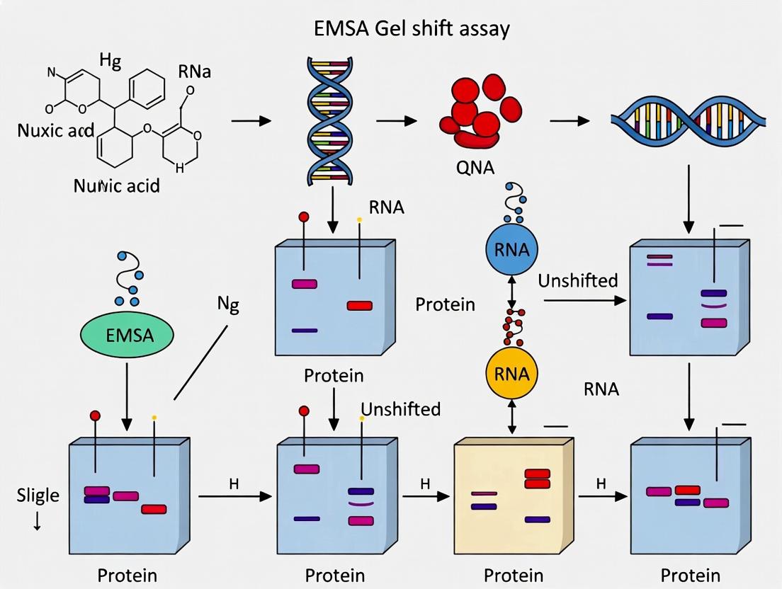
EMSA Gel Shift Assay: A Comprehensive Guide to Principles, Protocols, and Applications in Molecular Biology
This definitive guide to Electrophoretic Mobility Shift Assay (EMSA) provides researchers, scientists, and drug development professionals with a complete framework for studying protein-nucleic acid interactions.
EMSA Gel Shift Assay: A Comprehensive Guide to Principles, Protocols, and Applications in Molecular Biology
Abstract
This definitive guide to Electrophoretic Mobility Shift Assay (EMSA) provides researchers, scientists, and drug development professionals with a complete framework for studying protein-nucleic acid interactions. It begins with foundational principles explaining the core concept of mobility shifts and binding specificity. The article then details a step-by-step methodological protocol, including probe design, binding reaction setup, and gel electrophoresis. It addresses common troubleshooting issues like smearing, weak shifts, and high background, offering optimization strategies. Finally, it explores validation techniques, quantitative approaches, and comparisons with modern alternatives like fluorescence anisotropy and SPR, ensuring readers can design, execute, and interpret robust EMSA experiments for gene regulation and drug discovery research.
What is an EMSA? Unpacking the Core Principles of Protein-Nucleic Acid Interactions
Within the foundational research on Electrophoretic Mobility Shift Assays (EMSAs), the "band shift" phenomenon is the central, observable event that transformed the study of nucleic acid-protein interactions. This whitepaper details the core principles of this assay, its historical discovery, and provides a contemporary technical guide for its application in modern molecular biology and drug discovery contexts.
Historical Discovery and Core Principle
The EMSA, commonly called the gel shift or band shift assay, was independently pioneered in the early 1980s by two research groups: Revzin and Von Hippel, and Garner and Revzin. The seminal discovery was that a protein bound to a DNA or RNA fragment dramatically reduces the electrophoretic mobility of that nucleic acid during non-denaturing polyacrylamide or agarose gel electrophoresis. This results in a detectable "shift" of the band representing the nucleic acid to a higher molecular weight position (or, for large complexes, retention in the well). The assay's simplicity, sensitivity, and ability to quantify binding affinities and kinetics made it an immediate and enduring cornerstone of molecular biology.
The following table summarizes key quantitative parameters and outcomes from a typical EMSA experiment, as established in foundational and current research.
Table 1: Key Quantitative Parameters in a Standard EMSA Experiment
| Parameter | Typical Range/Value | Description & Significance |
|---|---|---|
| Nucleic Acid (Probe) | 0.1-10 nM (labeled) | Trace concentration to maintain pseudo-first-order binding kinetics; often 5'-end labeled with ³²P or a fluorophore. |
| Protein (Extract/Purified) | 0 - 1000 nM | Varied to generate a binding curve for affinity determination (Kd). |
| Poly(dI:dC) | 0.05-2 µg/reaction | Inert competitor DNA to suppress non-specific protein-probe interactions. |
| Electrophoresis Temperature | 4°C | Reduces complex dissociation during electrophoresis (gel running). |
| Gel Acrylamide % | 4-10% | Lower % for larger complexes (>500 bp); higher % for better resolution of smaller complexes. |
| Detection Limit (Protein) | ~10 fmol | Demonstrates high sensitivity for detecting DNA-binding proteins. |
| Apparent Kd (from EMSA) | pM to µM range | Equilibrium dissociation constant derived from quantitating free vs. bound probe across protein concentrations. |
Detailed Experimental Protocol
Protocol: Native EMSA for DNA-Protein Complex Analysis
A. Probe Preparation
- Labeling: Prepare a double-stranded DNA probe (20-50 bp) containing the target sequence. Use T4 Polynucleotide Kinase and [γ-³²P]ATP for 5'-end labeling, or a fluorophore-conjugated primer during PCR/synthesis.
- Purification: Purify the labeled probe using a spin column (e.g., G-25 Sephadex) to remove unincorporated nucleotides.
B. Binding Reaction
- Set up a 10-20 µL reaction mixture on ice:
- Binding Buffer (Final 1X): 10 mM HEPES (pH 7.5-8.0), 50-100 mM KCl/NaCl, 1 mM EDTA, 1 mM DTT, 0.1-0.5% NP-40/Triton X-100, 5-10% Glycerol.
- Non-specific Competitor: 0.5-1 µg of poly(dI:dC) or sheared salmon sperm DNA.
- Labeled Probe: 10,000-20,000 cpm (≈0.1-1 ng).
- Nuclear Extract/Purified Protein: Variable concentration (e.g., 0.5-10 µg of crude extract).
- Optional Controls: Include a 100-fold molar excess of unlabeled identical probe (specific competitor) or a non-specific DNA sequence (non-specific competitor) to confirm binding specificity.
- Incubate at room temperature or 30°C for 20-30 minutes.
C. Non-Denaturing Gel Electrophoresis
- Gel Preparation: Prepare a 4-8% non-denaturing polyacrylamide gel (29:1 acrylamide:bis) in 0.5X TBE or TAE buffer. Pre-run the gel at 100 V for 30-60 minutes at 4°C.
- Loading: Add a non-ionic loading dye (e.g., 10% glycerol, 0.01% bromophenol blue) to the binding reactions. Load samples onto the pre-run gel.
- Run Conditions: Run the gel at 80-120 V, constant voltage, in the cold room (4°C) for 1.5-2.5 hours until the dye front migrates 2/3 down the gel.
D. Detection & Analysis
- For radioactive probes: Dry the gel and expose to a phosphorimager screen or X-ray film.
- For fluorescent probes: Image the gel using an appropriate fluorescence scanner.
- Quantify the intensity of bands corresponding to the free probe and shifted complex(es) using image analysis software (e.g., ImageQuant, ImageJ). Plot bound/free or % bound vs. protein concentration to determine apparent Kd.
Visualization of Core Principles and Workflow
EMSAPrinciple Workflow
The Scientist's Toolkit: Essential Research Reagents & Materials
Table 2: Essential Reagent Solutions for EMSA
| Item | Function & Rationale |
|---|---|
| Purified Protein / Nuclear/Cytoplasmic Extract | Source of the DNA/RNA-binding protein of interest. Nuclear extract is standard for transcription factors. |
| Labeled DNA/RNA Probe | The detectable nucleic acid fragment containing the specific binding site. Radioisotopes (³²P) offer highest sensitivity; fluorophores enable safer, non-radioactive detection. |
| Poly(dI:dC) or similar non-specific DNA | Critical reagent to titrate out non-sequence-specific nucleic acid-binding proteins, reducing background and highlighting specific shifts. |
| 10X Binding Buffer | Provides optimal ionic strength (K⁺/Na⁺), pH (HEPES/Tris), reducing agent (DTT), and stabilizing agents (Glycerol, NP-40) for the interaction. |
| Non-denaturing Polyacrylamide Gel Mix | Matrix for separation based on size and charge of complexes. Lower acrylamide % for larger complexes. Must be non-denaturing (no SDS). |
| 0.5X TBE or TAE Running Buffer | Low-ionic-strength buffer prevents complex dissociation during electrophoresis and provides conductivity. |
| Gel Shift Loading Dye | Non-ionic dye (e.g., glycerol with bromophenol blue) to increase sample density for loading without disrupting non-covalent complexes. |
| Specific & Non-specific Competitor Oligos | Unlabeled oligonucleotides used in control reactions to demonstrate the sequence specificity of the observed band shift. |
| Phosphorimager System / Fluorescence Scanner | For detection and quantification of shifted and free probe bands. Essential for calculating binding affinity (Kd). |
Within the broader thesis on Electrophoretic Mobility Shift Assay (EMSA) basic principles, understanding the fundamental relationship between molecular binding and altered electrophoretic mobility is paramount. This whitepaper provides an in-depth technical guide on the core biophysical principles that govern this shift, which is the cornerstone of EMSA methodology. The assay's utility in quantifying protein-nucleic acid interactions, screening drug candidates, and studying transcriptional regulation hinges on a precise mechanistic understanding of this phenomenon.
Core Biophysical Principles
Electrophoretic mobility (μ) is defined by the equation: μ = q / (6πηr), where q is the net charge, η is the viscosity of the medium, and r is the Stokes radius (hydrodynamic radius). Binding events alter these parameters, leading to a measurable shift in migration through a gel matrix.
2.1. Primary Factors Altering Mobility:
- Change in Mass/Size (r): The binding of a protein to a nucleic acid probe increases the molecular mass and typically the Stokes radius. According to the equation, an increase in r decreases mobility.
- Change in Net Charge (q): Proteins carry a net positive charge under standard EMSA conditions (pH ~8.0), while nucleic acids are highly negative. The binding of a protein partially neutralizes the probe's negative charge, reducing q. This also decreases mobility.
- Conformational Change: Binding can induce significant bending or kinking in the nucleic acid, altering its hydrodynamic radius r without a proportional increase in mass. This conformational change can further retard migration.
The observed "gel shift" or "supershift" is the net result of these combined physical alterations.
Quantitative Data on Mobility Shifts
The following table summarizes typical experimental data from EMSA studies, illustrating the correlation between binding events and mobility reduction.
Table 1: Quantification of Electrophoretic Mobility Shifts in Model EMSA Experiments
| Probe Type (DNA/RNA) | Bound Protein (or Compound) | Approx. Complex Size (kDa) | % Reduction in Mobility (vs. free probe) | Primary Contributing Factor |
|---|---|---|---|---|
| 25-bp dsDNA (15.5 kDa) | p50 Transcription Factor (50 kDa) | 65.5 kDa | ~35-45% | Increased mass & charge neutralization |
| 30-nt RNA (10 kDa) | HuR (RNA-binding protein, 36 kDa) | 46 kDa | ~30-40% | Increased mass & charge neutralization |
| Bent DNA A-tract (20-bp) | HMG-box protein (25 kDa) | ~40 kDa | ~50-60% | Conformational change (bending) |
| dsDNA with drug site | Minor-groove binder (e.g., Netropsin, ~0.5 kDa) | ~16 kDa | ~5-15% | Conformational change & slight charge effect |
Detailed Experimental Protocol: A Standard EMSA
4.1. Key Research Reagent Solutions:
| Reagent/Material | Function & Critical Notes |
|---|---|
| 32P- or fluorescently-labeled nucleic acid probe | The target molecule whose mobility is monitored. Label must not interfere with protein binding. |
| Purified protein or nuclear extract | Source of the binding protein. Extract complexity may require specific competitors. |
| 10X Binding Buffer | Typically: 100 mM Tris, 500 mM KCl, 10 mM DTT, 50% Glycerol (pH 7.5-8.0). Provides optimal ionic strength and reducing conditions. |
| Non-specific competitor DNA (Poly(dI•dC)) | Suppresses protein binding to non-specific sequences, improving signal-to-noise. |
| Specific unlabeled competitor probe | Validates binding specificity by abolishing shift in a competition assay. |
| Non-denaturing Polyacrylamide Gel (4-6%) | Matrix for separation. Must be pre-run and run in low-ionic strength buffer (0.5X TBE) to maintain weak interactions. |
| Electrophoresis Buffer (0.5X TBE or TAE) | Provides continuous conductivity with minimal ionic strength to avoid disrupting complexes. |
| Gel Shift Stabilizer (e.g., 10% glycerol in gel) | Enhances complex stability during electrophoresis. |
| Antibody (for supershift) | Binds to the protein in the complex, causing a further mobility reduction ("supershift") for identification. |
4.2. Protocol:
- Binding Reaction: Combine in order:
- Nuclease-free water (to final volume of 20 μL).
- 10X Binding Buffer (2 μL).
- 1 μg/μL Poly(dI•dC) (1-2 μL) (optimize for each protein).
- Purified protein or nuclear extract (2-10 μg).
- Labeled nucleic acid probe (10-20 fmol).
- Incubate at room temperature or 4°C for 20-30 minutes.
- Competition/Supershift Controls:
- For specific competition: Add 50-200-fold molar excess of unlabeled probe before adding the labeled probe.
- For supershift: Add 1-2 μg of specific antibody after the initial binding incubation; incubate further 15-30 min.
- Electrophoresis:
- Pre-run a non-denaturing polyacrylamide gel in 0.5X TBE at 100V for 60 minutes at 4°C.
- Load binding reactions (mixed with non-denaturing loading dye) onto the gel.
- Run at constant voltage (80-100V) for 1.5-2 hours at 4°C until the dye front migrates 2/3 down the gel.
- Detection:
- For radioactive probes: Dry gel and expose to a phosphorimager screen.
- For fluorescent probes: Image directly using a suitable scanner.
Visualizing Principles and Workflows
Diagram 1: Factors Governing Mobility Shift in EMSA
Diagram 2: Standard EMSA Workflow
The Electrophoretic Mobility Shift Assay (EMSA) is a cornerstone technique for studying protein-nucleic acid interactions. Its fundamental principle relies on the detection of a retarded electrophoretic migration of a labeled nucleic acid probe when bound by a protein. This whitepaper delves into the three core components—the labeled probe, the protein extract, and competitors—which are critical for assay specificity, sensitivity, and interpretability. Mastery of these components is essential for rigorous research in gene regulation, drug discovery targeting transcription factors, and mechanistic biology.
The Triad of Core Components: A Detailed Technical Analysis
Labeled Nucleic Acid Probe
The probe is the detectable reporter molecule in the EMSA.
- Composition: Typically a short (20-30 bp), double-stranded DNA or RNA oligonucleotide containing a specific protein-binding sequence (e.g., a transcription factor response element).
- Labeling: The probe must be labeled for visualization. Common methods include:
- Radioactive (³²P): Traditional, high-sensitivity method.
- Fluorescent (Cy5, FAM): Non-radioactive, safer, with good sensitivity.
- Biotin: Followed by streptavidin-conjugate detection (chemiluminescent or colorimetric).
- Function: Serves as the ligand for the protein of interest. Its shift in mobility upon binding is the assay's readout.
Protein Extract
The source of the protein(s) that interact with the nucleic acid probe.
- Types: Nuclear extract (for transcription factors), whole-cell lysate, or purified recombinant protein.
- Critical Parameters: Extract preparation must preserve protein activity and native conformation. Protease and phosphatase inhibitors are mandatory. Protein concentration must be optimized to ensure binding is within the linear range.
Competitors
Unlabeled nucleic acids used to demonstrate binding specificity.
- Specific Competitor: An identical unlabeled version of the probe sequence. Successfully competes for protein binding, abolishing or diminishing the shifted band.
- Non-specific Competitor: An unrelated sequence (e.g., poly(dI-dC), sheared salmon sperm DNA). Added to absorb non-specific DNA-binding proteins, reducing background.
- Mutant Competitor: A probe sequence with a mutated binding site. Used to confirm sequence-specific binding when it fails to compete effectively.
Table 1: Common Probe Labeling Methods Comparison
| Method | Typical Sensitivity (fmol) | Stability | Safety & Regulation | Required Equipment |
|---|---|---|---|---|
| ³²P End-Labeling | 0.1 - 1 | Short (half-life) | High risk; Radioactive license | Phosphorimager, Geiger counter |
| Fluorescent (5'-end) | 1 - 10 | Long (months) | Safe | Fluorescence scanner/imager |
| Biotin (3'-end) | 0.5 - 5 | Long (years) | Safe | Standard gel imager (chemiluminescence) |
Table 2: Recommended Competitor Concentrations in EMSA Binding Reactions
| Competitor Type | Typical Working Concentration | Purpose | Expected Outcome on Shifted Band |
|---|---|---|---|
| Non-specific (poly(dI-dC)) | 0.05 - 0.2 µg/µL | Block non-specific interactions | Reduces smearing, sharpens specific band |
| Specific Unlabeled Probe | 10x - 100x molar excess over labeled probe | Prove sequence specificity | Significant reduction or elimination |
| Mutant Unlabeled Probe | 10x - 100x molar excess | Confirm sequence specificity | Minimal or no reduction |
Detailed Experimental Protocols
Protocol 4.1: EMSA Binding Reaction Setup
Objective: To form complexes between the protein extract and the labeled probe under controlled conditions. Reagents: Labeled probe, protein extract, binding buffer (10 mM HEPES, pH 7.9, 50 mM KCl, 1 mM DTT, 10% glycerol, 0.1% NP-40), poly(dI-dC), specific/non-specific competitors, nuclease-free water. Procedure:
- Prepare a master mix of binding buffer, glycerol, and poly(dI-dC) for the number of reactions +1.
- Aliquot the master mix into thin-walled PCR tubes or a microplate.
- Add competitors to appropriate tubes (e.g., 50-fold molar excess of unlabeled probe).
- Add protein extract (typically 2-10 µg). Include a "probe-only" control with no extract.
- Pre-incubate for 10 minutes at room temperature to allow competitor binding.
- Add a constant amount of labeled probe (e.g., 20 fmol) to each reaction.
- Incubate for 20-30 minutes at room temperature.
- Load samples directly onto a pre-run native polyacrylamide gel.
Protocol 4.2: Supershift Assay Protocol
Objective: To identify a specific protein within a complex using an antibody. Modification to 4.1:
- After step 5 (pre-incubation with competitor), add 1-2 µg of specific antibody or an isotype control antibody to the appropriate reactions.
- Incubate for an additional 30-60 minutes at 4°C before adding the labeled probe (step 6).
- A further retardation of the complex ("supershift") confirms the presence of the target protein.
Visualizations
Title: EMSA Experimental Workflow
Title: EMSA Specificity Control Decision Tree
The Scientist's Toolkit: EMSA Research Reagent Solutions
Table 3: Essential Materials for EMSA
| Item | Function & Importance | Example/Note |
|---|---|---|
| Chemiluminescent Nucleic Acid Labeling Kit | Non-radioactive, sensitive probe labeling. | Biotin 3' End Labeling Kits. |
| Nuclear Extract Kit | Standardized, high-quality active transcription factor source. | Kits with validated controls (e.g., from Active Motif, Thermo). |
| Poly(dI-dC) | Critical non-specific competitor to suppress background. | Supplied as a lyophilized powder or stock solution. |
| EMSABinding Buffer (5X) | Optimized, consistent buffer for complex formation. | Often includes salts, glycerol, DTT, and carrier. |
| Native PAGE Gel Kit | Pre-cast gels and matched buffers for optimal separation. | 4-20% gradient Tris-Glycine gels. |
| HRP-Streptavidin Conjugate | Detection agent for biotinylated probes. | Used with chemiluminescent substrate. |
| Super-shift Grade Antibodies | Antibodies that recognize native protein in the complex. | Validated for EMSA/supershift applications. |
| Phosphorimaging/ ChemiDoc System | High-sensitivity detection and quantification of bands. | Essential for quantitative EMSA. |
The Electrophoretic Mobility Shift Assay (EMSA), or gel shift assay, is a cornerstone technique for studying nucleic acid-protein interactions. Within the broader thesis of EMSA basic principles research, this guide details its application for probing three critical interaction types: sequence-specific binding of transcription factors (TFs) to DNA, the activity of diverse DNA-binding proteins (DBPs), and the formation of ribonucleoprotein (RNP) complexes. EMSA provides a direct, quantitative measure of binding affinity, stoichiometry, and specificity, forming the foundation for understanding gene regulatory networks and facilitating drug discovery targeting these interactions.
Core Quantitative Data on Studied Interactions
Table 1: Key Quantitative Parameters for EMSA-Based Interaction Studies
| Parameter | Transcription Factor-DNA | General DNA-Binding Protein-DNA | RNA-Protein Complex |
|---|---|---|---|
| Typical Probe Length | 15-40 bp (containing consensus sequence) | 20-1000+ bp (dependent on protein) | 50-500 nucleotides |
| Typical Kd Range | 10^-9 to 10^-12 M | 10^-6 to 10^-12 M (highly variable) | 10^-8 to 10^-12 M |
| Common EMSA Gel Type | Non-denaturing Polyacrylamide (4-6%) | Agarose (0.5-2%) or Polyacrylamide | Non-denaturing Polyacrylamide (4-8%) |
| Critical Buffer Components | Poly(dI:dC) as non-specific competitor, Mg2+ | Variable specific salt (e.g., Mg2+, Zn2+) | RNase inhibitors, Heparin competitor |
| Key Controls | Unlabeled specific competitor (cold probe), mutant probe, supershift with antibody | Substrate mutant, ion dependence, protease treatment | RNA sequence mutant, RNAse treatment, supershift |
| Primary Output | Binding affinity, complex stoichiometry, sequence specificity | Binding presence/absence, complex size, co-factor requirement | Complex stability, specificity, mapping binding region |
Table 2: Recent Advancements and Detection Limits (2020-2024)
| Advancement | Principle | Sensitivity Gain vs Classic EMSA | Applicable Interaction Type |
|---|---|---|---|
| Fluorescence Anisotropy EMSA | Measures polarization of fluorescently labeled probe | 10-100x (allows solution-based Kd) | TF-DNA, RNA-Protein |
| Digital EMSA (Microfluidics) | Single complex detection in nanochannels | Up to 1000x for rare complexes | All types, low-abundance samples |
| Infrared (IR) Dye Detection | Uses IR-labeled antibodies for supershift | 5-10x (reduced background) | TF-DNA (supershift specific) |
| Capillary Electrophoresis EMSA | CE-based separation with LIF detection | 50-100x (quantitative, automated) | RNA-Protein, TF-DNA |
Detailed Experimental Protocols
Protocol for Transcription Factor-DNA Binding EMSA
Objective: To detect and characterize the binding of a nuclear extract protein to a specific DNA consensus sequence.
Materials: Radioactively (γ-32P) or fluorescently (e.g., Cy5) end-labeled double-stranded DNA probe, purified TF or nuclear extract, binding buffer (10 mM HEPES pH 7.9, 50 mM KCl, 1 mM DTT, 2.5 mM MgCl2, 10% glycerol, 0.1% NP-40), poly(dI:dC) (1 µg/µL), 6% non-denaturing polyacrylamide gel (0.5x TBE), electrophoresis apparatus.
Method:
- Probe Preparation: Anneal complementary oligonucleotides containing the TF binding site. Label using T4 Polynucleotide Kinase and [γ-32P]ATP or purchase pre-labeled probes.
- Binding Reaction:
- Combine on ice: 4 µL binding buffer, 1 µL poly(dI:dC) (1 µg), 1-10 µg nuclear extract or 10-100 fmol purified TF, and nuclease-free water to 9 µL.
- Pre-incubate for 10 minutes at room temperature to block non-specific sites.
- Add 1 µL labeled probe (~20 fmol, 50,000 cpm). Final volume = 10 µL.
- Incubate 20-30 minutes at room temperature.
- Competition/Supershift Controls:
- Specific Competitor: Add 100-fold molar excess of unlabeled identical probe before adding labeled probe.
- Mutant Competitor: Add 100-fold molar excess of unlabeled probe with a scrambled/mutated binding site.
- Antibody Supershift: Add 1-2 µg of specific antibody after binding reaction; incubate additional 20 mins.
- Electrophoresis:
- Pre-run gel in 0.5x TBE buffer for 30-60 min at 100 V (4°C).
- Load samples (add 1 µL of 10x loading dye) directly onto running gel.
- Run at 100 V, 4°C, until dye front migrates 2/3 of the gel.
- Detection: For radioactive probes, dry gel and expose to phosphorimager screen. For fluorescent probes, scan gel using an appropriate imager.
Protocol for RNA-Protein Complex EMSA (RNP Gel Shift)
Objective: To analyze the interaction of a protein (e.g., splicing factor, miRNA-binding protein) with a specific RNA sequence.
Materials: In vitro transcribed RNA probe (labeled with 32P-α-UTP or fluorescent tag), purified RNA-binding protein (RBP), binding buffer (10 mM HEPES pH 7.5, 50 mM KCl, 1.5 mM MgCl2, 1 mM DTT, 0.01% Triton X-100, 5% glycerol, 40 U RNase inhibitor), competitor RNA (e.g., yeast tRNA, heparin), 6% non-denaturing polyacrylamide gel (0.5x TBE, pre-run).
Critical Modifications from DNA EMSA:
- RNase-Free Conditions: Use DEPC-treated water, RNase-free tubes and tips. Include RNase inhibitor (e.g., RNasin) in all steps.
- Probe Preparation: Generate uniformally labeled RNA by in vitro transcription with T7/SP6 RNA polymerase in the presence of [α-32P]UTP. Gel-purify the full-length transcript.
- Binding Reaction: Use heparin (1-5 µg/µL) or yeast tRNA as a non-specific competitor instead of poly(dI:dC). Include 1.5-5 mM MgCl2, which is often critical for RNA folding and RBP binding.
- Electrophoresis: Pre-run and run the gel at 4°C to maintain complex stability. The gel percentage may be increased (up to 8%) for smaller RNA probes.
- Detection: As with DNA EMSA. For supershift assays, ensure antibodies do not have RNase activity.
Signaling Pathways & Experimental Workflows
Title: Core EMSA Experimental Workflow
Title: TF Activation & DNA Binding Pathway
The Scientist's Toolkit: Essential Research Reagents & Materials
Table 3: Key Research Reagent Solutions for EMSA Experiments
| Reagent/Material | Function in EMSA | Key Considerations & Examples |
|---|---|---|
| Labeled Nucleic Acid Probe | Target molecule for detection. Allows visualization of free vs. bound states. | Choice: 32P (high sensitivity), Fluorescent dyes (safe, quantifiable), Biotin (chemiluminescent detection). Purity: Crucial for clean results. |
| Non-specific Competitor DNA/RNA | Binds and neutralizes non-specific binding proteins in extracts. Reduces background. | For DNA EMSA: Poly(dI:dC), sheared salmon sperm DNA. For RNA EMSA: Heparin, yeast tRNA, total cellular RNA. |
| Binding Buffer & Salts | Provides optimal ionic strength, pH, and co-factors for specific interaction. | Typical: HEPES or Tris buffer, KCl/NaCl, MgCl2 (essential for many DBPs/RBPs), DTT (reducing agent), glycerol (stabilizer, aids loading). |
| Non-denaturing Gel Matrix | Separates protein-nucleic acid complexes from free probe based on size/shape/charge. | Polyacrylamide (4-8%): High resolution for probes <500 bp. Agarose (0.5-2%): For very large complexes or long DNA probes. |
| Purified Protein or Cell Extract | Source of the DNA/RNA-binding protein. | Purified Protein: For Kd, stoichiometry. Nuclear/Cytoplasmic Extract: For studying endogenous protein activity; requires protease/phosphatase inhibitors. |
| Specific & Mutant Competitors | Demonstrates binding specificity. | Specific Cold Probe: Identical unlabeled sequence confirms saturable binding. Mutant Probe: Defines sequence requirements. |
| Antibodies (for Supershift) | Identifies protein in complex or disrupts binding (blocking antibody). | Confirms protein identity. Can cause "supershift" (slower migration) or disrupt shift. Must be suitable for native conditions. |
| RNase Inhibitors | Critical for RNA-protein EMSA only. Prevents degradation of RNA probe. | Essential in all steps. Common agents: RNasin (recombinant RNase inhibitor), SUPERase•In. |
This whitepaper is presented within the context of a broader thesis on Electrophoretic Mobility Shift Assay (EMSA) basic principles. EMSA, or gel shift assay, is a cornerstone technique for studying nucleic acid-protein interactions, pivotal in gene regulation research and drug discovery targeting transcription factors. The core interpretive challenge lies in accurately distinguishing and quantifying the shifted protein-bound complex band from the free probe band. This guide provides a technical framework for this critical analysis.
Fundamental Principles: The Basis of the Shift
The assay relies on the principle that a nucleic acid probe (DNA or RNA) bound by a protein exhibits reduced electrophoretic mobility through a non-denaturing polyacrylamide or agarose gel compared to the free probe. This results in a distinct band shift.
Key Band Patterns and Interpretations
Interpreting an EMSA gel requires analyzing the banding pattern. The table below summarizes common outcomes.
Table 1: Interpretation of EMSA Gel Band Patterns
| Band Pattern Observed | Interpretation | Biological Significance |
|---|---|---|
| Single band at the free probe position | No binding occurred. | Protein may not bind the probe sequence, binding conditions may be suboptimal, or protein is inactive. |
| One major higher molecular weight (shifted) band | Specific protein-probe complex formation. | Confirms interaction. Shift magnitude depends on protein size and conformational change. |
| Multiple shifted bands | Multiple discrete complexes (e.g., different oligomeric states, multiple proteins binding the same probe). | Suggests cooperative binding or presence of different protein complexes in the extract. |
| Smear above the free probe | Non-specific binding or multiple, unstable complexes. | Often indicates need for optimization of competitors (e.g., poly(dI-dC)) or binding conditions. |
| Disappearance of free probe band (supershift) | Antibody against the bound protein further retards the complex. | Confirms protein identity within the complex. |
| Reduction of shifted band intensity with unlabeled competitor | Specific competition for binding. | Validates sequence-specific nature of the interaction. |
Detailed Experimental Protocol for a Standard EMSA
Probe Preparation and Labeling
- End-Labeling with [γ-³²P] ATP: For 1 pmol of DNA oligonucleotide (annealed), use T4 Polynucleotide Kinase in provided buffer. Incubate at 37°C for 30 min, then purify using a microspin G-25 column.
- Alternative Non-Radioactive Labeling: Use biotin- or fluorophore-conjugated nucleotides followed by detection with streptavidin-HRP or direct fluorescence imaging.
Protein Sample Preparation
- Source: Purified recombinant protein or nuclear extract.
- Nuclear Extract Protocol (Brief): Harvest cells, lyse in hypotonic buffer, pellet nuclei, and extract proteins with high-salt buffer. Dialyze into low-salt storage buffer. Determine protein concentration (e.g., Bradford assay).
Binding Reaction Assembly
Combine components in order on ice:
- 1-10 µg Nuclear Extract or 10-100 ng Purified Protein
- 1-2 µg Poly(dI-dC) or other non-specific competitor DNA
- Binding Buffer (10 mM HEPES, 50 mM KCl, 1 mM DTT, 2.5 mM MgCl₂, 10% Glycerol, pH 7.9)
- Unlabeled specific competitor (for specificity controls; 50-100x molar excess)
- Labeled Probe (20,000-50,000 cpm)
- Total Volume: 20 µL
- Incubation: 20-30 minutes at room temperature.
Gel Electrophoresis and Detection
- Gel Preparation: Pre-run a 4-6% non-denaturing polyacrylamide gel (29:1 acrylamide:bis) in 0.5X TBE buffer at 100V for 60 min at 4°C.
- Loading: Add 5 µL of non-denaturing loading dye to reactions. Load entire sample.
- Electrophoresis: Run at 100V (constant voltage) in 0.5X TBE at 4°C until the bromophenol blue dye migrates ~2/3 of the gel.
- Detection:
- Radioactive: Transfer gel to blotting paper, dry, and expose to a phosphorimager screen.
- Biotin: Electro-transfer to positively charged nylon membrane, crosslink, and detect with chemiluminescent substrate.
Quantitative Analysis and Data Presentation
Quantification involves measuring the intensity of the free probe and bound complex bands. Key metrics are summarized below.
Table 2: Quantitative Metrics for EMSA Analysis
| Metric | Formula / Description | Purpose & Interpretation |
|---|---|---|
| Fraction Bound | Intensity(Bound Complex) / [Intensity(Bound) + Intensity(Free)] | Direct measure of binding activity under given conditions. |
| Dissociation Constant (Kd) | Derived from titration of protein against constant probe. [Protein] at half-maximal binding approximates Kd. | Affinity measurement. Lower Kd indicates tighter binding. |
| Inhibition Constant (IC₅₀) | Concentration of unlabeled competitor or drug that reduces complex formation by 50%. | Potency of a competitor or inhibitory compound. |
| Gel Shift Kinetics | Fraction bound plotted vs. time. | Determines association rate; can infer binding stability. |
The Scientist's Toolkit: Essential Research Reagent Solutions
Table 3: Key Reagents and Materials for EMSA
| Reagent/Material | Function/Purpose | Key Considerations |
|---|---|---|
| Purified Protein or Nuclear Extract | Source of DNA/RNA-binding protein. | Activity varies; use protease inhibitors, confirm functionality. |
| Labeled Nucleic Acid Probe | Detectable target for binding. | High specific activity (radioactive) or high sensitivity (biotin/fluor). Must be sequence-verified. |
| Non-Specific Competitor DNA (e.g., poly(dI-dC)) | Binds non-specific proteins to reduce background smearing. | Type and amount require optimization for each protein-probe pair. |
| Non-Denaturing Polyacrylamide Gel | Matrix for separation based on size/charge/shape. | Percentage (4-10%) affects resolution; low acrylamide:bis ratio (e.g., 29:1, 37.5:1) is common. |
| Specific Unlabeled Competitor Oligo | Validates specificity of the interaction. | Identical sequence to labeled probe. Should abolish the shifted band. |
| Antibody for Supershift | Confirms protein identity in the complex. | Must recognize native protein epitope; control IgG is essential. |
| Binding Buffer Components | Maintains pH, ionic strength, and stabilizing agents (DTT, glycerol). | Optimize divalent cations (Mg²⁺, Zn²⁺) and salt (KCl) for each system. |
| Detection System | Visualizes the separated bands. | Phosphorimager (³²P), CCD camera (fluorescent), or Chemiluminescence (biotin). |
Step-by-Step EMSA Protocol: From Probe Design to Autoradiography
Within the framework of research into Electrophoretic Mobility Shift Assay (EMSA) basic principles, the choice of probe labeling and detection method is fundamental. This technical guide provides an in-depth comparison of radiolabeled and non-radioactive (chemiluminescent/fluorescent) methods for nucleic acid probe design and labeling, critical for detecting protein-DNA/RNA interactions in EMSAs.
Core Principles of Probe Design
Regardless of detection method, an effective EMSA probe is a short, double-stranded DNA or single-stranded RNA oligonucleotide containing the specific protein-binding sequence. Key design considerations include sequence specificity, length (typically 20-40 bp), minimal secondary structure, and appropriate end-modification for labeling.
Labeling and Detection Methodologies
Radiolabeling (Typically with ³²P)
Principle: Incorporation of a radioactive isotope (e.g., [γ-³²P]ATP) via enzymatic reactions.
Detailed Protocol: 5' End-Labeling with T4 Polynucleotide Kinase
- Reaction Setup: In a sterile microcentrifuge tube, combine:
- 1–10 pmol of dephosphorylated, double-stranded DNA oligonucleotide probe.
- 2 µL of 10X T4 PNK Buffer (700 mM Tris-HCl, pH 7.6, 100 mM MgCl₂, 50 mM DTT).
- 20 µCi (≈10 pmol) of [γ-³²P]ATP.
- 10 units of T4 Polynucleotide Kinase.
- Nuclease-free water to a final volume of 20 µL.
- Incubation: Incubate at 37°C for 30 minutes.
- Termination: Heat-inactivate the enzyme at 65°C for 10 minutes.
- Purification: Remove unincorporated nucleotides using a spin column (e.g., Sephadex G-25) or ethanol precipitation. Resuspend in appropriate buffer.
- Quantification: Measure radioactivity using a scintillation counter. Optimal specific activity is >10⁸ cpm/µg.
Advantages: Extreme sensitivity (can detect sub-femtomole quantities), linear quantitative response over a wide range, and a well-established, straightforward protocol. Disadvantages: Requires specialized safety infrastructure (shielding, monitoring, waste disposal), isotopes have short half-lives (³²P: 14.3 days), and regulatory burdens.
Chemiluminescent Labeling
Principle: Probes are labeled with haptens (e.g., biotin, digoxigenin) and detected post-electrophoresis using enzyme-conjugated streptavidin or antibodies (e.g., Alkaline Phosphatase, HRP) that catalyze a light-emitting reaction.
Detailed Protocol: Biotin 3'-End Labeling Using Terminal Transferase
- Reaction Setup: Combine in a tube:
- 100 pmol of single-stranded DNA oligonucleotide.
- 4 µL of 5X Terminal Transferase Reaction Buffer.
- 1 µL of 1 mM Biotin-11-dUTP.
- 20 units of Terminal Deoxynucleotidyl Transferase (TdT).
- Nuclease-free water to 20 µL.
- Incubation: Incubate at 37°C for 60 minutes.
- Termination: Add 2 µL of 0.5 M EDTA to chelate Mg²⁺ and inactivate TdT.
- Purification: Purify via ethanol precipitation or spin column. Anneal to complementary strand.
- Detection Post-EMSA: Transfer gel to a nylon membrane via blotting. Block membrane, then incubate with Streptavidin-Alkaline Phosphatase (SA-AP) conjugate. Wash and incubate with chemiluminescent AP substrate (e.g., CDP-Star). Expose to X-ray film or CCD imager.
Fluorescent Labeling
Principle: Probes are directly conjugated to a fluorophore (e.g., Cy3, Cy5, FAM) during synthesis. Detection is via direct in-gel scanning using a fluorescence imager.
Detailed Protocol: Using Commercially Synthesized Fluorescent Probes
- Probe Preparation: Order HPLC-purified oligonucleotide with a 5' or 3' fluorophore modification. Resuspend to 100 µM stock in TE buffer.
- Annealing: Mix equimolar amounts of labeled and unlabeled complementary strands in annealing buffer. Heat to 95°C for 5 minutes and cool slowly to room temperature.
- EMSA & Direct Detection: Run EMSA as usual using a non-fluorescent gel tank and components. Scan the gel directly using a fluorescence gel scanner with appropriate excitation/emission filters. No blotting, blocking, or enzymatic development is required.
Quantitative Comparison of Methods
Table 1: Technical and Operational Comparison of Labeling Methods
| Parameter | Radiolabeling (³²P) | Chemiluminescent (Biotin/AP) | Fluorescent (Direct) |
|---|---|---|---|
| Typical Sensitivity | 0.1-1 fmol | 1-10 fmol | 5-50 fmol |
| Dynamic Range | >10⁴ | ~10³ | ~10³ |
| Detection Time | Minutes to hours (exposure) | 5-60 minutes (substrate rxn) | Instantaneous (scan) |
| Probe Stability | Short (isotope decay) | Years (at -20°C) | Years (protected from light) |
| Assay Workflow | Moderate | Lengthy (blotting required) | Fastest (no blotting) |
| Quantification | Excellent, linear | Good, non-linear at extremes | Good |
| Safety & Regulation | High (radioactive) | Low (standard chemicals) | Low |
| Primary Cost Driver | Isotope, waste disposal | Enzyme conjugates, substrate | Fluorophore, scanner |
| Re-probing Possibility | No (decay) | Possible (stripping difficult) | No (covalent label) |
Table 2: "The Scientist's Toolkit": Essential Reagents for EMSA Probe Labeling
| Reagent / Solution | Function in Probe Labeling & Detection |
|---|---|
| [γ-³²P]ATP | Radioactive phosphate donor for 5' end-labeling via T4 PNK. |
| T4 Polynucleotide Kinase (PNK) | Catalyzes transfer of γ-phosphate from ATP to 5'-OH of DNA. |
| Biotin-11-dUTP / DIG-dUTP | Hapten-labeled nucleotide for enzymatic incorporation into probe. |
| Terminal Deoxynucleotidyl Transferase (TdT) | Adds labeled nucleotides to the 3'-end of DNA. |
| Fluorophore-labeled Oligonucleotide | Synthesized probe with covalent fluorophore (e.g., Cy3) for direct detection. |
| Streptavidin-Alkaline Phosphatase (SA-AP) | Binds biotinylated probe; AP catalyzes chemiluminescent reaction. |
| CDP-Star / Lumi-Phos Plus | Chemiluminescent substrate for Alkaline Phosphatase. |
| Nylon Membrane (Positively Charged) | Membrane for transferring and immobilizing nucleic acids for chemiluminescent detection. |
| Blocking Buffer (e.g., with Casein) | Prevents non-specific binding of detection reagents to membrane. |
| Spin Columns (G-25/50) | For rapid removal of unincorporated nucleotides post-labeling. |
The choice of method hinges on the specific thesis context of EMSA optimization. Radiolabeling remains the gold standard for ultimate sensitivity and quantitative rigor where infrastructure permits. Chemiluminescence offers a safe, highly sensitive alternative but adds complexity with blotting. Fluorescence is the most streamlined, safe, and rapid method for high-throughput or qualitative applications, though with slightly lower sensitivity.
For foundational EMSA principles research, comparative studies using multiple methods on the same protein-nucleic acid interaction can yield invaluable insights into assay limitations and optimal configuration.
Visualizations
Title: EMSA Probe Labeling & Detection Method Decision Workflow
Title: Molecular Pathways for Three Probe Labeling & Detection Types
1. Introduction Within the framework of Electrophoretic Mobility Shift Assay (EMSA) research, the selection and preparation of the protein source are critical determinants of experimental success. EMSA, a cornerstone technique for studying protein-nucleic acid interactions, relies on the quality and specificity of the protein component. This guide provides an in-depth technical comparison of the three primary protein sources—nuclear extracts, recombinant proteins, and whole cell lysates—detailing their preparation protocols, advantages, limitations, and optimal use cases within EMSA-based research and drug discovery.
2. Comparative Analysis of Protein Sources The choice of protein source balances biological relevance, purity, yield, and experimental throughput.
Table 1: Quantitative Comparison of Protein Sources for EMSA
| Parameter | Nuclear Extracts | Recombinant Proteins | Whole Cell Lysates |
|---|---|---|---|
| Typical Total Protein Yield | 1-5 mg from 10⁷ cells | 0.1-10 mg/L culture | 2-10 mg from 10⁷ cells |
| Target Protein Abundance | Low (Requires enrichment) | High (Pure) | Very Low (Dilute) |
| Preparation Time | 2-4 hours | Days to weeks (incl. expression) | 30-60 minutes |
| Relative Cost | Moderate | High (setup), Low (scale) | Low |
| Biological Context | Native, nuclear-specific | Isolated, may lack PTMs | Native, cytoplasmic + nuclear |
| Key Advantage | Native nuclear complexes | High purity & specificity | Rapid, preserves some complexes |
| Primary Limitation | Complex mixture | May lack native PTMs/folding | High background interference |
3. Detailed Methodologies
3.1. Preparation of Nuclear Extracts Protocol adapted from Dignam et al. (1983) with contemporary modifications.
Reagents: Hypotonic Buffer (10 mM HEPES pH 7.9, 1.5 mM MgCl₂, 10 mM KCl, 0.5 mM DTT, protease inhibitors), Low-Salt Buffer (20 mM HEPES pH 7.9, 1.5 mM MgCl₂, 20 mM KCl, 0.2 mM EDTA, 25% glycerol, 0.5 mM DTT), High-Salt Buffer (as Low-Salt, but with 1.2 M KCl), Dialysis Buffer (20 mM HEPES pH 7.9, 100 mM KCl, 0.2 mM EDTA, 20% glycerol, 0.5 mM DTT).
Procedure:
- Harvest ~5x10⁷ adherent or suspension cells. Wash with cold PBS.
- Pellet cells (500 x g, 5 min, 4°C). Resuspend in 5x pellet volume of Hypotonic Buffer. Incubate on ice for 15 min.
- Centrifuge (500 x g, 5 min, 4°C). Discard supernatant.
- Resuspend pellet in 2x original pellet volume of Hypotonic Buffer. Lyse cells with 10-15 strokes in a Dounce homogenizer (tight pestle).
- Centrifuge (1000 x g, 10 min, 4°C). The pellet contains crude nuclei.
- Resuspend nuclear pellet in 0.5-1 mL of Low-Salt Buffer. While stirring, slowly add an equal volume of High-Salt Buffer. Stir gently for 30 min at 4°C.
- Centrifuge (25,000 x g, 30 min, 4°C). Transfer supernatant (nuclear extract) to dialysis tubing.
- Dialyze against 500 mL Dialysis Buffer for 3-5 hours at 4°C with one buffer change.
- Clarify by centrifugation (25,000 x g, 20 min, 4°C). Aliquot, snap-freeze, and store at -80°C. Determine concentration via Bradford assay.
3.2. Preparation of Recombinant Proteins General protocol for E. coli expressed, affinity-tagged proteins.
Reagents: Lysis Buffer (50 mM Tris-HCl pH 8.0, 300 mM NaCl, 10 mM imidazole, 1 mM PMSF, 1 mg/mL lysozyme), Wash Buffer (50 mM Tris-HCl pH 8.0, 300 mM NaCl, 20 mM imidazole), Elution Buffer (50 mM Tris-HCl pH 8.0, 300 mM NaCl, 250 mM imidazole), Storage Buffer (20 mM HEPES pH 7.9, 100 mM KCl, 10% glycerol, 0.5 mM DTT).
Procedure:
- Induce expression in 1 L bacterial culture. Harvest cells by centrifugation (5,000 x g, 15 min, 4°C).
- Resuspend pellet in 30 mL Lysis Buffer. Incubate on ice for 30 min.
- Sonicate on ice (6 cycles of 10 sec pulse, 20 sec rest). Centrifuge (20,000 x g, 30 min, 4°C).
- Incubate clarified lysate with 2 mL pre-equilibrated Ni-NTA resin for 1 hour at 4°C with gentle mixing.
- Load resin into a column. Wash with 20 mL Wash Buffer.
- Elute protein with 5 mL Elution Buffer. Collect 1 mL fractions.
- Analyze fractions by SDS-PAGE. Pool pure fractions.
- Dialyze against Storage Buffer to remove imidazole. Concentrate if necessary using a centrifugal concentrator. Aliquot and store at -80°C.
3.3. Preparation of Whole Cell Lysates Rapid preparation method for EMSA screening.
Reagents: Whole Cell Lysis Buffer (20 mM HEPES pH 7.9, 150 mM KCl, 1% NP-40, 0.5 mM DTT, protease/phosphatase inhibitors).
Procedure:
- Harvest 1-2x10⁶ cells. Wash with cold PBS.
- Lyse cells directly in 100-200 μL of Whole Cell Lysis Buffer. Vortex briefly.
- Incubate on ice for 15 min, vortexing intermittently.
- Centrifuge at maximum speed (≥16,000 x g, 15 min, 4°C) to pellet insoluble debris.
- Transfer supernatant (whole cell lysate) to a fresh tube. Determine protein concentration. Use immediately or aliquot and store at -80°C.
4. The Scientist's Toolkit: Essential Research Reagents
Table 2: Key Reagent Solutions for Protein Preparation & EMSA
| Reagent / Material | Function / Purpose |
|---|---|
| Protease & Phosphatase Inhibitor Cocktails | Preserves protein integrity by preventing degradation and maintaining PTMs during extraction. |
| DTT (Dithiothreitol) or β-Mercaptoethanol | Reducing agent that maintains cysteine residues in a reduced state, preventing improper disulfide bonds. |
| NP-40 / Igepal CA-630 | Mild non-ionic detergent for cell membrane disruption in whole cell and nuclear extract protocols. |
| Glycerol | Stabilizing agent added to storage buffers to prevent protein denaturation and maintain activity at -80°C. |
| HEPES Buffer | Biological pH buffer with minimal interference in protein-DNA binding interactions during EMSA. |
| Ni-NTA Agarose / Glutathione Sepharose | Affinity chromatography resins for purification of polyhistidine- or GST-tagged recombinant proteins. |
| Bradford / BCA Assay Kits | For accurate quantification of total protein concentration in complex mixtures. |
| Dialysis Tubing / Cassettes | For buffer exchange and removal of small molecules (e.g., salts, imidazole) from protein preparations. |
5. Visualizing Workflows and Pathways
Title: EMSA Protein Source Preparation Workflow
Title: Core EMSA Binding & Competition Principles
Within the framework of research on the basic principles of the Electrophoretic Mobility Shift Assay (EMSA), optimizing the binding reaction is paramount for achieving specific and high-affinity interactions between a target protein and a nucleic acid probe. This in-depth technical guide focuses on the three critical, interdependent variables: buffer composition, incubation time, and temperature. Precise optimization of these parameters is essential for generating reproducible, publication-quality data in transcriptional regulation studies, drug discovery screening, and mechanistic biochemistry.
The Critical Role of Binding Buffer Composition
The binding buffer establishes the chemical environment for the protein-nucleic acid interaction. Its components influence complex stability, specificity, and electrophoresis behavior.
Key Components & Their Functions:
- pH Buffer (e.g., Tris, HEPES): Maintains optimal pH for protein activity and binding, typically between 7.0 and 8.5.
- Monovalent Cations (KCl, NaCl): Moderate ionic strength to shield phosphate backbone charges without disrupting specific interactions. High concentrations can weaken binding.
- Divalent Cations (MgCl₂, ZnCl₂): Often crucial for the folding of certain DNA/RNA structures or for the catalytic site of DNA-binding proteins. Can be detrimental for some protein families.
- Polycations (Poly[dI-dC], spermine): Act as non-specific competitors to reduce protein binding to non-specific sequences on the probe or tube walls.
- Reducing Agents (DTT, β-mercaptoethanol): Maintain cysteine residues in reduced state, critical for the activity of many transcription factors.
- Non-ionic Detergents (NP-40, Tween-20): Reduce non-specific adsorption of protein to surfaces and prevent aggregation.
- Glycerol: Adds density for easy loading and can stabilize protein structure. Typically used at 5-10%.
- Carrier Protein (BSA): Stabilizes dilute proteins and further blocks non-specific binding.
Table 1: Standard & Optimized Binding Buffer Components
| Component | Typical Concentration Range | Primary Function | Optimization Consideration |
|---|---|---|---|
| Tris-HCl (pH 7.5-8.0) | 10-20 mM | pH stabilization | Adjust based on protein's pI and known optimal activity pH. |
| KCl or NaCl | 50-150 mM | Modulates ionic strength | High salt (>200 mM) often disrupts binding; titrate for best S:N. |
| MgCl₂ | 0-10 mM | Cofactor for structure/function | Essential for some proteins (e.g., zinc fingers), inhibitory for others. Test with/without. |
| Poly[dI-dC] | 0.05-0.2 µg/µL | Non-specific competitor | Amount is probe and protein-specific. Too little causes smearing; too much competes for specific binding. |
| DTT | 0.5-1.0 mM | Reducing agent | Mandatory for proteins with critical cysteine residues. Fresh preparation required. |
| NP-40 / Tween-20 | 0.05-0.1% | Reduces adsorption | Generally beneficial; included in most standard buffers. |
| Glycerol | 5-10% (v/v) | Stabilization, loading aid | Often included but not a universal requirement. |
| BSA | 0.1-0.5 µg/µL | Carrier/blocker | Useful for dilute protein extracts; may not be needed for purified proteins. |
Incubation Time & Temperature: Kinetics of Complex Formation
The incubation protocol dictates whether the reaction reaches equilibrium, impacting complex yield.
Time Course Experiment Protocol:
- Prepare a master binding reaction mixture containing buffer, probe, and protein extract.
- Aliquot equal volumes into separate tubes.
- Incubate all tubes at the chosen constant temperature (e.g., 20°C or 30°C).
- Stop reactions at staggered time points (e.g., 0, 5, 10, 20, 30, 45, 60 min) by immediately placing them on ice and adding a non-ionic loading dye.
- Load all samples onto a pre-run native polyacrylamide gel for EMSA analysis.
- Quantify the fraction of probe shifted. The time point where the signal plateaus indicates the minimum required incubation time.
Temperature Optimization Protocol:
- Prepare identical binding reaction mixtures.
- Incubate separate tubes at a range of temperatures (e.g., 4°C, 20°C, 30°C, 37°C) for the predetermined optimal time.
- Analyze by EMSA. Temperature affects reaction kinetics and complex stability. While 20-30°C is common, some complexes (e.g., involving large multi-subunit proteins) form better at physiological temperatures (37°C), while others are more stable at lower temperatures (4°C).
Table 2: Effects of Incubation Parameters on Binding
| Parameter | Typical Range | Effect on Binding Reaction | Recommended Optimization Strategy |
|---|---|---|---|
| Incubation Time | 10 - 60 minutes | Must be sufficient to reach binding equilibrium. Too short reduces yield; too long risks degradation. | Perform a time-course experiment at a moderate temperature (20-25°C). |
| Temperature | 4°C - 37°C | Higher temps accelerate kinetics but may destabilize complex or promote degradation. Lower temps favor stability but slow kinetics. | Test 4°C, 20°C, 30°C, and 37°C using the optimal time. Monitor for non-specific smearing at higher temps. |
Integrated Experimental Workflow for Optimization
Title: EMSA Binding Reaction Optimization Workflow
The Scientist's Toolkit: Research Reagent Solutions
Table 3: Essential Materials for EMSA Binding Optimization
| Item | Function & Rationale |
|---|---|
| High-Purity DNA/RNA Probe | Radiolabeled (³²P, ³³P) or fluorescently-labeled probe of precise sequence and high specific activity is critical for sensitive detection. |
| Recombinant Protein or Nuclear Extract | Source of DNA/RNA-binding protein. Purity influences required competitor amounts; extracts need more blocking. |
| Non-specific Competitor DNA | Poly[dI-dC] • dI-dC is the gold standard for many assays. Other polymers (e.g., salmon sperm DNA) may be used for specific proteins. |
| Nuclease-Free Water & Buffers | Prevents degradation of nucleic acid probes and protein samples during incubation. |
| Temperature-Controlled Incubation Blocks | Precise control (±0.5°C) over incubation temperature is necessary for reproducible time-course and temperature studies. |
| Pre-Cast or Hand-Cast Native Gels | Non-denaturing polyacrylamide gels (typically 4-10%) for separation of protein-nucleic acid complexes from free probe. |
| Electrophoresis & Transfer Systems | For running and, if required, blotting the EMSA gel. Cold room or chilled buffer systems help maintain complex stability during runs. |
| Imaging System | Phosphorimager for radioactive probes, or fluorescence/scanner systems for chemifluorescent/colorimetric detection. |
Troubleshooting Based on Binding Reaction Parameters
- No Shift Observed: Verify protein activity. Decrease salt concentration (e.g., from 150 mM to 50 mM KCl). Include essential divalent cations. Increase incubation time/temperature.
- High Background/Smearing: Increase the amount of non-specific competitor (Poly[dI-dC]). Include a non-ionic detergent (NP-40). Reduce incubation time or temperature to decrease non-specific interactions. Titrate protein amount downwards.
- Multiple Shifted Bands: Could indicate specific complex isoforms, proteolytic degradation, or multiple binding sites. Include protease inhibitors. Use a purified protein preparation. Perform a competition experiment with specific vs. mutant cold probe.
Systematic optimization of the binding reaction—through iterative adjustment of buffer constituents, incubation time, and temperature—forms the experimental foundation of a robust EMSA. This process is not a search for a universal condition but rather the precise calibration of the biochemical environment to the unique properties of the specific protein-nucleic acid complex under investigation. Data derived from a well-optimized EMSA, presented in clear quantitative formats, provides reliable, high-quality insights into molecular interactions central to gene regulation and drug discovery.
Native polyacrylamide gel electrophoresis (Native PAGE) is a fundamental technique for separating proteins under non-denaturing conditions, preserving their native conformation, biological activity, and interactions. Within the context of research on Electrophoretic Mobility Shift Assay (EMSA) basic principles, Native PAGE is the cornerstone physical method for visualizing protein-nucleic acid complexes. This guide details the technical specifications for successful EMSA execution.
Core Principles and Relevance to EMSA
In an EMSA, a radiolabeled or fluorescently-labeled nucleic acid probe is incubated with a protein sample. If the protein binds, a slower-migrating complex is formed. Native PAGE then resolves the free probe from the protein-bound probe, with the "gel shift" indicating interaction. The integrity of this complex is entirely dependent on the native gel's composition and electrophoretic conditions, which must maintain the protein's structure and the stability of the interaction.
Gel Composition
The gel matrix, typically composed of acrylamide and bis-acrylamide, provides a sieving effect. The key is to use a ratio that resolves complexes based on size and shape without denaturing them. No SDS or reducing agents are used.
Table 1: Common Native PAGE Gel Formulations for EMSA
| Gel Component | Resolving Gel (6%) | Resolving Gel (8%) | Stacking Gel (4%) | Function |
|---|---|---|---|---|
| Acrylamide:Bis (29:1) | 2.0 mL | 2.7 mL | 0.67 mL | Forms porous polymer network. |
| Tris-HCl (1.5 M, pH 8.8) | 1.25 mL | 1.25 mL | - | Maintains resolving gel pH. |
| Tris-HCl (0.5 M, pH 6.8) | - | - | 1.0 mL | Maintains stacking gel pH. |
| Glycerol (100%) | 0.5 mL | 0.5 mL | - | Stabilizes proteins and complexes. |
| 10% Ammonium Persulfate (APS) | 50 µL | 50 µL | 25 µL | Initiates polymerization. |
| Tetramethylethylenediamine (TEMED) | 5 µL | 5 µL | 5 µL | Catalyzes polymerization. |
| Deionized Water | To 10 mL | To 10 mL | To 5 mL | Solvent. |
Note: Gel percentage (e.g., 6-8%) is chosen based on complex size; higher % for smaller complexes. Glycerol aids loading and complex stability.
Electrophoresis Running Conditions
The running buffer, typically Tris-Glycine or Tris-Borate-EDTA (TBE) for EMSA, provides the necessary ions for conduction. The entire process must be conducted at 4°C to minimize complex dissociation and protease activity.
Table 2: Standard Native PAGE Running Conditions for EMSA
| Parameter | Condition | Rationale |
|---|---|---|
| Buffer System | 0.5X or 1X TBE, or Tris-Glycine (pH ~8.3) | Maintains native state; TBE provides better buffering capacity. |
| Voltage | 80-100 V constant voltage | Low voltage minimizes heat generation and complex disruption. |
| Temperature | 4°C (Cold room or chilled unit) | Stabilizes protein-DNA interactions. |
| Run Time | ~1.5-2 hours (or until dye front is near bottom) | Provides adequate separation. |
| Pre-Run | 30-60 min before loading | Equilibrates gel pH and temperature. |
Detailed EMSA Protocol Using Native PAGE
Protocol: EMSA for Protein-Nucleic Acid Binding Analysis
I. Probe Labeling & Purification (Example: 5' End-Labeling)
- Combine in a microcentrifuge tube: 1 µL (100 ng) of DNA oligo, 2 µL 10X T4 Polynucleotide Kinase (PNK) buffer, 1 µL [γ-³²P] ATP, 1 µL T4 PNK, and 15 µL nuclease-free water.
- Incubate at 37°C for 30 minutes.
- Stop reaction by heating to 65°C for 5 minutes.
- Purify labeled probe using a spin column (e.g., G-25 Sephadex) to remove unincorporated nucleotides.
II. Binding Reaction
- Set up a 20 µL reaction mixture: 1 µL labeled probe (~20 fmol), 2 µL 10X binding buffer (100 mM Tris, 500 mM KCl, 10 mM DTT, pH 7.5), 1 µg poly(dI-dC) as nonspecific competitor, varying amounts of protein extract/recombinant protein, and nuclease-free water to volume.
- Incubate at room temperature or 4°C for 20-30 minutes.
III. Native PAGE Electrophoresis
- Gel Casting: Prepare a 6-8% native polyacrylamide gel (see Table 1) in the appropriate running buffer (e.g., 0.5X TBE). Allow to polymerize completely.
- Pre-Run: Assemble gel apparatus in a cold room (4°C). Fill tanks with pre-chilled 0.5X TBE running buffer. Pre-run the gel at 100 V for 30-60 min to equilibrate.
- Sample Loading: Add 2-5 µL of native gel loading dye (e.g., 30% glycerol, 0.25% bromophenol blue) to each binding reaction. Load samples carefully into wells.
- Electrophoresis: Run the gel at 100 V constant voltage for approximately 1.5 hours, until the dye front is near the bottom. Maintain temperature at 4°C.
- Detection: Transfer gel to blotting paper, dry under vacuum, and expose to a phosphorimager screen or X-ray film.
The Scientist's Toolkit: EMSA Research Reagent Solutions
Table 3: Essential Materials for EMSA/Native PAGE
| Reagent/Material | Function in Experiment |
|---|---|
| Acrylamide/Bis-acrylamide (29:1) | Forms the cross-linked gel matrix for size-based separation. |
| Tris-Borate-EDTA (TBE) Buffer | Running buffer that maintains pH and ionic strength without denaturing complexes. |
| Non-specific Competitor DNA (poly(dI-dC)) | Blocks nonspecific protein interactions with the labeled probe, reducing background. |
| Radiolabeled (³²P) or Chemiluminescent Probe | Allows sensitive detection of free and bound nucleic acid species. |
| Recombinant Protein or Nuclear Extract | Source of the DNA/RNA-binding protein of interest. |
| Glycerol | Included in gel and loading dye to stabilize complexes and aid sample loading. |
| Cold Room or Cooling Unit | Critical for maintaining 4°C during electrophoresis to stabilize complexes. |
| Phosphorimager System | For high-sensitivity, quantitative detection of radiolabeled complexes. |
Visualizing the EMSA Workflow and Principles
EMSA Gel Shift Principle Diagram
Within the framework of research into Electrophoretic Mobility Shift Assay (EMSA) basic principles, the detection of protein-nucleic acid complexes is a critical step. Following gel electrophoresis, the resolved complexes must be visualized with high sensitivity and specificity. This technical guide details three core detection methodologies: autoradiography, chemiluminescence imaging, and fluorescence scanning. Each method presents distinct advantages in terms of sensitivity, safety, throughput, and quantitative capability, directly impacting the experimental outcomes in fundamental EMSA research and its applications in drug discovery.
Core Detection Methodologies
Autoradiography
Autoradiography employs radioactive isotopes (e.g., ³²P, ³³P, ³⁵S) to label nucleic acid probes. The decay energy from the isotope exposes a photographic film or an imaging plate placed in direct contact with the gel or membrane.
Key Experimental Protocol:
- Probe Labeling: End-label the DNA or RNA probe using T4 Polynucleotide Kinase (for 5' end) or Klenow fragment (for fill-in of recessed 3' ends) with [γ-³²P]ATP or [α-³²P]dNTPs.
- Purification: Remove unincorporated nucleotides using a spin column (e.g., Sephadex G-25) or gel filtration.
- EMSA Execution: Perform binding reaction and native gel electrophoresis.
- Transfer (Optional): For dried gel or vacuum-blot onto a membrane.
- Exposure: In a darkroom, place the sample in a cassette with a phosphor storage plate or X-ray film at -80°C (for film) to enhance sensitivity.
- Development: Develop the film using chemical developers and fixers, or scan the phosphor plate with a laser scanner (PhosphorImager).
Advantages: Exceptionally high sensitivity (can detect zeptomole amounts); direct, linear quantitation possible with phosphorimaging. Disadvantages: Health hazards; radioactive waste disposal; longer exposure times; regulatory burdens.
Chemiluminescence Imaging
This method uses enzyme-conjugated antibodies (e.g., Horseradish Peroxidase - HRP, or Alkaline Phosphatase - AP) that target a tag on the protein or probe. The enzyme catalyzes a reaction that produces light.
Key Experimental Protocol (Biotin-Streptavidin-HRP Example):
- Probe Labeling: Incorporate a biotinylated nucleotide during probe synthesis via PCR or enzymatic labeling.
- EMSA Execution: Perform binding and electrophoresis.
- Transfer: Electrophoretically transfer (blot) complexes from gel to a positively charged nylon membrane.
- Crosslinking: UV crosslink nucleic acids to the membrane.
- Blocking: Incubate membrane in blocking buffer (e.g., 5% non-fat dry milk in TBST) for 1 hour.
- Detection: Incubate with Streptavidin-HRP conjugate (1:10,000-1:20,000 dilution) for 30-60 minutes.
- Washing: Wash membrane 3-4 times with TBST.
- Substrate Incubation: Incubate with a chemiluminescent HRP substrate (e.g., Luminol/H₂O₂ enhancer). Reaction produces sustained light emission.
- Imaging: Capture signal using a cooled CCD camera-based imager. Multiple exposures may be taken.
Advantages: High sensitivity (approaching radioactivity); no radiation hazard; stable probes; membranes can be re-probed. Disadvantages: Requires efficient transfer; signal is enzyme-dependent and can be transient; potential for high background.
Fluorescence Scanning
Fluorescence detection uses directly fluorophore-labeled nucleic acid probes (e.g., Cy3, Cy5, FAM, TAMRA). The gel is scanned post-electrophoresis using a laser scanner that excites the fluorophore and detects emitted light.
Key Experimental Protocol:
- Probe Labeling: Purchase or synthesize probe with a covalent fluorophore attachment at the 5' or 3' end.
- EMSA Execution: Perform binding reaction with labeled probe. Note: Protect reaction from light post-labeling.
- Electrophoresis: Run native gel. Use low-fluorescence glass plates if possible.
- Scanning: Immediately after electrophoresis, place gel in a fluorescence gel scanner/imager. Set excitation/emission wavelengths appropriate for the fluorophore (e.g., 532 nm ex / 580 nm em for Cy3).
- Data Acquisition: Scan at a resolution of 50-100 µm. Use appropriate filters to minimize background from the gel matrix.
Advantages: Fastest method (no transfer, blocking, or development); direct gel scanning; multiplexing possible with different colored fluorophores; excellent quantitative linear range. Disadvantages: Lower absolute sensitivity than radioactivity or chemiluminescence; potential for fluorescent contaminants; background from gel plates or dust.
Quantitative Comparison of Detection Methods
Table 1: Comparative Analysis of EMSA Detection Methods
| Parameter | Autoradiography (³²P + PhosphorImager) | Chemiluminescence (HRP) | Fluorescence Scanning (Direct) |
|---|---|---|---|
| Typical Sensitivity | 0.1-1 fmol (highest) | 1-10 fmol | 10-100 fmol |
| Detection Dynamic Range | >10⁵ (excellent) | ~10⁴ (very good) | >10⁴ (excellent) |
| Typical Time to Result | Hours to days (exposure) | 2-4 hours (post-transfer) | <1 hour (post-electrophoresis) |
| Multiplexing Capability | Low (multiple isotopes difficult) | Low (sequential stripping/re-probing) | High (multiple fluorophores) |
| Quantitative Accuracy | Excellent (linear response) | Good (enzyme kinetics can affect) | Excellent (direct signal) |
| Probe Stability | Short (isotope half-life) | Long (years) | Long (years, protect from light) |
| Major Safety Concerns | High (ionizing radiation) | Low (chemical hazards) | Low (laser safety) |
| Primary Cost Driver | Radioisotopes, disposal, imaging plates | Enzymes, antibodies, substrate | Fluorophore probes, scanner |
| Best Suited For | Ultimate sensitivity, low-abundance complexes | Sensitive non-radioactive detection, publication | High-throughput screening, kinetics, multiplexing |
The Scientist's Toolkit: Key Research Reagent Solutions
Table 2: Essential Materials for EMSA Detection
| Item | Function & Key Consideration |
|---|---|
| ³²P-labeled ATP/dNTPs | Radioactive precursor for probe labeling via kinase or polymerase. Requires radiation safety protocol. |
| Biotin- or Digoxigenin-dUTP | Non-radioactive nucleotide for enzymatic incorporation into probes for chemiluminescence detection. |
| Fluorophore-labeled Oligonucleotides | Synthesized with Cy3, Cy5, FAM etc. for direct fluorescence detection. HPLC purification recommended. |
| T4 Polynucleotide Kinase | Catalyzes transfer of phosphate group to 5' end of DNA/RNA for radioactive end-labeling. |
| Streptavidin-HRP/AP Conjugate | High-affinity binding to biotinylated probe for chemiluminescent signal generation. |
| Chemiluminescent Substrate (e.g., ECL) | HRP or AP enzyme substrate that yields sustained light emission upon catalysis. |
| Phosphor Storage Screen | Reusable screen that stores latent energy from radioactive decay for laser scanning. |
| Low-Fluorescence Glass Plates | Minimize background autofluorescence for sensitive fluorescence gel scanning. |
| Positively Charged Nylon Membrane | For blotting and immobilizing nucleic acid complexes for chemiluminescence detection. |
| Cooled CCD Camera Imager | Captures low-light chemiluminescent and fluorescent signals with low noise. |
Visualizing EMSA Detection Workflows
Diagram 1: Decision Flow for EMSA Detection Method Selection
Diagram 2: Detailed Chemiluminescence EMSA Protocol Steps
Introduction Within the broader research on Electrophoretic Mobility Shift Assay (EMSA) basic principles, the technique’s true power is unlocked in its applied contexts. EMSA, a cornerstone of molecular interaction analysis, provides direct, quantitative evidence of nucleic acid-protein binding. This technical guide details its three pivotal applications, framed within the rigorous demands of modern biomedical research and drug discovery.
Identifying and Characterizing Binding Sites
EMSA is the definitive method for confirming suspected protein-binding regions (e.g., promoters, enhancers) on DNA or RNA. It maps binding sequences with high specificity.
Experimental Protocol: Binding Site Identification
- Probe Design & Labeling: Synthesize double-stranded oligonucleotides (~20-40 bp) spanning the wild-type suspected binding site. Label one strand at the 5’ end with a fluorophore (e.g., Cy5) or biotin for chemiluminescent detection. Create mutant probes with key sequence alterations as negative controls.
- Protein Preparation: Obtain the target protein (purified recombinant protein or nuclear extract).
- Binding Reaction: In a 20 µL volume, combine:
- Labeled probe: 0.1-1 nM (10-100 fmol)
- Protein source: 1-10 µg nuclear extract or 10-100 ng purified protein
- Poly(dI-dC): 1-2 µg (non-specific competitor)
- Binding Buffer (10 mM HEPES, 50 mM KCl, 1 mM DTT, 2.5 mM MgCl₂, 10% glycerol, pH 7.9)
- Incubate at room temperature for 20-30 minutes.
- Electrophoresis: Load samples onto a pre-run, native polyacrylamide gel (4-10%, depending on probe size) in 0.5X TBE buffer at 4°C (100-150 V, 60-90 min).
- Detection: Visualize using a fluorescence or chemiluminescence imager. A shifted band indicates a specific protein-DNA complex.
Data Presentation: Binding Affinity Comparison Table 1: Apparent Dissociation Constants (Kd) for Transcription Factor Mutants
| Protein Variant | Mutation Type | Apparent Kd (nM)* | Relative Binding Affinity |
|---|---|---|---|
| WT-NF-κB p50 | Wild-type | 2.5 ± 0.3 | 1.00 (Reference) |
| p50-R57A | DNA contact | 48.7 ± 5.1 | 0.05 |
| p50-Y60F | Dimerization | 12.1 ± 1.8 | 0.21 |
| p50-H64L | Minor groove | 5.8 ± 0.7 | 0.43 |
*Kd determined via EMSA by titrating protein against a constant probe concentration.
Title: EMSA Workflow for Binding Site Validation
Studying the Impact of Mutations
EMSA quantitatively assesses how genetic mutations in either the nucleic acid (cis) or the protein (trans) affect binding affinity and complex formation.
Experimental Protocol: Mutation Analysis
- Variable Component: For cis-effects, use labeled probes harboring specific nucleotide mutations. For trans-effects, use wild-type probes incubated with protein from mutant cell lines or recombinant mutant proteins.
- Competition EMSA: To quantify affinity changes, perform a binding reaction with constant protein and labeled wild-type probe, while titrating in increasing molar excess (e.g., 10x, 50x, 100x) of unlabeled competitor DNA (wild-type vs. mutant).
- Gel Shift & Quantification: Run EMSA as above. Quantify band intensities (free probe vs. shifted complex) using densitometry software.
- Data Analysis: For competition assays, calculate the percentage of bound probe remaining. Plot against competitor concentration to derive IC₅₀ values, indicating the competitor's relative binding strength.
Screening for Inhibitors in Drug Discovery
EMSA serves as a primary screen for compounds that disrupt pathogenic protein-nucleic acid interactions (e.g., viral protein-RNA, oncogenic transcription factor-DNA).
Experimental Protocol: Inhibitor Screening
- Establish Baseline: Optimize an EMSA producing a robust, specific shifted complex with 70-90% of probe bound.
- Compound Addition: In the binding reaction, include the candidate small molecule inhibitor (at varying concentrations, e.g., 1 µM to 100 µM) alongside the protein and probe. Pre-incubate protein with inhibitor for 10-15 minutes before adding the probe.
- Control Setup: Include controls: DMSO-only (vehicle), a known inhibitor (positive control), and a non-inhibitory compound (negative control).
- High-Throughput Adaptation: Assays can be scaled to 96-well plate formats using capillary or microfluidic electrophoresis systems for automated, quantitative analysis.
Data Presentation: Inhibitor Efficacy Table 2: EMSA-Based Screen of Putative NF-κB Inhibitors
| Compound ID | Chemical Class | IC₅₀ (µM)* | % Inhibition at 50 µM | Specificity (vs. AP-1) |
|---|---|---|---|---|
| NIB-01 | Sulfonamide | 4.2 ± 0.5 | 98.5 ± 2.1 | High |
| NIB-02 | Quinazolinone | 18.7 ± 2.3 | 85.3 ± 4.7 | Moderate |
| NIB-03 | Chalcone | >100 | 22.1 ± 8.5 | Low |
| DMSO Control | Vehicle | N/A | 0 ± 3.2 | N/A |
*IC₅₀: Concentration causing 50% reduction in protein-DNA complex formation.
Title: EMSA Mechanism for Inhibitor Screening
The Scientist's Toolkit: Essential EMSA Reagents
Table 3: Key Research Reagent Solutions for EMSA
| Reagent / Material | Function & Critical Notes |
|---|---|
| Purified Protein / Nuclear Extract | The binding partner. Purity is critical for specific shifts; nuclear extracts require non-specific competitor DNA. |
| Labeled Nucleic Acid Probe | EMSA readout. 5’-end labeling with biotin (chemiluminescence) or fluorophores (fluorescence) is standard. Must be HPLC-purified. |
| Non-specific Competitor (Poly(dI-dC)) | Blocks non-specific protein interactions with the probe, essential for clean backgrounds when using crude extracts. |
| Native Gel Matrix (Polyacrylamide) | Resolves complex from free probe. Acrylamide percentage (4-10%) is optimized by probe size. Must be run non-denaturing. |
| High-Salt Buffer Components (e.g., KCl, MgCl₂) | Maintain ionic strength and co-factor requirements for specific binding during the reaction and electrophoresis. |
| EMSA Gel Shift Buffer Kits | Commercial kits (e.g., from Thermo Fisher, Promega) provide optimized, reproducible buffers for labeling, binding, and detection. |
| Chemiluminescent Nucleic Acid Detection Module | Standardized streptavidin-HRP and substrates for sensitive, low-background detection of biotinylated probes. |
| Cold (Unlabeled) Competitor Oligos | Wild-type and mutant sequences for competition assays to confirm binding specificity and measure relative affinity. |
Solving Common EMSA Problems: Smears, No Shifts, and High Background
Within the broader thesis on Electrophoretic Mobility Shift Assay (EMSA) basic principles, a weak or absent gel shift remains a critical failure point, halting research in transcription factor analysis, drug discovery, and nucleic acid-protein interaction studies. This technical guide systematically addresses the three primary pillars of troubleshooting: protein activity, probe quality, and binding conditions, providing actionable protocols and data to restore experimental success.
Protein Activity: The Core Variable
A functionally active, properly folded protein is non-negotiable. Weak shifts often stem from protein degradation, misfolding, or lack of post-translational modifications.
Experimental Protocol: Verification of Protein Activity via Positive Control DNA
- Materials: Purified protein, verified positive control DNA probe (e.g., a known high-affinity consensus sequence), binding buffer, non-specific competitor DNA (e.g., poly(dI-dC)).
- Method:
- Prepare a standard 20 µL binding reaction with 1X binding buffer, 50-100 ng of purified protein, 0.1-0.5 µg of non-specific competitor, and 2-10 fmol of labeled positive control probe.
- Incubate at room temperature or 4°C for 20-30 minutes.
- Load onto a pre-run native polyacrylamide gel (6-8%) in 0.5X TBE buffer.
- Run at 100V at 4°C until the dye front migrates appropriately.
- Visualize via autoradiography or phosphorimaging.
- Interpretation: A clear shift with the positive control confirms basic protein activity. Failure here directs troubleshooting to protein production and handling.
Table 1: Quantitative Benchmarks for Protein Preparation
| Parameter | Optimal Range | Troubleshooting Action if Sub-Optimal |
|---|---|---|
| Purity (SDS-PAGE) | >90% single band | Re-optimize purification (affinity, ion-exchange). |
| Concentration | 10-500 nM in assay | Concentrate using centrifugal filters; avoid lyophilization. |
| Storage Buffer | Tris or HEPES pH 7.5-8.0, 10% glycerol, 150-300 mM KCl, 0.1 mM EDTA, 1 mM DTT | Dialyze into EMSA-compatible buffer; aliquot and store at -80°C. |
| Freeze-Thaw Cycles | ≤ 3 cycles | Make single-use aliquots. |
Probe Quality and Integrity
The nucleic acid probe must be of high specific activity and structurally intact. Poor labeling or probe degradation is a common culprit.
Experimental Protocol: Probe Quality Assessment
- Materials: End-labeled probe (³²P or fluorescent), 10% native PAGE gel, denaturing urea-PAGE gel for oligos.
- Method (Native Gel Check):
- Dilute 2 fmol of labeled probe in water.
- Load directly onto a 10% native PAGE gel alongside a bromophenol blue dye.
- Run at 100V for 30-45 min.
- Image immediately (Phosphorimager or fluorescence scanner).
- Interpretation: A single, tight band indicates intact probe. Smearing suggests degradation or improper purification. A secondary band may indicate incomplete labeling.
Table 2: Probe Labeling Efficiency Standards
| Labeling Method | Expected Efficiency | Critical Control |
|---|---|---|
| T4 Polynucleotide Kinase (γ-³²P-ATP) | ≥ 80% incorporation | Remove unincorporated nucleotides with spin columns or gel filtration. |
| Klenow Fill-in (α-³²P-dNTPs) | ≥ 90% incorporation | Ensure template is properly annealed. |
| Fluorescent 5'-Modification | As per HPLC/MS | Verify degree of labeling (DOL > 0.8). |
Binding Reaction Conditions Optimization
The biochemical environment dictates complex stability. Suboptimal conditions prevent or destabilize binding.
Experimental Protocol: Titration of Critical Components A systematic matrix experiment is required.
- Materials: Active protein, intact probe, stock solutions of salts (KCl, MgCl₂), non-specific competitors (poly(dI-dC), tRNA, salmon sperm DNA), carrier protein (BSA).
- Method:
- Set up a 4x4 matrix of 20 µL reactions varying Mg²⁺ (0, 1, 2, 5 mM) and KCl (50, 100, 150, 200 mM).
- Hold constant: 1X binding buffer, 10-20 fmol probe, 0.1 µg poly(dI-dC), 100 ng protein.
- Incubate and run on EMSA gel as described.
- Interpretation: Identify the combination yielding the strongest shift with minimal smearing. This identifies the ideal ionic strength for the specific interaction.
Table 3: Effect of Common Buffer Components on Shift Intensity
| Component | Typical Test Range | Impact on Shift |
|---|---|---|
| Monovalent Ion (KCl/NaCl) | 0 - 300 mM | High salt (>200 mM) often destabilizes weak interactions. |
| Divalent Ion (MgCl₂) | 0 - 10 mM | Can be essential for DNA bending proteins; can inhibit others. |
| Non-ionic Detergent (NP-40) | 0 - 0.1% | Reduces non-specific adhesion; >0.1% may disrupt complexes. |
| Non-specific Competitor | 0 - 2 µg/reaction | Too little: nonspecific smearing; too much: competes specific shift. |
| Carrier (BSA) | 0 - 0.1 mg/mL | Stabilizes dilute proteins; verify it doesn't interfere. |
The Scientist's Toolkit: EMSA Research Reagent Solutions
| Reagent/Material | Function & Rationale |
|---|---|
| High-Purity, Endotoxin-Free BSA | Carrier protein to prevent adsorption to tubes; does not interfere with nucleic acid binding. |
| Poly(dI-dC) / Poly(dA-dT) | Synthetic non-specific competitor DNA to sequester non-sequence-specific DNA-binding proteins. |
| Non-hydrolyzable ATP (e.g., ATPγS) | Controls for kinases in crude extracts; prevents phosphorylation-induced changes during assay. |
| Protease & Phosphatase Inhibitor Cocktails | Essential for crude lysates or nuclear extracts to maintain native protein state and modifications. |
| Chemiluminescent EMSA Kits (e.g., biotin-labeled probes) | Safer, stable alternative to radioisotopes with good sensitivity for many applications. |
| High-Binding-Retardation Gels (e.g., 4-6% acrylamide) | For large protein complexes or super-shifts, improves resolution of shifted bands. |
| Cold (Unlabeled) Specific Competitor Oligo | Critical control for sequence specificity of the observed shifted complex. |
Diagnostic Workflow and Pathway Diagrams
Diagram Title: EMSA Troubleshooting Decision Pathway
Diagram Title: Key Factors in EMSA Complex Formation
A methodical approach isolating protein, probe, and condition variables is essential for diagnosing EMSA failures. By adhering to the quantitative benchmarks, control experiments, and optimization protocols outlined, researchers can systematically overcome the challenge of weak or absent shifts, thereby advancing the core principles of reliable nucleic acid-protein interaction analysis.
Within the core thesis on Electrophoretic Mobility Shift Assay (EMSA) basic principles, the control of non-specific protein-nucleic acid interactions is paramount. The EMSA, or gel shift assay, is a foundational technique for studying sequence-specific DNA- or RNA-binding proteins. A central challenge is the propensity of many proteins, including the target of interest, to bind to nucleic acid backbones or sequences with low affinity in a non-specific manner. This non-specific binding can obscure the detection and quantification of specific complexes, leading to high background and misinterpretation of data. The strategic use of non-specific competitor DNA, most commonly Poly(deoxyinosinic-deoxycytidylic) acid (Poly dI•dC), is a critical experimental intervention to suppress these interactions, thereby revealing the specific binding events that are the assay's focus.
The Biochemical Rationale for Poly dI•dC
Poly dI•dC is a synthetic, double-stranded polynucleotide with a randomized sequence lacking known high-affinity binding sites for most specific transcription factors. Its effectiveness stems from its role as a "sacrificial" substrate for proteins that exhibit electrostatic or low-affinity sequence interactions with DNA.
- Electrostatic Competition: DNA-binding proteins often possess basic domains that interact with the negatively charged phosphate backbone of DNA. Poly dI•dC, as a long polymer, presents a high density of these non-specific charges, titrating out proteins that bind primarily through such interactions.
- Low-Affinity Sequence Competition: Some proteins may exhibit weak, transient binding to a variety of short, degenerate sequences. The randomized, repetitive nature of Poly dI•dC effectively competes for these proteins without competing for the high-affinity, specific site on the probe.
- Physicochemical Properties: Its synthetic nature ensures consistency between preparations, and it is nuclease-resistant, preventing degradation during the binding reaction.
Quantitative Impact on EMSA Results
The optimization of Poly dI•dC concentration is empirical and critical. Insufficient competitor leads to high background and smearing; excess competitor can sequester the specific protein of interest. The following table summarizes typical optimization data for a model system (e.g., NF-κB binding to an Ig-κB probe).
Table 1: Effect of Poly dI•dC Concentration on EMSA Signal-to-Noise
| Poly dI•dC (µg/rxn) | Specific Complex Intensity (Arbitrary Units) | Non-Specific/Background Signal | Probe Retained in Well | Recommended Use Case |
|---|---|---|---|---|
| 0 | 85 | Very High (85) | Low | None; high background invalidates assay. |
| 0.1 | 80 | High (65) | Low | Insufficient competition. |
| 0.5 | 78 | Moderate (30) | Low | May be used for very high-affinity interactions. |
| 1.0 | 75 | Low (10) | Low | Optimal for model system; clear specific shift. |
| 2.0 | 60 | Very Low (5) | Noticeable | Begin to see competition for specific protein. |
| 5.0 | 20 | Absent | High | Excessive; specific binding is severely inhibited. |
Table 2: Comparison of Common Non-Specific Competitors
| Competitor Type | Composition | Primary Mechanism | Best For | Potential Drawback |
|---|---|---|---|---|
| Poly dI•dC | Synthetic random polymer. | Electrostatic & low-affinity site competition. | Most transcription factors, general use. | Can inhibit some specific proteins at high conc. |
| Sheared Genomic DNA (e.g., salmon sperm) | Complex mixture of sequences. | Binds proteins with sequence preference. | Proteins with known non-specific sequence bias. | May contain unexpected specific sites; variable. |
| BSA or Other Proteins | Non-specific protein. | Binds sticky proteins, blocks tube surface. | Supplementary to nucleic acid competitors. | Does not compete for DNA-binding site directly. |
| Non-Specific Oligonucleotide | Short, defined sequence. | Competes for low-affinity sites. | When other competitors inhibit specific binding. | Limited electrostatic competition due to short length. |
Detailed Experimental Protocol: EMSA with Poly dI•dC Optimization
A. Materials & Reagents (The Scientist's Toolkit)
- Radioactive or Chemiluminescent Probe: A short, double-stranded DNA oligonucleotide containing the specific protein-binding site.
- Purified Protein or Nuclear Extract: Source of the DNA-binding protein.
- Poly dI•dC: Stock solution (1 µg/µL) in TE buffer or nuclease-free water.
- Binding Buffer (10X): Typically 100 mM HEPES, 500 mM KCl, 10 mM DTT, 10 mM EDTA, 50% Glycerol (pH 7.9).
- Non-Ionic Detergent: e.g., 1% NP-40.
- Carrier Protein: e.g., Bovine Serum Albumin (BSA, 100 µg/mL final).
- Polyacrylamide Gel (4-6%): Pre-cast in 0.5X TBE buffer.
- Electrophoresis & Detection System: Gel box, power supply, and imager (phosphorimager for radioactivity, CCD for chemiluminescence).
B. Step-by-Step Method
- Prepare the Binding Reactions: In a low-adhesion microcentrifuge tube, assemble the following master mix on ice:
- Nuclease-free water (to final volume of 20 µL)
- 2 µL 10X Binding Buffer
- 1 µL 1% NP-40 (optional, reduces protein aggregation)
- 2 µL BSA (100 µg/mL stock)
- 1 µL Poly dI•dC (variable concentration from 0-5 µg; the critical optimization variable)
- 1 µL Purified Protein or Nuclear Extract (e.g., 5-10 µg total protein)
- Pre-incubate for 10 minutes on ice. This step allows the competitor to pre-bind non-specific proteins.
- Add Labeled Probe: Add 1 µL of labeled DNA probe (20-50 fmol) to each reaction. Mix gently.
- Incubate: Incubate at room temperature (or specified temperature) for 20-30 minutes.
- Load and Run Gel: Add 5 µL of non-denaturing loading dye (without SDS) to each reaction. Load immediately onto a pre-run (100V, 1 hour) 4-6% native polyacrylamide gel in 0.5X TBE. Run at 100-150V, 4°C, until the dye front migrates 2/3 down the gel.
- Visualize: Disassemble gel, transfer to blotting paper, dry (for radioactive probes) or perform wet transfer to a membrane (for chemiluminescent probes). Detect using appropriate imaging.
Visualizing the Role of Competitor DNA in EMSA
Title: Competitor DNA Mechanism in EMSA (77 chars)
Title: Molecular Competition in EMSA Workflow (49 chars)
The empirical optimization of Poly dI•dC concentration is not a mere technical step but a fundamental application of competitive binding principles within EMSA methodology. By effectively saturating non-specific DNA-binding sites, it acts as a biochemical filter, allowing the specific, high-affinity interaction between a protein and its cognate DNA sequence to be visualized with clarity and quantified with confidence. A thorough understanding of its role and optimal use is essential for any research program relying on the integrity of EMSA data for studying gene regulation, protein function, or drug-DNA interactions.
Within the broader thesis on Electrophoretic Mobility Shift Assay (EMSA) basic principles, achieving clear, interpretable results is paramount. Smearing and poor band resolution directly compromise the assay's core function: to visualize specific protein-nucleic acid complexes. This technical guide addresses the primary gel and electrophoresis culprits behind these issues, providing a systematic troubleshooting framework grounded in current biophysical and biochemical principles.
Primary Causes and Quantitative Optimization
The quality of an EMSA gel is governed by a delicate balance of parameters. The following tables consolidate optimal ranges derived from current literature and experimental data.
Table 1: Gel Polymerization & Composition Parameters
| Parameter | Typical Problem Range | Optimal Range | Function & Rationale |
|---|---|---|---|
| Acrylamide:Bis Ratio | < 29:1 or > 80:1 | 29:1 to 60:1 | Controls pore size. Lower ratio (more crosslinker) creates smaller pores for better resolution of small complexes; higher ratio aids large complex entry. |
| Total Acrylamide % | < 4% or > 10% | 4% - 8% (non-denaturing) | Dictates gel matrix density. Lower % for large complexes (>500 kDa), higher % for small complexes (<100 kDa). |
| APS Concentration | < 0.5% or > 1.5% | 0.8% - 1.2% (w/v) | Ammonium persulfate initiates polymerization. Suboptimal concentrations lead to inconsistent polymerization and smearing. |
| TEMED Volume | Not scaled with APS | 0.05% - 0.1% (v/v) | Catalyzes polymerization. Must be fresh and proportionally matched to APS. |
| Polymerization Time | < 20 min | 30 - 60 min | Incomplete polymerization causes fine smearing and lane distortion. |
Table 2: Electrophoresis & Buffer Conditions
| Parameter | Problematic Practice | Optimal Protocol | Impact on Resolution |
|---|---|---|---|
| Buffer Ionic Strength | > 0.5x TBE/TAE | 0.25x - 0.5x TBE | High ionic strength generates excessive heat, causing band broadening and smearing. Low conductivity improves sharpness. |
| Pre-Electrophoresis | Skipped | 30-60 min at run voltage | Removes residual persulfate and equilibrates pH/gel temperature, preventing "smile" effects and smearing. |
| Running Voltage | > 15 V/cm | 6 - 10 V/cm | High voltage causes overheating, leading to complex dissociation, band smiling, and smearing. Constant voltage recommended. |
| Running Temperature | Uncontrolled (≥ 30°C) | 4°C - 10°C (cold room) | Maintains complex stability, reduces gel heating, and minimizes enzymatic degradation. |
| Loading Dye Glycerol | > 10% (v/v) | 2.5% - 5% (v/v) | Excessive glycerol causes dense loading and uneven migration, distorting bands. |
Detailed Experimental Protocols
Protocol 1: Preparation of High-Resolution Non-Denaturing Polyacrylamide Gels
This protocol is optimized for resolving protein-DNA complexes in the 10-200 kDa range.
- Gel Solution Assembly: For a 6% gel (29:1 acrylamide:bis), mix 3.0 mL of 30% acrylamide/bis solution (29:1), 5.0 mL of 5x TBE buffer, 16.8 mL of nuclease-free water, 200 µL of 10% ammonium persulfate (APS), and 20 µL of TEMED in a 50 mL conical tube. Note: Add APS and TEMED last.
- Immediate Casting: Swirl gently to mix and immediately pour between clean, sealed glass plates (1.0-1.5 mm spacers). Avoid introducing air bubbles.
- Consistent Polymerization: Insert a clean comb and allow the gel to polymerize for 45-60 minutes at room temperature in a low-dust environment. A distinct schlieren line will appear.
- Pre-Run Equilibration: After polymerization, place the gel in the electrophoresis tank filled with 0.5x TBE buffer. Carefully remove the comb. Pre-run the gel at 100 V for 45 minutes in a 4°C cold room to normalize pH and temperature.
Protocol 2: Optimized EMSA Electrophoresis Run
Follows complex formation and gel preparation.
- Sample Loading: Mix binding reaction with loading dye containing 2.5% glycerol and 0.01% xylene cyanol. Rinse wells thoroughly with running buffer using a syringe to remove urea and acrylamide debris. Load samples smoothly and steadily.
- Electrophoresis Conditions: Run the gel at a constant voltage of 80-100 V (approximately 8-10 V/cm) in 0.5x TBE buffer. Maintain the apparatus in a cold room (4°C) or using a recirculating chilled water bath to keep the buffer temperature below 10°C.
- Run Duration: Continue electrophoresis until the xylene cyanol dye (migrates similarly to a ~70 bp DNA fragment) has traveled 2/3 to 3/4 of the gel length. This typically takes 1.5-2.5 hours.
- Post-Run Processing: Disassemble the apparatus and transfer the gel gently to a fixation solution or directly to a blotting membrane if performing a transfer, following standard detection protocols.
Signaling and Workflow Visualization
Title: EMSA Smearing Diagnostic & Resolution Flowchart
The Scientist's Toolkit: Research Reagent Solutions
| Item | Function & Rationale | Key Consideration |
|---|---|---|
| High-Purity Acrylamide/Bis Solution (29:1, 40%) | Precursor for reproducible gel matrix. Lower impurity levels (acrylic acid) reduce smearing and background. | Use electrophoresis-grade, store at 4°C in dark, discard if yellowed. Pre-mixed solutions ensure consistent crosslinking. |
| Fresh Ammonium Persulfate (APS) | Free-radical initiator for gel polymerization. Degraded APS leads to incomplete polymerization and soft, smear-prone gels. | Prepare 10% (w/v) aliquots in water weekly; store at 4°C, or use single-use aliquots at -20°C. |
| Tetramethylethylenediamine (TEMED) | Catalyst for APS-initiated polymerization. Oxidation reduces efficacy. | Store at room temperature, tightly sealed; use from fresh bottles. |
| Molecular Biology Grade TBE Buffer (5x or 10x Stock) | Provides consistent ionic strength and buffering capacity (Tris-borate). Borate aids in heat dissipation. | Filter stock (0.22 µm) to prevent particles; dilute to 0.25-0.5x for running buffer to reduce joule heating. |
| Non-specific Competitor DNA (e.g., poly(dI-dC)) | Blocks non-specific protein binding to labeled probe, reducing background smearing. | Titration is critical; typical range 0.05-0.5 µg per reaction. Excess can compete for specific binding. |
| Chemiluminescent EMSA Kit | For high-sensitivity, non-radioactive detection. Includes optimized buffers for blocking, labeling, and detection. | Kits provide standardized reagents (e.g., HRP-streptavidin, luminol) that reduce protocol variability and increase signal-to-noise. |
| Cooled Electrophoresis Cell & Power Supply | Maintains low temperature (4-10°C) during run, critical for complex stability and preventing heat-induced band distortion. | A system with a built-in cooling core or compatibility with a recirculating chiller is ideal. |
The Electrophoretic Mobility Shift Assay (EMSA) is a cornerstone technique in molecular biology for studying protein-nucleic acid interactions, essential for research in transcriptional regulation and drug discovery targeting these interactions. A persistent challenge in obtaining publication-quality EMSA data is the presence of high background signal, which obscures specific shifted bands and compromises quantitative analysis. This technical guide, framed within a broader thesis on EMSA basic principles, focuses on two critical, often undervalued, procedural pillars for background reduction: rigorous probe purification and optimized washing steps. Effective implementation directly enhances signal-to-noise ratios, specificity, and the overall reliability of binding affinity calculations.
Probe Purification: The First Line of Defense
A poorly purified probe is a primary contributor to nonspecific background. Contaminants include unincorporated radioactive or fluorescent nucleotides, abortive transcription products, truncated DNA fragments, and salts.
Quantitative Impact of Purification Methods
The following table summarizes the efficiency of common probe purification techniques in reducing background, as quantified by the percentage reduction in nonspecific signal in the free probe lane and the consequent improvement in the signal-to-noise ratio (SNR) for the protein-bound complex.
Table 1: Efficacy of Nucleic Acid Probe Purification Methods for EMSA
| Purification Method | Principle | Nonspecific Background Reduction* | SNR Improvement* | Typical Probe Recovery | Best For |
|---|---|---|---|---|---|
| Native Polyacrylamide Gel Electrophoresis (PAGE) | Size-based separation in non-denaturing gel. | 85-95% | 10-15 fold | 50-70% | Gold standard for double-stranded DNA probes; removes all truncated species. |
| Denaturing PAGE (Urea-PAGE) | Size-based separation in denaturing conditions. | 90-98% | 15-25 fold | 40-60% | Single-stranded DNA/RNA probes; removes misfolded species. |
| Spin Column Chromatography (G-25/G-50) | Size-exclusion chromatography; removes nucleotides & salts. | 70-85% | 5-8 fold | >90% | Rapid cleanup of labeled probes; removes unincorporated label. |
| Ethanol Precipitation | Nucleic acid precipitation with salt & ethanol. | 30-60% | 2-4 fold | 70-90% | Bulk salt removal; often combined with other methods. |
*Compared to an unpurified probe. Data compiled from recent literature and technical notes.
Detailed Protocol: Native PAGE Purification of a DNA Probe
Materials:
- Radioactively (γ-32P) or fluorescently 5'-end-labeled DNA oligonucleotide.
- Complementary strand for annealing.
- 40% Acrylamide/Bis solution (29:1).
- 10x TBE buffer.
- TEMED, Ammonium persulfate (APS).
- Elution buffer: 0.5 M ammonium acetate, 1 mM EDTA, 0.1% SDS.
- Crush-soak elution device or electroelution apparatus.
Method:
- Annealing: Mix equimolar amounts of complementary oligonucleotides in annealing buffer (10 mM Tris, 50 mM NaCl, 1 mM EDTA, pH 8.0). Heat to 95°C for 5 min, then cool slowly to room temperature.
- Gel Casting: Prepare a 6-10% non-denaturing polyacrylamide gel (0.75-1.5 mm thick) in 0.5x or 1x TBE. Polymerize for 1 hour.
- Electrophoresis: Pre-run gel at 100 V for 30 min in 0.5x TBE. Load annealed probe mixed with non-denaturing loading dye. Run at constant voltage (∼10 V/cm) until the dye front migrates adequately (∼2/3 of the gel). Use low-specific-activity unlabeled probe as a visual marker.
- Localization: For radioactive probes, expose gel to a storage phosphor screen for 2-5 minutes. For fluorescent probes, use a gel scanner with appropriate laser/filter.
- Excision & Elution: Excise the gel slice containing the full-length probe. Elute using the crush-soak method (crush slice, soak in 400 µL elution buffer at 37°C overnight) or electroelution. Recover supernatant.
- Precipitation & Quantification: Precipitate the probe with 2.5 volumes of ethanol, wash with 70% ethanol, and resuspend in TE buffer or nuclease-free water. Determine specific activity (cpm/µL) or concentration.
Title: Workflow for High-Stringency Probe Purification via Native PAGE.
Optimizing Washing Steps: Eliminating Nonspecific Retention
Post-electrophoresis washing steps are critical for removing gel-embedded unbound probe and salts that contribute to diffuse background.
Comparison of Washing Buffer Strategies
The composition and duration of washes significantly impact background. The table below compares common washing protocols.
Table 2: Impact of Post-Electrophoresis Washing Steps on EMSA Background
| Wash Buffer Composition | Primary Function | Recommended Time & Temp | Background Reduction* | Risk of Complex Destabilization | Notes |
|---|---|---|---|---|---|
| 0.5x TBE (Standard) | Removes running buffer ions. | 10-15 min, RT | Low | None | Minimal effect; baseline wash. |
| 10% Methanol / 7% Acetic Acid | Fixes gel, removes urea/glycine. | 15-20 min, RT | Moderate | Low | Common for native gels; mild fixation. |
| SSC-Based Buffers (e.g., 0.25x SSC) | Low stringency; removes salts. | 20-30 min, RT | Moderate-High | Low-Medium | Ionic strength impacts stability. |
| SDS-Containing Buffer (e.g., 0.1% SDS) | Disrupts hydrophobic interactions, displaces loosely bound probe. | 2 x 10 min, RT | High | Medium-High | Highly effective but can weaken some complexes. Requires subsequent SDS removal wash. |
| Ternary Wash Systems (e.g., buffer1: 0.5% SDS; buffer2: 0.25x SSC; buffer3: 0.1x SSC) | Sequential increase in stringency. | 10 min each, RT | Very High | High (if overdone) | Maximum background reduction for robust complexes. |
*Estimated reduction in diffuse haze across the lane. RT = Room Temperature.
Detailed Protocol: Stringent Ternary Wash for Nitrocellulose Membranes (for Native EMSA)
Note: For non-radioactive EMSA where protein-nucleic acid complexes are transferred to a membrane.
Materials:
- EMSA gel transferred to positively charged nylon or nitrocellulose membrane.
- Wash Buffer 1: 0.5% (w/v) SDS in 2x SSC (300 mM NaCl, 30 mM sodium citrate, pH 7.0).
- Wash Buffer 2: 0.25x SSC with 0.1% SDS.
- Wash Buffer 3: 0.1x SSC.
- Orbital shaker or rocking platform.
Method:
- Immediately after transfer, place the membrane in a clean tray with ∼50 mL of Wash Buffer 1.
- Wash with gentle agitation on an orbital shaker (50-60 rpm) for 10 minutes at room temperature.
- Decant the buffer. Replace with ∼50 mL of Wash Buffer 2. Agitate for 10 minutes at room temperature.
- Decant the buffer. Replace with ∼50 mL of Wash Buffer 3. Agitate for 5-10 minutes at room temperature.
- Remove the membrane, briefly blot excess liquid on filter paper, and proceed to detection (e.g., Phosphorimager scanning, antibody incubation for supershift, or chemiluminescence).
Title: Sequential Stringency Washes for Membrane-Based EMSA Detection.
The Scientist's Toolkit: Key Reagent Solutions
Table 3: Essential Research Reagents for Low-Background EMSA
| Reagent / Material | Function in Background Reduction | Key Consideration |
|---|---|---|
| High-Purity Acrylamide/Bis | Minimizes gel-induced artifacts and smearing. | Use electrophoresis-grade; filter solution before casting. |
| UltraPure or Molecular Biology Grade Buffers (TBE, Tris, etc.) | Reduces ionic contaminants that cause aberrant migration. | Prepare with nuclease-free water for RNA EMSA. |
| Non-specific Competitor DNA (poly(dI-dC), salmon sperm DNA) | Quenches nonspecific protein binding sites on probe. | Titration is critical; too little causes background, too much competes specific binding. |
| BSA or Recombinant Albumin | Stabilizes specific protein-DNA complexes, reduces surface adhesion. | Use acetylated BSA for enzymes lacking nuclease activity. |
| High-Stringency Wash Buffers (SSC, SDS solutions) | Removes unbound and loosely associated probe post-electrophoresis. | Always test stringency on a known complex to avoid dissociation. |
| Probe Purification Spin Columns (e.g., G-50 MicroColumns) | Rapid removal of unincorporated labeled nucleotides. | Essential for end-labeling reactions before use or further purification. |
| Phosphor Storage Screens & Scanner | Enables quantitative, high-resolution imaging of radioactive probes with a wide dynamic range. | Superior to film for quantitation and detecting weak bands against background. |
The Electrophoretic Mobility Shift Assay (EMSA), or gel shift assay, is a foundational technique in molecular biology for detecting protein-nucleic acid interactions. Its basic principle—the retardation of a nucleic acid probe's electrophoretic mobility upon protein binding—provides a simple yet powerful readout. However, a core limitation of the basic EMSA is its inability to definitively identify the specific proteins within a protein complex bound to the probe. This gap is bridged by the supershift assay, an advanced optimization of the EMSA. This whitepaper details the methodology, optimization strategies, and applications of supershift assays, framed as a critical evolution within the broader thesis of EMSA basic principles research. The supershift assay directly addresses the "identification" challenge by incorporating specific antibodies to cause a further mobility retardation ("supershift"), thereby validating antibody specificity and identifying the protein component.
Core Principle of the Supershift Assay
A supershift assay extends the standard EMSA protocol. After forming the protein-nucleic acid complex, a specific antibody is added to the binding reaction. This antibody can be:
- Target-specific: Binds to the protein of interest within the complex.
- Tag-specific: Binds to an epitope tag (e.g., FLAG, HA, Myc) on a recombinant protein.
- Post-Translational Modification (PTM)-specific: Binds to a specific modification (e.g., phosphorylation, acetylation) on the protein.
If the antibody binds to its target within the protein-nucleic acid complex, it creates a larger ternary complex (protein-nucleic acid-antibody). This larger complex migrates even more slowly (or "shifts higher") during native gel electrophoresis than the original protein-nucleic acid complex, resulting in a "supershifted" band. The absence of a supershift, despite the presence of the antibody, suggests the target protein or epitope is not present in the DNA/RNA-binding complex.
Detailed Experimental Protocol
A. Materials and Reagents (The Scientist's Toolkit) Table 1: Key Research Reagent Solutions for Supershift Assays
| Item | Function & Specification |
|---|---|
| Labeled Nucleic Acid Probe | High-specific-activity probe (³²P, chemiluminescent, or fluorescent). Crucial for sensitive detection. |
| Nuclear Extract or Purified Protein | Source of DNA/RNA-binding proteins. Quality and concentration are critical for complex formation. |
| Specific Antibody for Supershift | Critical reagent. Must be validated for use in EMSA/supershift. Polyclonal often more effective than monoclonal. |
| Non-specific Control IgG | Isotype-matched antibody from same host species. Essential negative control to rule out non-specific effects. |
| Binding Buffer | Typically contains Tris/Hepes, KCl/NaCl, MgCl₂, DTT, glycerol, non-ionic detergent (NP-40), and poly(dI-dC). |
| Non-denaturing Polyacrylamide Gel | Typically 4-10% acrylamide:bis (29:1 or 37.5:1) in 0.5X TBE. Prepared freshly for optimal resolution. |
| Electrophoresis Running Buffer | 0.5X TBE, pre-chilled to 4°C. Maintains pH and ionic strength during electrophoresis. |
| Gel Transfer System | For non-radioactive detection, a blotting apparatus for capillary or semi-dry transfer to a nylon membrane. |
| Detection System | Phosphorimager (radioactive), CCD camera (chemiluminescence), or fluorescence scanner. |
B. Step-by-Step Methodology
Probe Preparation: Generate a double-stranded DNA or RNA probe (20-50 bp) containing the cognate binding sequence. Label using T4 polynucleotide kinase (³²P) or 3’-end labeling methods. Purify via gel electrophoresis or column.
Standard Protein-Binding Reaction:
- Assemble on ice: 2-10 µg nuclear extract or 10-200 ng purified protein, 1-2 µg poly(dI-dC) (non-specific competitor), in binding buffer (10-20 µL final volume).
- Pre-incubate for 10 minutes at room temperature.
- Add 20,000-50,000 cpm (or 10-50 fmol) of labeled probe.
- Incubate for 20-30 minutes at room temperature.
Antibody Addition for Supershift:
- Critical Optimization Step: After the initial protein-probe incubation, add 0.5-2 µg of the specific antibody or control IgG.
- Incubate for an additional 30-120 minutes on ice or at 4°C. This lower temperature favors antibody-antigen binding while minimizing complex dissociation or protease activity.
- Note: Some protocols add antibody concurrently or before the probe. Empirical optimization is required.
Gel Electrophoresis:
- Pre-run the native polyacrylamide gel in 0.5X TBE at 100V for 30-60 minutes at 4°C.
- Load samples (with non-ionic dye like loading buffer) without denaturation.
- Run at constant voltage (100-150V) for 1.5-2.5 hours, maintaining 4°C.
Detection:
- Radioactive: Dry gel and expose to phosphor storage screen.
- Chemiluminescent: Transfer to positively charged nylon membrane via semi-dry blotting, crosslink, and develop with appropriate substrate (e.g., Streptavidin-HRP for biotinylated probes).
- Fluorescent: Scan gel directly using an appropriate laser/excitation.
Optimization & Troubleshooting Data
Table 2: Optimization Parameters and Their Impact
| Parameter | Typical Range | Impact & Optimization Guidance |
|---|---|---|
| Antibody Amount | 0.1 - 5 µg/reaction | Too little: no supershift. Too much: can disrupt primary complex or cause non-specific trapping. Titrate. |
| Antibody Incubation Time | 30 min - 2 hrs (4°C) | Longer incubations may increase supershift efficiency but risk complex decay. |
| Antibody Incubation Temp. | 4°C vs. Room Temp. | 4°C is standard to preserve complexes. Some antibodies require RT. Test both. |
| Antibody Addition Order | Pre-, concurrent, or post-probe | Most common: add after complex formation. Adding before may block epitope. |
| Gel Percentage | 4% - 8% acrylamide | Lower % gels better resolve large supershifted complexes. Higher % gels resolve free probe. |
| IgG Control | Mandatory | Rules out non-specific supershifts caused by antibody interaction with gel matrix or proteins. |
| Competitor Controls | 50-200X molar excess unlabeled probe (specific) or non-specific DNA | Confirms sequence-specific binding of the original complex. |
Table 3: Common Troubleshooting Outcomes
| Observed Result | Potential Cause | Solution |
|---|---|---|
| No complex or supershift | Antibody disrupts protein-DNA interaction; epitope masked; inactive antibody. | Try adding antibody after complex forms. Validate antibody in other assays (WB, IP). |
| Loss of primary complex | Antibody binds epitope critical for DNA binding. | Use antibody targeting a different epitope or a PTM-specific antibody. |
| Supershift with control IgG | Non-specific interaction; protein A/G in extract binds IgG. | Use F(ab)₂ fragments; change antibody host species; include non-specific IgG in all reactions. |
| Smearing or poor resolution | Too much protein/antibody; gel ran too warm. | Titrate reagents down; ensure electrophoresis at 4°C; increase gel ionic strength slightly. |
Key Applications & Validation Data
Supershift assays serve two primary functions: Protein Identification and Antibody Validation.
Table 4: Quantitative Applications in Research & Development
| Application Context | Typical Readout | Utility in Drug Development |
|---|---|---|
| Identifying TF Complex Components | % of total complex supershifted by specific antibody. | Identifies specific transcription factors driving disease gene expression as potential drug targets. |
| Validating Antibody Specificity | Presence/Absence of a supershift band with the target protein. | Confirms antibody recognizes the native, folded protein in a functional complex—critical for assay development. |
| Detecting PTMs in Complexes | Supershift only with PTM-specific antibody (e.g., anti-p-NF-κB). | Monitors activation states of signaling pathways in response to therapeutic compounds. |
| Characterizing Mutant Proteins | Loss of supershift with epitope-specific antibody upon mutation. | Validates the impact of genetic variants on protein-DNA interactions (pharmacogenomics). |
Visualized Workflows and Pathways
Supershift Assay Core Workflow (100 chars)
Antibody Validation Logic Path (81 chars)
The supershift assay remains an indispensable, orthogonal technique built upon the foundational principles of the EMSA. It directly addresses the critical need for protein complex identification and functional antibody validation in basic research and drug development. While newer methods like chromatin immunoprecipitation (ChIP) offer in vivo insights, the supershift assay provides a robust, in vitro biochemical validation of specific protein-nucleic acid interactions. Successful implementation requires careful optimization of antibody use, stringent controls, and interpretation within the broader context of the protein-DNA interaction biology. Its integration into the EMSA workflow transforms a simple binding detection assay into a powerful tool for definitive molecular identification.
Beyond the Band Shift: Validating EMSA Data and Comparing Modern Alternatives
The Electrophoretic Mobility Shift Assay (EMSA) is a cornerstone technique for studying protein-nucleic acid interactions. Within the broader thesis on EMSA basic principles, this guide addresses the critical, subsequent step: validating the specificity and identity of the observed shifted complex. A simple shift in band mobility confirms an interaction, but not the specific interaction of interest. Nonspecific protein binding, complexes of alternative composition, or artifacts can produce similar results. This document provides an in-depth technical guide to three definitive validation experiments—competition, mutation, and antibody supershift—which are essential for rigorous interpretation of EMSA data in research and drug development contexts.
Core Validation Strategies: Principles and Applications
Competition Assays: The principle involves pre-incubating the DNA-/RNA-binding protein with an unlabeled nucleic acid competitor before adding the labeled probe.
- Self (Specific) Competitor: Identical unlabeled probe sequence. Successfully competes for binding, reducing signal from the specific shifted complex.
- Non-specific Competitor: Unrelated sequence (e.g., poly(dI-dC), mutated probe). Should not compete for specific binding. Used to block nonspecific protein interactions.
- Quantitative Application: A 50- to 200-fold molar excess of unlabeled specific competitor typically abolishes the specific shifted band.
Mutation Assays: The protein-binding site (consensus sequence) on the probe is mutated.
- Principle: Disruption of critical contact points between the protein and nucleic acid should abolish or significantly diminish complex formation.
- Design: Mutations are based on known consensus sequences (e.g., for NF-κB, AP-1, p53) or from preliminary mutagenesis scanning.
Antibody Supershift Assays: An antibody specific to the putative protein in the complex is added.
- Principle: Antibody binding to the protein further reduces the complex's mobility ("supershift") or, in some cases, disrupts the complex ("block" or "ablation").
- Application: Confirms the identity of the protein within the complex. Requires the antibody's epitope to be accessible in the DNA-bound form of the protein.
Table 1: Typical Molar Excess Ranges and Expected Outcomes in Competition EMSA
| Competitor Type | Typical Molar Excess (vs. labeled probe) | Expected Effect on Specific Complex | Expected Effect on Nonspecific Complex(es) |
|---|---|---|---|
| Unlabeled Specific Probe | 10x - 200x | Progressive reduction to complete ablation | Minimal to no reduction |
| Unlabeled Mutant Probe | 50x - 200x | Minimal to no reduction | Variable (may reduce if sequence similarity exists) |
| Non-specific DNA (e.g., poly(dI-dC)) | 0.1 - 10 µg/reaction | No reduction (used during pre-incubation) | Significant reduction/elimination |
Table 2: Interpretation of Antibody Supershift Assay Outcomes
| Observed Result | Interpretation | Required Control |
|---|---|---|
| Supershift (slower migrating band) | Antibody bound to protein in the complex. Confirms protein identity. | Isotype control antibody; antibody-only lane. |
| Ablation/Block (loss of shifted band) | Antibody binding disrupts protein-DNA interaction or epitope access. Suggests identity but is less definitive. | As above. |
| No Effect | Protein not present, epitope masked, or antibody non-functional in EMSA conditions. | Validate antibody in Western blot or immunoprecipitation. |
Detailed Experimental Protocols
Protocol 4.1: Competition EMSA
- Prepare Probe-Protein Mixes: Set up a series of standard EMSA binding reactions containing a constant amount of protein extract/nuclear lysate and labeled probe.
- Add Competitor: To each reaction tube, add increasing molar excesses (e.g., 0x, 10x, 50x, 100x, 200x) of unlabeled competitor DNA (specific or mutant). Critical: Add the unlabeled competitor to the protein/buffer mix BEFORE adding the labeled probe. Incubate this mixture for 10-15 minutes on ice to allow competition to occur.
- Initiate Binding: Add the constant amount of labeled probe to each tube. Proceed with the final binding incubation (20-30 min, room temp or on ice).
- Electrophoresis: Load samples onto a pre-run native polyacrylamide gel and run under standard EMSA conditions.
- Analysis: Visualize via autoradiography or phosphorimaging. Specific binding is demonstrated by dose-dependent competition with the unlabeled self probe, but not with the mutant probe.
Protocol 4.2: Mutation Assay EMSA
- Probe Design: Synthesize oligonucleotides matching the wild-type binding site. Design a mutant pair where key bases in the consensus sequence are altered (e.g., from GGACTTTCC to GGACTggCC for NF-κB).
- Probe Preparation: Label wild-type and mutant probe pairs in parallel reactions using [γ-³²P]ATP and T4 Polynucleotide Kinase. Purify identically.
- Parallel Binding Reactions: Perform EMSA binding reactions using identical protein amounts and binding conditions, with either the wild-type or mutant labeled probe.
- Analysis: Compare band intensity. A significant decrease or loss of shift with the mutant probe confirms sequence-specific binding.
Protocol 4.3: Antibody Supershift EMSA
- Standard Binding: First, set up the protein-DNA binding reaction with the labeled probe and incubate to allow complex formation (20-30 min).
- Antibody Addition: Add 1-2 µg of the specific antibody (or isotype control) to the completed binding reaction. Note: Some protocols add antibody before or simultaneously with the probe; this must be optimized.
- Secondary Incubation: Incubate the mixture further (typically 30-60 min on ice or at 4°C) to allow antibody-protein binding.
- Electrophoresis & Analysis: Load entire reaction on a native gel. A successful supershift appears as a band with lower mobility (higher in the gel) than the original protein-DNA complex. Antibody-induced ablation appears as loss of the original band.
Visualizations
Diagram 1: EMSA Specificity Validation Decision Flowchart
Diagram 2: Antibody Supershift Experimental Workflow
The Scientist's Toolkit: Key Research Reagent Solutions
Table 3: Essential Reagents and Materials for EMSA Validation Experiments
| Reagent/Material | Function & Purpose | Key Considerations for Specificity |
|---|---|---|
| Unlabeled Competitor Oligonucleotides | Cold probes for competition assays to demonstrate binding specificity. | Must be identical (specific) or mutated (control) sequences. High-purity HPLC grade recommended. |
| Mutant Consensus Sequence Probes | Labeled probes with disrupted binding sites to confirm sequence-specific interaction. | Mutations must be in critical bases of the known protein binding motif. |
| High-Affinity Specific Antibodies | For supershift/ablation assays to identify protein component(s) of the complex. | Must recognize native, DNA-bound protein. Monoclonal antibodies often preferred. |
| Isotype Control Antibodies | Negative control for supershift assays to rule out nonspecific antibody effects. | Same species, isotype, and concentration as the specific antibody. |
| Non-specific Carrier DNA (poly(dI-dC)) | Competes for and blocks non-sequence-specific DNA-binding proteins. | Optimization of amount is critical to suppress background without affecting specific complexes. |
| Modified Nucleotides (e.g., Digoxigenin-11-dUTP) | Non-radioactive label for probes used in chemiluminescent detection EMSA. | Essential for labs avoiding radioactivity; requires different blocking and detection buffers. |
| Premium-Grade T4 PNK & [γ-³²P]ATP | For efficient 5'-end radioactive labeling of EMSA probes. | High-specific-activity ATP and high-activity PNK ensure a hot probe, critical for sensitive detection. |
| Native Gel Electrophoresis System | To separate protein-nucleic acid complexes without denaturation. | Pre-cast native PAGE gels and compatible running buffers ensure reproducibility and sharp bands. |
Within the broader thesis on Electrophoretic Mobility Shift Assay (EMSA) basic principles, the quantitative determination of binding affinity stands as a critical advancement from simple qualitative "shift" detection. EMSA, or gel shift assay, traditionally identifies protein-nucleic acid interactions by observing a reduction in electrophoretic mobility of the labeled probe upon binding. Quantitative and semi-quantitative approaches transform this tool, enabling the precise calculation of the equilibrium dissociation constant (Kd), a fundamental parameter defining the strength of the interaction. This guide details the methodologies, data analysis, and practical considerations for researchers and drug development professionals aiming to extract rigorous binding constants from EMSA data.
Core Principle: From Shift to Scatchard
The core principle hinges on the law of mass action. By titrating a constant amount of labeled nucleic acid probe with increasing concentrations of the binding protein and quantifying the fraction bound versus free, one can derive binding isotherms. The Kd is the protein concentration at which half of the probe is bound. Semi-quantitative methods provide estimates, while fully quantitative approaches, employing careful internal controls and specific analysis models, yield precise Kd values.
Title: EMSA Binding Equilibrium Principle
Experimental Protocols
Quantitative EMSA for Kd Determination
Objective: To determine the equilibrium dissociation constant (Kd) for a sequence-specific DNA-protein interaction.
Key Reagents: Purified protein (e.g., transcription factor), 32P or fluorophore end-labeled DNA probe containing the binding site, non-specific competitor DNA (e.g., poly(dI-dC)), binding buffer, native polyacrylamide gel, electrophoresis apparatus.
Procedure:
- Prepare Binding Reactions: Set up a series of 20 µL binding reactions containing:
- Constant concentration of labeled probe (typically 0.1-0.5 nM; must be << Kd for accurate determination).
- Increasing concentrations of purified protein (e.g., 0, 0.1, 0.5, 1, 2, 5, 10, 20, 50 nM – a range bracketing the expected Kd).
- Constant amount of non-specific competitor DNA (to suppress non-specific binding).
- 1X binding buffer (commonly containing Tris-HCl, KCl, MgCl₂, DTT, glycerol, and non-ionic detergent).
- Incubation: Incubate reactions at room temperature or 4°C for 20-30 minutes to reach equilibrium.
- Electrophoresis: Load reactions onto a pre-run native polyacrylamide gel (4-10%). Run in 0.5X TBE buffer at 4-10°C (to stabilize complexes) at constant voltage (typically 100-150 V) until the free probe has migrated sufficiently.
- Detection: For radioactive probes, expose gel to a phosphorimager screen. For fluorescent probes, use a gel imager with appropriate excitation/emission settings.
- Quantification: Use image analysis software (e.g., ImageQuant, ImageJ) to quantify the intensity of the bands corresponding to the free probe and the protein-probe complex for each lane.
Competition EMSA (Semi-Quantitative IC50 Determination)
Objective: To determine the relative affinity (IC50) of a protein for different DNA sequences or the potency of a small-molecule inhibitor.
Procedure:
- Set up a binding reaction with a fixed, sub-saturating concentration of protein and labeled probe that yields approximately 80% of the probe bound.
- Include increasing concentrations of an unlabeled competitor DNA (identical sequence for self-competition, or mutated sequences for comparison) or a putative inhibitory compound.
- Run the EMSA gel as in 3.1.
- Quantify the decrease in the signal of the specific complex as a function of competitor concentration. The IC50 is the competitor concentration that reduces complex formation by 50%.
Data Analysis and Kd Calculation
Quantification and Normalization
For each lane i:
- Calculate Fraction Bound (θ): θ = Intensity(Complex) / [Intensity(Complex) + Intensity(Free Probe)].
- Correct for background intensity in each lane.
Fitting Binding Isotherms
Plot θ (or % Bound) versus the total protein concentration ([Protein]total). Fit the data to a one-site specific binding model (hyperbola) using non-linear regression (e.g., in GraphPad Prism, SigmaPlot). The fundamental equation for a simple 1:1 interaction is: θ = [Protein]free / (Kd + [Protein]free) Since [Protein]free ≈ [Protein]total when [Probe] << Kd, the fit yields the apparent Kd directly. If the probe concentration is significant relative to Kd, more complex quadratic equations accounting for depletion must be used.
Scatchard Plot (Historical/Legacy Method)
Plot θ / [Protein]free versus θ. The slope is -1/Kd, and the x-intercept gives the stoichiometry. This linear transformation is sensitive to errors and is largely superseded by non-linear fitting of the direct isotherm.
Table 1: Common Quantitative EMSA Analysis Methods
| Method | Plot Type | X-axis | Y-axis | Key Output | Notes |
|---|---|---|---|---|---|
| Direct Binding Isotherm | Hyperbolic | [Protein]total | Fraction Bound | Kd (from non-linear fit) | Preferred method; assumes [Probe] << Kd. |
| Scatchard Analysis | Linear | Fraction Bound (θ) | θ / [Protein]free | Kd = -1/slope | Sensitive to errors; can suggest cooperativity. |
| Competition Analysis | Sigmoidal (log scale) | [Competitor] (log) | % Complex Remaining | IC50 | Provides relative affinity, not absolute Kd. |
Critical Considerations & Validation
- Probe Concentration: Must be significantly below the expected Kd (ideally <0.1 * Kd) to avoid stoichiometric binding conditions, which lead to underestimation of Kd.
- Equilibrium Assurance: Incubation time and temperature must be sufficient for all reactions to reach equilibrium. This can be verified by time-course experiments.
- Gel Resolution: The gel must clearly resolve complex from free probe. Running conditions (gel %, temperature, buffer) must be optimized to prevent complex dissociation during electrophoresis ("band broadening").
- Specificity Controls: Include probes with mutated binding sites to demonstrate sequence specificity.
- Protein Activity & Quantification: Accurate protein concentration and functional activity are paramount. Use a functional assay (if available) to determine active protein fraction.
- Replication: Perform experiments with at least three independent replicates.
Title: Quantitative EMSA Workflow & Validation
The Scientist's Toolkit: Key Reagent Solutions
Table 2: Essential Materials for Quantitative EMSA
| Item | Function & Importance |
|---|---|
| High-Purity, Active Protein | Recombinant or purified protein with verified activity. Accurate concentration (via Bradford, absorbance, etc.) and knowledge of the active fraction are critical for correct Kd. |
| End-Labeled Nucleic Acid Probe | Short, double-stranded DNA/RNA containing the binding site. Radioactive (³²P, ³³P) or fluorescent (Cy5, FAM) labels allow sensitive detection. Must be of high specific activity and purity. |
| Non-Specific Competitor DNA | Poly(dI-dC), salmon sperm DNA, or sheared genomic DNA. Suppresses low-affinity, non-specific binding to the probe and protein, ensuring the observed shift is sequence-specific. |
| Optimized Binding Buffer | Typically contains buffering agent (e.g., Tris, HEPES), salts (KCl, NaCl), divalent cations (MgCl₂), reducing agent (DTT), glycerol (for loading), and non-ionic detergent (NP-40). Stabilizes the interaction and prevents non-specific sticking. |
| Pre-Cast or Hand-Cast Native Gels | Low-percentage (4-10%) polyacrylamide gels in TBE or TG buffer. Must be pre-run and run at 4-10°C to minimize complex dissociation during electrophoresis ("gel shift within a shift"). |
| Phosphorimager or Fluorescence Gel Scanner | For precise, linear quantification of band intensities. Phosphorimagers are standard for isotopes; laser-based scanners are used for fluorophores. |
| Quantification Software | Image analysis software (e.g., ImageQuant TL, ImageJ/Fiji) to quantify the pixel intensity of free and bound probe bands, generating data for fraction bound calculations. |
| Data Analysis Software | Non-linear regression software (e.g., GraphPad Prism, SigmaPlot, R) to fit the binding isotherm and calculate Kd with confidence intervals. |
Within the broader research into the fundamental principles of the Electrophoretic Mobility Shift Assay (EMSA or gel shift assay), understanding its comparative strengths and limitations against modern solution-based techniques is critical. This analysis contrasts the classical EMSA with Fluorescence Polarization/Anisotropy (FP/FA), a key homogeneous method for studying biomolecular interactions. Both techniques are pivotal for investigating DNA/RNA-protein interactions, a cornerstone in gene regulation research and drug discovery targeting transcription factors.
Core Principles and Theoretical Foundations
Electrophoretic Mobility Shift Assay (EMSA)
EMSA is a non-equilibrium, separation-based technique. It leverages the principle that a nucleic acid probe (DNA or RNA) bound to a protein exhibits a reduced electrophoretic mobility through a native polyacrylamide or agarose gel matrix compared to the free probe. The complex is "shifted" or retarded, allowing for visualization and semi-quantification.
Fluorescence Polarization/Anisotropy (FP/FA)
FP/FA is an equilibrium, solution-based technique grounded in the relationship between molecular rotation and emitted light. A small, fluorescently labeled probe is excited with plane-polarized light. The degree of polarization (P) or anisotropy (r) of the emitted light is inversely proportional to its rotational diffusion rate. Upon binding to a larger protein, the rotational tumbling slows dramatically, resulting in a measurable increase in polarization/anisotropy.
Quantitative Comparison of Key Parameters
The following tables summarize the core characteristics and performance metrics of both techniques.
Table 1: Fundamental Characteristics Comparison
| Parameter | EMSA (Gel Shift) | Fluorescence Polarization/Anisotropy (FP/FA) |
|---|---|---|
| Assay Format | Non-homogeneous (separation required) | Homogeneous (no separation) |
| Throughput | Low to Medium (gel-dependent) | High (96-/384-well plate) |
| Sample Consumption | Moderate to High (µg of protein) | Low (ng of protein, pM-nM probe) |
| Time to Result | Slow (hours: gel run, staining) | Fast (minutes after mixing) |
| Quantitative Nature | Semi-quantitative | Highly quantitative (Kd determination) |
| Equilibrium Measurement | No (complex may dissociate during run) | Yes (measures at equilibrium) |
| Detection Method | Radioactivity (³²P) or Chemiluminescence/Colorimetry | Fluorescence |
| Real-time Kinetics | No | Yes (for slower binding events) |
| Primary Output | Band shift on gel | mP (millipolarization) or Anisotropy (r) |
Table 2: Performance Metrics and Typical Data
| Metric | EMSA | FP/FA |
|---|---|---|
| Typical Kd Range | ~ nM - µM (qualitative) | pM - µM (precise) |
| Precision (CV) | 15-25% | 5-10% |
| Z'-Factor (HTS) | Not suitable | Often >0.5 (excellent for HTS) |
| Multiplexing | Difficult (multiple probes possible) | Challenging (spectral overlap) |
| Key Advantage | Visual confirmation of complex; detects multiple complexes; no label interference. | Solution-based, rapid, quantitative, low reagent use, automatable. |
| Key Limitation | Low throughput, non-equilibrium, hazardous reagents (radioactivity), difficult quantification. | Requires fluorescent probe; signal sensitive to environmental factors; size-limited (optimal for <20 kDa protein binding to small probe). |
Detailed Experimental Protocols
Standard EMSA Protocol for a DNA-Protein Interaction
Principle: Resolve protein-nucleic acid complexes from free probe via native PAGE.
Key Steps:
- Probe Preparation: A 20-40 bp dsDNA oligonucleotide containing the protein binding site is end-labeled with [γ-³²P] ATP using T4 Polynucleotide Kinase or filled in using Klenow fragment. Unincorporated nucleotides are removed via spin column purification.
- Binding Reaction: In a 10-20 µL volume, combine:
- 1x Binding Buffer (10 mM HEPES pH 7.9, 50 mM KCl, 0.5 mM DTT, 0.05% NP-40, 2.5% glycerol, 0.1 mg/mL BSA).
- 1-2 µg of poly(dI-dC) as non-specific competitor DNA.
- 1-10 fmol (10,000-20,000 cpm) of labeled DNA probe.
- 0.5-10 µg of nuclear extract or purified protein.
- Incubate at 20-25°C for 20-30 minutes.
- Gel Electrophoresis: Pre-run a 4-10% native polyacrylamide gel (29:1 acrylamide:bis) in 0.5x TBE buffer at 100 V for 60 minutes. Load samples (with non-ionic dye) and run at constant voltage (100-150 V) at 4°C until the dye front migrates ¾ down the gel.
- Detection: Transfer gel to blotting paper, dry under vacuum, and expose to a phosphorimager screen overnight. Analyze band intensity.
Standard FP Assay Protocol for Kd Determination
Principle: Measure increase in fluorescence polarization upon protein binding to a fluorescein-labeled oligonucleotide.
Key Steps:
- Probe & Protein Preparation: A 5'- or 3'-fluorescein-labeled dsDNA oligonucleotide (same sequence as EMSA probe) is prepared in assay buffer (e.g., 10 mM Tris pH 7.5, 50 mM NaCl, 1 mM DTT, 0.01% Tween-20). Recombinant purified protein is serially diluted in the same buffer.
- Plate Setup: In a black, low-volume 384-well plate, add a constant concentration of fluorescent probe (typically 0.1-1 nM, ~10x below expected Kd) to each well.
- Titration: Add increasing concentrations of protein to the probe-containing wells. A minimum of 12 data points across a range spanning 0 to >10x the estimated Kd is recommended. Include wells for probe-only (minimum polarization) and a positive control.
- Equilibration & Reading: Seal the plate, incubate in the dark at RT for 30-60 minutes to reach equilibrium. Read polarization (mP) on a plate reader equipped with FP optics (e.g., excitation ~485 nm, emission ~530 nm).
- Data Analysis: Plot mP vs. log[Protein]. Fit data to a one-site specific binding model (quadratic solution) using software (e.g., GraphPad Prism) to derive the equilibrium dissociation constant (Kd).
Visualization of Workflows
Diagram 1: EMSA Experimental Workflow
Diagram 2: FP Assay Experimental Workflow
Diagram 3: FP Principle: Size & Polarization
The Scientist's Toolkit: Key Research Reagent Solutions
Table 3: Essential Reagents for EMSA and FP/FA Assays
| Reagent Category | Specific Item (Example) | Function in EMSA | Function in FP/FA |
|---|---|---|---|
| Nucleic Acid Probe | Double-stranded oligonucleotide with specific binding sequence | Acts as the labeled target for protein binding. Visualized after gel shift. | Fluorescently labeled (e.g., FAM, TAMRA) target. Serves as the reporter for polarization change. |
| Labeling System | [γ-³²P] ATP & T4 PNK; or Biotin/Chromogenic Tags | Introduces radiolabel or other detectable moiety onto the DNA probe for visualization. | Fluorescent dye NHS ester for covalent conjugation to oligonucleotide. |
| Protein Source | Nuclear Extract or Purified Recombinant Protein | Provides the DNA-binding protein of interest. | Must be highly purified for accurate Kd determination. |
| Non-specific Competitor | Poly(dI-dC) • Poly(dI-dC) | Competes for non-specific DNA-binding proteins, reducing background in the gel. | Often omitted or used at low concentration to avoid interfering with equilibrium. |
| Binding Buffer | HEPES/KCl/DTT/Glycerol/BSA | Provides optimal ionic strength, pH, and reducing environment for the binding reaction. | Similar function, but often with lower glycerol and may include detergent (Tween-20) to prevent plate adhesion. |
| Separation Matrix | Native Polyacrylamide Gel (29:1 or 37.5:1) | Medium for electrophoretic separation of bound vs. free probe based on size/charge. | Not applicable (homogeneous assay). |
| Detection Reagent | Phosphorimager Screen & Scanner; or Streptavidin-HRP | For detecting radiolabeled or chemiluminescent signals from gel. | Fluorescent plate reader with polarizing filters (ex./em. appropriate for dye). |
| Positive Control | Cold (unlabeled) Competitor Oligo (same sequence) | Confirms specificity of shift by showing dose-dependent competition. | Unlabeled competitor oligo used in competition assays to confirm binding specificity. |
This analysis is framed within a broader thesis on the basic principles of the Electrophoretic Mobility Shift Assay (EMSA). While EMSA remains a foundational, non-perturbative technique for detecting nucleic acid-protein interactions based on size and charge separation in a gel matrix, modern drug development demands quantitative kinetics and affinity data. This guide provides a comparative analysis of EMSA with two label-free, real-time biosensor techniques: Surface Plasmon Resonance (SPR) and Bio-Layer Interferometry (BLI).
Core Principles & Quantitative Comparison
Table 1: Fundamental Characteristics Comparison
| Feature | EMSA (Gel Shift) | Surface Plasmon Resonance (SPR) | Bio-Layer Interferometry (BLI) |
|---|---|---|---|
| Core Principle | Gel electrophoresis separation based on complex size/charge. | Optical measurement of refractive index change near a sensor chip surface. | Optical measurement of interference pattern shift at a biosensor tip surface. |
| Detection Method | Labeled (radioactive/fluorescent) probe, post-electrophoresis. | Label-free, real-time. | Label-free, real-time. |
| Output Data | Qualitative/Semi-quantitative (complex formation). | Quantitative: ka, kd, KD, specificity. | Quantitative: ka, kd, KD, specificity. |
| Throughput | Low to medium (multiple samples per gel). | Medium (automated microfluidics). | High (parallel, tip-based). |
| Sample Consumption | Moderate to High (µg range). | Low (nanoliter flow cell). | Low (µL volumes in plates). |
| Assay Time | Hours (gel run, imaging). | Minutes to hours per cycle. | Minutes to hours, multiple in parallel. |
| Regeneration/Reuse | No. Single-use gel. | Yes. Sensor chip can often be regenerated. | Yes. Biosensor tips can often be regenerated. |
| Key Limitation | Non-equilibrium, low quantitative precision, no kinetics. | Requires immobilization, bulk refractive index sensitivity. | Lower sensitivity vs. SPR, tip cost. |
Table 2: Typical Kinetic & Affinity Data Range
| Parameter | EMSA | SPR | BLI |
|---|---|---|---|
| Affinity (KD) | ~nM - µM (estimated) | pM - mM | pM - mM |
| Association Rate (ka) | Not determined | 103 - 107 M-1s-1 | 103 - 107 M-1s-1 |
| Dissociation Rate (kd) | Not determined | 10-6 - 10-1 s-1 | 10-6 - 10-1 s-1 |
| Typical Assay Duration | 3-5 hours | 15-30 min per cycle | 10-20 min per sensor |
Detailed Experimental Protocols
Protocol 1: Standard EMSA for DNA-Protein Interaction
Objective: To detect and confirm specific binding of a transcription factor to its cognate DNA sequence. Key Reagents: Labeled DNA probe, nuclear extract/purified protein, poly(dI:dC), non-specific competitor DNA, EMSA gel (4-6% polyacrylamide, 0.5x TBE). Procedure:
- Binding Reaction: Combine 10-20 fmol labeled DNA probe, 2-10 µg nuclear extract, 1-2 µg poly(dI:dC) (non-specific competitor), in binding buffer (10 mM HEPES, 50 mM KCl, 0.5 mM EDTA, 1 mM DTT, 10% glycerol). Incubate 20-30 min at room temperature.
- Electrophoresis: Load samples onto pre-run native polyacrylamide gel in 0.5x TBE buffer. Run at 100-150 V for 60-90 min at 4°C.
- Detection: Transfer gel to filter paper, dry, and expose to phosphorimager screen (radioactive) or directly image with a fluorescence scanner.
Protocol 2: SPR Kinetics Experiment
Objective: To determine the kinetic rate constants and affinity of a protein-DNA interaction. Key Reagents: Biotinylated DNA oligo, streptavidin sensor chip, running buffer (with surfactant, e.g., HBS-EP+), analyte protein in serial dilutions. Procedure:
- Immobilization: Inject a solution of biotinylated DNA over a streptavidin-coated sensor chip channel to achieve a desired immobilization level (typically 50-100 Response Units).
- Association Phase: Inject a series of analyte protein concentrations (e.g., 0.5x, 1x, 2x, 5x estimated KD) over the DNA surface at a constant flow rate (e.g., 30 µL/min) for 2-3 minutes.
- Dissociation Phase: Switch flow to running buffer only and monitor signal decay for 5-10 minutes.
- Regeneration: Inject a mild regeneration solution (e.g., 10-50 mM NaOH) for 30s to remove bound analyte, restoring the surface.
- Data Analysis: Double-reference (reference surface & buffer blank) the data. Fit the association and dissociation curves globally to a 1:1 binding model to extract ka and kd. Calculate KD = kd/ka.
Protocol 3: BLI Assay for Binding Specificity
Objective: To screen multiple protein constructs for binding to an immobilized DNA target. Key Reagents: Biotinylated DNA oligo, streptavidin (SA) biosensors, 96-well plate with assay buffer, buffer blanks, and analyte proteins. Procedure:
- Baseline: Hydrate SA biosensors in assay buffer for 10 min.
- Loading: Immerse tips in a well containing biotinylated DNA solution (5-50 µg/mL) for 300s to load DNA onto the tip surface.
- Baseline 2: Move to a buffer well for 60s to establish a stable baseline.
- Association: Move to wells containing different analyte protein samples for 180s to monitor binding.
- Dissociation: Move back to a buffer well for 300s to monitor complex dissociation.
- Regeneration: Briefly dip in a mild regeneration solution (e.g., low pH glycine) if needed for next cycle.
- Analysis: Align curves to the start of association. Response at a given time point can be used for comparative analysis; full kinetics can be extracted with proper design.
Visualizations
Title: EMSA Gel Shift Assay Workflow
Title: Optical Principles of SPR and BLI
Title: Decision Tree for Technique Selection
The Scientist's Toolkit: Research Reagent Solutions
Table 3: Essential Materials for Nucleic Acid-Protein Interaction Studies
| Item | Function | Typical Example/Supplier |
|---|---|---|
| Biotinylated DNA Oligos | For immobilization on SPR chips/BLI tips with streptavidin surfaces. | HPLC-purified oligos from IDT or Sigma. |
| Streptavidin Sensor Chips (SPR) | Gold surface with covalently attached streptavidin for capturing biotinylated ligand. | Series S SA chip (Cytiva). |
| Streptavidin Biosensors (BLI) | Fiber optic tips coated with streptavidin for capturing biotinylated ligand. | Streptavidin (SA) Biosensors (Sartorius). |
| EMSA Gel Shift Kits | Optimized buffers, dyes, and control reagents for reliable EMSA. | LightShift Chemiluminescent EMSA Kit (Thermo). |
| HBS-EP+ Buffer | Standard SPR running buffer (HEPES, NaCl, EDTA, surfactant). | Cytiva or Teknova. |
| Kinetics Buffer | Low-protein binding, matched buffer for BLI/SPR dilutions. | PBS or Tris with 0.01-0.1% BSA/Tween. |
| Poly(dI:dC) | Non-specific competitor DNA to reduce non-specific protein binding in EMSA. | Sigma-Aldrich. |
| Regeneration Solutions | Mild acidic/basic or high-salt buffers to remove bound analyte without damaging the surface. | 10 mM Glycine pH 2.0-3.0, 10-50 mM NaOH. |
| High-Binding 96-Well Plates (BLI) | Plates for holding samples, buffers, and ligands during a BLI run. | Black 96-well plates (Greiner or Falcon). |
Within a broader thesis on EMSA gel shift assay basic principles, it is essential to contextualize this classic in vitro technique against modern in-cell methodologies like Chromatin Immunoprecipitation (ChIP). This analysis compares their core principles, applications, data output, and technical requirements, providing a framework for selecting the appropriate tool in gene regulation and drug discovery research.
Core Principles & Context
EMSA (Electrophoretic Mobility Shift Assay): An in vitro technique used to detect protein-nucleic acid (DNA or RNA) interactions. It is based on the principle that a protein-bound nucleic acid probe migrates more slowly through a non-denaturing polyacrylamide or agarose gel than a free probe.
Chromatin Immunoprecipitation (ChIP): An in vivo technique used to identify where a particular protein (e.g., transcription factor, histone modification) binds to the genome in living cells. It involves cross-linking proteins to DNA, shearing chromatin, immunoprecipitating with a specific antibody, and analyzing the co-precipitated DNA.
Quantitative Comparison
Table 1: Core Technical Comparison of EMSA and ChIP
| Parameter | EMSA (Gel Shift Assay) | Chromatin Immunoprecipitation (ChIP) |
|---|---|---|
| Primary Objective | Detect direct protein-nucleic acid binding in vitro. | Map genomic binding sites of proteins in vivo. |
| Experimental Context | Cell-free, in vitro. | In living cells (in vivo/in situ). |
| Throughput | Low to medium (one probe per gel). | Medium to high (genome-wide with ChIP-seq). |
| Quantitative Output | Semi-quantitative (band intensity). | Quantitative (e.g., % input, enrichment folds). |
| Key Readout | Gel band shift (mobility change). | Enriched DNA sequences (qPCR or sequencing). |
| Resolution | Binding site defined by probe sequence (bp). | ~200 bp (ChIP-chip) to single base pair (ChIP-exo). |
| Time to Result | 1-2 days. | 2-4 days (standard), longer for sequencing. |
| Required Key Reagent | Purified protein and labeled nucleic acid probe. | Specific antibody for target protein. |
Table 2: Data Characteristics and Applications
| Aspect | EMSA | ChIP |
|---|---|---|
| Binding Specificity | Assesses direct, sequence-specific binding (via competition). | Identifies in vivo binding sites; may include indirect associations. |
| Kinetics/Affinity | Can measure dissociation constants (Kd) via titration. | Cannot directly measure affinity. |
| Functional Context | No cellular context (e.g., chromatin, co-factors). | Native chromatin context with modifications and complexes. |
| Primary Applications | Confirming binding to a specific sequence, testing mutant probes, supershift for protein ID. | Mapping genome-wide binding profiles, studying epigenetic marks, transcriptional networks. |
| Drug Development Utility | Screen for compounds disrupting specific protein-DNA/RNA interactions. | Assess compound effects on transcription factor occupancy or epigenetic state in cells. |
Experimental Protocols
Detailed EMSA Protocol
1. Probe Preparation:
- Design complementary oligonucleotides containing the suspected protein-binding sequence.
- Anneal oligonucleotides to form double-stranded DNA probe.
- Label the probe at the 5' end with [γ-³²P] ATP using T4 Polynucleotide Kinase or use a non-radioactive label (e.g., biotin, fluorophore).
- Purify the labeled probe using a spin column.
2. Protein Sample Preparation:
- Use purified recombinant protein or nuclear extract from cells/tissues.
- Determine protein concentration and aliquot.
3. Binding Reaction:
- Set up a 10-20 µL reaction mixture containing:
- 10-20 fmol labeled probe.
- 1-10 µg nuclear extract or ng amounts of purified protein.
- Binding buffer (10 mM HEPES, 50 mM KCl, 1 mM DTT, 2.5 mM MgCl₂, 10% glycerol, 0.05% NP-40).
- Poly(dI-dC) (1-2 µg) as non-specific competitor.
- Optional: unlabeled specific competitor probe (for specificity) or antibody (for supershift).
- Incubate at room temperature for 20-30 minutes.
4. Non-Denaturing Gel Electrophoresis:
- Pre-run a 4-6% polyacrylamide gel (29:1 acrylamide:bis) in 0.5X TBE buffer at 100V for 30-60 min at 4°C.
- Load samples (with non-reactive loading dye) onto the gel.
- Run at 100V (constant voltage) in 0.5X TBE at 4°C until the dye front nears the bottom.
5. Detection:
- For radioactive probes: Dry gel and expose to a phosphorimager screen or X-ray film.
- For non-radioactive probes: Transfer to nylon membrane, cross-link, and detect using streptavidin-HRP (biotin) or direct fluorescence imaging.
Detailed ChIP Protocol (Standard)
1. Cross-linking:
- Treat cells with 1% formaldehyde for 8-10 minutes at room temperature to cross-link proteins to DNA.
- Quench with 125 mM glycine.
2. Cell Lysis and Chromatin Shearing:
- Lyse cells in SDS lysis buffer.
- Shear chromatin by sonication to fragments of 200-1000 bp. Optimize for each cell type.
- Centrifuge to remove debris.
3. Immunoprecipitation:
- Dilute sheared chromatin in ChIP dilution buffer.
- Pre-clear with Protein A/G beads for 1 hour.
- Incubate an aliquot (Input control) is set aside.
- Add target-specific antibody (or IgG control) to the main sample and incubate overnight at 4°C.
- Add Protein A/G beads and incubate for 2 hours to capture antibody-protein-DNA complexes.
4. Washing, Elution, and Reversal of Cross-linking:
- Wash beads sequentially with low salt, high salt, LiCl, and TE buffers.
- Elute complexes in elution buffer (1% SDS, 0.1M NaHCO₃).
- Reverse cross-links by adding NaCl (to 200 mM) and heating at 65°C for 4-6 hours (or overnight).
5. DNA Purification and Analysis:
- Treat with Proteinase K, then purify DNA using phenol-chloroform extraction or spin columns.
- Analyze enriched DNA by qPCR (ChIP-qPCR) with primers for regions of interest or by sequencing (ChIP-seq) for genome-wide analysis.
Visualizations
The Scientist's Toolkit: Research Reagent Solutions
Table 3: Essential Reagents for EMSA and ChIP
| Reagent | Technique | Function & Importance |
|---|---|---|
| Purified Recombinant Protein | EMSA | Provides a defined, concentrated source of the DNA/RNA-binding protein for in vitro binding assays. |
| Nuclear Extract Kit | EMSA | Provides a complex mixture of nuclear proteins from cells, useful for studying endogenous transcription factors. |
| ³²P or Biotin Labeling Kit | EMSA | Enables sensitive detection of the nucleic acid probe after gel electrophoresis. |
| Non-Specific Competitor DNA (poly(dI-dC)) | EMSA | Blocks non-specific protein binding to the probe, reducing background and improving specificity. |
| High-Affinity Specific Antibody | ChIP, EMSA Supershift | Critical for immunoprecipitating the target protein-DNA complex (ChIP) or identifying a protein in a shifted band (EMSA supershift). |
| Protein A/G Magnetic Beads | ChIP | Solid-phase support for efficient antibody capture, washing, and elution of immune complexes. |
| Chromatin Shearing Enzyme/System | ChIP (Enzymatic) | Provides a consistent, low-energy method (e.g., MNase) for shearing chromatin, alternative to sonication. |
| ChIP-Seq Library Prep Kit | ChIP-seq | Provides all reagents for converting immunoprecipitated DNA into a sequenced-ready library, including end-repair, adapter ligation, and PCR amplification. |
| Cross-Linking Reversal Buffer | ChIP | Typically contains high salt and Proteinase K to efficiently reverse formaldehyde cross-links and digest proteins prior to DNA purification. |
| SYBR Green qPCR Master Mix | ChIP-qPCR | Enables quantitative measurement of DNA enrichment at specific genomic loci following immunoprecipitation. |
EMSA and ChIP serve complementary roles in the study of gene regulation. EMSA remains the gold standard for demonstrating direct, sequence-specific protein-nucleic acid interactions in a controlled in vitro environment, allowing for precise biochemical characterization. In contrast, ChIP provides a powerful in vivo snapshot of protein-genome interactions within the native chromatin landscape, enabling the discovery of novel binding sites and regulatory networks. The choice between them hinges on the specific research question—ranging from mechanistic biochemistry (EMSA) to functional genomics (ChIP). A comprehensive thesis on EMSA principles is strengthened by clearly delineating its capabilities and limitations relative to these pivotal in-cell techniques.
The Electrophoretic Mobility Shift Assay (EMSA), or gel shift assay, remains a cornerstone technique for studying protein-nucleic acid interactions. Within the broader thesis of EMSA basic principles research, this guide provides a technical evaluation of its role in the modern molecular biology toolkit. EMSA's core principle—the detection of slowed electrophoretic migration of a nucleic acid probe upon binding by a protein—provides direct evidence of interaction, but its application requires careful consideration of strengths, limitations, and complementary methodologies.
Core Principles and Quantitative Performance Metrics
The fundamental readout of an EMSA is the quantifiable shift in migration. Key performance parameters, derived from recent literature and technical manuals, are summarized below.
Table 1: Quantitative Performance Characteristics of Standard EMSA
| Parameter | Typical Range/Value | Notes & Impact on Experiment |
|---|---|---|
| Detection Sensitivity (Protein) | 0.1-10 nM | Highly dependent on probe specific activity and affinity (Kd). |
| Binding Affinity (Kd) Measurement Range | ~1 nM - 1 µM | Best for moderate-to-high affinity interactions. |
| Resolution (Complex vs. Free Probe) | High | Dependent on gel percentage and electrophoresis conditions. |
| Sample Throughput (Manual) | 10-20 samples/day | Labor-intensive gel casting, running, and imaging. |
| Reproducibility (Inter-assay CV) | 10-25% | Influenced by gel uniformity, electrophoresis stability, and probe consistency. |
| Probe Size Optimal Range | 20-60 bp | Larger probes may show nonspecific binding; smaller probes may lack context. |
Strengths and Inherent Limitations
Key Strengths
- Direct Visualization: Provides tangible, gel-based evidence of a specific complex.
- Complex Characterization: Can resolve multiple complexes (e.g., dimers, tetramers, co-factor complexes) based on distinct mobility shifts.
- Native Conditions: Interactions occur in a non-denaturing environment, preserving native protein conformation.
- Specificity Assays: Competition with unlabeled (cold) probes confirms sequence specificity. Antibody "supershifts" confirm protein identity.
- No Requirement for Specialized Equipment: Utilizes standard vertical gel electrophoresis apparatus.
Core Limitations
- Equilibrium Disturbance: The binding equilibrium is disturbed during gel loading and electrophoresis (caging effect), potentially skewing results for very low-affinity or fast-dissociating complexes.
- Low Throughput: Inherently low-throughput and difficult to automate fully.
- Quantification Challenges: Accurate densitometry requires linear detection range, careful background subtraction, and often radioactive probes.
- Material Intensive: Requires purified or partially purified protein, often in significant amounts.
- Misses Transient Interactions: Very weak or transient interactions may not survive electrophoresis.
Detailed Protocol: A Standard EMSA for Transcription Factor Binding
1. Probe Preparation:
- Design complementary oligonucleotides containing the putative binding site, with 5-10 bp flanking sequences.
- Anneal oligos by mixing in equimolar ratio (10 µM each) in 10 mM Tris, 50 mM NaCl, 1 mM EDTA, pH 8.0. Heat to 95°C for 5 min, then cool slowly to room temperature.
- Label 50-100 fmol of annealed probe with [γ-32P]ATP using T4 Polynucleotide Kinase in 1X T4 PNK buffer for 1 hr at 37°C. Purify using a spin column (e.g., G-25 Sephadex) to remove unincorporated nucleotides.
- Verify specific activity (cpm/µL).
2. Binding Reaction:
- Assemble a 20 µL reaction on ice:
- 1X Binding Buffer (10 mM HEPES pH 7.9, 50 mM KCl, 1 mM DTT, 2.5 mM MgCl2, 0.05% NP-40, 10% glycerol).
- 1 µg poly(dI-dC) as nonspecific competitor.
- Purified protein (e.g., 1-10 µg nuclear extract or 10-200 ng recombinant protein).
- (Optional) Unlabeled competitor probe (50-200-fold molar excess) for specificity control.
- Radiolabeled probe (~20 fmol, ~20,000 cpm).
- Incubate at room temperature for 20-30 minutes.
3. Electrophoresis:
- Pre-run a 6% non-denaturing polyacrylamide gel (29:1 acrylamide:bis) in 0.5X TBE buffer at 100 V for 30-60 min at 4°C.
- Load samples (with minimal dye) and run at 100-150 V in 0.5X TBE at 4°C until the bromophenol blue dye is ~2/3 down the gel.
4. Detection:
- Transfer gel to blotting paper, dry under vacuum, and expose to a phosphorimager screen overnight.
- Analyze using imaging software to quantify shifted vs. free probe.
Complementary and Alternative Techniques
EMSA is best interpreted in concert with other methods.
Table 2: Complementary Techniques to EMSA
| Technique | Principle | Key Advantage vs. EMSA | Key Disadvantage vs. EMSA |
|---|---|---|---|
| Surface Plasmon Resonance (SPR) | Real-time measurement of biomolecular interactions on a sensor chip. | Kinetics: Provides association (kon) and dissociation (koff) rates. Label-free. | Requires immobilization, which may affect activity; expensive instrumentation. |
| Isothermal Titration Calorimetry (ITC) | Measures heat change upon binding in solution. | Thermodynamics: Provides Kd, ΔH, ΔS, and stoichiometry in a true solution. | Requires large amounts of sample; low throughput. |
| Fluorescence Anisotropy/Polarization | Measures change in rotational speed of a fluorescent probe upon protein binding. | Solution-based, high-throughput: No separation step; adaptable to 384-well plates. | Requires fluorescent labeling; can be affected by inner filter effect. |
| DNA-Protein Crosslinking (e.g., ChIP) | Crosslinks proteins to DNA in vivo, followed by immunoprecipitation. | In vivo context: Identifies genomic binding sites within chromatin. | Indirect; requires specific antibodies; complex workflow. |
Experimental Workflow and Pathway Integration
Decision Workflow for Protein-Nucleic Acid Interaction Studies
NF-κB Pathway and EMSA Probe Target
The Scientist's Toolkit: Essential Research Reagent Solutions
Table 3: Key Reagents for EMSA Experiments
| Reagent/Material | Function & Critical Features | Recommended Example/Consideration |
|---|---|---|
| Purified Protein | The binding partner of interest. Source dictates assay conditions. | Recombinant protein (full-length or DBD), in vitro translated protein, or nuclear extract. |
| Double-Stranded DNA Probe | Contains the specific target sequence for binding. | Chemically synthesized, annealed oligonucleotides (20-60 bp). Critical for specificity controls. |
| Isotopic Label ([γ-32P]ATP) | High-sensitivity detection of probe. Requires radiation safety protocols. | Traditionally used; specific activity >6000 Ci/mmol. Consider biotin/chemiluminescent alternatives. |
| T4 Polynucleotide Kinase (PNK) | Catalyzes transfer of 32P from [γ-32P]ATP to 5' terminus of DNA. | Essential for probe labeling. Includes reaction buffer. |
| Non-Specific Competitor DNA | Blocks non-specific protein-DNA interactions to reduce background. | Poly(dI-dC), sheared salmon sperm DNA, or other carrier DNA. Type/amount requires optimization. |
| Non-Denaturing Polyacrylamide Gel | Matrix for separation of protein-nucleic acid complexes from free probe. | Typically 4-8% acrylamide (29:1 or 37.5:1 acryl:bis). Must be pre-run for consistency. |
| Electrophoresis Buffer (0.5X TBE) | Conducts current and maintains pH during separation. Low ionic strength preserves complexes. | Tris-Borate-EDTA. Running at 4°C is standard to stabilize complexes. |
| Antibody for Supershift | Confirms identity of protein in the shifted complex. | Specific, high-affinity antibody that recognizes the native protein. Causes a further mobility shift or ablation. |
The EMSA gel shift assay is a powerful, direct method for detecting specific protein-nucleic acid interactions, but its value is maximized when researchers critically understand its thermodynamic and practical constraints. Its strengths in complex visualization and specificity testing are counterbalanced by limitations in throughput and equilibrium disturbance. Therefore, within a rigorous research thesis, EMSA should not stand alone. It serves as a foundational in vitro validation tool, the results of which guide and should be integrated with data from complementary solution-based (e.g., ITC, FP) and in vivo (e.g., ChIP) techniques. Choosing the right tool requires aligning the question—be it kinetic, thermodynamic, or biological context—with the appropriate methodology, using EMSA as a proven starting point for detailed molecular interaction analysis.
Conclusion
The EMSA gel shift assay remains a cornerstone technique for the direct, visual study of protein-nucleic acid interactions due to its simplicity, specificity, and relatively low cost. Mastering its foundational principles, as explored in the first intent, is essential for experimental design. A rigorous methodological approach, detailed in the second intent, ensures reproducible results. Proactively addressing the troubleshooting challenges outlined in the third intent is key to obtaining clean, interpretable data. Finally, understanding EMSA's position within the modern toolkit—as validated through complementary assays and compared to higher-throughput or in-vivo methods—allows researchers to leverage its unique strengths appropriately. For biomedical and clinical research, EMSA continues to be invaluable for elucidating gene regulatory mechanisms, characterizing disease-associated mutations in binding sites, and screening for therapeutic compounds that disrupt pathogenic interactions. Future directions include increased integration with quantitative digital imaging and adaptation for high-throughput screening formats, ensuring EMSA's continued relevance in the era of systems biology and targeted drug development.