EMSA Explained: The Complete Guide to Transcription Factor Detection and Analysis for Modern Research
This comprehensive guide explores the Electrophoretic Mobility Shift Assay (EMSA), a foundational technique for detecting and analyzing protein-DNA interactions.
EMSA Explained: The Complete Guide to Transcription Factor Detection and Analysis for Modern Research
Abstract
This comprehensive guide explores the Electrophoretic Mobility Shift Assay (EMSA), a foundational technique for detecting and analyzing protein-DNA interactions. We cover the core principles of EMSA, from DNA probe design and nuclear extraction to gel electrophoresis and detection. The article provides a detailed, step-by-step protocol for researchers, addresses common troubleshooting and optimization challenges, and compares EMSA with modern alternatives like ChIP-seq and AlphaScreen. Aimed at scientists and drug development professionals, this resource synthesizes current best practices and future directions for studying transcription factor binding in gene regulation research and therapeutic development.
What is EMSA? Understanding the Core Principles of Transcription Factor Binding Analysis
Within the broader thesis on transcription factor detection methodologies, the Electrophoretic Mobility Shift Assay (EMSA), also known as the gel shift assay, remains the foundational, gold-standard technique for the qualitative and semi-quantitative analysis of protein-nucleic acid interactions in vitro. This whitepaper provides an in-depth technical examination of EMSA's principles, contemporary protocols, and critical applications in transcriptional regulation research and drug discovery targeting DNA-binding proteins.
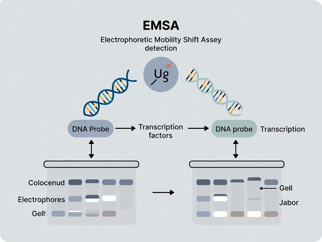
EMSA detects complex formation between a protein and a nucleic acid probe based on the reduction of the probe's electrophoretic mobility through a non-denaturing polyacrylamide or agarose gel. The protein-bound DNA or RNA migrates more slowly than the free probe, resulting in a distinct "shifted" band. This simple principle allows researchers to ascertain binding specificity, affinity, stoichiometry, and the presence of necessary co-factors.
The following tables consolidate key quantitative parameters for successful EMSA execution and analysis.
Table 1: Core Experimental Parameters for a Standard EMSA
| Parameter | Typical Range / Value | Notes |
|---|---|---|
| Probe Length | 20-50 bp | Optimal for most TFs; longer for multi-factor complexes. |
| Polyacrylamide Gel % | 4-10% (non-denaturing) | Lower % for larger complexes (>200 kDa). |
| Electrophoresis Buffer | 0.5x TBE or 0.25x TAE | Low ionic strength minimizes complex dissociation. |
| Electrophoresis Temp | 4°C | Stabilizes complexes during run. |
| Protein Amount | 0.1-10 µg nuclear extract / 1-100 ng purified protein | Titration is required. |
| Incubation Time | 20-30 min at RT or 4°C | |
| Cold Competitor DNA | 10-200x molar excess | For specificity verification. |
| Antibody for "Supershift" | 1-2 µg per reaction | Confirms protein identity. |
Table 2: Comparison of Probe Labeling Methods
| Method | Sensitivity | Stability | Key Applications |
|---|---|---|---|
| End-labeling with [γ-³²P] ATP | Very High (autorad) | Radioactive decay | Gold standard for detection of low-abundance factors. |
| Biotin End-labeling | High (chemiluminescence) | Very Stable | Non-radioactive; ideal for labs without radioisotope facilities. |
| Fluorescent Dye Labeling | Moderate (laser scanner) | Very Stable | Multiplexing potential; direct detection without secondary steps. |
| Digoxigenin (DIG) Labeling | High (chemiluminescence) | Very Stable | Similar to biotin; common in commercial kits. |
Detailed Experimental Protocols
Core EMSA Protocol for Transcription Factor Detection
Materials:
- Purified protein or nuclear extract.
- ³²P- or biotin-labeled double-stranded DNA probe containing putative binding site.
- Unlabeled competitor DNA (specific and non-specific).
- Poly(dI-dC) or other non-specific carrier DNA.
- Binding buffer (10 mM HEPES, pH 7.9, 50 mM KCl, 1 mM DTT, 0.5 mM EDTA, 5% glycerol).
- 4-10% non-denaturing polyacrylamide gel (29:1 acrylamide:bis).
- 0.5x TBE electrophoresis buffer.
- Electrophoresis and transfer apparatus, and detection system (phosphorimager, X-ray film, or chemiluminescence imager).
Method:
- Probe Preparation: Anneal complementary oligonucleotides. Label 5' ends with T4 polynucleotide kinase and [γ-³²P]ATP or biotin-11-UTP. Purify using a spin column.
- Binding Reaction:
- In a final volume of 10-20 µL, combine:
- Binding buffer.
- 1 µg poly(dI-dC) (to reduce non-specific binding).
- 10-20 fmol labeled probe (~10,000 cpm for ³²P).
- Protein extract (amount determined by titration).
- For competition assays: include 50-100x molar excess of unlabeled competitor DNA.
- For supershift assays: add 1-2 µg of specific antibody after initial binding.
- Incubate at room temperature for 20-30 minutes.
- In a final volume of 10-20 µL, combine:
- Gel Electrophoresis:
- Pre-run the non-denaturing polyacrylamide gel in 0.5x TBE at 100V for 30-60 min at 4°C.
- Load samples (with a minimal dye like bromophenol blue) directly onto the running gel.
- Run at 100-150V, constant voltage, for 1-2 hours at 4°C until the dye front is near the bottom.
- Detection:
- For radioactive probes: Transfer gel to filter paper, dry, and expose to a phosphor screen or X-ray film.
- For biotinylated probes: Electroblot to a positively charged nylon membrane. Crosslink DNA to membrane via UV. Detect using a streptavidin-HRP and chemiluminescent substrate system.
Supershift Assay Protocol
This protocol is an extension of the core EMSA, used to confirm the identity of a protein within the shifted complex.
- Perform the standard binding reaction (steps 1-2 above) and incubate for 20 min.
- Add 1-2 µg of an antibody specific to the suspected transcription factor. Use an isotype control antibody for a negative control.
- Incubate further for 20-60 min at 4°C.
- Proceed with gel electrophoresis (step 3 above). A successful supershift will result in a further retardation of the shifted band (a "supershifted" band) or its disappearance due to antibody-mediated complex disruption or stabilization.
Visualizations
Diagram 1: EMSA Principle and Result Interpretation
Diagram 2: EMSA Experimental Workflow
The Scientist's Toolkit: Essential Research Reagents
Table 3: Key Reagent Solutions for EMSA
| Item | Function / Purpose | Example / Specification |
|---|---|---|
| Chemically Synthesized Oligonucleotides | Source of specific DNA binding site probe. | HPLC-purified, complementary strands, 20-50 nt each. |
| [γ-³²P] ATP or Biotin Labeling Kit | Enables sensitive detection of the DNA probe. | T4 PNK for ³²P; Biotin 3' End Labeling Kit. |
| Non-Specific Carrier DNA | Reduces non-specific protein-DNA interactions. | Poly(dI-dC), sheared salmon sperm DNA. |
| Cold Competitor Oligos | Validates binding specificity. | Unlabeled identical (specific) or mutated (non-specific) oligos. |
| Transcription Factor-Specific Antibody | For supershift assays to confirm protein identity. | Monoclonal or polyclonal, EMSA/supershift validated. |
| Nuclear Extraction Kit | Isolates active transcription factors from cells. | Commercial kits with protease/phosphatase inhibitors. |
| Non-Denaturing Acrylamide/Bis Solution | Forms the sieving matrix for complex separation. | 29:1 or 37.5:1 acrylamide:bis ratio, high purity. |
| Electrophoresis Buffer (10x TBE) | Provides conductive ions for electrophoresis. | 890 mM Tris, 890 mM Boric Acid, 20 mM EDTA, pH ~8.3. |
| Chemiluminescent Nucleic Acid Detection Module | Detects biotin- or DIG-labeled probes. | Includes streptavidin-HRP, substrate, blotting membrane. |
As a pillar technique within the thesis on transcription factor detection, EMSA's enduring status as the gold standard is justified by its directness, adaptability, and capacity for mechanistic insight. While newer high-throughput methods exist for binding site discovery, EMSA provides an indispensable, orthogonal validation tool. Its utility in demonstrating direct, sequence-specific binding in vitro forms the critical biochemical foundation upon which hypotheses regarding transcriptional regulation in vivo are built, making it an essential component of the molecular biologist's and drug developer's arsenal for target validation and mechanistic studies.
Within the broader thesis of Electrophoretic Mobility Shift Assay (EMSA) research for transcription factor (TF) detection, the core principle of mobility shift analysis stands as a foundational technique. This guide details the biochemical and biophysical principles by which the retardation of nucleic acid electrophoretic migration indicates protein binding, enabling the study of gene regulation, protein-DNA interactions, and drug targeting.
Transcription factors regulate gene expression by binding to specific DNA sequences. EMSA, also known as gel shift assay, is the primary method for detecting and quantifying these interactions in vitro. The core principle hinges on the observation that a protein-nucleic acid complex migrates more slowly through a non-denaturing polyacrylamide or agarose gel than the free nucleic acid probe due to increased mass and altered charge-to-mass ratio. This mobility shift is the direct, observable readout of binding.
Table 1: Key Quantitative Parameters in a Standard EMSA Experiment
| Parameter | Typical Range / Value | Explanation / Impact on Shift |
|---|---|---|
| Polyacrylamide Gel Concentration | 4-10% (non-denaturing) | Higher % retards all migration; optimizes complex separation. |
| DNA Probe Length | 20-50 base pairs | Longer probes migrate slower; minimal impact on shift magnitude. |
| Binding Reaction Incubation | 20-30 min, 20-25°C | Ensures equilibrium binding. Temperature affects kinetics. |
| Electrophoresis Voltage | 80-150 V (constant) | Higher voltage causes heating and complex dissociation. |
| Electrophoresis Buffer (TBE/TAE) Ionic Strength | 0.5x or 1x | Low ionic strength can promote non-specific binding. |
| Apparent Kd (Determined by EMSA) | pM to nM range | Measure of binding affinity derived from band intensity quantification. |
Table 2: Controls and Their Expected Outcomes in EMSA
| Control Type | Components | Expected Gel Result (Purpose) |
|---|---|---|
| Free Probe | Labeled DNA only | Single fast-migrating band (baseline mobility). |
| Specific Competition | TF + labeled DNA + excess unlabeled specific DNA | Diminished shifted band (confirms sequence specificity). |
| Non-specific Competition | TF + labeled DNA + excess unlabeled non-specific DNA (e.g., poly(dI·dC)) | Shifted band remains (confirms binding specificity). |
| Antibody Supershift | TF + labeled DNA + specific anti-TF antibody | Further retardation (supershift) confirms TF identity. |
| Mutant Probe | TF + labeled mutant DNA | No or reduced shifted band (confirms sequence requirement). |
Detailed Experimental Protocol: Standard EMSA
A. Probe Preparation and Labeling
- Design: Synthesize complementary oligonucleotides containing the predicted TF binding site.
- Annealing: Mix equimolar amounts of each oligonucleotide in annealing buffer (10 mM Tris, 50 mM NaCl, 1 mM EDTA). Heat to 95°C for 5 min, then cool slowly to room temperature.
- End-Labeling: Use T4 Polynucleotide Kinase to transfer the γ-(^{32})P-ATP phosphate group to the 5'-ends of the DNA. Purify labeled probe using a spin column to remove unincorporated nucleotides.
B. Protein Extraction and Quantification
- Nuclear Extract Preparation: Use a hypotonic buffer to swell cells, followed by mechanical disruption and centrifugation to isolate nuclei. Lyse nuclei with a high-salt buffer (e.g., 400 mM KCl). Clarify by centrifugation.
- Quantification: Determine protein concentration using the Bradford or BCA assay.
C. Binding Reaction
- Assemble reactions on ice (typical volume: 20 µL):
- 1-10 µg nuclear extract or purified TF protein.
- Labeled DNA probe (20,000-50,000 cpm).
- Binding Buffer: 10 mM HEPES, 50 mM KCl, 1 mM DTT, 2.5 mM MgCl₂, 10% glycerol, 0.05% NP-40.
- Poly(dI·dC) (1-2 µg) as non-specific competitor.
- Incubate at room temperature for 20-30 minutes.
D. Non-Denaturing Gel Electrophoresis
- Gel Preparation: Cast a 4-10% polyacrylamide gel (29:1 acrylamide:bis) in 0.5x TBE buffer. Pre-run at 100 V for 60 min at 4°C.
- Loading & Run: Add loading dye (without SDS) to reactions. Load samples. Run in 0.5x TBE at 100 V, 4°C, until the dye front nears the bottom.
- Visualization: Transfer gel to filter paper, dry, and expose to a phosphorimager screen or X-ray film.
Visualization of Core Principles and Workflow
Title: Core EMSA Principle: Binding Causes Mobility Shift
Title: Standard EMSA Experimental Workflow
The Scientist's Toolkit: Key Research Reagent Solutions
Table 3: Essential Materials for EMSA Experiments
| Reagent / Material | Function / Purpose in EMSA |
|---|---|
| γ-(^{32})P-ATP or Chemiluminescent Labeling Kits | Radioactive or non-radioactive label for sensitive detection of DNA probe. |
| T4 Polynucleotide Kinase | Enzyme for end-labeling DNA probes with (^{32})P. |
| Poly(dI·dC) / Salmon Sperm DNA | Non-specific competitor DNA to reduce non-specific protein-DNA interactions. |
| HEPES or Tris-Based Binding Buffer | Provides optimal pH and ionic conditions for specific TF-DNA binding. |
| Non-Denaturing Acrylamide/Bis Solution (29:1, 40%) | For casting gels that separate based on size/shape, not denatured state. |
| 0.5x TBE Electrophoresis Buffer | Low ionic strength buffer run at 4°C minimizes complex dissociation during run. |
| Nuclear Extract Kit / Homemade Lysis Buffers | For preparation of protein extracts enriched for nuclear TFs. |
| TF-Specific Antibodies | For supershift assays to confirm TF identity within the complex. |
| Phosphorimager System / X-ray Film | For detection and quantification of shifted bands. |
This technical guide provides an in-depth examination of the three core components central to the Electrophoretic Mobility Shift Assay (EMSA), a cornerstone technique in transcription factor (TF) detection research. Framed within a broader thesis on advancing EMSA methodologies for drug discovery and mechanistic studies, this whiteparesents the latest technical specifications, optimized protocols, and critical considerations for generating robust, reproducible data.
The Electrophoretic Mobility Shift Assay (EMSA), or gel shift assay, remains a fundamental in vitro technique for studying protein-nucleic acid interactions, primarily the binding of transcription factors to specific DNA sequences. Its utility in confirming putative binding sites, assessing binding affinity and specificity, and detecting active TFs in cellular models underpins research in gene regulation, signaling pathway dissection, and therapeutic drug development targeting transcriptional dysregulation.
Core Component 1: The DNA Probe
The DNA probe is the labeled DNA fragment containing the specific cis-regulatory element (e.g., promoter or enhancer sequence) of interest.
Design & Synthesis
- Sequence: Typically 20-40 base pairs, with the consensus binding site positioned centrally.
- Labeling: Probes are end-labeled with a reporter molecule.
- Controls: An unlabeled, identical "cold" probe is essential for competition experiments. A mutant probe with scrambled/disrupted consensus sequence serves as a negative control for binding specificity.
Table 1: Common DNA Probe Labeling Methods
| Method | Reporter | Detection Mode | Sensitivity | Stability |
|---|---|---|---|---|
| Radioactive | γ-³²P-ATP | Autoradiography/Phosphorimaging | Very High (zeptomole) | Short (~10-14 day half-life) |
| Chemiluminescent | Biotin | Chemiluminescence (Streptavidin-HRP) | High (attomole) | Long (months) |
| Fluorescent | Fluorophore (e.g., Cy5, FAM) | Fluorescence Imaging | Moderate-High | Long (months) |
Protocol: DNA Probe Preparation (Biotin End-Labeling)
- Annealing: Combine complementary single-stranded oligonucleotides in annealing buffer (10 mM Tris, 50 mM NaCl, 1 mM EDTA, pH 8.0). Heat to 95°C for 5 min, then cool slowly to room temperature.
- End-Labeling Reaction:
- 1 µg double-stranded oligonucleotide
- 5 µL 10X T4 Polynucleotide Kinase Buffer
- 5 µL Biotin-11-dUTP (or similar)
- 10 U T4 Polynucleotide Kinase
- Nuclease-free water to 50 µL.
- Incubate at 37°C for 60 minutes.
- Purification: Purify the labeled probe using a spin column (e.g., G-25 Sephadex) to remove unincorporated nucleotides. Verify concentration via spectrophotometry.
Core Component 2: Nuclear Extract
The nuclear extract is the source of transcription factors, providing a complex mixture of nuclear proteins from cells or tissues of interest.
Preparation Principles
The goal is to isolate intact, functional nuclear proteins while minimizing contamination from cytoplasmic components and proteolytic degradation.
Table 2: Key Reagents in Nuclear Extract Preparation
| Reagent | Function | Critical Note |
|---|---|---|
| Hypotonic Buffer | Swells cells, weakens cytoplasmic membrane. | Contains DTT and protease inhibitors fresh. |
| Detergent (e.g., NP-40) | Lyses plasma membrane, releases cytoplasmic contents. | Concentration is cell-type critical; too high lyses nuclei. |
| High-Salt Buffer | Dissociates proteins from nuclear chromatin. | Typically 400-500 mM NaCl or KCl. Salt concentration affects final TF activity. |
| Protease/Phosphatase Inhibitors | Preserves protein integrity & modification state. | Must be added fresh to all buffers immediately before use. |
Protocol: Rapid Nuclear Extraction (Adapted from Andrews & Faller)
- Harvest & Wash: Pellet ~10⁷ cells, wash with ice-cold PBS.
- Hypotonic Lysis: Resuspend in 400 µL cold Hypotonic Buffer (10 mM HEPES pH 7.9, 1.5 mM MgCl₂, 10 mM KCl, 0.5 mM DTT, inhibitors). Incubate on ice 10 min.
- Detergent Lysis: Add 25 µL of 10% NP-40. Vortex 10 sec vigorously.
- Nuclear Pellet: Centrifuge at 16,000 × g for 30 sec at 4°C. Pellet contains nuclei.
- Nuclear Extraction: Resuspend nuclear pellet in 50 µL ice-cold High-Salt Buffer (20 mM HEPES pH 7.9, 25% glycerol, 400 mM NaCl, 1.5 mM MgCl₂, 0.2 mM EDTA, 0.5 mM DTT, inhibitors). Rock at 4°C for 30 min.
- Clarification: Centrifuge at 16,000 × g for 10 min at 4°C. Aliquot supernatant (nuclear extract) and store at -80°C. Determine protein concentration (Bradford assay).
Core Component 3: The Binding Reaction
This step facilitates the specific interaction between the TF in the nuclear extract and the labeled DNA probe under controlled conditions.
Reaction Optimization
The reaction must balance conditions that promote specific binding while minimizing non-specific interactions.
Table 3: Standard Binding Reaction Components & Functions
| Component | Typical Concentration/Range | Purpose |
|---|---|---|
| Labeled Probe | 0.1-1.0 ng (20-100 fmol) per reaction | Substrate for TF binding. |
| Nuclear Extract | 2-10 µg protein per reaction | Source of transcription factor(s). |
| Poly(dI:dC) | 0.05-0.5 µg/µL | Inert competitor DNA to reduce non-specific binding. |
| Binding Buffer | 10 mM Tris, 50 mM KCl, 1 mM DTT, 5% Glycerol, pH 7.5 | Provides ionic strength, pH, and stabilizing agents. |
| MgCl₂/EDTA | Variable (0-5 mM) | Divalent cations can be crucial for some TF-DNA interactions. |
| Non-ionic Detergent | 0.01-0.1% (e.g., NP-40) | Redvents protein aggregation. |
Protocol: Standard EMSA Binding Reaction
- Master Mix: For n reactions, combine on ice:
- Nuclease-free water to final volume.
- 2 µL 10X Binding Buffer
- 1 µL 1 µg/µL Poly(dI:dC)
- 1 µL 50% Glycerol (if not in buffer)
- Optional: 1 µL 100 mM MgCl₂.
- Add Protein: Add x µL of nuclear extract (2-10 µg total protein). Mix gently.
- Pre-incubation: Incubate on ice for 10 minutes to allow competitor DNA to bind non-specific proteins.
- Initiate Reaction: Add 1 µL of labeled DNA probe (0.1-1 ng) to each tube. Mix gently.
- Binding Incubation: Incubate at room temperature or 4°C for 20-30 minutes.
- Load Sample: Add 1 µL of 10X gel loading dye (non-denaturing, without SDS) and load immediately onto a pre-run native polyacrylamide gel.
The Scientist's Toolkit: Essential Research Reagent Solutions
Table 4: Key Reagents for EMSA Experiments
| Item | Function & Rationale |
|---|---|
| Biotin-11-dUTP / ³²P-ATP | Reporter molecule for labeling DNA probe; enables detection. |
| T4 Polynucleotide Kinase | Catalyzes the transfer of phosphate group to 5' end of DNA for labeling. |
| Protease Inhibitor Cocktail (EDTA-free) | Prevents proteolytic degradation of TFs during extract prep; EDTA-free allows metal-dependent binding later. |
| Phosphatase Inhibitors (e.g., NaF, β-glycerophosphate) | Preserves the phosphorylation state of TFs, critical for their activity. |
| Poly(dI:dC) | Synthetic double-stranded nucleic acid used as a non-specific competitor to absorb non-sequence-specific DNA-binding proteins. |
| Non-ionic Detergent (NP-40/Tween-20) | Reduces non-specific hydrophobic interactions and protein aggregation in binding reactions. |
| HEPES Buffer, pH 7.9 | Biological pH buffer for nuclear extraction and binding; more stable than Tris at cold temperatures. |
| High-Binding Capacity Streptavidin-HRP Conjugate | For chemiluminescent detection; binds biotinylated probe with high affinity. |
| Native Gel Electrophoresis System | Provides the matrix (typically 4-6% acrylamide:bis) to separate protein-DNA complexes from free probe based on size/charge under non-denaturing conditions. |
| Chemiluminescent Substrate (e.g., Luminol-based) | Generates light signal upon HRP catalysis for sensitive detection of biotinylated complexes. |
Experimental Workflow & Pathway Visualization
Historical Context and Enduring Relevance of EMSA in Molecular Biology
The Electrophoretic Mobility Shift Assay (EMSA), also known as the gel shift assay, has been a cornerstone technique in molecular biology for over four decades. First described in 1981 by Garner and Revzin, and independently by Fried and Crothers, EMSA provided the first simple, rapid method to detect sequence-specific DNA-binding proteins. This whitepaper examines its historical development, core principles, and its enduring, adapted relevance within modern transcription factor (TF) research and drug discovery. Framed within a thesis on EMSA for TF detection, we argue that while foundational, its utility persists through technical evolution and integration with high-throughput methodologies.
Historical Context & Evolution
EMSA emerged from the need to study protein-nucleic acid interactions directly, moving beyond purely genetic or biochemical inferences. Its invention paralleled the cloning era, allowing researchers to marry recombinant DNA with functional biochemistry.
Key Historical Milestones:
- 1981: First descriptions of the assay using crude nuclear extracts.
- Late 1980s - 1990s: Widespread adoption for cloning TFs via screening expression libraries. Establishment of "supershift" assays using antibodies.
- 2000s: Adaptation to fluorescence (F-EMSA) and capillary electrophoresis, enabling quantification.
- 2010s - Present: Integration with high-throughput platforms (e.g., microfluidic EMSA) and combination with mass spectrometry for TF complex identification.
Core Principle & Technical Framework
EMSA detects complexes based on reduced electrophoretic mobility of a nucleic acid probe (DNA or RNA) when bound by a protein. The complex migrates more slowly through a non-denaturing polyacrylamide or agarose gel than the free probe.
Detailed Standard EMSA Protocol
Reagents & Materials:
- Purified Protein or Nuclear Extract: Source of the DNA-binding protein.
- Labeled DNA Probe: 20-50 bp dsDNA containing the putative binding site. Typically end-labeled with ³²P (traditional) or a fluorophore (e.g., Cy5, FAM).
- Non-specific Competitor DNA: Poly(dI-dC) or sheared salmon sperm DNA to suppress non-specific binding.
- Binding Buffer: Typically contains Tris-HCl (pH 7.5), KCl or NaCl, MgCl₂, DTT, EDTA, glycerol (for loading), and non-ionic detergent (e.g., NP-40).
- Non-denaturing Polyacrylamide Gel (4-6%): Cast and run in 0.5X TBE or TAE buffer.
- Electrophoresis & Detection System: Apparatus capable of cold-room or ice-water cooling. Phosphorimager (radioactive) or fluorescence scanner/CCD imager.
Step-by-Step Method:
- Probe Preparation: Anneal complementary oligonucleotides. Label via T4 Polynucleotide Kinase (³²P) or use pre-labeled oligos.
- Binding Reaction:
- In a 10-20 µL reaction, combine:
- Binding Buffer (1X final concentration)
- Non-specific competitor DNA (0.5-2 µg)
- Protein extract (2-10 µg) or purified protein (fmol-nmol range)
- Labeled probe (10-50 fmol)
- Incubate at 20-25°C for 20-30 minutes.
- In a 10-20 µL reaction, combine:
- Gel Electrophoresis:
- Pre-run the non-denaturing gel in cold buffer at 100-150 V for 30-60 min.
- Load samples (with glycerol/dye) directly without heating.
- Run at constant voltage (100-150 V) with cooling until the free probe is 2/3 down the gel.
- Detection:
- For ³²P: Dry gel and expose to a Phosphor screen or X-ray film.
- For fluorescence: Image gel directly using appropriate excitation/emission settings.
- Controls: Always include "free probe" lane and competition lanes with excess unlabeled specific (cold) and non-specific oligonucleotides.
Quantitative Data Analysis (Typical Range):
| Parameter | Typical Value / Range | Notes |
|---|---|---|
| Probe Length | 20 - 50 bp | Must contain binding motif; shorter probes give better resolution. |
| Protein Amount | 0.1 - 10 µg (crude extract) | Titration is required for optimal signal. |
| Incubation Time | 20 - 30 min | Longer times may promote non-specific binding. |
| Affinity Range (Kd) Detectable | ~10⁻⁹ - 10⁻¹² M | Suitable for medium to high-affinity interactions. |
| Gel Run Time | 1 - 3 hours | Dependent on probe size and gel percentage. |
| Sensitivity (Detection Limit) | ~0.1-1 fmol of complex (³²P) | Fluorescence methods are generally less sensitive. |
Advanced Modifications & Current Protocols
Fluorescence EMSA (F-EMSA)
Uses fluorophore-labeled probes, eliminating radioactivity. Enables multiplexing and direct quantification via in-gel fluorescence scanning.
Protocol Modification:
- Use IRDye800, Cy5, or FAM-labeled oligonucleotides.
- Post-electrophoresis, scan gel in a dedicated fluorescence imager (e.g., Li-COR Odyssey, Typhoon).
- Allows precise quantification of bound vs. free fraction for Kd calculation.
Supershift EMSA
An antibody specific to the TF or an epitope tag is added to the binding reaction. A further reduction in mobility ("supershift") confirms the identity of the TF in the complex.
Protocol Addition:
- After initial protein-probe incubation, add 1-2 µg of specific or control antibody.
- Incubate for an additional 30-60 minutes on ice before loading.
- A new, higher-molecular-weight band appears if the antibody binds the protein-DNA complex.
Microscale Thermophoresis (MST) & EMSA Alternative Data
While not EMSA, MST is a modern solution-phase method for quantifying binding affinities.
| Method | Throughput | Sample Consumption | Kd Measurement | Key Advantage |
|---|---|---|---|---|
| Classic EMSA | Low | Moderate (~10 µL) | Yes, via titration | Visual proof of complex; accessible. |
| F-EMSA | Medium | Moderate (~10 µL) | Yes, more precise | Safer, quantifiable, multiplexable. |
| MST | High | Very Low (< 1 µL) | Yes, direct in solution | No separation step; rapid Kd. |
The Scientist's Toolkit: Key Reagent Solutions
| Item | Function & Rationale |
|---|---|
| Nuclear Extraction Kit | Isolates transcription factors from cell nuclei with preserved DNA-binding activity. |
| Biotin- or Fluorescence-End Labeled Oligonucleotides | Provides non-radioactive, stable probes for detection via chemiluminescence or fluorescence. |
| Poly(dI-dC) | A synthetic, non-specific competitor DNA that binds and sequesters non-sequence-specific nucleic acid-binding proteins. |
| HEK-293T Transient Transfection Reagents | For overexpression of epitope-tagged TFs, enabling supershift assays with tag-specific antibodies. |
| Non-denaturing Acrylamide/Bis Mix (29:1, 37.5:1) | Forms the porous matrix of the native gel, critical for separating complexes based on size/shape. |
| Phosphor Storage Screen & Imager | For high-sensitivity, quantitative detection of ³²P-labeled probes. |
| Cooled Electrophoresis Cell | Maintains gel at 4°C during the run, preventing complex dissociation and ensuring sharp bands. |
| TF-Specific & Isotype Control Antibodies | Essential for supershift assays to confirm TF identity within the shifted complex. |
Visualization of Concepts & Workflows
Title: Core EMSA Experimental Workflow
Title: Historical Evolution of EMSA Methodology
Title: EMSA in Transcription Factor Drug Discovery
Despite the advent of sophisticated techniques like ChIP-seq and AlphaScreen, EMSA retains critical relevance. Its strengths are directness, simplicity, and cost-effectiveness. It remains the gold standard for in vitro validation of TF-DNA interactions, mapping binding sites via mutagenesis, and preliminary screening for small-molecule inhibitors that disrupt these interactions. Modern adaptations (F-EMSA, high-throughput formats) address historical limitations of throughput and safety, ensuring EMSA's continued place in the molecular biologist's and drug developer's arsenal. It serves not as a standalone answer, but as an indispensable, orthogonal validation tool within a comprehensive thesis on transcription factor biology.
Electrophoretic Mobility Shift Assay (EMSA) is a foundational technique for the direct detection and quantification of transcription factor (TF)-DNA interactions. This whitepaper positions EMSA not as an isolated method, but as a critical entry point into a systems-level investigation of cellular processes. By confirming specific protein-nucleic acid binding, EMSA provides the initial, crucial evidence that anchors deeper exploration into three core biological and pharmacological arenas: the logic of gene regulation, the dynamics of signal transduction pathways, and the mechanistic basis of drug action. This document serves as a technical guide for leveraging EMSA findings to design sophisticated, follow-on experiments in these interconnected domains.
Deconstructing Gene Regulatory Networks
EMSA identifies a TF binding to a cis-regulatory element. The subsequent research trajectory involves characterizing the transcriptional outcome and mapping the network.
Quantitative Data from Gene Regulation Studies
Table 1: Common Quantitative Outputs in Gene Regulation Studies Following EMSA Validation
| Parameter | Measurement Technique | Typical Readout | Biological Significance |
|---|---|---|---|
| Binding Affinity (Kd) | EMSA with titration, Surface Plasmon Resonance (SPR) | 1 nM - 1 µM | Strength of TF-DNA interaction. |
| Transcript Abundance | qRT-PCR, RNA-seq | Fold-change (e.g., 5.2x upregulation) | Functional consequence of TF binding. |
| Promoter/Enhancer Activity | Luciferase Reporter Assay | Relative Luminescence Units (RLU) | Functional impact of the bound cis-element. |
| Chromatin Accessibility | ATAC-seq | Peak Counts/Read Depth | Status of DNA region (open/closed). |
| In Vivo Binding Occupancy | ChIP-seq | Peak Enrichment (e.g., 15-fold over IgG) | Genomic footprint of the TF. |
Experimental Protocol: Chromatin Immunoprecipitation (ChIP) for In Vivo Validation
Objective: To confirm EMSA-identified TF binding occurs on native chromatin within cells.
- Crosslinking: Treat cells with 1% formaldehyde for 10 min at room temperature to fix protein-DNA complexes.
- Cell Lysis & Sonication: Lyse cells and shear chromatin via sonication to fragments of 200-1000 bp.
- Immunoprecipitation: Incubate chromatin with antibody specific to the TF of interest. Use Protein A/G beads to capture antibody complexes.
- Washing & Elution: Wash beads stringently. Reverse crosslinks by heating at 65°C with high salt.
- DNA Purification: Recover DNA using a column-based kit.
- Analysis: Quantify target DNA sequence by qPCR (ChIP-qPCR) or sequence entire population (ChIP-seq).
Research Reagent Solutions: Gene Regulation Toolkit
Table 2: Essential Reagents for Gene Regulation Studies
| Reagent / Material | Function |
|---|---|
| Biotinylated or ³²P-labeled DNA Probe | For EMSA; allows detection of protein-bound DNA. |
| Recombinant Transcription Factor | Purified protein for in vitro binding assays (EMSA, SPR). |
| TF-Specific Antibody (ChIP-grade) | For immunoprecipitation of chromatin complexes. |
| Dual-Luciferase Reporter System | Measures transcriptional activity of a cloned promoter/enhancer. |
| Next-Generation Sequencing Kit | For genome-wide analysis (ChIP-seq, ATAC-seq). |
Diagram 1: From EMSA to Gene Regulatory Networks
Mapping Signal Transduction Cascades
TF activation is often the terminal event of a signaling pathway. EMSA can monitor TF activation states (e.g., phosphorylation-induced DNA binding) to trace upstream signaling events.
Quantitative Data from Signaling Studies
Table 3: Key Metrics in Signal Transduction Pathway Analysis
| Parameter | Measurement Technique | Typical Readout | Biological Significance |
|---|---|---|---|
| Kinase/Phosphatase Activity | Phospho-Specific EMSA, Western Blot | Phosphorylation Level | Proximal regulator of TF activity. |
| Second Messenger Concentration | FRET-based sensors, ELISA | [cAMP], [Ca²⁺], etc. | Intracellular signal propagation. |
| Protein-Protein Interaction | Co-IP, FRET, BRET | Interaction Strength/Proximity | Complex assembly in pathway. |
| Ligand-Receptor Binding | Radioligand Binding, SPR | Binding Affinity (Ki, IC50) | Initial signal event. |
| Pathway Activity | Phospho-antibody arrays, RTK assays | Multiplexed Phospho-signature | Integrated pathway output. |
Experimental Protocol: Phospho-Specific EMSA for Signaling Inference
Objective: To determine if a signaling event (e.g., cytokine treatment) alters TF DNA-binding capacity.
- Cell Stimulation: Treat one cell population with pathway agonist (e.g., TNF-α) and leave another as unstimulated control.
- Nuclear Extract Preparation: Lyse cells, isolate nuclei, and extract nuclear proteins in high-salt buffer.
- EMSA with Inhibitors: Pre-incubate extracts with or without a phosphatase (e.g., lambda phosphatase) for 30 min.
- Standard EMSA: Perform EMSA using the target DNA probe.
- Analysis: A supershift or enhanced band intensity in the stimulated sample indicates phosphorylation-enhanced binding. Phosphatase treatment should abolish this effect.
Research Reagent Solutions: Signal Transduction Toolkit
Table 4: Essential Reagents for Signaling Studies
| Reagent / Material | Function |
|---|---|
| Pathway-Specific Agonists/Antagonists | To activate or inhibit specific nodes (e.g., PMA, Forskolin, kinase inhibitors). |
| Phosphatase Inhibitor Cocktails | Preserves phosphorylation state of proteins during extraction. |
| Phospho-Specific Antibodies | Detects activated (phosphorylated) forms of signaling proteins and TFs. |
| FRET/BRET Biosensor Plasmids | Live-cell reporting of second messenger dynamics or protein interactions. |
| Active Kinase/Phosphatase Enzymes | For in vitro modification of TFs to test direct effects on DNA binding. |
Diagram 2: Signaling to Altered TF-DNA Binding
Elucidating Drug Mechanisms of Action (MoA)
EMSA is pivotal in drug discovery for compounds targeting gene expression. It can directly test if a drug disrupts or stabilizes a specific TF-DNA complex.
Quantitative Data in Drug Mechanism Studies
Table 5: Core Data for Quantifying Drug Effects on TF Function
| Parameter | Measurement Technique | Typical Readout | Pharmacological Significance |
|---|---|---|---|
| IC50 for Binding Disruption | Competitive EMSA | Concentration (e.g., IC50 = 5 µM) | Potency in inhibiting TF-DNA interaction. |
| EC50 for Pathway Modulation | Reporter Assay + Drug Titration | Concentration (e.g., EC50 = 0.1 µM) | Functional cellular potency. |
| Binding Constant (Kd) to TF | SPR, ITC, DSF | Affinity (Kd) | Direct physical engagement of target. |
| Target Occupancy | Cellular Thermal Shift Assay (CETSA) | ΔTm (°C) | Drug engagement in cells. |
| Transcriptomic Signature | RNA-seq post-treatment | Differential Gene Expression | On-target vs. off-target effects. |
Experimental Protocol: Competitive EMSA for Drug Screening
Objective: To test if a small molecule inhibitor disrupts the formation of a specific TF-DNA complex.
- Form Complex: Incubate recombinant TF with labeled DNA probe to form complex.
- Drug Competition: Pre-incubate the TF with a titration series of the candidate drug (e.g., 0.1, 1, 10, 100 µM) for 30 min before adding the probe.
- Run EMSA: Perform standard EMSA.
- Analysis: Quantify band intensity of the shifted complex. Plot % complex formation vs. log[drug concentration] to calculate the IC50 value.
Research Reagent Solutions: Drug Mechanism Toolkit
Table 6: Essential Reagents for Drug Mechanism Studies
| Reagent / Material | Function |
|---|---|
| Candidate Drug Compounds | Small molecules or biologics for testing. |
| Fluorescent DNA Probes (e.g., Cy5) | Safer, stable alternative to radioisotopes for HTS EMSA. |
| SPR or Microscale Thermophoresis (MST) Chip/Capillaries | Label-free quantification of drug-protein binding affinity. |
| CETSA/LIP Assay Kits | Confirms target engagement in a cellular context. |
| High-Content Screening System | For image-based, multi-parameter analysis of drug effects. |
Diagram 3: Drug Mechanisms Disrupting TF-DNA Complex
A Step-by-Step EMSA Protocol: From Probe Design to Image Analysis
The Electrophoretic Mobility Shift Assay (EMSA) is a cornerstone technique for studying protein-nucleic acid interactions, central to elucidating transcriptional regulation. The core of a successful EMSA experiment is a high-quality DNA probe that accurately represents the transcription factor binding site. The choice between radioactive and non-radioactive labeling is a critical, Phase 1 decision that impacts experimental sensitivity, safety, cost, and downstream applications. This guide details the design, synthesis, and labeling strategies for DNA probes within the framework of transcription factor detection research.
Probe Design Fundamentals
A well-designed probe is typically a double-stranded oligonucleotide, 20-40 base pairs in length, containing the consensus or putative binding sequence. Flanking sequences should be inert and facilitate labeling. Critical parameters include:
- Specificity: Must match the known binding motif.
- Length: Optimal for binding while minimizing non-specific interactions.
- GC Content: Affects annealing stability and probe behavior.
Labeling Methodologies: Technical Comparison
Radioactive Labeling (³²P)
Protocol: End-Labeling with T4 Polynucleotide Kinase (PNK)
- Reaction Setup: In a microcentrifuge tube, combine:
- 1–10 pmol of dephosphorylated, single-stranded oligonucleotide.
- 2 µL of 10X T4 PNK buffer.
- 20 µCi of [γ-³²P]ATP.
- 10 units of T4 Polynucleotide Kinase.
- Nuclease-free water to 20 µL.
- Incubation: Incubate at 37°C for 30-60 minutes.
- Termination: Heat-inactivate at 65°C for 10 minutes.
- Annealing: Add a 1.5x molar excess of the complementary strand, heat to 95°C for 5 min, and slowly cool to room temperature.
- Purification: Purify the double-stranded probe using a spin column (e.g., Sephadex G-25) to remove unincorporated nucleotides.
Non-Radioactive Labeling
Protocol: Enzymatic Tagging with Biotin or Digoxigenin
- Template Design: Synthesize oligonucleotide with a modified base (e.g., amino-allyl dUTP) or use a tailing strategy.
- Biotinylation (Example - 3' End Tailing):
- Combine 1–5 pmol of double-stranded DNA probe, 1X Terminal Deoxynucleotidyl Transferase (TdT) buffer, 0.25 mM Biotin-11-dUTP, and 20 units of TdT enzyme in a 50 µL reaction.
- Incubate at 37°C for 1 hour.
- Terminate with 2.5 µL of 0.2M EDTA and purify via ethanol precipitation.
- Detection: Post-EMSA, transfer DNA to a positively charged nylon membrane via capillary or electroblotting. Probe is detected using Streptavidin-Horseradish Peroxidase (HRP) conjugate and a chemiluminescent substrate.
Quantitative Comparison: Radioactive vs. Non-Radioactive
Table 1: Comparative Analysis of DNA Probe Labeling Strategies for EMSA
| Parameter | Radioactive (³²P) | Non-Radioactive (Biotin/Digoxigenin) |
|---|---|---|
| Sensitivity | Extremely High (attomole range) | High (femtomole range) |
| Spatial Resolution | Excellent | Very Good |
| Signal Stability | Short (half-life-dependent) | Long (years, stable conjugates) |
| Exposure Time | Minutes to Hours | Seconds to Minutes |
| Hazard Level | High (Ionizing Radiation) | Low/None |
| Regulatory Burden | Significant (Licensing, Waste) | Minimal |
| Direct Quantification | Possible (Phosphorimaging) | Semi-quantitative |
| Cost per Experiment | Low (Reagents) | Moderate to High |
| Infrastructure Cost | High (Shielding, Scanners) | Low |
| Probe Stability | Short (Weeks) | Long (Years) |
| Multiplexing Potential | Low | Moderate |
The Scientist's Toolkit: Key Reagent Solutions
Table 2: Essential Materials for DNA Probe Preparation and Labeling
| Reagent/Material | Function in Probe Preparation |
|---|---|
| T4 Polynucleotide Kinase (PNK) | Catalyzes the transfer of a phosphate group from [γ-³²P]ATP to the 5'-end of DNA. |
| [γ-³²P]ATP | Radioactive donor molecule for 5' end-labeling. |
| Terminal Deoxynucleotidyl Transferase (TdT) | Adds labeled nucleotides (e.g., Biotin-dUTP) to the 3'-end of DNA. |
| Biotin-11-dUTP / Digoxigenin-11-dUTP | Modified nucleotides for enzymatic incorporation of non-radioactive tags. |
| Streptavidin-Horseradish Peroxidase (HRP) | High-affinity binding conjugate for detecting biotinylated probes. |
| Chemiluminescent Substrate (e.g., Luminol) | HRP substrate that produces light upon oxidation for film/imager detection. |
| Nylon Membrane (Positively Charged) | Solid support for transferring and immobilizing DNA post-EMSA for non-radioactive detection. |
| Sephadex G-25 Spin Columns | Size-exclusion chromatography for removing unincorporated nucleotides from labeling reactions. |
Decision Pathway and Experimental Integration
Title: Decision Pathway for Probe Labeling Method in EMSA
Title: EMSA Workflow Integrating Probe Labeling Phase
Within the context of Electrophoretic Mobility Shift Assay (EMSA) for transcription factor detection research, the preparation of high-quality nuclear or whole cell extracts is the critical foundational step. The integrity, purity, and biological activity of these extracts directly determine the specificity and sensitivity of downstream EMSA experiments. This technical guide details current, optimized methodologies for extract preparation, emphasizing quantitative quality control metrics and standardized protocols to ensure reproducibility in research and drug development.
Transcription factors (TFs) are often present in low abundance and can be sequestered in specific cellular compartments. Successful EMSA detection relies on extracts that preserve TF DNA-binding activity without introducing contaminants that cause non-specific shifts or background interference. Nuclear extracts are preferred for studying DNA-binding proteins that reside predominantly in the nucleus, while whole cell extracts provide a broader profile, useful for cytoplasmic-nuclear shuttling proteins.
Methodologies for Extract Preparation
Preparation of Whole Cell Extracts (Modified Bradford Protocol)
This method is suitable for adherent and suspension cells, focusing on rapid lysis to preserve protein complexes.
Protocol:
- Cell Harvest & Washing: Pellet 1x10^7 cells. Wash twice with 10 mL of ice-cold 1X Phosphate-Buffered Saline (PBS). Centrifuge at 500 x g for 5 min at 4°C.
- Lysis: Resuspend cell pellet in 1 mL of Lysis Buffer (20 mM HEPES pH 7.9, 0.4 M NaCl, 1 mM EDTA, 1 mM EGTA, 1 mM DTT, 1 mM PMSF, 1X Protease Inhibitor Cocktail). Vortex vigorously for 15 seconds.
- Freeze-Thaw: Snap-freeze the cell suspension in liquid nitrogen for 1 min, then thaw at 37°C for 5 min. Repeat for a total of 3 cycles.
- Clarification: Centrifuge the lysate at 12,000 x g for 15 min at 4°C.
- Dialyzation & Storage: Transfer the supernatant (whole cell extract) to dialysis tubing (6-8 kDa MWCO). Dialyze against 500 mL of Storage Buffer (20 mM HEPES pH 7.9, 100 mM KCl, 0.2 mM EDTA, 0.2 mM EGTA, 1 mM DTT, 20% glycerol) overnight at 4°C. Aliquot, snap-freeze in liquid nitrogen, and store at -80°C.
Preparation of Nuclear Extracts (Gold Standard Dignam Protocol)
This is the benchmark method for obtaining highly active nuclear proteins, involving cytoplasmic separation and high-salt nuclear protein extraction.
Protocol:
- Cell Harvest: Collect 5x10^7 cells. Wash twice with 10 mL ice-cold PBS.
- Hypotonic Lysis: Resuspend pellet in 1 mL of Hypotonic Buffer (10 mM HEPES pH 7.9, 1.5 mM MgCl2, 10 mM KCl, 0.5 mM DTT, 0.2 mM PMSF). Incubate on ice for 15 min.
- Detergent Lysis: Add 50 μL of 10% Nonidet P-40 (NP-40). Vortex for 10 sec.
- Nuclei Pellet: Centrifuge at 3,300 x g for 15 min at 4°C. The supernatant is the cytoplasmic fraction (store separately if needed). The pellet contains intact nuclei.
- Nuclear Extraction: Resuspend the nuclear pellet in 200 μL of High-Salt Extraction Buffer (20 mM HEPES pH 7.9, 1.5 mM MgCl2, 420 mM NaCl, 0.2 mM EDTA, 25% glycerol, 0.5 mM DTT, 0.2 mM PMSF). Rock gently at 4°C for 30 min.
- Clarification: Centrifuge at 21,000 x g for 30 min at 4°C.
- Dialysis & Storage: Transfer supernatant (nuclear extract) to dialysis tubing. Dialyze against 500 mL of Storage Buffer for 4-6 hours. Aliquot, snap-freeze, and store at -80°C.
Quantitative Quality Control Metrics
The following parameters are essential for validating extract quality prior to EMSA experiments.
Table 1: Quality Control Metrics for Nuclear & Whole Cell Extracts
| Parameter | Target Specification | Assay Method | Impact on EMSA |
|---|---|---|---|
| Protein Concentration | 2-10 mg/mL | Bradford / BCA Assay | Ensures consistent loading in binding reactions. |
| Cytoplasmic Contamination (Nuclear Extracts) | LDH Activity <5% of total | Lactate Dehydrogenase (LDH) Assay | High contamination leads to non-specific background. |
| Nuclear Integrity | DNA-bound Histone H3 >90% retained | Western Blot (Histone H3) | Indicates successful nuclear isolation. |
| Protease Activity | Degradation of BSA Standard <10% in 1 hr at 37°C | Fluorescent Protease Assay | Proteolysis degrades TFs, abolishing DNA binding. |
| Nuclease Activity | Supercoiled Plasmid DNA >80% intact after 1 hr incubation | Agarose Gel Electrophoresis | Degrades probe DNA, causing failed shift detection. |
| Transcription Factor Activity | Specific binding signal in control EMSA | EMSA with consensus oligonucleotide | Final functional validation of extract viability. |
The Scientist's Toolkit: Essential Research Reagents
Table 2: Key Research Reagent Solutions for Extract Preparation
| Reagent | Function & Rationale | Critical Notes |
|---|---|---|
| HEPES Buffer (pH 7.9) | Maintains physiological pH during extraction; superior to Tris for protein-DNA interactions. | Must be prepared fresh and pH-adjusted at room temperature. |
| Protease Inhibitor Cocktail (EDTA-free) | Inhibits serine, cysteine, and metalloproteases to prevent TF degradation. | EDTA-free versions are essential if extracts will be used in Mg²⁺-dependent EMSA reactions. |
| Phenylmethylsulfonyl Fluoride (PMSF) | Irreversible serine protease inhibitor (e.g., against trypsin, chymotrypsin). | Unstable in aqueous solution; add to buffers immediately before use. |
| Dithiothreitol (DTT) | Reducing agent that maintains cysteine residues in TFs in a reduced, active state. | More stable than β-mercaptoethanol; add fresh to buffers. |
| Glycerol (20-25%) | Cryoprotectant that stabilizes protein structure and prevents ice crystal formation during storage. | Ensures long-term activity retention at -80°C. |
| Nonidet P-40 (NP-40) | Mild non-ionic detergent for plasma membrane lysis without disrupting nuclear membranes. | Critical for clean separation of cytoplasmic and nuclear fractions. |
| High-Grade NaCl & KCl | Provide ionic strength for cell lysis (high salt) and maintain protein solubility during dialysis. | Use molecular biology grade to avoid heavy metal contaminants. |
| Dialysis Tubing (6-8 kDa MWCO) | Removes small molecules, salts, and inhibitors, allowing buffer exchange into optimal storage conditions. | Must be pre-treated per manufacturer's instructions to remove contaminants. |
Experimental Workflow and Pathway Visualization
Nuclear Extract Preparation Workflow
From Cell Stimulus to EMSA Detection Pathway
Within the context of a comprehensive thesis on Electrophoretic Mobility Shift Assay (EMSA) for transcription factor (TF) detection, Phase 3 represents the critical juncture where initial binding events are refined into robust, specific, and reproducible interactions. This phase systematically interrogates the biochemical environment—buffer composition, competitor nucleic acids, and incubation time—to maximize the signal-to-noise ratio of the protein-nucleic acid complex. Optimization here is paramount for downstream applications in mechanistic studies, drug discovery targeting TF-DNA interactions, and diagnostic assay development.
Optimizing Buffer Conditions
The binding buffer establishes the ionic and chemical milieu for the TF-probe interaction. Key components influence complex stability and specificity.
Key Buffer Components & Optimization Targets:
- pH & Buffering Agent: Typically Tris-HCl or HEPES (10-20 mM, pH 7.5-8.5). Maintains protein stability and can affect binding affinity.
- Monovalent Salts (KCl, NaCl): (50-150 mM). Higher concentrations reduce non-specific electrostatic interactions.
- Divalent Cations (Mg²⁺, Zn²⁺): (0-10 mM). Essential for TFs with metal-binding domains (e.g., zinc fingers). Can promote non-specific binding if excessive.
- Glycerol: (5-10%). Stabilizes proteins and reduces adsorption to tubes.
- Non-ionic Detergents (NP-40, Tween-20): (0.01-0.1%). Minimizes non-specific protein and probe adhesion.
- Carrier Proteins (BSA, milk proteins): (0.1-1 mg/mL). Blocks non-specific binding sites.
- Reducing Agents (DTT, β-mercaptoethanol): (1-5 mM). Prevents oxidation of cysteine residues in the TF.
Experimental Protocol: Buffer Matrix Titration
- Prepare a master mix containing the labeled DNA probe (e.g., 1 fmol/µL), poly(dI-dC) (0.05 µg/µL), and nuclease-free water.
- Aliquot equal volumes of the master mix into separate tubes.
- Add an equal volume of serially varied 2x concentrated binding buffers differing in one component (e.g., KCl: 0 mM, 50 mM, 100 mM, 150 mM, 200 mM final concentration).
- Initiate reactions by adding a constant amount of nuclear extract or purified TF.
- Incubate at room temperature for 30 minutes.
- Load samples onto a pre-run native polyacrylamide gel for EMSA analysis.
- Quantify the shifted band intensity. Optimal condition yields the highest specific complex intensity with minimal smearing or non-specific bands.
Table 1: Representative Buffer Optimization Data (Hypothetical TF)
| Buffer Component | Tested Range | Optimal Concentration (Final) | Effect on Complex Yield |
|---|---|---|---|
| KCl | 0 - 200 mM | 75 mM | Yield peaked at 75 mM; higher concentrations disrupted complex. |
| MgCl₂ | 0 - 5 mM | 2 mM | Essential for complex formation; >3 mM increased non-specific background. |
| DTT | 0 - 10 mM | 2 mM | No effect up to 5 mM; omitted in some purifications. |
| NP-40 | 0 - 0.2% | 0.05% | Reduced probe trapping in wells; no impact on specific shift. |
| BSA | 0 - 2 mg/mL | 0.5 mg/mL | Improved reproducibility and complex stability. |
Diagram 1: Buffer components impact on EMSA binding.
Selecting and Titrating Competitor Nucleic Acids
Competitors are unlabeled nucleic acids (poly(dI-dC), salmon sperm DNA, specific mutant oligonucleotides) added to sequester non-sequence-specific DNA-binding proteins.
Experimental Protocol: Competitor Titration
- Set up standard binding reactions with optimal buffer.
- Include a titration series of a competitor (e.g., poly(dI-dC): 0, 0.01, 0.05, 0.1, 0.5, 1.0 µg per reaction).
- For specificity ("cold competition"), include reactions with a 50x and 200x molar excess of unlabeled identical probe or a mutant probe.
- Incubate and analyze by EMSA.
- The ideal non-specific competitor amount suppresses background without diminishing the specific shift. Specific competitor should abolish the shift, while mutant should not.
Table 2: Competitor Effects on EMSA Signal
| Competitor Type | Purpose | Typical Working Amount | Expected Outcome |
|---|---|---|---|
| Poly(dI-dC) | Bind non-specific, charged-interaction proteins. | 0.05 - 0.5 µg/rxn | Reduces smearing; clarifies specific shift. |
| Salmon Sperm DNA | Bind a broader range of DNA-binding proteins. | 0.1 - 1.0 µg/rxn | Can be more effective for some extracts; may require titration. |
| Unlabeled Specific Probe (Cold) | Confirm binding specificity. | 10x - 200x molar excess | Dose-dependent reduction/abolition of shifted band. |
| Unlabeled Mutant Probe | Confirm sequence specificity. | 50x - 200x molar excess | Minimal or no reduction of shifted band. |
Diagram 2: Competitor selection and validation logic.
Determining Incubation Time & Temperature
Kinetics of complex formation must reach equilibrium for consistent results. Temperature affects binding kinetics and complex stability.
Experimental Protocol: Time Course Experiment
- Set up multiple identical binding reactions on ice using optimal buffer and competitors.
- Simultaneously start incubation at the chosen temperature (e.g., 25°C or 4°C).
- Remove aliquots at various time points (e.g., 0, 5, 10, 15, 20, 30, 45, 60 min) and immediately place on ice.
- Load all samples onto a native gel simultaneously to process equally.
- Plot complex intensity vs. time to identify the plateau phase. Choose an incubation time within this plateau.
- Repeat at 4°C for temperature-sensitive TFs.
Table 3: Time & Temperature Optimization Results
| Incubation Temperature | Time to Reach Plateau | Relative Complex Stability | Recommended Use Case |
|---|---|---|---|
| 4°C (on ice) | 20 - 40 minutes | High; less degradation. | Standard for crude nuclear extracts; less stable TFs. |
| Room Temp (25°C) | 10 - 20 minutes | Moderate. | For purified, stable proteins; may increase kinetics. |
| 30°C | 5 - 15 minutes | Lower; risk of degradation. | For high-affinity interactions; requires stable TF. |
The Scientist's Toolkit: Key Research Reagent Solutions
| Reagent/Material | Function in EMSA Optimization | Example Product/Note |
|---|---|---|
| HEPES or Tris-HCl Buffer | Provides stable pH environment for binding reaction. | Molecular biology grade, nuclease-free. |
| Nuclease-Free Water | Prevents degradation of DNA probe and RNA if used. | Certified DEPC-treated or ultrafiltered. |
| Poly(dI-dC)•Poly(dI-dC) | Synthetic non-specific competitor DNA. | Aliquot to avoid freeze-thaw cycles. |
| Salmon Sperm DNA | Alternative natural non-specific competitor. | Requires shearing and denaturing before use. |
| BSA (Acetylated) | Carrier protein to reduce non-specific adsorption. | Acetylated BSA is less likely to bind DNA. |
| Protease Inhibitor Cocktail | Preserves transcription factor integrity in extracts. | Add fresh to lysis/binding buffers. |
| DTT or β-Mercaptoethanol | Reducing agent to maintain cysteine residues. | Prepare fresh stock solutions. |
| Non-ionic Detergent (NP-40) | Reduces hydrophobic interactions and adhesion. | Use high-purity grade. |
| Magnetic Separation Beads | For rapid EMSA (EMSA-MSAs) or complex purification. | Streptavidin-coated if using biotinylated probes. |
| High-Binding Microplates | For plate-based EMSA or high-throughput screening. | Used in conjunction with fluorescence detection. |
Within the broader framework of an Electrophoretic Mobility Shift Assay (EMSA) for transcription factor detection and drug discovery research, Phase 4—the separation step via non-denaturing (native) gel electrophoresis—is the critical juncture where protein-nucleic acid complexes are resolved from unbound probes. This phase directly visualizes the thermodynamic equilibrium established during binding, providing qualitative and quantitative data on transcription factor activity, affinity, and the impact of pharmacological inhibitors.
Core Principles of Native Gel Electrophoresis for EMSA
Unlike denaturing SDS-PAGE, native gel electrophoresis preserves the three-dimensional structure and non-covalent interactions of biomolecules. For EMSA, this allows the separation of a faster-migrating, unlabeled nucleic acid probe from a slower-migrating complex formed when a transcription factor binds. The migration shift is due to the increased molecular weight and altered charge-to-mass ratio of the complex.
Key Quantitative Parameters for Gel Preparation: Optimal separation depends on precise control of gel composition and electrophoretic conditions. The following table summarizes standard parameters:
Table 1: Standardized Conditions for EMSA Native Gels
| Parameter | Typical Range | Common Optimal Value | Function/Rationale |
|---|---|---|---|
| Acrylamide:%Bis-acrylamide | 4-6% (29:1 or 37.5:1) | 5% (37.5:1) | Resolves complexes in 10-200 kDa range; lower % for larger complexes. |
| Gel Thickness | 0.5 - 1.5 mm | 1.0 mm | Balances resolution, sample capacity, and staining/drying time. |
| Buffer System | 0.25X or 0.5X TBE; Tris-Glycine | 0.5X TBE | Maintains pH and ionic strength; TBE offers better buffering capacity. |
| Glycerol in Gel | 0-5% (v/v) | 2.5% (v/v) | Increases sample density for clean loading; stabilizes complexes. |
| Pre-run Conditions | 30-100 V, 30-60 min | 100 V, 60 min at 4°C | Stabilizes pH, temperature, and ionic conditions before sample entry. |
| Running Conditions | Constant Voltage: 80-150 V | 100 V constant | Maintains complex stability; lower voltage reduces heating. |
| Run Temperature | 4-10°C | 4°C (cold room) | Minimizes complex dissociation during electrophoresis. |
| Run Time | 60-90 minutes | ~75 minutes (until dye front is ~3/4 down) | Sufficient separation of bound vs. free probe. |
Detailed Experimental Protocol
Protocol: Casting and Running a Non-Denaturing Polyacrylamide Gel for EMSA
Materials:
- Vertical gel electrophoresis system with glass plates, spacers (1.0 mm), and comb.
- Acrylamide/Bis-acrylamide stock solution (29:1 or 37.5:1 ratio).
- 10X Tris-Borate-EDTA (TBE) Buffer: 890 mM Tris base, 890 mM boric acid, 20 mM EDTA, pH ~8.3.
- Ammonium persulfate (APS): 10% (w/v) solution in water (fresh or stored at 4°C for <1 week).
- Tetramethylethylenediamine (TEMED).
- Non-denaturing Loading Dye (5X): 20% Ficoll-400, 0.05% Bromophenol Blue, 0.05% Xylene Cyanol in dH₂O.
- Electrophoresis Power Supply.
- Cooling system or cold room.
Method:
- Clean and Assemble: Thoroughly clean glass plates and spacers. Assemble the cassette according to manufacturer's instructions, ensuring a leak-proof seal.
- Prepare Gel Solution: For a 5% gel (10 mL volume), mix:
- 1.67 mL Acrylamide/Bis (37.5:1, 30% stock)
- 0.5 mL 10X TBE
- 0.25 mL Glycerol (100%)
- 7.58 mL dH₂O Degas the solution for 10 minutes to prevent bubble formation during polymerization.
- Catalyze Polymerization: Add 50 µL of 10% APS and 10 µL of TEMED. Swirl gently to mix. Note: Polymerization begins immediately; work swiftly.
- Pour the Gel: Using a pipette, pour the gel solution between the plates. Insert the comb carefully, avoiding bubbles. Allow to polymerize for 30-45 minutes at room temperature.
- Set Up and Pre-run: Once polymerized, place the gel cassette into the electrophoresis tank. Fill both the upper and lower chambers with pre-chilled 0.5X TBE running buffer. Carefully remove the comb and rinse the wells with buffer. Pre-run the gel at 100 V for 60 minutes in a cold room (4°C) to establish equilibrium and cool the system.
- Load Samples: During pre-run, prepare EMSA binding reactions. Mix each reaction with 1/5 volume of 5X non-denaturing loading dye. After pre-run, turn off the power, flush wells with buffer, and load samples and appropriate controls (free probe control is essential).
- Run the Gel: Run the gel at a constant voltage of 100 V (~10 V/cm) until the Bromophenol Blue dye front has migrated to approximately ¾ of the gel length (~75 minutes). Maintain temperature at 4°C throughout.
- Post-Run Processing: After electrophoresis, disassemble the apparatus. The gel is now ready for downstream detection (e.g., autoradiography for radioactive probes, fluorescence imaging, or wet/dry transfer for chemiluminescent detection).
Visualization of EMSA Phase 4 Workflow
Diagram Title: EMSA Phase 4: Native Gel Electrophoresis Workflow
The Scientist's Toolkit: Key Reagent Solutions for EMSA Native Gels
Table 2: Essential Reagents for the Separation Step
| Reagent Solution | Composition & Preparation | Critical Function in Phase 4 |
|---|---|---|
| 10X Tris-Borate-EDTA (TBE) Buffer | 108 g Tris base, 55 g Boric acid, 7.44 g Na₂EDTA·2H₂O in 1L dH₂O, pH ~8.3. Dilute to 0.25X or 0.5X for running buffer. | Provides consistent ionic strength and pH during electrophoresis, crucial for complex stability and reproducible migration. |
| Native Gel Loading Dye (5X) | 20% Ficoll-400, 0.05% Bromophenol Blue, 0.05% Xylene Cyanol in dH₂O. Filter sterilize. | Increases sample density for clean well loading; contains inert dyes to visually monitor electrophoretic progress. |
| Acrylamide/Bis Stock | 30% (w/v) Acrylamide, with Bis-acrylamide at a 37.5:1 or 29:1 ratio (Acrylamide:Bis). Filter and store dark at 4°C. | The matrix-forming component of the gel. The ratio and percentage determine pore size and resolution range for complexes. |
| Polymerization Catalysts | APS: 10% (w/v) Ammonium Persulfate in dH₂O (fresh). TEMED: Liquid, stored at 4°C. | Initiates (APS) and accelerates (TEMED) free-radical polymerization of acrylamide monomers into a cross-linked gel matrix. |
| High-Purity Water | Nuclease-free, deionized water (e.g., Milli-Q grade). | Used for all buffer and gel solutions to prevent contaminating nucleases or ions from degrading samples or altering migration. |
| Gel Staining Solutions | SYBR Green or Gold: For fluorescent detection of nucleic acid probe. Ethidium Bromide: Traditional intercalating dye. | Allows visualization of the nucleic acid component (both bound and free) post-electrophoresis, confirming run success. |
Within the framework of an Electrophoretic Mobility Shift Assay (EMSA) thesis for transcription factor detection, the selection of an appropriate detection method is critical for sensitivity, safety, and quantification. This whitepaper provides an in-depth technical comparison of the three principal detection modalities: autoradiography, chemiluminescence, and fluorescence. We detail protocols, present comparative quantitative data, and outline essential reagents to guide researchers and drug development professionals in optimizing their nucleic acid-protein interaction studies.
Following the electrophoretic separation of protein-nucleic acid complexes in EMSA, specific detection of the shifted band is required. The evolution from radioactive to non-radioactive methods has expanded experimental flexibility. This phase is pivotal, influencing the assay's limit of detection, dynamic range, safety profile, and suitability for downstream applications like supershift assays or quantitative analysis.
Core Detection Methodologies
Autoradiography
Principle: Utilizes a radioisotope (typically ³²P)-labeled nucleic acid probe. Emission of beta particles exposes an X-ray film or a phosphor storage screen, which is subsequently developed or scanned. Key Protocol: The EMSA gel is dried and placed in direct contact with a storage phosphor screen for a period (30 minutes to several days) depending on signal strength. The screen is then scanned in a phosphorimager to quantify band intensity.
Chemiluminescence
Principle: Relies on an enzyme (commonly Horseradish Peroxidase - HRP or Alkaline Phosphatase - AP) conjugated to the probe or a secondary antibody. The enzyme catalyzes a reaction that produces light, which is captured on X-ray film or by a CCD camera. Key Protocol (Biotin-Streptavidin-HRP): Following transfer to a positively charged nylon membrane, the biotinylated probe is detected with streptavidin-HRP conjugate. The membrane is incubated with a luminol-based substrate (e.g., Luminol/Enhancer + H₂O₂). Light emission is captured over 1-10 minutes.
Fluorescence
Principle: Employs a fluorophore (e.g., Cy3, Cy5, FAM) directly conjugated to the nucleic acid probe. The gel or membrane is imaged using a laser-based scanner with appropriate excitation and emission filters. Key Protocol: The fluorescently labeled probe is used directly in the binding reaction. Post-electrophoresis, the gel is scanned in situ using a fluorescence imager. No transfer, development, or substrate addition steps are required.
Quantitative Comparison of Detection Methods
The following table summarizes the core performance characteristics of each method within the EMSA context.
Table 1: Comparative Analysis of EMSA Detection Methods
| Parameter | Autoradiography (³²P) | Chemiluminescence | Fluorescence |
|---|---|---|---|
| Typical Sensitivity (LOD) | ~0.1-1 fmol | ~1-10 fmol | ~1-50 fmol |
| Dynamic Range | 3-4 orders of magnitude | 3-4 orders of magnitude | 3-5 orders of magnitude |
| Exposure/Imaging Time | Hours to days | Seconds to minutes | Seconds to minutes |
| Signal Stability | Decays with isotope t½ | Transient (minutes) | Stable (months, if protected) |
| Quantitation Ease | Excellent (Phosphorimager) | Good (CCD) | Excellent (Scanner) |
| Multiplexing Potential | No | Possible with multiple substrates | Yes (multiple fluorophores) |
| Hazard/Safety | High (Ionizing Radiation) | Low (Chemical Hazards) | Very Low |
| Probe Re-usability | No | Limited (Stripping possible) | Yes (Direct, no stripping) |
| Cost per Experiment | Low (reagent), High (waste) | Moderate | Moderate to High (labeled probe) |
| Best For | Maximum sensitivity, publication legacy data | High sensitivity without radioactivity | Quantitative, multiplexed, fast workflows |
Detailed Experimental Protocols
Protocol: Chemiluminescent Detection for Biotinylated Probes
- Transfer: Following EMSA, electrophoretically transfer nucleic acids from gel to a positively charged nylon membrane (0.45 µm) at 380 mA for 1 hour in 0.5x TBE.
- Crosslinking: UV-crosslink the nucleic acid to the membrane (1200 J/m²).
- Blocking: Incubate membrane in 10 mL Blocking Buffer (5% non-fat dry milk in 1x TBST) for 1 hour at RT with gentle agitation.
- Conjugate Binding: Dilute Streptavidin-HRP conjugate 1:20,000 in Blocking Buffer. Incubate membrane in 10 mL of this solution for 30 minutes at RT.
- Washing: Wash membrane 4 x 5 minutes with 15 mL of 1x TBST.
- Substrate Incubation: Mix equal parts Luminol and Peroxide solutions. Incubate membrane in substrate for 5 minutes.
- Imaging: Drain excess substrate, wrap membrane in clear plastic, and expose to a CCD imager for 1-10 minutes.
Protocol: Direct Fluorescence Detection
- Probe Labeling: Use HPLC-purified oligonucleotide probe with a 5' or 3' fluorophore (e.g., Cy5).
- Binding & EMSA: Perform standard binding reaction and run gel in 0.5x TBE buffer. Do not add ethidium bromide.
- In-Gel Imaging: Place the gel directly on the scanner bed of a fluorescence gel imager. Use appropriate laser/excitation and emission filters for the fluorophore.
- Scanning: Acquire image at a resolution of 50-100 µm. Adjust PMT voltage or laser power to avoid saturation.
Visualizing EMSA Detection Workflows
Diagram Title: EMSA Detection Method Selection Workflow
The Scientist's Toolkit: Essential Reagents & Materials
Table 2: Key Research Reagent Solutions for EMSA Detection
| Reagent/Material | Function in Detection | Typical Example/Brand |
|---|---|---|
| ³²P-γ-ATP | Radioactive labeling of DNA probe via kinase reaction. | PerkinElmer, Hartmann Analytic |
| Biotin-11-dUTP | Non-radioactive incorporation into probe for chemiluminescent detection. | Thermo Fisher Scientific |
| Fluorophore-labeled Oligo | Directly labeled probe for fluorescence detection; requires no secondary steps. | IDT, Sigma-Aldrich (Cy3, Cy5, FAM) |
| Streptavidin-HRP Conjugate | Binds biotinylated probe to enable chemiluminescent signal generation. | Cell Signaling Technology, Thermo |
| Chemiluminescent Substrate | Luminol/peroxide mixture oxidized by HRP to produce light. | SuperSignal (Thermo), Clarity (Bio-Rad) |
| Phosphor Storage Screen | Captures and stores latent image from radioactive decay for phosphorimager scanning. | GE Healthcare, Bio-Rad |
| Positively Charged Nylon Membrane | Binds negatively charged nucleic acids for chemiluminescent detection post-transfer. | Hybond-N+, Amersham |
| Fluorescence Gel Imager | Scanner with lasers and filters to excite and capture emission from in-gel fluorophores. | Typhoon (Cytiva), Azure (Azure Biosystems) |
| Blocking Agent (Non-fat Milk/BSA) | Prevents non-specific binding of detection conjugates to the membrane. | Standard Laboratory Reagents |
| Crosslinker (UV Chamber) | Covalently attaches nucleic acid to nylon membrane to prevent wash-off. | Stratalinker (Stratagene) |
1. Introduction
Within the broader methodological framework of the Electrophoretic Mobility Shift Assay (EMSA) for transcription factor (TF) detection research, establishing the biological relevance of a protein-nucleic acid complex is paramount. A shifted band on a native gel indicates binding, but not the identity of the bound protein or the specificity of the interaction. Super-shift and competition assays are two critical, orthogonal techniques used to confirm the specificity and identity of the bound TF, transforming an observation into a defensible scientific conclusion.
2. The Super-Shift Assay: Confirming Protein Identity
The super-shift assay, or antibody supershift, is used to confirm the presence of a specific protein within the protein-DNA complex. This is achieved by incubating the protein extract with an antibody specific to the suspected TF prior to EMSA.
Principle: If the antibody binds to the TF (either before or after it binds the probe), it creates a larger, higher molecular weight "supershifted" complex. This complex migrates more slowly (higher in the gel) than the original protein-DNA complex. A successful supershift confirms the identity of the TF.
Detailed Protocol:
- Binding Reaction Setup: Prepare a standard EMSA binding reaction with your labeled probe, nuclear extract, and binding buffer.
- Antibody Addition: Add 1-2 µg of the specific antibody (or a species/isotype-matched control IgG) to the reaction mix. The antibody can be added simultaneously with the extract or after a pre-incubation of extract and probe (15-20 min on ice).
- Incubation: Incubate the complete reaction for 30-60 minutes on ice or at 4°C.
- Electrophoresis: Load the reaction onto a pre-run native polyacrylamide gel and run under standard EMSA conditions.
- Analysis: Visualize via autoradiography or phosphorimaging. Look for the disappearance or reduction of the original shifted band and the appearance of a new, higher-molecular-weight supershifted band.
3. The Competition Assay: Establishing Binding Specificity
Competition assays determine the sequence specificity of the observed protein-DNA interaction. They assess whether binding can be outcompeted by unlabeled (cold) DNA molecules.
Principle: An excess of unlabeled competitor DNA is included in the binding reaction. If the competitor contains the specific TF binding site, it will compete for the limited TF, reducing the signal from the labeled probe. A mutated or nonspecific competitor should have little to no effect.
Detailed Protocol:
- Competitor Design: Prepare three types of unlabeled double-stranded oligonucleotides:
- Specific Competitor: Identical to the labeled probe.
- Mutant Competitor: Contains mutations known to disrupt TF binding.
- Nonspecific Competitor: An unrelated DNA sequence (e.g., AP-1 site when studying NF-κB).
- Binding Reaction Setup: Set up a series of binding reactions with constant amounts of labeled probe and protein extract.
- Competitor Addition: Add increasing molar excesses (e.g., 5x, 25x, 50x, 100x) of each competitor to separate reactions. Include a no-competitor control.
- Pre-incubation (Optional but Recommended): Pre-incubate the protein extract with the competitor for 10-15 minutes on ice before adding the labeled probe. This favors competitor binding.
- Incubation & Electrophoresis: Complete the binding reaction and run the gel as per standard EMSA.
- Competitor Design: Prepare three types of unlabeled double-stranded oligonucleotides:
4. Data Presentation and Analysis
Table 1: Expected Results from Integrated Super-Shift and Competition Assays
| Assay Type | Condition | Expected Gel Result | Interpretation |
|---|---|---|---|
| Super-Shift | No Antibody | Standard shifted band (Band B) | Baseline complex formation. |
| Specific Antibody | Reduction of Band B; appearance of a slower-migrating band (Band SS) | Confirms identity of TF in Band B. | |
| Control IgG | Band B unchanged; no Band SS | Rules out nonspecific antibody effects. | |
| Competition | No Competitor | Full intensity of Band B | Baseline complex formation. |
| Specific Cold Competitor (50x) | >80% reduction of Band B intensity | Binding is sequence-specific. | |
| Mutant Cold Competitor (50x) | <20% reduction of Band B intensity | Mutations abolish competitive binding. | |
| Nonspecific Competitor (100x) | <10% reduction of Band B intensity | Binding is specific to the target sequence. |
Table 2: Quantitative Analysis of a Typical Competition Assay
| Competitor Type | Molar Excess (fold) | % Band Intensity (Relative to 0x) | Specificity Index* |
|---|---|---|---|
| Specific | 0 | 100% | - |
| 5 | 45% | 12.2 | |
| 25 | 12% | 13.3 | |
| 50 | 5% | 14.0 | |
| Mutant | 0 | 100% | - |
| 5 | 95% | - | |
| 25 | 90% | - | |
| 50 | 88% | - |
*Specificity Index = (100 - % Intensity with Mutant) / (100 - % Intensity with Specific) at same molar excess.
5. Visualizing Workflows and Logical Relationships
Super-Shift Assay Workflow (65 chars)
Competition Assay Logic (55 chars)
EMSA Validation Decision Pathway (56 chars)
6. The Scientist's Toolkit: Essential Research Reagent Solutions
Table 3: Key Reagents for Super-Shift and Competition Assays
| Reagent | Function & Critical Detail | Example/Note |
|---|---|---|
| High-Quality Specific Antibody | For super-shift. Must recognize the native, non-denatured conformation of the TF. Mouse monoclonal or rabbit polyclonal are common. | Anti-NF-κB p65, Anti-p53. Control IgG from same host species is mandatory. |
| Unlabeled Competitor Oligonucleotides | For competition. Must be HPLC- or gel-purified, annealed to perfect dsDNA. Critical tool for defining specificity. | Specific, mutant (2-3 base pair mismatch in core motif), and unrelated nonspecific sequences. |
| Radioactive (γ-32P/33P) or Chemiluminescent-Labeled Probe | Provides detection signal. High specific activity is crucial for clean, low-background competition assays. | 32P-ATP for kinase end-labeling; biotin- or digoxigenin-labeled probes for non-radioactive detection. |
| Ultrapure Nuclear Extract | Source of transcription factors. Quality and lack of degradation are paramount for complex integrity. | Commercial extracts from stimulated cells (e.g., TNF-α treated for NF-κB) or in-house prepared extracts. |
| Carrier DNA (Poly dI:dC) | Nonspecific competitor to reduce protein binding to non-target sequences in the probe. Concentration must be titrated for each extract. | Typically used at 0.05-0.2 µg/µL in the binding reaction. |
| EMSAGel-Shift Buffer Systems | Optimized binding buffers often include salts (KCl), glycerol, Mg2+, DTT, and non-ionic detergents to promote specific interactions. | Commercial 5x EMSA buffers or lab-made recipes (e.g., with or without MgCl2). |
Solving Common EMSA Problems: A Troubleshooting and Optimization Handbook
In the context of Electrophoretic Mobility Shift Assay (EMSA) research for transcription factor (TF) detection, the failure to observe a gel shift ("no-shift") represents a critical diagnostic challenge. This guide provides a systematic framework for troubleshooting, grounded in the core principle that a successful EMSA requires functional protein, a valid probe, and appropriate binding conditions.
1. Quantitative Data Summary: Common Culprits and Diagnostic Markers
Table 1: Key Variables and Their Impact on EMSA Shift Formation
| Variable Category | Specific Parameter | Optimal Range/State for EMSA | Effect if Suboptimal |
|---|---|---|---|
| Protein (TF) Source | Recombinant Protein Purity | >90% (SDS-PAGE verification) | Non-specific binding; degradation. |
| Nuclear Extract Protein Concentration | 2-10 µg/µL (Bradford assay) | Insufficient TF for detection. | |
| TF Post-Translational Modifications | Phosphorylation state appropriate for probe | Loss of DNA-binding affinity. | |
| Probe (DNA) | Specific Activity (³²P or Chemilum.) | 50,000-100,000 cpm/µL | Signal too weak for detection. |
| Double-Stranded Completeness | Confirmed by native PAGE | No binding site for TF. | |
| Consensus Sequence Fidelity | Matches known TF motif (e.g., JASPAR) | Drastically reduced affinity. | |
| Binding Conditions | Poly(dI:dC) Concentration | 0.05-0.5 µg/µL in reaction | High background; masked specific shift. |
| Mg²⁺/Zn²⁺ (Cofactors) | 1-5 mM (ion-dependent TFs) | Loss of structural integrity for TF. | |
| Incubation Time & Temperature | 20-30 min at 20-25°C | Incomplete binding or protein denaturation. | |
| Gel Conditions | Polyacrylamide Percentage | 4-6% for large complexes | Complexes do not enter or run off gel. |
| EDTA in Gel/Buffer | Avoid if TF requires divalent cations | Dissociation of complex during run. | |
| Electrophoresis Temperature | 4°C (cold room) | Complex dissociation due to heat. |
Table 2: Control Experiments for Systematic Diagnosis
| Control Experiment | Purpose | Expected Result if System is Functional | Interpretation of "No-Shift" |
|---|---|---|---|
| Positive Control Probe | Use a well-characterized DNA probe for a different, abundant TF (e.g., SP1). | Visible shifted band. | Problem lies with protein or general conditions. |
| Competition (Cold Probe) | Add 100x molar excess unlabeled specific probe to reaction. | Shifted band intensity drastically reduced. | Protein is active; shift is sequence-specific. Absence of competition suggests non-specific binding. |
| Antibody Supershift | Add antibody against the target TF post-binding. | Band shifts higher (supershift) or disappears. | Confirms presence of specific TF in complex. No change suggests TF absent. |
| Mutant Probe Control | Use probe with mutated consensus sequence. | No shifted band formed. | Validates binding specificity. A shift suggests non-specific interaction. |
2. Detailed Diagnostic Experimental Protocols
Protocol 1: Verification of Protein (TF) Activity via Western Blot and DNA-Affinity Pulldown
- Method: Prepare nuclear extract or recombinant protein. Run an aliquot on SDS-PAGE and immunoblot for the target TF to confirm presence. In parallel, incubate 200 µg nuclear extract with 2 µg biotinylated double-stranded consensus oligonucleotide bound to streptavidin magnetic beads for 1 hour at 4°C in binding buffer. Wash beads, elute bound protein with high-salt buffer, and analyze by Western blot.
- Interpretation: Positive Western from extract confirms TF presence. Positive pulldown Western confirms TF retains DNA-binding capability. If pulldown is negative, the TF is inactive.
Protocol 2: Probe Integrity and Competence Assay
- Method: Anneal labeled oligonucleotides to form the EMSA probe. Run 20,000 cpm on a 10% native polyacrylamide gel at 100V for 60 min. Image for radioactivity/chemiluminescence. A single, tight band indicates proper annealing. Perform a competition test: in separate tubes, add increasing molar excess (10x, 50x, 100x) of unlabeled identical probe to a standard binding reaction with a known active TF (e.g., nuclear extract positive control).
- Interpretation: A single band confirms probe quality. Dose-dependent reduction in shift intensity with cold competitor confirms the probe is competent for specific protein binding.
Protocol 3: Optimization of Critical Binding Conditions
- Method: Set up a matrix of binding reactions with constant protein and probe. Systematically vary: A) Poly(dI:dC) concentration (0, 0.1, 0.25, 0.5 µg/µL), B) Divalent cation (1-5 mM MgCl₂, ZnCl₂, or none), C) Glycerol concentration (0%, 5%, 10% v/v for complex stabilization). Run full EMSA.
- Interpretation: Identifies the precise condition that minimizes non-specific background while stabilizing the specific TF-DNA complex.
3. Visualizing the Diagnostic Pathway
Title: Systematic EMSA No-Shift Diagnostic Decision Tree
Title: EMSA Workflow with Critical Failure Points Highlighted
4. The Scientist's Toolkit: Key Research Reagent Solutions
Table 3: Essential Reagents for EMSA Development and Troubleshooting
| Reagent / Kit | Primary Function in EMSA | Key Consideration for Troubleshooting |
|---|---|---|
| Nuclear Extraction Kit (e.g., NE-PER) | Isolate active transcription factors from cells. | Ensure protease/phosphatase inhibitors are fresh to maintain TF activity and modifications. |
| Biotin 3’-End DNA Labeling Kit | Generate non-radioactive, labeled EMSA probes. | Check labeling efficiency via dot-blot; low efficiency causes weak signal. |
| Chemiluminescent Nucleic Acid Detection Module | Detect biotinylated probes on membrane. | Sensitivity must be high enough for low-abundance complexes. |
| Poly(dI-dC) • Poly(dI-dC) | Non-specific competitor DNA to reduce background. | Titration is critical; too much can compete away specific binding. |
| Transcription Factor (Specific) Antibody | For supershift or Western blot confirmation. | Must be validated for supershift; not all binding-site antibodies work. |
| EMSAGel Shift Kits | Pre-cast native gels & optimized buffers. | Provides standardized platform to rule out gel/buffer issues. |
| Positive Control DNA & Extract (e.g., SP1) | Validates entire EMSA workflow. | Essential control to isolate problem to target TF system vs. general technique. |
| Protease & Phosphatase Inhibitor Cocktails | Preserve TF integrity during extraction. | Omission leads to degraded or inactivated TF. |
In the context of Electrophoretic Mobility Shift Assay (EMSA) for transcription factor detection research, achieving high signal-to-noise ratios is paramount. Non-specific binding and high background compromise data interpretation, leading to false positives and inaccurate quantification of protein-DNA interactions. This guide details current, evidence-based strategies to enhance specificity, enabling more reliable identification and characterization of transcription factors in drug development and basic research.
Core Challenges in EMSA Specificity
Non-specific binding in EMSA primarily arises from electrostatic interactions between positively charged protein regions and the negatively charged DNA backbone, or from low-complexity protein domains interacting with probe sequences. Background stems from probe degradation, incomplete separation of complexes, or suboptimal electrophoresis conditions.
The following table consolidates quantitative data from recent studies on common additives used to mitigate non-specific binding in EMSA.
Table 1: Efficacy of Common Competitors & Additives in EMSA
| Agent | Typical Concentration Range | % Reduction in Non-Specific Signal (Mean ± SD)* | Primary Mechanism | Potential Drawback |
|---|---|---|---|---|
| Poly(dI-dC) | 0.05–0.2 mg/mL | 78 ± 12 | Competes for non-specific electrostatic binding | Can inhibit weak specific interactions |
| Salmon Sperm DNA | 0.1–1.0 mg/mL | 65 ± 15 | Competes for sequence-neutral DNA binding | Higher variability between preparations |
| BSA (Acetylated) | 0.1–0.5 mg/mL | 45 ± 10 | Blocks protein adhesion to tubes/gel | Minimal effect on direct DNA binding |
| Non-ionic Detergent (e.g., NP-40) | 0.1–1.0% (v/v) | 30 ± 8 | Reduces hydrophobic aggregation | Can destabilize some multi-protein complexes |
| DTT (or β-ME) | 1–5 mM | 25 ± 7 | Prevents oxidation-related aggregation | Must be added fresh |
| MgCl₂ | 1–5 mM | -20 to +15 | Can stabilize specific complexes | May increase non-specific binding for some TFs |
Data synthesized from comparative studies published 2021-2023. *Can variably increase or decrease background.
Detailed Experimental Protocols
Protocol 1: Systematic Optimization of Binding Conditions
Objective: To empirically determine the optimal combination of competitors and salts to maximize specificity for a novel transcription factor.
- Prepare Probe: Generate a 20-30 bp dsDNA probe containing the putative binding site, end-labeled with γ-³²P-ATP.
- Setup Competition Matrix: In a 20 μL binding reaction (10 mM HEPES, pH 7.9, 50 mM KCl, 1 mM DTT, 10% glycerol), keep nuclear extract volume constant. Set up a matrix of reactions varying:
- Poly(dI-dC): 0, 0.05, 0.1, 0.2 mg/mL.
- MgCl₂: 0, 1, 2, 5 mM.
- Include a 100-fold molar excess of unlabeled specific competitor in control lanes.
- Incubation & Electrophoresis: Incubate 20 min at 25°C. Load onto a pre-run 6% native polyacrylamide gel (0.5x TBE, 4°C). Run at 100V for 60-90 min.
- Analysis: Expose gel to a phosphorimager. The optimal condition yields a sharp, intense shifted band for the specific complex that is completely comporable, with minimal smear or non-specific bands.
Protocol 2: Supershift/Competition EMSA for Specificity Confirmation
Objective: To unequivocally confirm the identity of the protein in the shifted complex and validate binding specificity.
- Perform Standard EMSA: As optimized in Protocol 1.
- Antibody Supershift: For selected reactions, add 1-2 μg of antibody specific to the transcription factor of interest after the initial 20 min binding incubation. Incubate further for 30-60 min on ice. A successful supershift will cause the complex to migrate higher or "smear" into the well.
- Cold Competition: In parallel reactions, include increasing molar excesses (10x, 50x, 100x) of:
- Unlabeled Specific Competitor: Identical oligonucleotide to the probe.
- Unlabeled Mutant Competitor: Oligonucleotide with a scrambled or mutated binding site.
- Interpretation: Specific binding is demonstrated by dose-dependent disappearance of the shifted band with the specific, but not the mutant, competitor. The supershift confirms protein identity.
Visualization of Strategies and Workflows
Title: Strategic Workflow to Mitigate EMSA Background
Title: Optimized Step-by-Step EMSA Protocol for Specificity
The Scientist's Toolkit: Research Reagent Solutions
Table 2: Essential Reagents for High-Specificity EMSA
| Item | Function & Rationale | Recommended Product/Specification |
|---|---|---|
| Non-specific Competitor DNA | Competes for non-specific DNA-binding proteins. Poly(dI-dC) is standard; sequence complexity affects stringency. | Poly(deoxyinosinic-deoxycytidylic) acid, ammonium salt. |
| Acetylated Bovine Serum Albumin (BSA) | Blocks non-specific protein binding to reaction tubes and gel wells without interacting with DNA. | Molecular biology grade, acetylated to remove nuclease activity. |
| Non-ionic Detergent | Reduces hydrophobic protein-protein aggregation that can trap probe non-specifically. | NP-40 or Triton X-100, 10% stock solution. |
| Dithiothreitol (DTT) | Maintains transcription factors in reduced state, preventing oxidation-induced aggregation. | Prepare fresh 1M stock, store in aliquots at -20°C. |
| High-Purity Nucleotides | For probe labeling and competition. Impurities inhibit kinases and polymerases. | Ultrapure dNTPs and ATP solutions, PCR grade. |
| [γ-³²P] ATP or Chemiluminescent Label | Probe labeling. Chemiluminescent systems (e.g., biotin-streptavidin-HRP) now offer low background and long shelf-life. | PerkinElmer Chemiluminescent Nucleic Acid Detection Module. |
| Transcription Factor-Specific Antibody | For supershift assays. Must be capable of recognizing native, DNA-bound protein. | Validated for EMSA/supershift by supplier (e.g., Santa Cruz "sc-xxxxX" for EMSA). |
| Pre-cast Native PAGE Gels | Ensure consistency in gel matrix, critical for reproducible migration. | 6-8% Tris-Borate-EDTA (TBE) native gels, 10- or 15-well. |
| High-Binding Tubes | Minimize protein loss on tube walls. | Low-retention, siliconized microcentrifuge tubes. |
Mitigating high background and non-specific binding in EMSA requires a multi-faceted approach grounded in an understanding of macromolecular interactions. By systematically optimizing binding conditions with empirical titration of competitors and salts, employing rigorous specificity controls, and adhering to a meticulous cold electrophoresis protocol, researchers can achieve the high-specificity data required for robust transcription factor research. These strategies are indispensable for generating reliable data that can inform drug discovery targeting transcriptional pathways.
Thesis Context: This technical guide is framed within a doctoral thesis investigating the optimization of Electrophoretic Mobility Shift Assays (EMSA) for the sensitive and quantitative detection of low-abundance transcription factors in mammalian cell nuclear extracts. Achieving high-resolution, smear-free gels is paramount for accurately quantifying protein-DNA interactions and interpreting competitive binding or supershift assays.
Poor resolution and smearing in native polyacrylamide gels severely compromise EMSA data, leading to ambiguous band identification, inaccurate quantification, and failed supershift experiments. These artifacts stem from suboptimal gel composition, improper electrophoresis conditions, or sample quality issues. Within transcription factor research, where complex formation may be transient or involve large macromolecular assemblies, optimization is critical.
Table 1: Optimization of Polyacrylamide Gel Composition for EMSA
| Parameter | Standard Range | Optimized for High MW Complexes | Optimized for Low MW Complexes | Function & Rationale |
|---|---|---|---|---|
| Acrylamide:Bis Ratio | 29:1 to 37.5:1 | 37.5:1 (Larger pore size) | 29:1 (Smaller pore size) | Controls gel pore size. Higher bis-crosslinking creates smaller pores for better separation of small complexes. |
| Total Acrylamide % | 4-10% | 4-6% | 6-10% | Percent concentration defines sieving properties. Lower % for large complexes/supershifts; higher % for small, sharp bands. |
| Gel Thickness | 0.5-1.5 mm | 1.0-1.5 mm | 0.75-1.0 mm | Thinner gels run cooler, provide sharper bands but are more delicate. |
| Glycerol (in Gel) | 0-5% (v/v) | 2.5% | 5% | Stabilizes complexes and improves loading. Higher % can reduce smearing. |
Table 2: Electrophoresis Buffer and Running Conditions
| Parameter | Typical Condition | Optimized Condition | Impact on Resolution |
|---|---|---|---|
| Buffer Ionic Strength | 0.5X TBE or 0.25X TAE | 0.5X TBE (preferred) | Higher ionic strength (TBE) minimizes non-specific protein-DNA aggregation and smearing. |
| Buffer Recirculation | No | Yes (for runs >1.5 hrs) | Prevents pH gradient formation (cathode becomes basic, anode acidic) which causes band distortion. |
| Running Voltage | 10 V/cm | 8-10 V/cm (constant voltage) | Higher voltage causes heating, leading to complex dissociation and smearing. Use a cold room or cooling system. |
| Pre-Electrophoresis | 0-60 min | 30-60 min, 100V | Equilibrates gel pH and removes ammonium persulfate radicals that can degrade complexes. |
| Run Temperature | Ambient | 4°C | Maintains complex stability, reduces gel heating. Critical for labile interactions. |
Detailed Experimental Protocols
Protocol 1: Casting an Optimized, High-Resolution Native Polyacrylamide Gel
- Gel Solution Preparation (for 6%, 37.5:1 gel, 10 ml):
- 1.5 ml 40% Acrylamide/Bis (37.5:1 stock)
- 1.0 ml 5X TBE Buffer (54 g Tris base, 27.5 g boric acid, 20 ml 0.5M EDTA, pH 8.3, per liter)
- 0.5 ml Glycerol (100%)
- 6.95 ml Nuclease-Free Water
- 50 µl 10% Ammonium Persulfate (APS) (freshly prepared)
- 10 µl Tetramethylethylenediamine (TEMED)
- Casting: Mix components in the order listed, adding TEMED last. Swirl gently and immediately pour between clean, sealed glass plates (1.0 mm spacers). Insert a well comb without introducing bubbles.
- Polymerization: Allow to polymerize for 45-60 minutes at room temperature. A distinct refractive line will appear.
Protocol 2: Optimized EMSA Electrophoresis Run
- Setup: Mount the polymerized gel in the electrophoresis unit. Fill both chambers with pre-chilled 0.5X TBE running buffer.
- Pre-Run: Carefully remove the comb. Flush wells with running buffer using a syringe. Pre-run the gel for 30-60 minutes at 100V in a cold room (4°C) with buffer recirculation (or manually mixing buffer chambers every 15 minutes).
- Sample Loading: Prepare EMSA binding reactions as standard. Add 1/10 volume of 10X native loading dye (e.g., 30% glycerol, 0.25% bromophenol blue). Do not use SDS or heat denature.
- Run: Turn off power, flush wells again, and load samples. Run the gel at 8-10 V/cm (e.g., ~80-100V for a 10 cm gel) until the dye front migrates 2/3 down the gel. Maintain temperature at 4°C.
- Post-Run: Disassemble apparatus and proceed to gel transfer (for blotting) or direct staining.
The Scientist's Toolkit: Research Reagent Solutions
| Item | Function & Rationale |
|---|---|
| High-Purity, Electrophoresis-Grade Acrylamide/Bis | Ensures reproducible polymerization and avoids ionic contaminants that cause smearing. |
| Nuclease-Free Water | Prevents degradation of labeled DNA probe by contaminating nucleases. |
| Non-Hydrolyzed TBE Buffer (5X Stock) | Borate can precipitate over time; fresh stock prevents conductivity changes. |
| Carrier DNA (poly(dI-dC)) | Critical for blocking non-specific protein-DNA interactions. Optimal amount (0.05-0.1 µg/µl) must be empirically determined for each extract. |
| Protease & Phosphatase Inhibitors (in extract prep) | Prevents degradation or modification of transcription factors that can cause band heterogeneity/smearing. |
| High-Binding DNA-Binding Plates (for probe prep) | Maximizes recovery of purified, labeled oligonucleotide probe, reducing contaminants. |
| Cooled Circulator Electrophoresis Unit | Actively maintains temperature at 4°C throughout the run, critical for complex stability. |
Visualizations
Diagram 1: EMSA Troubleshooting and Optimization Decision Pathway
Diagram 2: Core Optimized EMSA Experimental Workflow
Electrophoretic Mobility Shift Assay (EMSA) remains a cornerstone technique for studying protein-nucleic acid interactions, particularly for transcription factor (TF) detection and characterization. A core challenge in this field is the generation of weak, inconsistent, or high-background signals, which compromise data reliability and quantification. This technical guide addresses the critical upstream variables of probe labeling and detection sensitivity, framed within the broader thesis that optimizing these foundational steps is paramount for advancing quantitative, reproducible TF research and subsequent drug discovery targeting transcriptional pathways.
Quantitative Analysis of Probe Labeling Methods
The choice of labeling method directly impacts specific activity, stability, and compatibility with detection systems. The following table summarizes key quantitative metrics for contemporary labeling strategies.
Table 1: Comparative Analysis of DNA Probe Labeling Methods for EMSA
| Labeling Method | Typical Specific Activity (cpm/fmol) | Optimal Stability | Detection Limit (fmol) | Relative Cost | Key Advantage | Primary Limitation |
|---|---|---|---|---|---|---|
| End-Labeling (T4 PNK, [γ-³²P]ATP) | 1 x 10⁶ - 5 x 10⁶ | 10-14 days (⁵⁰) | 0.1 - 0.5 | Low | High sensitivity, gold standard | Radioactive hazard, short half-life |
| 3'-End Labeling (Terminal Transferase, [α-³²P]ddATP) | 5 x 10⁵ - 2 x 10⁶ | 10-14 days (⁵⁰) | 0.5 - 1.0 | Low | Labels any 3'-end | Lower specific activity than PNK |
| PCR Incorporation (Biotin-11-dUTP) | N/A | Years | 5 - 15 | Medium | Extremely stable, safe, high yield | Lower sensitivity than radioisotopes |
| Chemical Modification (Psoralen-Biotin) | N/A | Years | 10 - 20 | Medium | Labels dsDNA without enzymes | Non-uniform labeling, potential interference |
| Fluorescent Dye Incorporation (CY5-dCTP) | N/A | Years | 15 - 30 | High | Safe, multiplexing capability | Highest equipment cost, susceptible to quenching |
Experimental Protocols for Enhanced Labeling & Detection
Protocol 3.1: High-Efficiency T4 Polynucleotide Kinase (PNK) End-Labeling
Objective: Maximize specific activity of ⁵⁷P-labeled probes for ultimate sensitivity. Reagents: DNA oligonucleotide (10 pmol/µL), [γ-³²P]ATP (6000 Ci/mmol, 150 µCi/µL), T4 PNK (10 U/µL), 10X PNK Buffer A. Procedure:
- In a microcentrifuge tube, mix: 1 µL DNA oligo (10 pmol), 2 µL 10X PNK Buffer A, 14 µL nuclease-free water, 2 µL [γ-³²P]ATP (300 µCi), 1 µL T4 PNK (10 U).
- Incubate at 37°C for 45 minutes.
- Add 1 µL of 0.5 M EDTA (pH 8.0) to stop the reaction.
- Purify the labeled probe using a micro Bio-Spin P-30 column (Tris buffer) to remove unincorporated nucleotides. Pre-spin the column at 1000g for 2 minutes before sample application.
- Elute by centrifugation at 1000g for 4 minutes. Measure radioactivity by scintillation counting (Cerenkov method). Aliquot and store at -20°C.
Protocol 3.2: High-Sensitivity Chemiluminescent Detection of Biotinylated Probes
Objective: Achieve sub-femtomole detection of biotinylated EMSA probes with low background. Reagents: Biotinylated DNA probe, Streptavidin-Horseradish Peroxidase (SA-HRP) conjugate (1 mg/mL), Chemiluminescent Substrate (e.g., Luminol/Enhancer), Blocking Buffer (5% BSA in TBST), Nylon Membrane (positively charged). Procedure:
- After EMSA electrophoresis, electroblot DNA-protein complexes onto a nylon membrane in 0.5X TBE buffer at 380 mA for 1 hour at 4°C.
- Crosslink DNA to the membrane using a UV crosslinker (120 mJ/cm²).
- Block the membrane with 5% BSA in TBST for 1 hour with gentle agitation.
- Dilute SA-HRP 1:20,000 in blocking buffer. Incubate the membrane for 30 minutes.
- Wash the membrane 4 x 5 minutes with TBST.
- Incubate with chemiluminescent substrate for 5 minutes, protected from light.
- Image using a digital imaging system with cooled CCD camera. Use multiple exposures (30s, 2min, 5min) to capture optimal signal-to-noise ratio.
Visualization of Pathways and Workflows
Diagram Title: EMSA Signal Optimization Decision Pathway
Diagram Title: Enhanced Sensitivity EMSA Experimental Workflow
The Scientist's Toolkit: Research Reagent Solutions
Table 2: Essential Reagents for High-Sensitivity EMSA
| Item | Function & Rationale | Example Product/Catalog |
|---|---|---|
| HPLC-Purified Oligonucleotides | Ensures probe sequence fidelity and eliminates truncated products that cause high background. | IDT Ultramer DNA Oligos, Sigma Genosys HPLC-purified. |
| [γ-³²P]ATP, 6000 Ci/mmol | High specific activity radioisotope for maximum sensitivity in traditional EMSA. | PerkinElmer BLU002Z. |
| Biotin-11-dUTP | Non-radioactive label for incorporation via PCR; offers long-term stability. | Thermo Fisher Scientific R0081. |
| Streptavidin-Horseradish Peroxidase (SA-HRP) | High-affinity conjugate for chemiluminescent detection of biotinylated probes. | Cytiva RPN1231V. |
| Chemiluminescent Substrate (Luminol-based) | Provides sustained, bright light emission for sensitive CCD-based imaging. | Thermo Fisher Scientific SuperSignal West Pico PLUS. |
| T4 Polynucleotide Kinase (PNK) | Enzyme for efficient 5'-end labeling of DNA probes with ⁵⁷P. | NEB M0201S. |
| Non-Specific Carrier DNA | Suppresses non-specific TF binding to probe (e.g., poly(dI-dC)). | Sigma-Aldrich P4929. |
| Mobility Shift Assay 5X Binding Buffer | Optimized buffer system to promote specific TF-DNA interactions. | Thermo Fisher Scientific 20148. |
| Positively Charged Nylon Membrane | Essential for efficient transfer and retention of negatively charged DNA complexes. | Roche 11209299001. |
| Phosphor Storage Screen & Imager | For quantitative, high dynamic range detection of radioisotopic signals. | GE Healthcare Typhoon FLA 9500. |
Within the broader thesis on advancing the reliability and quantitative power of the Electrophoretic Mobility Shift Assay (EMSA) for transcription factor detection research, the optimization of binding reaction conditions stands as a foundational pillar. The core objective is to maximize specific protein-nucleic acid complex formation while systematically minimizing non-specific interactions. This guide delves into the three most critical and interdependent optimization parameters: salt concentration, non-specific competitor DNA (poly dI:dC), and incubation time. Mastery of these parameters is non-negotiable for researchers, scientists, and drug development professionals aiming to generate reproducible, high-fidelity data for studying gene regulation, characterizing novel transcription factors, or screening therapeutic compounds.
Salt Concentration: The Ionic Balance
The ionic strength of the binding reaction, primarily governed by KCl or NaCl concentration, directly influences electrostatic interactions. Transcription factors bind DNA via a combination of hydrogen bonding and electrostatic interactions between basic amino acid residues (e.g., arginine, lysine) and the negatively charged DNA phosphate backbone.
- Low Salt Concentration (<50 mM KCl): Promotes non-specific binding. The reduced ionic strength fails to shield the strong electrostatic forces, allowing proteins to stick to DNA sequences they do not specifically recognize.
- Optimal Salt Concentration (50-150 mM KCl): Provides the ideal balance. It sufficiently shields non-electrostatic interactions, permitting the specific hydrogen-bonding and van der Waals contacts of the protein's DNA-binding domain to dominate.
- High Salt Concentration (>200 mM KCl): Can disrupt specific binding by out-competing the protein-DNA ionic interactions, leading to diminished or absent complex formation.
Poly dI:dC: The Non-Specific Competitor
Poly dI:dC is a synthetic double-stranded polynucleotide used as a non-specific competitor DNA. Its function is to "soak up" proteins that bind DNA in a sequence-independent manner (e.g., histones, nucleases, other DNA-binding proteins in crude extracts).
- Insufficient Poly dI:dC: Leads to high background, smearing, and retention of probe in the well due to non-specific protein-DNA complexes.
- Excessive Poly dI:dC: Can sequester the very transcription factor of interest, especially if it has low abundance or affinity, resulting in a loss of the specific shifted band.
- Optimization is Context-Dependent: The required amount scales with protein extract complexity. Nuclear extracts require significantly more poly dI:dC than purified recombinant proteins.
Incubation Time: Kinetics of Equilibrium
The incubation time allows the binding reaction to reach equilibrium. This parameter is often overlooked but is crucial for quantitative comparisons.
- Too Short: Equilibrium is not reached, leading to an underestimation of binding affinity and poor reproducibility.
- Optimal Time: Typically 20-30 minutes at room temperature or 15-20 minutes on ice for sensitive complexes. Must be determined empirically.
- Too Long: Risk of protein or probe degradation, especially in crude extracts containing proteases or nucleases, leading to decreased complex stability.
Table 1: Optimization Parameter Ranges and Effects
| Parameter | Typical Test Range | Low-End Effect | High-End Effect | Optimal Starting Point |
|---|---|---|---|---|
| Salt (KCl) | 0 - 300 mM | High non-specific binding, smearing | Disruption of specific complex | 75 mM |
| Poly dI:dC | 0.05 - 2 µg/µL* | High background, probe trapping | Loss of specific signal | 0.1 µg/µL (pure protein); 0.5 µg/µL (nuclear extract) |
| Incubation Time | 10 - 60 min | Sub-equilibrium, variable results | Complex degradation | 25 min at RT |
*Amount per 20 µL binding reaction. Amount in µg is more common (e.g., 0.5-2 µg total per reaction for nuclear extracts).
Table 2: Example Optimization Grid Results (Hypothetical Data for a Novel TF)
| Condition (KCl / Poly dI:dC) | Specific Complex Intensity | Non-Specific Background | Probe Depletion | Result Assessment |
|---|---|---|---|---|
| 50 mM / 0.1 µg | Moderate | High | High | Poor - High background |
| 50 mM / 0.5 µg | Low | Moderate | Low | Poor - Specific signal lost |
| 100 mM / 0.5 µg | High | Low | Low | Optimal |
| 100 mM / 1.0 µg | Moderate | Very Low | Very Low | Acceptable - Signal reduced |
| 150 mM / 0.5 µg | Low | Very Low | Very Low | Poor - Salt inhibits binding |
Experimental Protocols
Protocol A: Systematic Optimization of Salt and Poly dI:dC
- Prepare 5X Binding Buffer Master Mix: 20% glycerol, 5 mM MgCl₂, 2.5 mM EDTA, 2.5 mM DTT, 250 mM HEPES (pH 7.9). Store at -20°C.
- Set Up Reaction Matrix: In a series of 0.5 mL tubes, create a matrix varying KCl (50, 75, 100, 125, 150 mM final) and poly dI:dC (0.1, 0.25, 0.5, 1.0 µg per reaction).
- Assemble Binding Reactions (20 µL final):
- Nuclease-free water (to volume)
- 4 µL 5X Binding Buffer
- KCl stock solution (to achieve desired final concentration)
- 1 µg nuclear extract protein or 10-50 ng purified protein
- Poly dI:dC (variable, from 1 mg/mL stock)
- 20 fmol labeled DNA probe
- Incubate: 25 minutes at room temperature.
- Load and Run: Add 2 µL of 10X gel loading dye (non-denaturing). Immediately load onto a pre-run 6% native polyacrylamide gel (0.5X TBE, 4°C). Run at 100V for 60-90 minutes.
- Analyze: Visualize via autoradiography or phosphorimaging.
Protocol B: Determining Optimal Incubation Time
- Prepare Master Mix: Combine all reaction components (with optimized salt and poly dI:dC) except the labeled probe for sufficient volume for n+1 reactions.
- Initiate Reactions: Aliquot the master mix into individual tubes. Add probe to each tube at staggered start times (e.g., -60, -45, -30, -20, -10, -5 minutes).
- Stop Reactions: At time zero, add loading dye to each tube in the reverse order they were started.
- Load and Run: Load all samples simultaneously onto the same gel. This eliminates gel-to-gel variability.
- Quantify: Measure complex intensity. The time at which intensity plateaus represents the minimum time to reach equilibrium.
Visualizations
Diagram 1: EMSA Parameter Optimization Logic Flow (99 chars)
Diagram 2: Molecular Interactions in EMSA Binding Reaction (96 chars)
The Scientist's Toolkit: Key Research Reagent Solutions
Table 3: Essential Materials for EMSA Optimization
| Item | Function & Rationale | Example/Note |
|---|---|---|
| High-Purity Poly dI:dC | Synthetic, repetitive competitor DNA. Absorbs non-specific DNA-binding proteins, critical for clean results with crude extracts. | Store at -20°C. Avoid repeated freeze-thaw. |
| DTT (Dithiothreitol) | Reducing agent. Maintains transcription factors in a reduced, active state by preventing oxidation of cysteine residues. | Prepare fresh 1M stock; add to buffer just before use. |
| Non-Denatured Protein Extract | Source of transcription factor. Quality is paramount. Must be free of nucleases and prepared with protease inhibitors. | Nuclear extract for TFs; purified protein for characterization. |
| 32P or Chemiluminescent DNA Probe | Provides detection signal. Must be high-specific-activity and purified (e.g., via gel electrophoresis) to remove unincorporated nucleotides. | Alternative: Fluorescent or biotin-labeled probes. |
| Native Gel Electrophoresis System | Separates protein-DNA complexes from free probe based on size/charge shift. Must be run cold (4°C) to maintain complex stability. | 4-6% polyacrylamide, 0.5X TBE buffer. |
| HEPES-based Binding Buffer | Buffering agent. Maintains stable pH (~7.9) during reaction. Superior to Tris in protein-DNA interaction studies. | Avoid phosphate buffers with divalent cations. |
| Carrier DNA (e.g., salmon sperm DNA) | Non-specific competitor, often used in combination with poly dI:dC for complex nuclear extracts. | Requires shearing or sonication to a uniform size. |
Best Practices for Reproducible and Publication-Quality EMSA Results
Within the broader thesis on Electrophoretic Mobility Shift Assay (EMSA) for transcription factor detection research, achieving reproducible and publication-quality results is paramount. This technical guide details best practices encompassing probe design, sample preparation, electrophoresis, detection, and data analysis to ensure robust and reliable data for scientific and drug development applications.
Key Experimental Protocols and Methodologies
Probe Design and Labeling
- Probe Design: Design oligonucleotides containing the consensus binding sequence (typically 20-40 bp) with 5-10 bp flanking regions. Verify specificity using databases like JASPAR. HPLC-purify all oligonucleotides.
- Labeling Protocol (Biotinylation): a. Resuspend the sense strand oligonucleotide to 100 µM in nuclease-free water. b. Prepare a reaction mix: 4 µL oligonucleotide, 4 µL 5X TdT buffer, 4 µL 5 µM Biotin-11-dUTP, 7 µL nuclease-free water, 1 µL Terminal Deoxynucleotidyl Transferase (TdT, 20 U/µL). c. Incubate at 37°C for 60 minutes. d. Purify using a spin column per manufacturer's instructions. e. Anneal with complementary strand by heating to 95°C for 5 min and cooling slowly to room temperature.
Nuclear Extract Preparation (Standard Method)
Binding Reaction and Electrophoresis
Detection (Chemiluminescent)
Table 1: Critical Parameters for Reproducible EMSA
| Parameter | Optimal Range | Impact on Result |
|---|---|---|
| Protein Amount | 2-10 µg nuclear extract | Too little: No shift. Too much: Non-specific shifts, smearing. |
| Probe Amount | 10-40 fmol per reaction | Must be in excess over protein for quantitation. |
| Poly(dI-dC) | 0.5-2.0 µg per reaction | Suppresses non-specific binding; titrate for each extract. |
| Incubation Time | 20-40 minutes | Ensures equilibrium binding. |
| Gel Temperature | 4-10°C | Maintains complex stability during electrophoresis. |
| Acrylamide % | 4-8% (non-denaturing) | Lower % improves separation of large complexes. |
| Salt in Binding Buffer | 50-100 mM KCl | Affects stringency; higher salt reduces non-specific binding. |
Table 2: Troubleshooting Common EMSA Artifacts
| Artifact | Possible Cause | Solution |
|---|---|---|
| No Shift | Inactive extract, no TF present, incorrect probe. | Verify extract activity, confirm TF expression, check probe sequence. |
| Smearing | Too much protein, degraded probe, inappropriate buffer. | Titrate protein, check probe integrity, optimize salt/pH. |
| Multiple Bands | Multiple TFs binding, proteolytic cleavage. | Use specific competitor, add fresh protease inhibitors. |
| High Background | Incomplete blocking, overexposed film. | Increase blocking time, titrate antibody/streptavidin-HRP. |
| Probe degradation | Nuclease contamination. | Use nuclease-free reagents, include RNase/DNase inhibitors. |
Visualizing the EMSA Workflow and Principle
Title: EMSA Experimental Workflow & Essential Controls
Title: EMSA Principle: TF Binding Reduces DNA Probe Mobility
The Scientist's Toolkit: Essential Research Reagent Solutions
| Item | Function & Importance in EMSA |
|---|---|
| High-Purity, HPLC-Grade Oligonucleotides | Ensures probe sequence fidelity and eliminates truncated products that cause background smearing. |
| Biotin-11-dUTP or Fluorescent Dy-Labeled dUTP | Stable, non-radioactive labels for probe detection, suitable for publication and high-sensitivity imaging. |
| Terminal Deoxynucleotidyl Transferase (TdT) | Enzyme for efficient 3'-end labeling of DNA probes with modified nucleotides. |
| Poly(dI-dC) or Poly(dA-dT) | Non-specific competitor DNA critical for blocking non-sequence-specific protein binding to the probe. |
| Protease & Phosphatase Inhibitor Cocktails | Preserves transcription factor activity and modification state during nuclear extraction and binding. |
| Non-Denaturing Acrylamide/Bis Solution (29:1 or 37.5:1) | Forms the matrix for native gel electrophoresis; consistent purity and ratio are vital for reproducible migration. |
| Positively Charged Nylon Membrane | Robust binding of negatively charged DNA for efficient transfer and subsequent detection steps. |
| Chemiluminescent Substrate (e.g., Luminol/Enhancer) | Provides high sensitivity and dynamic range for detecting HRP-conjugated streptavidin. |
| Recombinant Transcription Factor Protein | Essential positive control for assay validation and optimization when nuclear extracts are variable. |
| TF-Specific Antibody for Supershift | Confirms the identity of the protein in the shifted complex, adding specificity to the publication. |
Beyond EMSA: Validation Strategies and Comparison with Modern Techniques
Within a broader thesis on Electrophoretic Mobility Shift Assay (EMSA) for transcription factor (TF) detection, validation of in vitro DNA-protein interactions is paramount. EMSA alone indicates binding but cannot confirm functional, sequence-specific activity in a cellular context. This technical guide details the imperative integration of luciferase reporter assays and site-directed mutagenesis to establish functional correlation, transforming observational EMSA data into biologically verified conclusions.
Core Validation Strategy: An Integrated Workflow
The validation pipeline progresses from in vitro binding confirmation to in vivo functional analysis.
Diagram Title: Integrated EMSA Validation Workflow
Detailed Experimental Protocols
EMSA with Wild-type and Mutant Probes
Objective: To confirm the sequence specificity of the TF-DNA interaction observed in initial EMSA.
- Probe Design: Generate complementary oligonucleotides containing the predicted wild-type (WT) binding sequence and a mutant (MUT) version with 3-5 key base-pair substitutions in the core consensus.
- Labeling: Label probes (WT and MUT) at the 5' end with IRDye 800CW or Cy5 for near-infrared fluorescence detection (or use traditional biotin/chemiluminescence).
- Binding Reaction: Set up parallel reactions:
- Reaction 1: Nuclear extract + labeled WT probe.
- Reaction 2: Nuclear extract + labeled WT probe + 100x excess unlabeled WT competitor (specific competition).
- Reaction 3: Nuclear extract + labeled WT probe + 100x excess unlabeled MUT competitor (non-specific competition control).
- Reaction 4: Nuclear extract + labeled MUT probe.
- Electrophoresis & Analysis: Run on a 6% non-denaturing polyacrylamide gel in 0.5x TBE. A specific interaction is confirmed by a shifted band with the WT probe that is abolished by cold WT competitor but not by cold MUT competitor, and is absent with the labeled MUT probe.
Luciferase Reporter Assay
Objective: To test if the TF binding site is functionally active in driving transcription within living cells.
- Reporter Plasmid Construction: Clone a ~200-500 bp genomic fragment containing the putative TF binding site (WT or MUT) upstream of a minimal promoter (e.g., SV40 or TK) driving the firefly luciferase gene in a vector like pGL4.
- Transfection: Co-transfect cultured mammalian cells (e.g., HEK293, HeLa) with:
- Test plasmid (pGL4-WT-promoter or pGL4-MUT-promoter).
- Control plasmid (e.g., pRL-TK Renilla luciferase for normalization).
- Optional: An expression plasmid for the TF of interest to test sufficiency, or a siRNA targeting the TF to test necessity.
- Measurement & Analysis: After 24-48 hours, lyse cells and measure firefly and Renilla luciferase activity using a dual-luciferase reporter assay system. Normalize firefly luminescence to Renilla luminescence for each sample.
Site-Directed Mutagenesis of the Cis-Element
Objective: To create the mutant promoter fragment for the reporter assay.
- Method (PCR-based): Use overlap extension PCR or a commercial site-directed mutagenesis kit (e.g., Q5 Site-Directed Mutagenesis Kit, NEB).
- Primer Design: Design primers that incorporate the desired base-pair substitutions in the TF binding site core sequence.
- Verification: The final reporter construct must be fully sequenced to confirm the mutation and rule off-PCR errors.
Data Presentation: Correlation Analysis
Quantitative data from EMSA and luciferase assays must be analyzed for statistical correlation.
Table 1: Correlation of EMSA Band Shift Intensity with Luciferase Activity
| Experimental Condition | EMSA: % Probe Shifted (Mean ± SEM, n=3) | Luciferase: Relative Light Units (Normalized, Mean ± SEM, n=6) | Interpretation |
|---|---|---|---|
| Wild-Type (WT) Probe/Construct | 65.2 ± 4.1% | 100.0 ± 8.5% | Baseline binding & activity |
| Mutant (MUT) Probe/Construct | 8.7 ± 1.5%* | 15.3 ± 3.2%* | Loss-of-function confirms specificity |
| WT + Specific Competitor | 9.8 ± 2.1%* | N/A | Confirms competitive binding |
| WT + TF Overexpression | 85.1 ± 3.8%* | 320.5 ± 25.7%* | TF is sufficient for binding & activation |
| WT + TF siRNA Knockdown | 22.4 ± 3.3%* | 35.6 ± 5.1%* | TF is necessary for binding & activation |
*: p < 0.01 vs. WT control (Student's t-test).
The Scientist's Toolkit: Essential Research Reagents
Table 2: Key Reagent Solutions for Validation Experiments
| Reagent / Kit / Material | Primary Function in Validation |
|---|---|
| IR800CW or Cy5-labeled Oligonucleotides | Fluorescently labeled EMSA probes for sensitive, non-radioactive detection. |
| Nuclear Extract Kit (e.g., NE-PER) | Prepares protein extracts enriched for nuclear transcription factors. |
| Commercial EMSA Kit (e.g., LightShift) | Provides optimized buffers, protocols, and detection methods for EMSA. |
| Site-Directed Mutagenesis Kit (e.g., Q5) | Efficiently introduces specific base-pair mutations into DNA constructs. |
| Dual-Luciferase Reporter Assay System | Enables sequential measurement of firefly and Renilla luciferase from a single sample. |
| pGL4 Luciferase Reporter Vectors | Backbone plasmids with optimized firefly luc genes for reporter assays. |
| pRL Control Vectors (TK, SV40) | Provide constitutive Renilla luc expression for normalization of transfection efficiency. |
| Lipid-based Transfection Reagent (e.g., Lipofectamine 3000) | Delivers plasmid DNA into mammalian cells for reporter assays. |
Pathway and Logical Relationship
The following diagram illustrates the logical decision pathway for validating an EMSA-identified TF-DNA interaction.
Diagram Title: EMSA Validation Decision Logic
Robust validation of EMSA findings requires a convergent, multi-assay approach. Correlating in vitro binding data (EMSA with mutant probes) with in vivo functional output (luciferase reporter assays) through targeted mutagenesis establishes not only the specificity but also the biological relevance of a transcription factor-DNA interaction. This integrated framework is essential for progressing from initial binding observations to conclusions with high confidence, a critical step in gene regulation research and therapeutic target validation.
Electrophoretic Mobility Shift Assay (EMSA) has long been the cornerstone in vitro method for studying transcription factor (TF)-DNA interactions. Its primary strength lies in demonstrating direct, sequence-specific binding with purified components. However, a comprehensive thesis on EMSA must acknowledge its fundamental limitation: it operates outside the cellular context. This directly necessitates a comparison with Chromatin Immunoprecipitation (ChIP), the premier in vivo technique for mapping protein-genome interactions. This whitepaper provides a technical guide contrasting these methodologies, framing ChIP as the essential in vivo validation tool for hypotheses generated by in vitro EMSA studies.
Core Comparative Analysis
The following table summarizes the quantitative and qualitative differences between the two techniques.
Table 1: Comparative Summary of EMSA and ChIP
| Aspect | EMSA (Electrophoretic Mobility Shift Assay) | ChIP (Chromatin Immunoprecipitation) |
|---|---|---|
| Primary Insight | Direct, biophysical protein-nucleic acid binding. | In vivo occupancy of a protein on genomic DNA. |
| Experimental Context | In vitro (cell-free system). | In vivo (within fixed cells/tissues). |
| Key Output | Binding affinity, kinetics, complex stoichiometry. | Genomic localization map of protein binding sites. |
| Typical Resolution | Single, defined DNA probe (~20-40 bp). | Genome-wide (ChIP-seq) or locus-specific (ChIP-qPCR). |
| Throughput | Low to medium (one probe per gel). | High (genome-wide with sequencing). |
| Quantitative Data | Apparent Kd (dissociation constant), relative binding affinity. | Enrichment fold-change over control (peak calling). |
| Typical Assay Duration | 1-2 days. | 3-5 days. |
| Critical Requirement | Purified/partially purified protein; labeled DNA probe. | High-quality, specific antibody for the target protein. |
| Ability to Detect | Direct binding to naked DNA. | Binding within chromatin context; requires accessibility. |
Table 2: Quantitative Data from Representative Studies
| Study Focus | EMSA Data (Typical) | ChIP Data (Typical) |
|---|---|---|
| TF-DNA Affinity | Apparent Kd in nM range (e.g., p53 binding to its consensus: ~1-10 nM). | Enrichment fold (e.g., p53 ChIP-seq peak at target gene: 8-15x over IgG control). |
| Binding Specificity | >100-fold difference in Kd for consensus vs. mutant probe. | Significant peak (q-value < 0.01) at known response elements. |
| Multiprotein Complexes | Supershift with specific antibody; altered complex size. | Co-binding profiles (via Re-ChIP or ChIP-seq colocalization analysis). |
Detailed Methodologies
Protocol: Core EMSA for Transcription Factor Binding
- Probe Preparation: A double-stranded DNA oligonucleotide containing the suspected TF binding site (20-40 bp) is labeled with [γ-³²P] ATP using T4 Polynucleotide Kinase or with a non-radioactive tag (e.g., biotin).
- Binding Reaction: Combine 2-10 fmol of labeled probe with 0.1-10 µg of nuclear extract or purified TF protein in a binding buffer (10-20 mM HEPES, pH 7.9, 50-100 mM KCl, 1 mM DTT, 0.1-0.5 µg/µL poly(dI·dC), 5-10% glycerol). Incubate 20-30 min at room temperature.
- Electrophoresis: Load the reaction onto a pre-run, non-denaturing polyacrylamide gel (4-6%) in 0.5X TBE buffer. Run at 100-150 V at 4°C until the free probe has migrated sufficiently.
- Detection: For radioactive probes, dry gel and expose to a phosphorimager screen. For chemiluminescent detection, transfer DNA to a nylon membrane and crosslink prior to detection steps.
Protocol: Native ChIP (N-ChIP) for Histone Modifications
Note: This protocol is for abundant chromatin proteins like histones. X-ChIP (crosslinking) is standard for TFs.
- Chromatin Preparation: Isolate nuclei from cells. Digest chromatin with Micrococcal Nuclease (MNase) to yield primarily mononucleosomes.
- Immunoprecipitation: Dilute chromatin in low-salt IP buffer. Incubate with 1-5 µg of specific antibody against the histone modification (e.g., H3K4me3) overnight at 4°C with rotation.
- Recovery: Add Protein A/G beads, incubate, and wash extensively with low-salt, high-salt, and LiCl wash buffers.
- Elution & Reversal: Elute complexes with elution buffer (1% SDS, 0.1M NaHCO₃). Add RNase A and Proteinase K. Reverse crosslinks (if any) at 65°C overnight.
- DNA Purification: Purify DNA using phenol-chloroform extraction or spin columns. Analyze by qPCR or sequencing.
Protocol: Crosslinking ChIP (X-ChIP) for Transcription Factors
- Crosslinking: Treat cells with 1% formaldehyde for 8-10 min at room temperature to crosslink proteins to DNA. Quench with glycine.
- Sonication: Lyse cells and sonicate chromatin to shear DNA to fragments of 200-500 bp. This requires optimization.
- Immunoprecipitation: Dilute sonicated lysate. Pre-clear with beads. Incubate supernatant with TF-specific antibody (2-5 µg) overnight.
- Wash, Elute, Reverse: Follow steps similar to N-ChIP, using stringent wash buffers (often including RIPA buffer).
- DNA Analysis: Purify DNA. Analyze via qPCR for specific loci or prepare libraries for high-throughput sequencing (ChIP-seq).
Visualization of Workflows and Relationships
Title: EMSA In Vitro Experimental Workflow
Title: Crosslinking ChIP In Vivo Workflow
Title: Complementary Roles of EMSA and ChIP in TF Research
The Scientist's Toolkit: Essential Research Reagents
Table 3: Key Reagent Solutions for EMSA and ChIP
| Reagent/Material | Primary Function | Critical Application |
|---|---|---|
| Poly(dI·dC) | Non-specific competitor DNA. | Reduces non-sequence-specific binding in EMSA reactions. |
| [γ-³²P] ATP or Biotin-labeled Nucleotides | Radiolabel or tag for DNA probes. | Enables detection of EMSA probe after gel electrophoresis. |
| T4 Polynucleotide Kinase (PNK) | Transfers phosphate to 5' end of DNA. | Labels EMSA probes for radioactive detection. |
| Formaldehyde (37%) | Crosslinking agent. | Covalently links TFs to DNA in vivo for X-ChIP. |
| Micrococcal Nuclease (MNase) | Digests linker DNA between nucleosomes. | Generates mononucleosomes for N-ChIP. |
| Protein A/G Magnetic Beads | Binds antibody Fc region. | Captures and isolates antibody-target complexes in ChIP. |
| ChIP-Grade Antibody | High-specificity, high-affinity antibody. | Immunoprecipitates the target protein in its native chromatin context. |
| Sonicator (Cup Horn or Probe) | Shears chromatin by acoustic energy. | Fragments crosslinked chromatin to 200-500 bp for X-ChIP. |
| Protease Inhibitor Cocktail (PIC) | Inhibits endogenous proteases. | Preserves protein integrity during all stages of ChIP and EMSA extract prep. |
| ChIP-Seq Library Prep Kit | Prepares sequencing libraries. | Converts immunoprecipitated DNA into sequencer-compatible libraries. |
Within the broader thesis on Electrophoretic Mobility Shift Assay (EMSA) for transcription factor (TF) detection research, this guide provides a technical comparison between this classical, low-throughput technique and modern high-throughput methods, namely Chromatin Immunoprecipitation Sequencing (ChIP-seq) and Systematic Evolution of Ligands by EXponential enrichment (SELEX). Understanding their complementary trade-offs is critical for experimental design in basic research and drug discovery.
Core Methodologies and Trade-offs
Electrophoretic Mobility Shift Assay (EMSA)
Purpose: To detect direct protein-nucleic acid interactions in vitro, typically using purified proteins and labeled DNA/RNA probes.
Detailed Protocol:
- Probe Labeling: A synthetic double-stranded oligonucleotide containing the suspected TF binding site is end-labeled with γ-³²P-ATP using T4 polynucleotide kinase or fluorescent/chemiluminescent tags.
- Protein Preparation: Purified recombinant TF or nuclear extract is prepared in binding buffer (typically containing Tris-HCl, KCl, NaCl, MgCl₂, DTT, glycerol, and non-specific competitor DNA like poly(dI-dC)).
- Binding Reaction: Labeled probe (0.1-10 fmol) is incubated with the protein sample for 20-30 minutes at room temperature.
- Non-Denaturing Gel Electrophoresis: The reaction mixture is loaded onto a pre-run 4-10% polyacrylamide or 0.5-2% agarose gel in 0.5X TBE buffer at 4-10°C.
- Detection: The gel is dried and exposed to X-ray film (radioactive) or imaged directly (fluorescent/chemiluminescent). A shifted band indicates a protein-bound probe.
Chromatin Immunoprecipitation Sequencing (ChIP-seq)
Purpose: To identify genome-wide binding sites of a protein of interest (e.g., a TF) in vivo.
Detailed Protocol:
- Crosslinking: Cells are treated with formaldehyde (1% final concentration) for 5-10 minutes to crosslink proteins to DNA.
- Cell Lysis & Chromatin Shearing: Cells are lysed, and chromatin is fragmented to 200-600 bp fragments via sonication or enzymatic digestion (MNase).
- Immunoprecipitation: The fragmented chromatin is incubated with a specific antibody against the target TF. Antibody-chromatin complexes are captured using protein A/G beads.
- Washing & De-crosslinking: Beads are washed stringently. Crosslinks are reversed by incubation at 65°C with high salt, and proteins are digested with Proteinase K.
- DNA Purification & Library Prep: Recovered DNA is purified. Sequencing libraries are constructed via end-repair, A-tailing, adapter ligation, and PCR amplification.
- High-Throughput Sequencing & Analysis: Libraries are sequenced, and reads are aligned to a reference genome. Peak-calling algorithms identify enriched genomic regions.
Systematic Evolution of Ligands by EXponential Enrichment (SELEX)
Purpose: To discover high-affinity nucleic acid binding sequences (aptamers) for a target protein in vitro.
Detailed Protocol (HT-SELEX):
- Initial Library Creation: A synthetic single-stranded DNA (ssDNA) or RNA library is generated, containing a central randomized region (~20-40 nt) flanked by constant primer-binding regions.
- Selection Cycle: a. Binding: The library is incubated with the immobilized target TF under defined buffer conditions. b. Partitioning: Unbound sequences are washed away. Bound sequences are eluted (e.g., by heat or high salt). c. Amplification: Eluted sequences are amplified by PCR (for DNA) or RT-PCR (for RNA) to create an enriched pool for the next round.
- Iteration: Steps 2a-c are repeated for 5-15 rounds with increasing selection stringency.
- High-Throughput Sequencing & Motif Discovery: The final enriched pool is sequenced. Computational tools (e.g., MEME, STREME) identify consensus binding motifs.
Comparative Analysis: Quantitative Data
Table 1: Core Characteristics and Trade-offs
| Feature | EMSA | ChIP-seq | SELEX |
|---|---|---|---|
| Throughput | Low (single probe/protein) | High (genome-wide) | High (comprehensive sequence space) |
| Context | In vitro (controlled) | In vivo (cellular context) | In vitro (controlled) |
| Primary Output | Confirmation of binding; affinity/kinetics | Genome-wide binding sites | High-affinity consensus motif |
| Quantification | Semi-quantitative (band intensity) | Quantitative (read counts) | Quantitative (sequence enrichment) |
| Time Required | 1-2 days | 3-5 days to weeks | 2-4 weeks |
| Cost per Sample | Low ($50-$200) | High ($500-$2000+) | Medium-High ($300-$1000+) |
| Key Requirement | Purified protein / specific antibody | High-quality ChIP-grade antibody | Purified protein, no antibody needed |
| Pros | Direct binding proof; tests mutants; measures affinity/specificity | Maps binding in native chromatin; identifies novel sites | Discovers binding motif de novo; no antibody needed |
| Cons | Low throughput; no genomic location; potential false positives in vitro | Indirect; antibody-dependent; lower resolution; complex data analysis | No genomic location; may select non-physiological sequences |
Table 2: Typical Experimental Parameters
| Parameter | EMSA | ChIP-seq | SELEX (HT) |
|---|---|---|---|
| Input Material | 1-10 µg nuclear extract / 10-100 ng purified protein | 1-10 million cells per IP | 1-10 µg purified protein |
| Data Generated | Gel image with bands | 20-50 million sequencing reads | 1-10 million sequences per round |
| Resolution | Single binding site | 100-200 bp (peak regions) | Single-nucleotide (motif) |
| Binding Affinity Range | nM to µM (measurable) | Biologically relevant in vivo binding | pM to nM (selected) |
Visualizing Workflows and Relationships
Title: Strategic Selection of TF Binding Assays
Title: Comparative Experimental Workflows
The Scientist's Toolkit: Key Research Reagent Solutions
Table 3: Essential Materials for Featured Experiments
| Assay | Reagent/Material | Function & Critical Notes |
|---|---|---|
| EMSA | Purified TF or Nuclear Extract | Source of the DNA-binding protein. Quality and purity directly impact specificity. |
| ³²P- or Fluorescent-labeled Oligonucleotide | Probe for detecting the DNA-protein complex. Must contain putative binding site. | |
| Poly(dI-dC) or other carrier DNA | Non-specific competitor to reduce background from non-specific protein binding. | |
| Non-denaturing Polyacrylamide Gel | Matrix to separate bound (shifted) from unbound (free) probe based on size/charge. | |
| ChIP-seq | High-quality, ChIP-validated Antibody | Specificity is paramount; must recognize the native, crosslinked TF. Major failure point. |
| Protein A/G Magnetic Beads | For efficient capture and washing of antibody-TF-DNA complexes. | |
| Sonication Device or MNase | For consistent chromatin shearing to optimal fragment size (200-600 bp). | |
| DNA Library Prep Kit (Illumina-compatible) | To prepare the immunoprecipitated DNA for high-throughput sequencing. | |
| SELEX | Randomized ssDNA/RNA Library | Starting pool of ~10¹⁴ unique sequences to query the TF's binding preference. |
| Immobilization Matrix (e.g., Strep-tag on TF) | Allows partitioning of bound/unbound sequences (e.g., via streptavidin beads). | |
| High-Fidelity PCR/RT-PCR Reagents | For accurate amplification of selected pools without introducing biases. | |
| High-Throughput Sequencing Service | To analyze the sequence enrichment over multiple selection rounds. |
EMSA remains the gold standard for direct, quantitative validation of specific TF-DNA interactions in vitro, serving as a critical orthogonal method within a thesis focused on TF detection. Its strength lies in biochemical validation. ChIP-seq provides the indispensable in vivo genomic context but relies heavily on antibody quality and complex bioinformatics. SELEX excels at de novo motif discovery without prior sequence knowledge. The modern researcher must understand these trade-offs, often employing EMSA to validate findings from high-throughput screens like ChIP-seq and SELEX, thereby constructing a robust, multi-faceted understanding of transcription factor biology.
Electrophoretic Mobility Shift Assay (EMSA) has long been a cornerstone technique for studying transcription factor (TF)-DNA interactions. However, its limitations—including being endpoint, semi-quantitative, and lacking true thermodynamic and kinetic parameters—have driven the search for robust, real-time, in-solution alternatives. Surface Plasmon Resonance (SPR) and Microscale Thermophoresis (MST) have emerged as powerful biophysical techniques that address these gaps. This whitepaper provides an in-depth technical comparison and guide for integrating SPR and MST into transcription factor research, moving beyond the qualitative confines of EMSA.
Core Principles & Technical Foundations
Surface Plasmon Resonance (SPR)
SPR measures real-time biomolecular interactions by detecting changes in the refractive index near a sensor surface. A transcription factor (analyte) is flowed over a sensor chip coated with immobilized DNA (ligand). Binding events alter the plasmon resonance angle, providing a direct measure of association and dissociation.
Key Parameters: Association rate constant (ka), Dissociation rate constant (kd), Equilibrium dissociation constant (KD), Binding stoichiometry.
Microscale Thermophoresis (MST)
MST quantifies biomolecular interactions by measuring the directed movement of molecules in a microscopic temperature gradient. The thermophoretic mobility of a fluorescently labeled molecule (e.g., TF or DNA) changes upon binding. This change is used to determine binding affinities directly in solution.
Key Parameters: Equilibrium dissociation constant (KD), Stoichiometry, Enthalpy and entropy contributions inferred from temperature-dependent measurements.
Quantitative Comparison: SPR vs. MST vs. EMSA
The following table summarizes the core capabilities and performance metrics of each technique in the context of TF-DNA interaction studies.
| Parameter | SPR | MST | Traditional EMSA |
|---|---|---|---|
| Measured Output | Resonance units (RU) vs. time | Fluorescence change vs. [Ligand] | Band shift on gel |
| Primary Binding Metric | KD, ka, kd | KD | Apparent KD (semi-quantitative) |
| Typical KD Range | pM – mM | pM – mM | nM – µM (low resolution) |
| Sample Consumption | Moderate (µg) | Very Low (ng-pg) | Moderate (µg) |
| Throughput | Medium | High | Low |
| Real-time Kinetics | Yes | No (endpoint) | No |
| In-solution / Label-free | No (one molecule immobilized) | Yes (fully in-solution) | No (gel-based) |
| Key Advantage | Full kinetic profiling | Minimal immobilization, native conditions | Accessibility, visual confirmation |
| Key Disadvantage | Immobilization artifacts, mass transport issues | Requires fluorescent label | Qualitative, no kinetics, gel artifacts |
Data synthesized from current manufacturer specifications (Biacore, Nicoya, Monolith) and recent peer-reviewed comparative studies (2023-2024).
Detailed Experimental Protocols
SPR Protocol for TF-DNA Binding Kinetics
Objective: Determine the kinetic rate constants (ka, kd) and equilibrium affinity (KD) for a transcription factor binding to a specific DNA sequence.
Key Research Reagent Solutions:
| Item | Function |
|---|---|
| SPR Sensor Chip (e.g., SA, NTA, Carboxylated) | Provides a surface for stable, oriented immobilization of biotinylated or modified DNA. |
| Running Buffer (e.g., HEPES + EDTA + Surfactant) | Maintains physiological pH and ionic strength, minimizes non-specific binding. |
| Biotinylated Double-Stranded DNA Probe | The ligand; biotin allows for specific capture on streptavidin (SA) chips. |
| Transcription Factor (Purified) | The analyte in serial dilutions. |
| Regeneration Solution (e.g., mild acid or salt) | Gently removes bound TF without damaging the immobilized DNA layer. |
Methodology:
- Chip Preparation: Prime the SPR instrument with degassed, filtered running buffer.
- DNA Immobilization: Inject the biotinylated dsDNA probe over a streptavidin (SA) sensor chip flow cell. Aim for a low ligand density (50-100 RU) to minimize mass transport limitations.
- Analyte Binding: Inject a series of TF concentrations (e.g., 0.5 nM to 200 nM) over the DNA surface and a reference flow cell at a constant flow rate (e.g., 30 µL/min). Monitor the association phase for 2-3 minutes.
- Dissociation: Switch to running buffer and monitor dissociation for 5-10 minutes.
- Regeneration: Inject a short pulse (30 sec) of regeneration solution (e.g., 10 mM glycine pH 2.0 or 1M NaCl) to remove all bound TF.
- Data Analysis: Subtract the reference cell sensorgram. Fit the concentration series globally to a 1:1 Langmuir binding model using the instrument's software to extract ka, kd, and KD (KD = kd/ka).
MST Protocol for TF-DNA Binding Affinity
Objective: Measure the equilibrium dissociation constant (KD) for a TF-DNA interaction in solution.
Key Research Reagent Solutions:
| Item | Function |
|---|---|
| Capillaries | High-quality glass capillaries for holding nanoliter-scale samples. |
| MST-Optimized Buffer | Low-fluorescence buffer, often with carrier proteins (BSA) to prevent adhesion. |
| Fluorescently Labeled Molecule | Either the TF (via dye labeling) or the DNA (via 5'- or internal fluorescent dye). |
| Unlabeled Binding Partner | Titrated against the constant concentration of labeled molecule. |
Methodology:
- Sample Labeling: Label the TF with a compatible fluorescent dye (e.g., RED-NHS 2nd generation) following manufacturer protocols, or use a fluorescently-tagged DNA probe.
- Sample Preparation: Prepare a constant, low concentration of the labeled molecule (e.g., 10 nM fluorescent TF). Create a serial dilution of the unlabeled DNA ligand (e.g., from 100 µM to 30 pM, 16 two-fold dilutions) in the same buffer.
- Mixing: Mix a constant volume of the labeled molecule with each concentration of the unlabeled ligand. Include a "no ligand" control.
- Loading & Measurement: Load each sample into a capillary. Place capillaries in the instrument. The instrument heats a localized spot with an IR-laser, creating a temperature gradient. It records the fluorescence of the labeled molecule before, during, and after the IR-laser is turned on.
- Data Analysis: The software calculates the normalized fluorescence (Fnorm) for each capillary. Plot Fnorm or the thermophoresis parameter (T-Jump or ΔFnorm) vs. ligand concentration. Fit the curve to a law of mass action model to determine the KD.
Visualization of Workflows and Data Integration
SPR Workflow for TF-DNA Kinetics
MST Workflow for TF-DNA Affinity
Decision Guide: SPR vs. MST for TF Studies
SPR and MST offer complementary, quantitative advantages over EMSA for transcription factor research. SPR excels in providing full kinetic characterization of interactions, crucial for understanding dynamic binding mechanisms. MST offers unparalleled sensitivity with minimal sample consumption and operates in a fully solution-based, near-native environment, avoiding immobilization biases. The choice between them depends on the specific research question—whether detailed kinetics or true solution-phase affinity is paramount. Integrating these techniques provides a robust, multi-faceted biophysical framework that significantly surpasses the informational limits of EMSA, enabling deeper insights into gene regulatory networks and facilitating drug discovery targeting transcription factors.
Integrating EMSA into a Holistic Workflow for Transcription Factor Characterization
Within a comprehensive thesis on Electrophoretic Mobility Shift Assay (EMSA) for transcription factor (TF) detection, this document positions EMSA not as a standalone endpoint, but as a critical, integrative component within a holistic characterization workflow. The core thesis is that EMSA's true power is realized when its qualitative binding data is combined with orthogonal techniques to build a multidimensional profile of TF activity, specificity, and function. This guide provides a technical framework for this integration, targeting researchers and drug development professionals seeking to validate targets or screen therapeutic modulators.
The Holistic EMSA-Centric Workflow
A robust characterization pipeline proceeds from in silico prediction to cellular validation. EMSA serves as the central biochemical confirmation step, bridging computational and cellular analyses.
Title: A Holistic EMSA-Driven Workflow for TF Characterization
Quantitative Data from Integrated Techniques
EMSA provides semi-quantitative data (Kd app). When combined with other methods, a comprehensive quantitative profile emerges.
Table 1: Quantitative Outputs from an Integrated TF Characterization Workflow
| Technique | Primary Readout | Quantifiable Parameter | Typical Range/Value | Role Complementing EMSA |
|---|---|---|---|---|
| EMSA | Nucleic Acid-Protein Complex Migration | Apparent Dissociation Constant (Kd app) | 10-9 to 10-12 M | Direct, quantitative measure of binding affinity under defined conditions. |
| Surface Plasmon Resonance (SPR) | Real-time Biomolecular Interaction | Association Rate (kon), Dissociation Rate (koff), Equilibrium KD | kon: 103-107 M-1s-1koff: 10-1-10-5 s-1 | Provides kinetic parameters, validating EMSA's equilibrium Kd. |
| Microscale Thermophoresis (MST) | Fluorescence-based Binding in Solution | Equilibrium KD | 10-3 to 10-15 M | Confirms affinity in a label-free or mild-labeling solution environment. |
| Chromatin Immunoprecipitation (ChIP-qPCR) | In Vivo DNA Occupancy | Fold Enrichment over Control | 2-fold to >100-fold enrichment | Validates that in vitro EMSA binding correlates with in vivo chromatin occupancy. |
Detailed Experimental Protocols
Protocol 1: Core EMSA for Holistic Workflow Integration
This protocol is optimized for downstream data correlation.
Key Materials:
- Recombinant TF: Full-length or DNA-binding domain (DBD), purified to >90% homogeneity.
- Biotin- or Cy5-labeled dsDNA Probe: 20-40 bp containing predicted consensus sequence.
- Binding Buffer: 10 mM HEPES, 50 mM KCl, 5 mM MgCl2, 1 mM DTT, 0.05% NP-40, 2.5% Glycerol, pH 7.5.
- Poly(dI·dC): Non-specific competitor DNA (50-200 ng/µL final).
- Native PAGE Gel: 6-8% acrylamide:bis (29:1) in 0.5X TBE, pre-run for 60 min at 100V.
- Detection System: Chemiluminescence (Streptavidin-HRP) or fluorescence scanner.
Procedure:
- Binding Reaction: Mix 5-20 fmol labeled probe with recombinant TF (0.1-100 nM) in binding buffer + 50 ng/µL poly(dI·dC). Incubate 20-30 min at 25°C.
- Electrophoresis: Load samples onto pre-run native gel. Run in 0.5X TBE at 4°C, 100V for 60-90 min (until free probe migrates ~2/3 of gel).
- Transfer & Detection: For biotin probes, electroblot to positively charged nylon membrane (0.5X TBE, 100 mA, 60 min). Crosslink (UV, 120 mJ/cm²). Block and detect with Streptavidin-HRP/chemiluminescent substrate.
- Quantification: Use image analysis software to quantify band intensity. Plot fraction bound vs. TF concentration to determine Kd app.
Protocol 2: Sequential EMSA-ChIP Validation Pipeline
This protocol links in vitro binding with cellular function.
A. EMSA-Guided Probe Design for ChIP:
- Perform EMSA with wild-type and mutant promoter probes.
- Confirm sequence-specific binding shift only with wild-type probe.
- Design qPCR primers flanking the validated EMSA-binding site (amplicon 80-150 bp).
B. Chromatin Immunoprecipitation (ChIP) Protocol:
- Crosslinking: Treat cells (1-5 x 10⁶) with 1% formaldehyde for 10 min at 25°C. Quench with 125 mM glycine.
- Sonication: Lyse cells and sonicate chromatin to 200-500 bp fragments. Verify size by agarose gel.
- Immunoprecipitation: Incubate chromatin with 2-5 µg of validated anti-TF antibody or species-matched IgG control overnight at 4°C. Capture with protein A/G beads.
- Elution & Reverse Crosslink: Elute complexes, add NaCl (200 mM final), and incubate at 65°C for 4-6 hours.
- DNA Purification & qPCR: Purify DNA with silica columns. Perform qPCR with primers designed from EMSA data. Calculate % input and fold enrichment over IgG control.
The Scientist's Toolkit: Essential Reagent Solutions
Table 2: Key Research Reagents for an EMSA-Integrated Workflow
| Reagent/Material | Function & Role in Holistic Workflow |
|---|---|
| High-Purity Recombinant TF (Full-length/DBD) | Ensures specific binding in EMSA; essential for SPR/MST affinity/kinetic studies. Source affects post-translational modifications. |
| Chemically Modified DNA Probes (Biotin, Cy5, 6-FAM) | Enable sensitive, non-radioactive EMSA detection. Fluorescent labels allow direct transition to MST. |
| Sequence-Specific & Mutant Competitor Oligos | Validate binding specificity in EMSA competition assays. Critical for defining the exact consensus motif. |
| Validated ChIP-Grade Antibody | Required for transitioning from in vitro (EMSA) to in vivo (ChIP) validation. Specificity is paramount. |
| Poly(dI·dC) or Salmon Sperm DNA | Suppresses non-specific protein-DNA interactions in EMSA, ensuring shift specificity. |
| Native Gel Electrophoresis System | The core separation platform for EMSA. Low ionic strength and cool temperature preserve complexes. |
| Mobility Shift-Compatible Buffers | Optimized buffers (e.g., with glycerol, mild detergents) stabilize transient TF-DNA complexes during EMSA. |
Integrating Data into a Cohesive Model
The final step is synthesizing data from all modules. EMSA confirms direct binding, SPR/MST refines affinity/kinetics, and ChIP confirms cellular relevance. Discrepancies (e.g., high in vitro affinity but low in vivo enrichment) guide new hypotheses about regulation (e.g., chromatin accessibility, co-factors).
Title: Data Integration Synthesizes a Cohesive TF Activity Model
This integrated workflow, with EMSA at its core, transforms simple binding detection into a powerful engine for comprehensive transcription factor characterization, directly supporting target validation and drug discovery efforts.
Within the broader thesis on transcription factor (TF) detection methodologies, the Electrophoretic Mobility Shift Assay (EMSA) occupies a unique and enduring niche. While advanced techniques like ChIP-seq and protein-binding microarrays offer genome-wide profiling, EMSA remains indispensable for its direct, in vitro proof of specific protein-nucleic acid interactions. This whitepaper details the core advantages of EMSA—experimental simplicity, direct visualization of complexes, and quantitative potential—framing it as a critical, orthogonal validation tool in transcriptional regulation research and drug discovery.
The central thesis of TF detection research asserts that a multi-method approach is required to move from correlative genomic data to mechanistic causality. High-throughput in vivo techniques identify potential binding sites, but in vitro validation is crucial. EMSA provides this validation through a simple principle: protein-bound nucleic acid probes exhibit reduced mobility in a non-denaturing polyacrylamide gel. This direct biophysical assay confirms specific binding, informs binding affinity, and can characterize disruptions by small molecules, making it fundamental for both basic research and pharmaceutical development.
Core Advantages: Simplicity, Visualization, Quantification
Advantage 1: Experimental and Interpretive Simplicity
EMSA requires minimal specialized equipment beyond standard gel electrophoresis apparatus. The workflow is rapid, from binding reaction to result, allowing for high-throughput screening of conditions or compounds.
Key Protocol: Basic EMSA for TF Binding
- Probe Preparation: A 20-40 bp DNA oligonucleotide containing the putative binding site is labeled with γ-³²P-ATP (or a non-radioactive tag like biotin) using T4 Polynucleotide Kinase.
- Binding Reaction: Combine in a 10-20 µL volume:
- Labeled probe (10-20 fmol)
- Nuclear extract or purified protein (2-10 µg)
- Poly(dI•dC) (1-2 µg) as non-specific competitor
- Binding buffer (10 mM HEPES, 50 mM KCl, 1 mM DTT, 2.5 mM MgCl₂, 0.1 mM EDTA, 10% glycerol)
- Incubate 20-30 minutes at room temperature.
- Electrophoresis: Load reaction onto a pre-run 4-10% non-denaturing polyacrylamide gel in 0.5x TBE buffer. Run at 100-150V at 4°C for 60-90 minutes.
- Detection: Visualize via autoradiography (radioactive) or chemiluminescence (biotin).
Advantage 2: Direct Visualization of Complexes
EMSA provides a snapshot of all species in the binding reaction: free probe, specific protein-probe complexes, and higher-order complexes. Specificity is confirmed through competition (excess unlabeled wild-type vs. mutant probe) and supershift assays (antibody against the TF).
Diagram Title: EMSA Workflow from Reaction to Result
Advantage 3: Quantification of Binding Parameters
Densitometric analysis of gel bands allows for the quantification of bound vs. free probe, enabling the determination of apparent dissociation constants (Kd) and binding stoichiometry.
Quantitative EMSA Protocol:
- Perform a series of binding reactions with constant labeled probe concentration and increasing amounts of purified protein.
- Analyze gel images with software (e.g., ImageJ, ImageQuant).
- Calculate fraction bound = (Intensitycomplex) / (Intensitycomplex + Intensityfree probe).
- Plot fraction bound vs. protein concentration and fit data to a hyperbolic binding isotherm (for single site) or the Hill equation (for cooperative binding) to derive Kd.
Table 1: Quantitative Binding Data from a Model EMSA Experiment (NF-κB p50 with κB site probe)
| Protein Concentration (nM) | Free Probe Intensity | Complex Intensity | Fraction Bound |
|---|---|---|---|
| 0 | 10000 | 0 | 0.00 |
| 2 | 8200 | 1800 | 0.18 |
| 5 | 5500 | 4500 | 0.45 |
| 10 | 3300 | 6700 | 0.67 |
| 20 | 1800 | 8200 | 0.82 |
| 50 | 500 | 9500 | 0.95 |
| Derived Kd | ~5.2 nM |
The Scientist's Toolkit: Essential Reagent Solutions
Table 2: Key Research Reagents for EMSA
| Reagent / Material | Function & Importance |
|---|---|
| Labeled DNA Probe | Target sequence for TF binding. Radioactive (³²P) offers high sensitivity; non-radioactive (biotin/fluor) is safer. |
| Nuclear Extract | Source of native, functionally active transcription factors from cultured cells or tissues. |
| Purified Recombinant TF | Essential for quantitative Kd measurements and definitive binding attribution. |
| Non-specific Competitor | Poly(dI•dC) or sheared salmon sperm DNA; absorbs non-specific nucleic acid-binding proteins. |
| Antibody (for Supershift) | Confirms TF identity by binding to the protein in the complex, causing a further mobility shift ("supershift"). |
| Non-denaturing PAGE Gel | Matrix that separates complexes based on size/charge without disrupting non-covalent protein-DNA interactions. |
| Cold Competitor Probes | Unlabeled wild-type (specific) and mutant (non-specific) oligonucleotides to confirm binding specificity. |
Advanced Applications & Pathway Integration
EMSA is pivotal in mapping disruption points in signaling cascades. For instance, to test if a drug inhibits NF-κB DNA binding, cells are treated, nuclear extracts are prepared, and EMSA assesses binding to a κB site probe.
Diagram Title: EMSA Measures NF-κB Pathway Output
Within the comprehensive thesis of TF research, EMSA's strength is not in genomic scale but in biochemical rigor and clarity. Its simplicity ensures accessibility, its direct visualization builds intuitive understanding, and its quantitative capacity provides fundamental binding metrics. As a cornerstone technique, EMSA continues to validate genomic discoveries and drive mechanistic interrogation in both academic and drug development laboratories.
Conclusion
EMSA remains an indispensable, direct, and quantitative tool for probing transcription factor-DNA interactions in vitro, despite the rise of high-throughput genomic methods. Its strength lies in its simplicity, visual confirmation of binding, and ability to assess binding specificity and affinity under controlled conditions. For foundational discovery and mechanistic validation—especially in drug discovery targeting specific transcription factors—EMSA provides critical data. Future directions involve increased integration with bioinformatics, coupling with more sensitive detection platforms, and its continued role as a validation cornerstone for data from genomic-scale assays. Mastering EMSA's principles, execution, and interpretation equips researchers with a powerful skill for unraveling the complexities of gene regulation in health and disease.