EMSA (Electrophoretic Mobility Shift Assay): A Comprehensive Guide to Advantages, Limitations, and Best Practices for Researchers
This article provides a definitive overview of the Electrophoretic Mobility Shift Assay (EMSA), a cornerstone technique for studying protein-nucleic acid interactions.
EMSA (Electrophoretic Mobility Shift Assay): A Comprehensive Guide to Advantages, Limitations, and Best Practices for Researchers
Abstract
This article provides a definitive overview of the Electrophoretic Mobility Shift Assay (EMSA), a cornerstone technique for studying protein-nucleic acid interactions. Tailored for researchers, scientists, and drug development professionals, it systematically explores the fundamental principles of EMSA, its core methodological applications in gene regulation and drug discovery, common troubleshooting and optimization strategies, and its validation against modern alternatives. By synthesizing current information, the guide offers a balanced assessment to empower informed experimental design and data interpretation in biomedical research.
What is EMSA? Core Principles, Historical Context, and Fundamental Applications
Within the broader context of evaluating the advantages and limitations of biomolecular interaction assays, the Electrophoretic Mobility Shift Assay (EMSA), also known as the gel shift assay, remains the foundational, gold-standard technique for detecting and quantifying sequence-specific interactions between proteins and nucleic acids (DNA or RNA). Its enduring relevance lies in its direct visualization of complexes, simplicity, and adaptability, despite the emergence of newer technologies.
Core Principle
EMSA leverages the principle that a protein-nucleic acid complex migrates more slowly through a non-denaturing polyacrylamide or agarose gel than the free nucleic acid probe due to increased size and potential conformational changes. This "shift" in electrophoretic mobility is the assay's hallmark.
Experimental Protocol: A Standard EMSA Workflow
Probe Preparation
- Labeling: A short, specific DNA or RNA oligonucleotide (typically 20-30 bp) is labeled, usually at the 5' or 3' end. Radioisotopes (γ-³²P-ATP) offer high sensitivity, while non-radioactive labels (biotin, fluorophores, digoxigenin) are now prevalent.
- Purification: The labeled probe is purified (e.g., via spin column) to remove unincorporated nucleotides.
Binding Reaction
- Components are mixed in a binding buffer and incubated (20-30 min, room temp or 4°C):
- Labeled Probe: 0.1-1 ng (10,000-20,000 cpm for radioactive).
- Protein Source: 1-20 µg of nuclear extract or purified recombinant protein.
- Binding Buffer: Typically contains Tris-HCl (pH 7.5), KCl, NaCl, MgCl₂, DTT, glycerol, and non-specific carrier (e.g., poly(dI-dC)).
- Competitors: For specificity assessment (see Table 1).
Electrophoresis
- The reaction mixture is loaded onto a pre-run non-denaturing polyacrylamide gel (4-10%).
- Electrophoresis is performed at low constant voltage (80-150 V, 1-2 hrs) in a low-ionic-strength buffer (0.5X TBE) at 4°C to maintain complex stability.
Detection & Analysis
- Radioactive: Gel is dried and exposed to a phosphorimager screen or X-ray film.
- Non-Radioactive: Gel is transferred to a nylon membrane via electroblotting, and the label is detected (e.g., chemiluminescence for biotin).
- Signal intensity of shifted vs. free probe bands is quantified to determine binding affinity or stoichiometry.
Key Applications and Validation Controls
Critical controls validate specificity and characterize interactions.
Table 1: Essential EMSA Controls and Their Interpretation
| Control Type | Components Added | Purpose & Expected Result |
|---|---|---|
| No Protein | Probe + Buffer | Identifies migration of free, unbound probe. |
| Specific Competitor | Protein + Labeled Probe + Unlabeled identical probe (in excess) | Confirms sequence-specific binding; shifted band should decrease/intensity ("cold competition"). |
| Non-specific Competitor | Protein + Labeled Probe + Unlabeled non-specific DNA (e.g., poly(dI-dC)) | Demonstrates specificity; shifted band should not decrease. |
| Antibody Supershift | Protein + Labeled Probe + Antibody against the protein | Confirms protein identity; complex may shift higher ("supershift") or be disrupted. |
| Mutant Probe | Protein + Labeled mutant probe (with disrupted binding site) | Confirms binding site specificity; shifted band should be absent or weakened. |
Table 2: Quantitative Output Parameters from EMSA
| Parameter | How It's Derived | Typical Range / Value |
|---|---|---|
| Dissociation Constant (Kd) | Plot of % probe bound vs. protein concentration, fitted to a binding isotherm. | pM to nM range for high-affinity interactions. |
| Binding Stoichiometry | Molar ratio of protein to probe at which all probe is shifted; determined by titration. | Often 1:1 for transcription factors, can be higher for multi-subunit complexes. |
| Inhibition Constant (Ki) | From competition assays; concentration of unlabeled competitor that displaces 50% of labeled probe. | Used to compare relative affinities of different competitor sequences. |
Title: EMSA Experimental Decision Workflow
The Scientist's Toolkit: Key Research Reagent Solutions
Table 3: Essential Materials for EMSA
| Item | Function & Critical Notes |
|---|---|
| Purified Protein / Nuclear Extract | Source of DNA/RNA-binding protein. Quality and concentration are paramount. Commercial extraction kits ensure reproducibility. |
| Labeled Nucleic Acid Probe | The target sequence. Chemically synthesized, precision-labeled probes (biotin, fluorophores) from vendors reduce handling hazards. |
| EMSA/Gel Shift Kits | Commercial kits (e.g., Thermo Fisher LightShift, Roche DIG Gel Shift) provide optimized buffers, controls, and detection reagents, standardizing the assay. |
| Non-specific Carrier DNA | Poly(dI-dC) or sheared salmon sperm DNA. Competes for non-specific protein interactions, reducing background. |
| Non-denaturing Gel Matrix | Pre-cast polyacrylamide gels (4-10%) ensure consistent pore size and save time. |
| Electrophoresis System | A cooling unit is critical to maintain complex stability during the run. |
| High-Sensitivity Detection System | For non-radioactive EMSA: cooled CCD cameras or chemiluminescent imagers. For radioactive: phosphorimagers. |
Title: EMSA Gel Band Interpretation Schema
Advantages and Limitations in the Modern Context
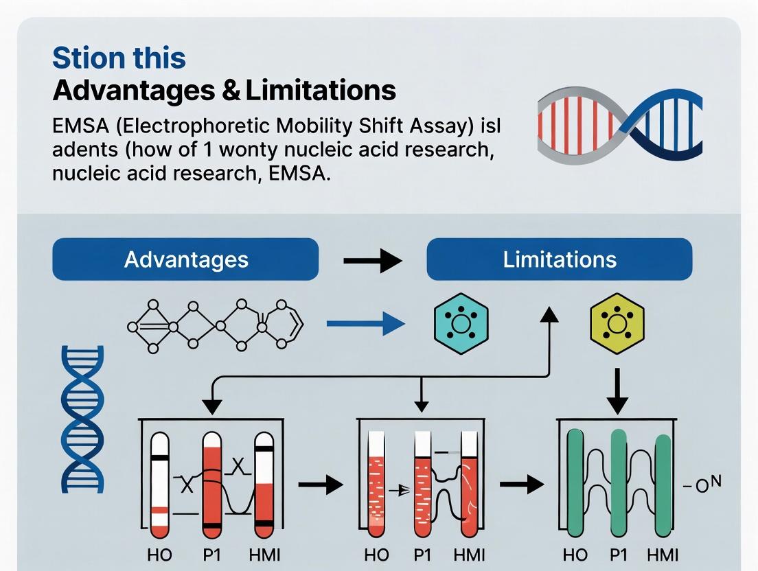
Advantages: Direct, visual proof of complex formation; adaptable to various nucleic acid structures; relatively low-cost; semi-quantitative; can assess multi-protein complexes. Limitations: Low-throughput; equilibrium can be disturbed during electrophoresis; requires optimization; absolute affinity measurements can be challenging; not truly native (gel matrix effects).
As a cornerstone technique, EMSA's strength lies in its unambiguous, direct detection of protein-nucleic acid complexes. Its integration with robust controls and quantitative analysis provides reliable data on binding specificity and affinity. While newer high-throughput methods exist, EMSA's simplicity, visual clarity, and ability to validate more complex assays solidify its enduring role as the gold standard in the molecular biologist's arsenal.
Electrophoretic Mobility Shift Assay (EMSA), also termed gel shift assay, is a cornerstone technique in molecular biology for detecting and quantifying specific protein-nucleic acid interactions. Within the broader thesis of evaluating EMSA's advantages and limitations, this whitepaper details the core biophysical principle enabling this simple yet powerful method.
Core Principle and Biophysical Basis
The fundamental principle of EMSA is that the electrophoretic mobility of a nucleic acid (DNA or RNA) probe through a native polyacrylamide or agarose gel is retarded or "shifted" upon binding to a protein or other ligand. This shift occurs because the resulting complex has:
- Increased Molecular Mass: The protein adds mass to the probe.
- Altered Charge-to-Mass Ratio: The protein's charge modifies the overall charge of the complex.
- Conformational Change: Protein binding can induce bending or looping of the nucleic acid, further hindering migration.
The assay is performed under non-denaturing (native) conditions to preserve non-covalent interactions. The degree of retardation is observable as a distinct band higher in the gel than the free probe. Competition experiments using unlabeled specific or non-specific oligonucleotides confirm binding specificity. Supershift assays, employing antibodies against the protein of interest, provide further verification and can identify specific proteins within a complex.
The utility of EMSA spans qualitative detection to quantitative analysis. Key quantitative parameters are summarized below.
Table 1: Key Quantitative Parameters in EMSA Analysis
| Parameter | Typical Range/Value | Significance & Notes |
|---|---|---|
| Probe Length (DNA) | 20-50 bp | Optimal for resolution; longer fragments can exhibit multiple binding events or non-specific binding. |
| Polyacrylamide Gel Concentration | 4-10% | Lower % for larger complexes (>500 kDa); higher % for better resolution of smaller shifts. |
| Electrophoresis Temperature | 4-10°C | Maintained to stabilize protein-nucleic acid interactions during separation. |
| Binding Affinity (Kd) Measurement Range | 10^-9 to 10^-12 M | Determined by titrating protein against a constant probe concentration and quantifying bound/free fractions. |
| Detection Sensitivity (Chemiluminescence) | Low femtomole (10^-15 mol) | Varies with probe label (radioactive 32P is most sensitive, followed by chemiluminescent and fluorescent dyes). |
| Dynamic Range for Quantification | ~2 orders of magnitude | Limited by gel resolution and detection method linearity. |
Table 2: Comparative Analysis of EMSA Probe Labeling Strategies
| Label Type | Sensitivity | Stability | Safety & Handling | Required Equipment | Best For |
|---|---|---|---|---|---|
| Radioactive (³²P) | Very High (fmol) | Short (half-life ~14 days) | High risk; requires special licensing, shielding, and waste disposal. | Phosphorimager or X-ray film | Highest sensitivity applications; precise Kd determinations. |
| Chemiluminescent | High (fmol-amol) | High (months to years) | Safe; standard lab handling. | CCD imager or X-ray film | Routine detection; labs avoiding radioactivity. |
| Fluorescent (Cy5, FAM) | Moderate (pmol) | High (months to years) | Safe; standard lab handling. | Fluorescence scanner or imager | Multiplexing (multiple probes); real-time kinetics not common. |
| Biotin | Moderate (pmol) | High (months to years) | Safe; standard lab handling. | Requires streptavidin-enzyme conjugate and chemiluminescent/colorimetric substrate. | Labs with established colorimetric workflows. |
Detailed Experimental Protocols
Protocol 1: Standard EMSA for DNA-Protein Interaction
- Purpose: To detect sequence-specific binding of a nuclear extract protein to a double-stranded DNA probe.
- Key Reagents: Labeled DNA probe, nuclear extract, poly(dI-dC) as non-specific competitor, binding buffer, native polyacrylamide gel.
- Probe Preparation: A 25-30 bp oligonucleotide containing the predicted protein-binding site is annealed to its complement. The duplex is end-labeled using [γ-³²P]ATP and T4 Polynucleotide Kinase, or purchased pre-labeled.
- Binding Reaction:
- Combine in order: 14 μL of nuclease-free water, 4 μL of 5X binding buffer (typically: 50 mM Tris, 250 mM NaCl, 5 mM DTT, 5 mM EDTA, 20% glycerol, pH 7.5), 1 μL of poly(dI-dC) (1 μg/μL), 10 μg of nuclear extract protein, and 1 μL of labeled probe (~20 fmol).
- Incubate at room temperature for 20-30 minutes.
- Gel Electrophoresis:
- Pre-run a 6% native polyacrylamide gel (29:1 acrylamide:bis) in 0.5X TBE buffer at 100V for 30-60 minutes at 4°C.
- Load samples (with non-ionic dye like 6X loading buffer) and run at 100-150V in the cold room (4°C) until the dye front migrates 2/3 down the gel.
- Detection:
- Transfer gel to blotting paper, dry under vacuum, and expose to a phosphor storage screen overnight.
- Image using a phosphorimager.
Protocol 2: Supershift Assay
- Purpose: To confirm the identity of a protein within a shifted complex.
- Key Reagents: Specific antibody against the suspected DNA-binding protein.
- Perform the standard binding reaction as in Protocol 1.
- After the initial 20-minute incubation, add 1-2 μg of the specific antibody (or an isotype control antibody) to the reaction.
- Incubate for an additional 30-60 minutes on ice or at room temperature.
- Load and run the gel as described. The antibody binding to the protein-DNA complex creates a larger "supershifted" complex with even slower mobility, appearing higher in the gel.
Protocol 3: Competition EMSA
- Purpose: To demonstrate binding specificity.
- Key Reagents: Unlabeled ("cold") specific competitor oligonucleotide and non-specific/mutant control oligonucleotide.
- Set up a series of standard binding reactions.
- Prior to adding the labeled probe, add increasing molar excesses (e.g., 10x, 50x, 100x, 200x) of unlabeled competitor DNA to the appropriate tubes.
- Add a constant amount of labeled probe to all tubes.
- Incubate and run the gel. Specific binding is demonstrated by the disappearance of the shifted band with the specific competitor, but not with the non-specific control.
Visualizing EMSA Principles and Workflows
Title: EMSA Core Principle: Binding Causes Gel Shift
Title: Standard EMSA Experimental Workflow
Title: EMSA Verification Assays: Competition & Supershift
The Scientist's Toolkit: Key Research Reagent Solutions
Table 3: Essential Materials for EMSA
| Item | Function | Critical Considerations |
|---|---|---|
| Chemiluminescent Nucleic Acid Labeling Kit | Non-radioactive probe labeling. Reagents for end-labeling with biotin or digoxigenin. | Offers safety and stability; sensitivity is sufficient for most applications. |
| Non-Radioactive EMSA Detection Kit | Detection of biotin- or digoxigenin-labeled probes via streptavidin-HRP/conjugate and chemiluminescent substrate. | Enables complete workflow without radioactivity. |
| EMSA/Gel-Shift Binding Buffer (5X) | Optimized buffer providing correct ionic strength, pH, and stabilizers (glycerol, DTT) for protein-nucleic acid interactions. | Consistency and convenience; reduces optimization time. |
| Non-Specific Competitor DNA (poly(dI-dC)) | Inert nucleic acid polymer that binds non-specific proteins, reducing background and highlighting specific shifts. | Concentration must be titrated for each new protein extract. |
| Native PAGE Gel Kit | Pre-cast native polyacrylamide gels and compatible running buffer. | Ensures reproducibility and saves time in gel preparation. |
| High-Quality Nuclear Extract | Source of DNA-binding proteins (e.g., transcription factors) from specific cell lines or tissues. | Activity and protein concentration are key; prepare fresh or use validated commercial extracts. |
| Transcription Factor-Specific Antibody | For supershift assays to confirm protein identity within the complex. | Must be verified for use in EMSA/supershift; should recognize native protein. |
| Cold/Unlabeled Competitor Oligonucleotides | Specific wild-type and mutant sequences for competition assays. | Confirms sequence specificity of the observed interaction. |
Historical Evolution of the Gel Shift Assay from Basic Research to Mainstream Tool
Within the broader thesis analyzing the advantages and limitations of the Electrophoretic Mobility Shift Assay (EMSA), understanding its historical evolution is critical. This technical guide traces the assay's development from a novel method for studying protein-DNA interactions to an indispensable, mainstream tool in molecular biology, biochemistry, and drug development.
Historical Development and Key Milestones
Foundational Period (1980s)
The gel shift assay, or EMSA, was pioneered in the early 1980s as a direct method to detect sequence-specific DNA-binding proteins. It moved the field beyond indirect footprinting techniques.
Mainstream Adoption (1990s-2000s)
Widespread adoption was driven by the study of transcription factors, gene regulation, and signal transduction. Advancements like supershift assays (using specific antibodies) and the use of fluorescent or chemiluminescent probes standardized the method for broader labs.
Modern Innovations (2010s-Present)
Current evolution focuses on high-throughput capabilities, quantitative analysis via digital imaging, and integration with microfluidic platforms. It is now a staple in drug discovery for screening compounds that modulate protein-nucleic acid interactions.
Quantitative Evolution of EMSA Applications
Table 1: Historical Shift in EMSA Application Prevalence in Literature (PubMed)
| Decade | Primary Application Focus | Estimated % of Total Nucleic Acid Interaction Papers | Key Technological Driver |
|---|---|---|---|
| 1980-1989 | Detection of novel DNA-binding proteins | ~5% | Radiolabeling (³²P) |
| 1990-1999 | Transcription factor analysis & mutation studies | ~15% | Antibody supershift; Chemiluminescence |
| 2000-2009 | Kinetics & complex composition studies | ~22% | Fluorescent dyes (Cy5, FAM); Densitometry |
| 2010-Present | High-throughput screening; Quantitative diagnostics | ~30% | Capillary electrophoresis; Digital EMSA |
Table 2: Performance Metrics Comparison Across EMSA Generations
| Parameter | Classic Radioactive EMSA | Modern Fluorescent EMSA | High-Throughput Capillary EMSA |
|---|---|---|---|
| Assay Time | 6-24 hours | 2-4 hours | < 1 hour |
| Detection Sensitivity | High (fmol) | Moderate-High (fmol-pmol) | High (fmol) |
| Quantitation Accuracy | Moderate (manual) | High (digital) | Very High (automated) |
| Throughput (samples/day) | 10-20 | 50-100 | 384+ |
| Safety & Waste Concern | High | Low | Very Low |
Detailed Experimental Protocols
Protocol 1: Classic Radioactive EMSA for Transcription Factor Binding
Objective: To detect and confirm the binding of a nuclear extract protein to a specific DNA consensus sequence. Reagents: ³²P-end-labeled DNA probe, purified protein or nuclear extract, poly(dI-dC) nonspecific competitor, EMSA binding buffer (10 mM Tris, 50 mM KCl, 1 mM DTT, 2.5% glycerol, 0.05% NP-40, pH 7.5), 4-6% non-denaturing polyacrylamide gel, 0.5X TBE running buffer. Procedure:
- Prepare Reaction Mix: Combine 1-10 fmol labeled probe, 2-5 µg nuclear extract, 1-2 µg poly(dI-dC), and binding buffer to 20 µL final volume.
- Incubate: 20-30 minutes at room temperature.
- Load & Run: Add loading dye, load onto pre-run gel. Run at 100V, 4°C in 0.5X TBE until dye front migrates 2/3 down.
- Transfer & Detect: Transfer gel to filter paper, dry, and expose to X-ray film or phosphorimager screen.
Protocol 2: Modern Fluorescent EMSA for Competitive Binding Studies
Objective: To determine the dissociation constant (Kd) or screen for inhibitory compounds using a fluorescent probe. Reagents: 5'-FAM or Cy5-labeled DNA probe, purified recombinant protein, unlabeled specific competitor DNA, EMSA buffer, 6% DNA retardation gel (commercial), fluorescence-compatible imager. Procedure:
- Titration Setup: Hold probe concentration constant (e.g., 1 nM) while titrating protein across a range (e.g., 0.1 nM to 1 µM) in separate reactions.
- Competition Variation: For inhibitor screening, hold protein and labeled probe constant while adding increasing amounts of unlabeled competitor or test compound.
- Incubate: 30 minutes at 25°C in the dark.
- Electrophoresis: Run on pre-cast gel in dark conditions at 100V for 45-60 min.
- Quantification: Image gel using a fluorescence scanner. Quantify free and bound probe bands to calculate % shift and Kd.
Experimental Workflow and Pathway Diagrams
Diagram Title: EMSA Core Experimental Workflow
Diagram Title: EMSA Detects Key Signaling Output
The Scientist's Toolkit: Essential Research Reagent Solutions
Table 3: Key Reagents and Materials for Modern EMSA
| Item | Function & Rationale |
|---|---|
| Chemiluminescent Nucleic Acid Labeling Kit | Non-radioactive, sensitive probe labeling using biotin or digoxigenin. Safer and stable. |
| Recombinant Purified Transcription Factor | Provides consistent, high-activity protein source for binding studies and inhibitor screening. |
| Non-Specific Competitor DNA (poly dI-dC) | Blocks non-specific protein-DNA interactions, reducing background and improving specificity. |
| Pre-Cast DNA Retardation Gels (6-8%) | Ensure reproducibility, save time, and provide optimal matrix for complex separation. |
| Fluorescent Scanner or Imager | Enables quantitative analysis of fluorescent or chemiluminescent signals with wide dynamic range. |
| EMSA-Specific Binding Buffer (10X) | Optimized salt, glycerol, and detergent concentration to promote specific interactions. |
| Supershift/Antibody Incursion Antibodies | Antibodies specific to the DNA-binding protein to confirm identity and induce "supershift". |
| Capillary Electrophoresis EMSA Kit | For high-throughput, automated size-separation and quantification, minimizing manual steps. |
The gel shift assay's evolution from a basic research technique to a mainstream tool encapsulates the drive for quantitative, safe, and high-throughput methods in life sciences. Its enduring utility within the thesis framework on EMSA's advantages and limitations lies in its direct visualization capability, adaptability to various detection modalities, and its critical role in validating interactions central to gene regulation and drug discovery. Future developments will likely integrate further with '-omics' platforms and real-time binding analysis.
Within the broader thesis evaluating the advantages and limitations of the Electrophoretic Mobility Shift Assay (EMSA), a critical analysis of its core components is essential. EMSA remains a cornerstone technique for studying protein-nucleic acid interactions, pivotal in gene regulation research and drug discovery targeting transcription factors. This technical guide deconstructs the triumvirate of key components: the labeled probe, the nuclear/cellular extract, and the non-denaturing gel. The integrity and optimization of each directly dictate the assay's specificity, sensitivity, and validity, factors central to any assessment of EMSA's utility in modern molecular biology.
The Labeled Probe
The probe is the labeled nucleic acid fragment (DNA or RNA) containing the specific protein-binding sequence of interest.
Design & Labeling: Probes are typically 20-40 base pairs long, incorporating the consensus sequence. Modern labeling predominantly uses fluorophores (e.g., Cy5, FAM) for direct detection or biotin for chemiluminescent detection, having largely replaced radioactive (³²P) methods due to safety and waste concerns.
Key Quantitative Parameters:
- Specific Activity: Critical for sensitivity. A 2023 comparative study reported the following detection limits:
| Label Type | Optimal Specific Activity | Approximate Detection Limit (fmol of complex) | Relative Cost |
|---|---|---|---|
| ³²P (γ-ATP) | ~6000 Ci/mmol | 0.1 - 0.5 | Low |
| Biotin | 1-3 biotin molecules/probe | 1 - 5 | Moderate |
| Fluorophore (Cy5) | 1 fluorophore/probe | 2 - 10 | High |
- Probe Concentration: Typically 0.1-1 nM (10-100 fmol) per binding reaction to ensure label excess without self-competition.
Protocol: Fluorescent Probe Labeling via PCR
- Primer Design: Design forward and reverse primers to amplify the target sequence. The 5' end of one primer is synthesized with the desired fluorophore.
- PCR Reaction: Set up a 50 µL reaction: 10 ng template DNA, 0.2 µM labeled primer, 0.2 µM unlabeled primer, 200 µM dNTPs, 1X PCR buffer, 1.5 mM MgCl₂, 1.25 U high-fidelity DNA polymerase.
- Thermocycling: Standard amplification: 95°C for 2 min; 35 cycles of (95°C for 30s, 60°C for 30s, 72°C for 30s/kb); 72°C for 5 min.
- Purification: Purify the PCR product using a spin column to remove unincorporated labeled primers. Verify concentration and labeling efficiency via spectrophotometry (absorbance at 260 nm and fluorophore-specific wavelength).
Nuclear/Cellular Extract
The source of the protein(s) of interest. Nuclear extracts are standard for transcription factor studies.
Preparation Principles: The goal is to isolate active proteins while maintaining native interactions and preventing degradation. All steps are performed at 4°C with protease and phosphatase inhibitors.
Protocol: Rapid Nuclear Extract Preparation (Mini-scale)
- Harvest & Wash: Pellet ~2x10⁶ cells. Wash with 1 mL ice-cold PBS.
- Hypotonic Lysis: Resuspend pellet in 400 µL of Hypotonic Buffer (10 mM HEPES pH 7.9, 1.5 mM MgCl₂, 10 mM KCl, 0.5 mM DTT, 0.2% NP-40, 1X protease inhibitors). Incubate on ice for 10 min.
- Centrifuge: Spin at 3,000 x g for 10 min. The supernatant is the cytoplasmic fraction. Retain if needed.
- Nuclear Lysis: Resuspend the nuclear pellet in 50 µL of High-Salt Extraction Buffer (20 mM HEPES pH 7.9, 420 mM NaCl, 1.5 mM MgCl₂, 0.2 mM EDTA, 25% glycerol, 0.5 mM DTT, 1X protease inhibitors).
- Extract: Rock at 4°C for 30 min. Centrifuge at 20,000 x g for 15 min.
- Aliquot & Store: Collect supernatant (nuclear extract). Determine protein concentration (Bradford assay), aliquot, and store at -80°C. Avoid >2 freeze-thaw cycles.
Quantitative Considerations:
- Yield: Typical yield is 1-5 µg of nuclear protein per 10⁶ mammalian cells.
- Amount in EMSA: 2-10 µg of total nuclear protein per 20 µL binding reaction is standard. Excess can cause non-specific shifts.
The Non-Denaturing Gel
The matrix that separates protein-nucleic acid complexes from free probe based on size and charge, without disrupting non-covalent interactions.
Composition: Polyacrylamide (typically 4-8%) in 0.5X Tris-Borate-EDTA (TBE) or Tris-Glycine buffer. Glycerol (2-5%) is often added to the gel to enhance complex stability and facilitate loading.
Critical Electrophoresis Parameters:
| Parameter | Typical Condition | Rationale & Impact |
|---|---|---|
| Acrylamide % | 6% | Optimal resolution for complexes 10-200 kDa. Higher % retards migration. |
| Crosslinker Ratio | 29:1 or 37.5:1 (Acrylamide:Bis) | Standard for native gels. |
| Buffer System | 0.5X TBE | Lower ionic strength than 1X provides better resolution and cooler running. |
| Running Temperature | 4°C (cold room or with cooling apparatus) | Minimizes complex dissociation during electrophoresis. |
| Voltage | 80-100 V constant | Higher voltage generates heat, causing "smiling" and complex denaturation. |
| Run Time | 1.5 - 2.5 hours | Until free probe migrates ~2/3 down the gel. |
Protocol: Casting and Running a 6% Non-Denaturing Gel
- Assemble Cassette: Clean glass plates and spacers (1.0-1.5 mm thick).
- Mix Gel Solution: For 20 mL: 3.0 mL 40% acrylamide/bis (29:1), 2.0 mL 5X TBE, 14.8 mL dH₂O, 1.0 mL 80% glycerol. Degas for 10 min.
- Polymerize: Add 200 µL 10% ammonium persulfate and 20 µL TEMED. Pour immediately, insert comb, and allow to polymerize for 45-60 min.
- Pre-run: Place gel in tank with 0.5X TBE running buffer. Pre-electrophorese at 100 V for 45-60 min at 4°C to remove residual APS and equilibrate pH.
- Load & Run: Mix binding reactions with 2-5 µL of native loading dye (no SDS/beta-mercaptoethanol). Load samples. Run at 100 V constant voltage in the cold until the dye front (bromophenol blue) is near the bottom.
- Detection: For fluorescent probes, scan gel directly using an appropriate imaging system. For biotin probes, transfer to a nylon membrane and develop with streptavidin-HRP.
The Scientist's Toolkit: Research Reagent Solutions
| Item | Function in EMSA | Key Considerations |
|---|---|---|
| Fluorophore-labeled Oligonucleotides | High-sensitivity, non-radioactive probe generation. | Order HPLC-purified. Common dyes: Cy5 (647 nm), FAM (495 nm). Store in dark. |
| Nuclear Extract Kit | Commercial reagent for rapid, standardized extract prep from cells/tissues. | Ensures reproducibility. Often includes optimized buffers and inhibitors. Cost-effective for small sample numbers. |
| Protease/Phosphatase Inhibitor Cocktails | Preserves protein activity and modification state during extract preparation. | Use broad-spectrum, EDTA-free cocktails. Add fresh to lysis buffers. |
| Non-specific Competitor DNA | Suppresses non-specific protein-probe binding (e.g., poly(dI-dC), salmon sperm DNA). | Titration is critical; excess can compete for specific binding. Typical: 0.05-0.1 µg/µL in reaction. |
| Native Gel Stain (SYBR Green, GelRed) | Post-electrophoresis staining for total nucleic acid visualization. | Useful for checking gel integrity and loading uniformity. Less sensitive than specific probe detection. |
| Chemiluminescent Nucleic Acid Detection Module | For biotinylated probe detection post-electrophoresis/transfer. | Includes streptavidin-HRP and stable peroxide/luminol reagents. Provides high sensitivity and signal-to-noise. |
Visualization of EMSA Workflow and Critical Controls
EMSA Workflow & Validation Pathway
Molecular Interactions in EMSA Complexes
This technical guide details the primary applications of the Electrophoretic Mobility Shift Assay (EMSA) within a broader research thesis examining its advantages and limitations. EMSA remains a cornerstone in vitro technique for directly probing protein-nucleic acid interactions, fundamental to deciphering transcriptional regulatory networks. This document provides current methodologies, data interpretation frameworks, and technical resources to empower research in gene regulation and drug discovery.
Table 1: Key Quantitative Parameters in a Standard EMSA Experiment
| Parameter | Typical Range / Value | Notes / Impact on Results |
|---|---|---|
| Probe Length (DNA/RNA) | 20-50 base pairs | Longer probes may permit multiple, non-specific protein interactions. |
| Probe Concentration | 0.1 - 10 nM (labeled) | Below Kd of interaction; ensures sensitivity while minimizing non-specific binding. |
| Protein (TF) Amount | 0.5 - 20 µg of nuclear extract or 1-100 ng recombinant protein | Must be titrated to observe clear shift without probe depletion. |
| Poly[dI•dC] Competitor | 0.05 - 2 µg/µL | Suppresses non-specific binding; optimal amount is protein-source dependent. |
| Electrophoresis Conditions | 4-10°C, 80-150 V, 1-2 hours | Low temperature stabilizes complexes; voltage/time adjusted for gel % and complex resolution. |
| Binding Reaction Incubation | 20-30 minutes at 20-25°C (RT) | Allows equilibrium binding. Ice incubation may favor some complexes. |
| Detection Limit (Chemiluminescence) | Low femtomole (fmol) range | Dependent on label efficiency and exposure time. |
Table 2: Comparative Analysis of EMSA Variations
| Assay Variation | Key Differentiator | Primary Application Advantage | Typical Resolution/Complexity |
|---|---|---|---|
| Standard EMSA | Radioactive (³²P) or fluorescently labeled probe. | Direct detection of protein-nucleic acid complexes. | Single shifted band(s). |
| Supershift EMSA | Addition of antibody specific to the TF or epitope tag. | Confirms TF identity within the complex. | Band shift to higher molecular weight ("supershift") or ablation. |
| Competition EMSA | Inclusion of unlabeled competitor DNA (wild-type vs. mutant). | Determines binding specificity and affinity. | Titrated reduction of shifted band intensity. |
| Fluorescent EMSA (FEMSA) | Cy5, FAM, or similar fluorescent probes. | Safer, faster; enables multiplexing and gel-based quantification. | Similar to standard, with multi-color potential. |
Detailed Experimental Protocols
Protocol 1: Standard EMSA for Nuclear Extract Analysis
A. Probe Preparation
- Design & Labeling: Synthesize complementary oligonucleotides containing the putative TF binding site with 5-10 bp flanking sequences. Anneal to form double-stranded probe. Label using T4 Polynucleotide Kinase and [γ-³²P]ATP or a 5'-end fluorescent tag kit.
- Purification: Purify labeled probe using a spin column (e.g., Sephadex G-25) to remove unincorporated nucleotides.
B. Binding Reaction
- Prepare a 20 µL reaction mix on ice:
- 1X Binding Buffer (e.g., 10 mM HEPES pH 7.9, 50 mM KCl, 1 mM DTT, 2.5 mM MgCl₂, 10% glycerol, 0.05% NP-40).
- 1 µg Poly[dI•dC] (or alternative non-specific competitor).
- Nuclear extract (e.g., 5-10 µg total protein) or recombinant TF.
- Nuclease-free water to volume.
- Pre-incubate on ice for 10 minutes to block non-specific sites.
- Add 0.5-1 ng (20,000-50,000 cpm) of labeled probe.
- Incubate at room temperature for 20-30 minutes.
C. Non-Denaturing Gel Electrophoresis
- Gel Preparation: Prepare a 4-6% polyacrylamide gel (29:1 acrylamide:bis) in 0.5X TBE buffer. Pre-run gel at 100 V for 30-60 minutes at 4°C.
- Loading: Add 5X native loading dye (without SDS) to each reaction. Load samples immediately.
- Run: Electrophorese at 80-150 V (constant voltage) in 0.5X TBE at 4°C until the bromophenol blue dye is near the bottom.
- Detection: For radioactive probes, dry gel and expose to a phosphorimager screen. For fluorescent probes, image directly using an appropriate scanner.
Protocol 2: Supershift EMSA
- Follow the Standard EMSA binding reaction setup.
- After the initial 20-minute binding incubation, add 1-2 µg of specific antibody or an isotype control antibody.
- Incubate the reaction for an additional 30-60 minutes on ice or at room temperature (optimize for antibody).
- Proceed with gel electrophoresis as in Protocol 1. A successful supershift is indicated by a retarded band of higher molecular weight or the ablation of the original shifted band.
Protocol 3: Competition EMSA
- Set up a series of standard binding reactions with nuclear extract.
- Include increasing molar excesses (e.g., 10x, 50x, 100x, 200x) of unlabeled double-stranded oligonucleotide competitor:
- Specific Competitor: Identical to the probe sequence.
- Mutant Competitor: Contains scrambled or point-mutated binding site.
- Add competitors before adding the labeled probe (incubate 10 minutes).
- Add labeled probe and complete the binding reaction.
- Specific binding is demonstrated by dose-dependent reduction of the shifted band with the wild-type, but not the mutant, competitor.
Signaling Pathway & Workflow Visualizations
Diagram 1: Core EMSA Experimental Workflow (7 steps)
Diagram 2: TF Activation & DNA Binding in Gene Regulation
The Scientist's Toolkit: Research Reagent Solutions
Table 3: Essential Materials for EMSA Experiments
| Reagent / Material | Function & Purpose | Key Considerations |
|---|---|---|
| Chemiluminescent Nucleic Acid Labeling Kit | Non-radioactive, high-sensitivity probe labeling. | Kits using biotin or digoxigenin are common. Requires specific blocking/detection buffers. |
| Recombinant Transcription Factor | Pure protein source for defining specific interactions. | Allows precise control of protein concentration and avoids contaminating activities. |
| Nuclear Extraction Kit | Isolates nuclear proteins, including TFs, from cultured cells or tissues. | Critical for studying endogenous, post-translationally modified TFs in their native state. |
| EMSA Gel Shift Kits (Commercial) | Provide optimized buffers, competitors, and control DNA/protein. | Reduces optimization time; ensures reproducibility for standard assays. |
| High-Affinity, Sequence-Specific TF Antibodies | For supershift/ablation experiments. | Must be validated for use in EMSA; epitope must be accessible in DNA-bound complex. |
| Non-Specific Competitor DNA (Poly[dI•dC]) | Blocks non-specific electrostatic interactions between proteins and probe. | Amount is critical; too little leads to smearing, too much can disrupt specific binding. |
| Non-Denaturing Acrylamide/Bis Mix (29:1 or 37.5:1) | Forms the matrix for resolving protein-nucleic acid complexes. | Lower percentage gels (4%) better for large complexes; higher (6%) for sharper bands. |
| Phosphor Storage Screen & Imager | High-resolution, quantitative detection of radioisotopic signals. | Superior dynamic range and sensitivity compared to X-ray film. |
| Fluorescent Gel Scanner/Imager | Required for FEMSA; enables multiplexing. | Must have appropriate excitation/emission filters for chosen fluorophores. |
Mastering EMSA Protocol: Step-by-Step Guide, Variations, and Advanced Applications
The Electrophoretic Mobility Shift Assay (EMSA) is a cornerstone technique for studying protein-nucleic acid interactions in vitro. Within the broader thesis on EMSA's advantages and limitations, this protocol details the standard workflow, focusing on the critical choice between radioactive and chemiluminescent probe labeling. The selection impacts sensitivity, safety, cost, and required instrumentation, which are central to evaluating the technique's applicability in modern research and drug development.
Probe Design and Preparation
The DNA or RNA probe typically contains the specific target sequence (20-30 bp) flanked by nonspecific sequence. For competition assays, an unlabeled identical oligonucleotide (cold competitor) and a mutated version (non-specific competitor) are essential controls.
Probe Labeling: Radioactive vs. Chemiluminescent
The choice of labeling method is fundamental. Quantitative comparisons are summarized in Table 1.
Table 1: Quantitative Comparison of Probe Labeling Methods
| Parameter | Radioactive (γ-32P/γ-33P ATP) | Chemiluminescent (Biotin/Streptavidin-HRP) |
|---|---|---|
| Typical Sensitivity (detection limit) | 0.1-1 fmol | 1-10 fmol |
| Signal Stability (Half-life) | Physical half-life of isotope (e.g., 14.3 days for ³²P) | Months to years (stable conjugate) |
| Exposure/Detection Time | 30 min to overnight | 1-5 minutes |
| Assay Time Post-Electrophoresis | Slow (drying, exposure) | Fast (transfer, blotting, detection) |
| Relative Hazard | High (ionizing radiation) | Low |
| Regulatory & Waste Cost | High (licensing, disposal) | Low |
| Equipment Cost | High (phosphorimager/Geiger counter) | Moderate (standard gel imager with chemiluminescence) |
| Spatial Resolution | Excellent | Very Good |
| Quantification Linear Range | ~4-5 orders of magnitude | ~3-4 orders of magnitude |
Detailed Protocol: Radioactive End-Labeling with γ-32P ATP
- Materials: DNA oligonucleotide probe, T4 Polynucleotide Kinase (PNK), 10X PNK buffer, γ-32P ATP, Nuclease-free water, Micro Bio-Spin P-30 columns.
- Method:
- In a microcentrifuge tube, combine: 1 µL oligonucleotide (100 nM), 2 µL 10X PNK buffer, 1 µL T4 PNK (10 U), 15.5 µL nuclease-free water, and 5 µL γ-32P ATP (50 µCi).
- Incubate at 37°C for 30 minutes.
- Terminate the reaction by heating at 65°C for 5 minutes.
- Purify the labeled probe using a spin column per manufacturer's instructions to remove unincorporated nucleotides.
- Determine labeling efficiency by scintillation counting.
Detailed Protocol: Chemiluminescent End-Labeling with Biotin
- Materials: Biotin 3'-End DNA Labeling Kit (e.g., Thermo Fisher), or 5'-Biotin-modified oligonucleotide.
- Method (3' End Labeling):
- Assemble reaction: 1 µL oligonucleotide (100 nM), 5 µL 5X Terminal Transferase (TdT) buffer, 5 µL Biotin-11-ddUTP, 3 µL TdT enzyme, and 11 µL nuclease-free water.
- Incubate at 37°C for 60 minutes.
- Stop with 2.5 µL of 0.2M EDTA.
- Purify using a spin column or ethanol precipitation.
Protein Binding and Electrophoresis
- Binding Reaction: Combine 2-10 µg nuclear extract (or purified protein), 1-2 µL poly(dI-dC) (1 µg/µL) in binding buffer (10 mM HEPES, 50 mM KCl, 0.5 mM EDTA, 1 mM DTT, 5% Glycerol, pH 7.9). Incubate on ice for 10 minutes. Add labeled probe (20,000-50,000 cpm for radioactive; ~20 fmol for biotin). Bring total volume to 20 µL. Incubate at room temp for 20-30 min.
- Gel Loading: Add 4 µL of 6X non-denaturing loading dye (30% glycerol, 0.25% bromophenol blue).
- Electrophoresis: Pre-run a 4-6% non-denaturing polyacrylamide gel (29:1 acrylamide:bis) in 0.5X TBE at 100V for 60 min in the cold room. Load samples and run at 100V for 90-120 min until dye migrates ~2/3 down.
Post-Electrophoresis Detection
Radioactive Detection Protocol
- Gel Drying: Transfer gel to filter paper, cover with plastic wrap, and dry under vacuum at 80°C for 60 min.
- Imaging: Expose dried gel to a phosphor storage screen for 1-12 hours. Scan the screen with a phosphorimager.
Chemiluminescent Detection Protocol
- Electroblotting: Transfer protein-DNA complexes from gel to a positively charged nylon membrane at 380 mA for 30-60 min in 0.5X TBE at 4°C.
- Crosslinking: UV crosslink the nucleic acids to the membrane (1200 J/m², 254 nm).
- Blocking & Detection: Block membrane with blocking buffer for 15 min. Incubate with Streptavidin-Horseradish Peroxidase (HRP) conjugate (1:3000 dilution) for 15 min. Wash thoroughly.
- Imaging: Incubate with chemiluminescent substrate (e.g., Luminol/H₂O₂) and image with a CCD-based gel documentation system.
The Scientist's Toolkit: Key Research Reagent Solutions
| Item | Function |
|---|---|
| Poly(dI-dC) | Non-specific competitor DNA to reduce background from non-sequence-specific protein binding. |
| T4 Polynucleotide Kinase (PNK) | Catalyzes the transfer of the terminal (γ) phosphate from ATP to the 5'-OH of DNA/RNA for radioactive labeling. |
| γ-32P ATP | Radioactive ATP donor providing the high-sensitivity label for probe detection. |
| Biotin-11-ddUTP | A modified nucleotide used by Terminal Transferase to add a single biotin label to the 3' end of DNA. |
| Streptavidin-HRP Conjugate | High-affinity binding to biotin, coupled to HRP enzyme for chemiluminescent signal generation. |
| Non-denaturing Polyacrylamide Gel Mix | Matrix for separating protein-nucleic acid complexes based on size/shift while maintaining native interactions. |
| Positively Charged Nylon Membrane | Binds negatively charged nucleic acids with high affinity for chemiluminescent blotting procedures. |
| Chemiluminescent HRP Substrate | Enzyme substrate that produces light upon oxidation by HRP for film or CCD-based detection. |
Visualization Diagrams
Title: EMSA Standard Protocol Workflow Decision Tree
Title: Chemiluminescent EMSA Detection Signaling Pathway
Supershift and Antibody-Based EMSA for Specific Protein Identification
Within the broader research context of analyzing the advantages and limitations of the Electrophoretic Mobility Shift Assay (EMSA), this technical guide focuses on two powerful refinement techniques: the supershift assay and antibody-based EMSA. The standard EMSA, while excellent for detecting protein-nucleic acid interactions, lacks inherent specificity for identifying the exact protein constituent within a complex. Supershift and antibody-based approaches solve this critical limitation by incorporating specific antibodies, thereby confirming protein identity and offering insights into multiprotein complexes. This guide details the protocols, data interpretation, and practical toolkit for implementing these advanced EMSA methods.
Core Principles & Mechanisms
A standard EMSA detects binding via reduced electrophoretic mobility of a nucleic acid probe upon protein binding. The supershift assay extends this principle by including an antibody that specifically recognizes the bound protein. This can result in a ternary complex (protein-nucleic acid-antibody) with an even greater reduction in mobility ("supershifted" band). Alternatively, if the antibody epitope is blocked by nucleic acid binding or if the antibody disrupts the interaction, it can prevent complex formation ("blocking" or "ablation").
Diagram 1: Supershift EMSA Principle & Outcomes
Detailed Experimental Protocols
Protocol: Standard EMSA with Supershift
Objective: To confirm the identity of a protein in a DNA-protein complex using a specific antibody.
Materials: (See "Scientist's Toolkit" Section 5). Procedure:
- Prepare Binding Reactions: Set up standard EMSA binding reactions (20 µL final volume) containing appropriate buffer, poly(dI-dC), nuclear extract or purified protein, and labeled probe. Incubate at room temperature (RT) for 20 min.
- Add Antibody: To the supershift sample, add 1-2 µg of the specific antibody (or matched control IgG). For the control, add an equal volume of antibody dilution buffer or control IgG.
- Incubate for Supershift: Continue incubation at RT for 30-60 min, or at 4°C overnight for higher affinity antibodies.
- Load and Run: Add 2-5 µL of non-denaturing loading dye. Load the entire reaction onto a pre-run 4-6% native polyacrylamide gel. Run in 0.5x TBE buffer at 100V (constant voltage) at 4°C until the dye front is near the bottom.
- Visualize: Transfer gel to blotting paper, dry, and expose to a phosphorimager screen or X-ray film. Alternatively, use a fluorescent scanner for fluorescently-labeled probes.
Critical Controls:
- Antibody Specificity: Include reactions with a non-specific, isotype-matched IgG.
- Competition Control: Include a reaction with 100x molar excess of unlabeled probe (cold competition) to demonstrate binding specificity.
- Antibody-Only Control: Probe + antibody only, to rule out non-specific antibody-probe interactions.
Protocol: Antibody-Based EMSA (Ab-EMSA) for Disruption
Objective: To determine if a specific protein is essential for complex formation.
Procedure:
- Pre-incubate Antibody with Protein: Incubate the nuclear extract or purified protein with the specific antibody (2-5 µg) for 30-60 min on ice before adding the labeled probe. This allows antibody binding and potential epitope blockade.
- Probe Addition: Add the labeled probe and poly(dI-dC) to the pre-incubated mixture. Perform the standard binding incubation (20-30 min, RT).
- Gel Electrophoresis: Proceed with gel loading and electrophoresis as in 3.1.
Interpretation: A significant reduction or ablation of the original protein-probe complex band indicates the antibody successfully disrupted the interaction, implicating the target protein as critical for binding.
Data Presentation & Analysis
Table 1: Interpretation of Supershift/Ab-EMSA Results
| Observed Band Pattern | Interpretation | Potential Caveat |
|---|---|---|
| New, higher molecular weight band ("supershift") | Antibody bound to the protein in the complex, confirming its presence. | Antibody binding may be indirect (e.g., to a co-factor). Does not prove direct DNA contact. |
| Reduction/Ablation of original complex band | Antibody blocked the protein's DNA-binding domain or disrupted complex integrity. | Steric hindrance; does not distinguish between direct binding protein and essential accessory factor. |
| No change in complex mobility | Target antigen not present in the complex, or antibody epitope is inaccessible. | Inconclusive; requires validation with alternative antibodies or methods. |
| Supershift + Residual original complex | Partial complex composition; only a fraction of complexes contain the target protein. | Indicates heterogeneous complexes or sub-stoichiometric protein presence. |
Table 2: Quantitative Analysis of Supershift EMSA: Representative Data
| Sample Condition | % Free Probe | % Original Complex | % Supershifted Complex | Interpretation |
|---|---|---|---|---|
| Probe Only | 98.5 | 0 | 0 | Baseline. |
| Probe + Protein (NF-κB) | 45.2 | 54.8 | 0 | Efficient complex formation. |
| Probe + Protein + control IgG | 44.8 | 55.2 | 0 | No non-specific antibody effect. |
| Probe + Protein + α-p65 Ab | 46.1 | 18.7 | 35.2 | p65 subunit confirmed in ~64% of complexes. |
| Probe pre-incubated with α-p65 Ab | 92.3 | 7.7 | 0 | Antibody disrupts p65-DNA binding. |
The Scientist's Toolkit: Research Reagent Solutions
| Item | Function & Critical Specification |
|---|---|
| High-Affinity, EMSA-Validated Antibodies | Monoclonal or affinity-purified polyclonal antibodies, preferably recognizing native conformation. Must be validated for use in supershift assays. |
| Control IgGs | Isotype-matched immunoglobulins from the same host species as the specific antibody. Critical for identifying non-specific band shifts. |
| Chemiluminescent/Fluorescent Nucleic Acid Labels | Non-radioactive alternatives (e.g., biotin, digoxigenin, Cy5) for probe labeling. Require specific detection modules (streptavidin-HRP, anti-dig antibodies). |
| EMSA Grade Poly(dI-dC) | Non-specific competitor DNA to suppress protein binding to non-specific sequences. Optimal concentration must be titrated. |
| Non-Denaturing Gel Systems | Pre-cast or hand-cast native polyacrylamide gels (4-8%) with high purity reagents for optimal complex resolution. |
| High-Sensitivity Imaging Systems | Phosphorimagers, chemiluminescence imagers, or fluorescence scanners capable of detecting weak signals and quantifying band intensity. |
| Cold Competitor Oligonucleotides | Unlabeled, identical (specific) or mutant (non-specific) oligonucleotides for competition assays to confirm binding specificity. |
Integrated Workflow & Decision Pathway
Diagram 2: Supershift/Ab-EMSA Experimental Workflow
The supershift and antibody-based EMSA techniques are indispensable for moving beyond mere detection of nucleic acid-protein interactions to achieving specific protein identification within the context of EMSA research. While they significantly address the specificity limitation of standard EMSA, they introduce their own considerations, such as antibody quality, epitope accessibility, and potential disruption of native interactions. When executed with rigorous controls and interpreted within their technical constraints, these methods powerfully complement the EMSA toolkit, providing definitive evidence for the involvement of specific proteins and contributing to a more comprehensive understanding of gene regulatory mechanisms.
Within the context of a comprehensive thesis examining the advantages and limitations of the Electrophoretic Mobility Shift Assay (EMSA), competitive EMSA stands out as a critical methodological refinement. This technique directly addresses fundamental questions of binding specificity and relative affinity, which are central to the validation of any EMSA result. While standard EMSA demonstrates that a protein can bind a nucleic acid probe, it cannot alone prove that the interaction is sequence-specific or functionally relevant. Competitive EMSA resolves this by introducing unlabeled competitor nucleic acids into the binding reaction, thereby providing a powerful and quantitative tool to characterize protein-DNA/RNA interactions in detail.
Core Principles and Quantitative Interpretation
The foundational principle of competitive EMSA is the competition between a labeled probe and an unlabeled competitor molecule for a limited number of protein binding sites. The key quantitative readout is the concentration of competitor required to reduce the signal of the protein-bound probe complex by 50% (IC₅₀). This value allows for comparative assessment of binding affinities.
Table 1: Types of Competitors and Their Interpretive Significance
| Competitor Type | Description | Functional Interpretation in Competitive EMSA |
|---|---|---|
| Specific (Cold) Competitor | Unlabeled DNA/RNA identical in sequence to the labeled probe. | Validates specificity. Effective competition confirms that binding is saturable and sequence-specific. |
| Mutant Competitor | Unlabeled DNA/RNA with point mutations in the putative binding site. | Defines sequence specificity. Failure to compete effectively indicates the mutated bases are critical for protein binding. |
| Non-specific Competitor | Unlabeled, unrelated DNA/RNA (e.g., poly(dI-dC), tRNA, salmon sperm DNA). | Controls for non-specific electrostatic interactions. Added to all reactions to suppress protein binding to non-specific sequences. |
| Heterologous Competitor | Unlabeled DNA/RNA from a different gene or regulatory region. | Tests biological specificity. Ability to compete may indicate related binding sites or a common binding factor. |
Table 2: Quantitative Analysis of Competitive EMSA Data
| Parameter | Definition | Method of Determination | Significance for Affinity |
|---|---|---|---|
| IC₅₀ | Concentration of unlabeled competitor that reduces bound probe signal by 50%. | Plot % bound probe vs. log[competitor]. Fit sigmoidal dose-response curve. | Lower IC₅₀ indicates higher relative affinity of the protein for that competitor sequence. |
| Relative Affinity (K_rel) | Ratio of affinities for two different sequences. | K_rel ≈ IC₅₀(Mutant) / IC₅₀(Wild-type) | A value >>1 indicates strong preference for the wild-type sequence. |
| Dissociation Constant (K_d)* | Equilibrium dissociation constant for the probe. | Derived from IC₅₀ and known probe concentration (Cheng-Prusoff approximation for EMSA: Kd = IC₅₀ / (1 + [Probe]/Kd_probe)). | Requires independent measurement of K_d for the labeled probe. |
Note: Accurate K_d determination via competition requires the binding reaction to be at equilibrium and the probe concentration to be near or below its K_d.
Detailed Experimental Protocol
Protocol 1: Standard Competitive EMSA for Specificity Determination
Objective: To confirm the sequence-specific binding of a protein to a labeled DNA probe.
Key Research Reagent Solutions:
- Labeled Probe: 5'-end radioactively (³²P) or fluorescently labeled double-stranded oligonucleotide containing the putative binding site. Purified via gel electrophoresis or HPLC.
- Unlabeled Specific Competitor: Identical in sequence to the labeled probe, annealed from complementary oligonucleotides.
- Unlabeled Mutant Competitor: Annealed oligonucleotides with critical binding site bases mutated (e.g., consensus to scramble).
- Non-specific Competitor: Poly(dI-dC) or sheared genomic DNA, to absorb non-specific DNA-binding proteins.
- Binding Buffer: Typically contains Tris/HCl (pH 7.5), KCl or NaCl, MgCl₂, DTT, glycerol, and a non-ionic detergent (e.g., NP-40).
- Recombinant Protein or Nuclear Extract: Source of the DNA-binding protein of interest.
- Native Polyacrylamide Gel: 4-10% acrylamide:bis-acrylamide (29:1 or 37.5:1) in 0.5x Tris-Borate-EDTA (TBE) or Tris-Glycine buffer, pre-run at 4°C.
Methodology:
- Reaction Setup: In a series of tubes, prepare a master mix containing binding buffer, a constant amount of non-specific competitor (e.g., 1 µg poly(dI-dC)), and a constant, limiting amount of protein extract.
- Competition Series: To individual tubes, add increasing molar excesses of unlabeled competitor (e.g., 0x, 1x, 5x, 10x, 50x, 100x, 200x relative to the labeled probe). Include a no-competitor control and a probe-only (no protein) control.
- Pre-incubation: Incubate the protein + competitor mixture on ice for 10-15 minutes. This allows the competitor to interact with the protein first.
- Probe Addition: Add a constant, low amount (e.g., 1-10 fmol) of labeled probe to each tube. Incubate at room temperature or on ice for 20-30 minutes to reach binding equilibrium.
- Electrophoresis: Load reactions directly onto the pre-run native polyacrylamide gel. Run at constant voltage (e.g., 100-150 V) in the cold room (4°C) until the free probe has migrated sufficiently.
- Detection & Analysis: Visualize complexes via autoradiography (radioactive) or fluorescence scanning. Quantify the signal intensity of the protein-probe complex. Plot the percentage of bound probe (relative to the no-competitor control) against the log of competitor concentration to generate competition curves and determine IC₅₀ values.
Protocol 2: Quantitative Affinity Determination (Relative K_d)
Objective: To determine the relative binding affinity of a protein for two different DNA sequences (e.g., wild-type vs. mutant).
Methodology:
- Perform Protocol 1 in parallel for two competitors: the unlabeled wild-type sequence (identical to probe) and an unlabeled mutant sequence.
- Ensure all reaction conditions (protein concentration, probe concentration, incubation time, gel conditions) are identical between the two competition series.
- Quantify the bound complex signals and generate two competition curves.
- Determine the IC₅₀ for each competitor from their respective curves.
- Calculate the relative affinity: Krel = IC₅₀ (mutant) / IC₅₀ (wild-type). A Krel of 50, for example, indicates the protein binds the wild-type sequence 50-fold more tightly than the mutant.
Visualizing the Workflow and Data Interpretation
Competitive EMSA Workflow and Specificity Decision Logic
Competition Curve Analysis for Relative Affinity
The Scientist's Toolkit: Key Reagents & Materials
Table 3: Essential Research Reagent Solutions for Competitive EMSA
| Item | Function & Description | Critical Notes |
|---|---|---|
| Chemically Synthesized Oligonucleotides | Source of probe and competitor sequences. High-purity, HPLC-purified oligos are essential for consistent results. | Must be accurately annealed to form double-stranded DNA. Concentration must be determined spectroscopically. |
| Labeling System | (a) T4 Polynucleotide Kinase & [γ-³²P]ATP for radioactive labeling. (b) Fluorescent dye-labeled oligos or kits for non-radioactive detection. | Radioactive methods offer higher sensitivity. Fluorescent methods are safer and allow multiplexing. |
| Non-specific Competitor DNA | Poly(deoxyinosinic-deoxycytidylic) acid [poly(dI-dC)] or similar inert DNA. | Binds and sequesters non-specific DNA-binding proteins, reducing background. Optimal amount must be titrated. |
| High-Purity Recombinant Protein | Purified protein of interest for definitive, clean results. | Removes complexities of crude extracts. Enables accurate K_d determination. |
| Native Gel Electrophoresis System | Apparatus, buffers, and reagents for casting and running non-denaturing polyacrylamide gels. | Gels must be run at 4°C to maintain complex stability. Buffer composition affects complex mobility and stability. |
| Precision Micropipettes & Low-Bind Tubes | For accurate dispensing of small volumes of reagents. | Binding interactions can be affected by adsorption to tube walls. Low-bind tubes minimize losses. |
| Signal Quantification Software | ImageJ, Quantity One, or similar software for densitometry/fluorescence quantification of gel bands. | Essential for converting gel images into quantitative data for IC₅₀ and K_rel calculations. |
In conclusion, integrated into a thesis on EMSA, competitive EMSA is presented as an indispensable technique that transforms a simple binding observation into a rigorous, quantitative analysis. It directly addresses core limitations of the standard assay by providing concrete evidence for specificity and a pathway to measure relative binding affinities. The protocols and interpretive frameworks outlined here provide researchers and drug development professionals with a clear roadmap to implement this powerful technique, thereby strengthening conclusions drawn from EMSA-based studies of gene regulatory mechanisms or drug-target interactions.
Reverse EMSA and Other Innovative Variations for Specialized Research Questions
Within the broader thesis examining the advantages and limitations of the traditional Electrophoretic Mobility Shift Assay (EMSA), it is clear that while EMSA remains a gold standard for detecting protein-nucleic acid interactions, its conventional format presents constraints. These include the inability to identify unknown proteins binding a known probe, poor suitability for high-throughput screening (HTS), and challenges with quantifying weak or transient interactions. This whitepaper details advanced variations, including Reverse EMSA, developed to address these specialized research questions in drug development and mechanistic biology.
Core Innovative Methodologies
Reverse EMSA (rEMSA)
Principle: Unlike traditional EMSA, which uses a labeled nucleic acid probe to detect a protein of interest, rEMSA employs a labeled, purified protein of interest to screen against a library of unlabeled, potential DNA or RNA binding sequences. This is particularly valuable for discovering the binding site motif for a characterized protein, such as a novel transcription factor or RNA-binding protein.
Detailed Protocol:
- Protein Labeling: Purify the recombinant protein. Label it chemically (e.g., with fluorescent dyes like Cy5 or Alexa Fluor 647) using amine- or cysteine-reactive chemistries, ensuring the label does not disrupt the functional domain.
- Library Preparation: Synthesize a randomized oligonucleotide library (e.g., 40-mer with a central 20-nucleotide random region). Amplify by PCR and gel-purify.
- Binding Reaction: Incubate the labeled protein (10-100 nM) with the unlabeled, double-stranded oligonucleotide library (1-10 µM total concentration) in binding buffer (10 mM HEPES, 50 mM KCl, 1 mM DTT, 2.5% glycerol, 0.05% NP-40, pH 7.5) with poly(dI·dC) as non-specific competitor for 30 minutes at 4°C.
- Electrophoresis & Recovery: Resolve the reaction on a native polyacrylamide gel (6-8%) at 4°C. Expose the gel to a fluorescence imager (for fluorescent labels) or autoradiography film. Excise the retarded band corresponding to the protein-DNA complex.
- Elution & Amplification: Elute DNA from the gel slice. Amplify the recovered DNA by PCR. This constitutes one round of selection.
- Sequencing & Analysis: Repeat steps 3-5 for 3-6 rounds to enrich high-affinity binders. Clone the final PCR product and sequence individual clones or perform high-throughput sequencing to identify consensus binding motifs.
Diagram: Reverse EMSA (rEMSA) Workflow
Fluorescence Anisotropy/ Polarization EMSA (FA/FP-EMSA)
Principle: This solution-phase assay measures the change in rotational speed of a fluorescently labeled oligonucleotide upon binding to a protein. Binding increases the molecular size, slowing rotation, and increasing the measured anisotropy/polarization. It is ideal for real-time kinetics, equilibrium binding constants, and HTS for inhibitors.
Detailed Protocol:
- Probe Preparation: Use a HPLC-purified oligonucleotide labeled at the 5’ or 3’ end with a fluorophore (e.g., FAM, TAMRA).
- Titration Experiment: Prepare a fixed, low concentration of the fluorescent probe (0.1-5 nM) in a binding buffer (low background fluorescence is critical). Aliquot into a black 384-well plate.
- Protein Addition: Titrate in increasing concentrations of the purified protein (from pM to µM range) across the wells. Include controls (probe only, unlabeled competitor).
- Measurement & Analysis: Incubate to equilibrium (15-30 min). Measure fluorescence anisotropy (excitation/emission appropriate for the fluorophore) using a plate reader. Fit the titration data to a quadratic binding equation to determine the dissociation constant (Kd).
Microfluidic Mobility Shift Assay (MMSA)
Principle: This automated, capillary-based system separates free and bound probe with superior resolution and sensitivity while using minimal sample volumes (nL-pL). It is excellent for analyzing precious samples or performing rapid, high-resolution analyses.
Detailed Protocol:
- Chip Priming: Use a commercial microfluidic system (e.g., LabChip GX). Prime the chip with the proprietary separation matrix and buffer.
- Sample Preparation: Perform standard binding reactions in 5-10 µL volumes. Include an internal ladder.
- Automated Analysis: Load samples onto the chip plate. The system automatically loads, separates, and detects fluorescently labeled species via laser-induced fluorescence.
- Data Quantification: Proprietary software calculates peak areas for free and bound probe, providing precise quantification of binding percentages without manual gel analysis.
Quantitative Data Comparison
Table 1: Comparison of EMSA Methodologies for Key Parameters
| Parameter | Traditional EMSA | Reverse EMSA (rEMSA) | FA/FP-EMSA | Microfluidic MMSA |
|---|---|---|---|---|
| Primary Application | Detect protein binding known sequence | Identify sequence bound by known protein | Determine affinity & kinetics, HTS | High-resolution, low-volume analysis |
| Throughput | Low (gel-based) | Medium (multi-round selection) | Very High (plate-based) | High (automated chip) |
| Sample Consumption | High (µg protein) | Medium (µg protein) | Low (ng-pg protein) | Very Low (nL volumes) |
| Quantitative Output | Semi-quantitative (band intensity) | Qualitative (sequence motif) | Precise (Kd, Ki, kinetics) | Highly Quantitative (% shifted) |
| Ability to Determine Kd | Approximate (EC50) | No | Yes (direct measurement) | Yes (via titration) |
| Real-Time Kinetic Data | No | No | Yes | No |
| Approximate Assay Time | 4-6 hours | Days to weeks (for selection) | 1-2 hours | 0.5-1 hour (for 96 samples) |
Table 2: Typical Performance Metrics for Quantitative EMSA Variations
| Metric | FA/FP-EMSA | Microfluidic MMSA |
|---|---|---|
| Dynamic Range for Kd Measurement | 0.1 nM – 1 µM | 1 nM – 100 nM (optimal separation) |
| Sample Volume Per Data Point | 20 – 100 µL | 5 – 10 nL |
| Coefficient of Variation (CV) | 5 – 10% | 3 – 8% |
| Z'-Factor for HTS Suitability | 0.5 – 0.8 (Excellent) | 0.4 – 0.7 (Good) |
| Detection Limit (Protein Amount) | ~10 fmol | ~0.1 fmol |
The Scientist's Toolkit: Key Research Reagent Solutions
Table 3: Essential Materials for Advanced EMSA Workflows
| Reagent / Kit | Primary Function | Key Consideration |
|---|---|---|
| Fluorescent DNA Labeling Kits (e.g., Cy5/Alexa Fluor ULYSIS) | Covalently labels oligonucleotides for FA/FP or fluorescent EMSA. | Choose NHS-ester or maleimide chemistry based on available functional groups. |
| High-Sensitivity DNA Stain (e.g., SYBR Green) | Detects unlabeled nucleic acids in gels/chips with low background. | Critical for visualizing low-abundance probes in rEMSA recovery steps. |
| Recombinant Protein Purification Kits (Nickel/NTA, GST-tag) | Produces high-purity, active protein for labeling (rEMSA) or titration (FA). | Ensure tag placement does not interfere with nucleic acid binding domain. |
| Microfluidic Protein Assay Kits (e.g., PerkinElmer LabChip) | Optimized reagents for separation and detection on MMSA platforms. | System-specific kits ensure reproducibility and high sensitivity. |
| Poly(dI·dC) or ssDNA Competitor | Reduces non-specific binding in all EMSA formats. | Titration is crucial; too much can compete away specific binding. |
| Anisotropy Buffer Kits | Optimized, low-fluorescence buffers for FA/FP-EMSA. | Minimizes background signal drift, improving data quality and Z'-factor. |
| Next-Gen Sequencing Library Prep Kit | For sequencing enriched pools from rEMSA. | Allows deep sequencing of selected oligonucleotides for motif discovery. |
Pathway and Interaction Mapping
Diagram: Integrating EMSA Variants in Drug Discovery Pathways
The evolution of EMSA into specialized formats like Reverse EMSA, FA/FP-EMSA, and MMSA directly addresses the limitations outlined in the overarching thesis. These innovations transform EMSA from a purely confirmatory, low-throughput technique into a versatile toolkit capable of de novo discovery, precise biophysical quantification, and high-throughput screening. For researchers and drug developers, the strategic selection of these methodologies enables a more comprehensive approach to interrogating protein-nucleic acid interactions, from initial target characterization to lead compound validation.
The Electrophoretic Mobility Shift Assay (EMSA) remains a cornerstone technique for studying protein-nucleic acid interactions. Within drug development, its primary advantage lies in its direct, quantitative, and relatively rapid ability to detect small molecule-mediated disruption of pathogenic complexes, such as those involving viral regulatory proteins, oncogenic transcription factors, or prion-like proteins bound to RNA. This guide frames EMSA within a thesis overviewing its advantages—including minimal equipment requirements, adaptability to high-throughput screening formats, and provision of quantitative binding affinity data (Kd)—and its limitations—such as potential for false positives from non-specific inhibition, reliance on in vitro conditions, and inability to provide atomic-resolution structural data. Despite the emergence of advanced techniques like SPR and Cryo-EM, EMSA's simplicity and cost-effectiveness secure its role in primary screening campaigns.
EMSA detects changes in the electrophoretic mobility of a nucleic acid probe (DNA or RNA) upon protein binding. A compound that successfully disrupts the interaction will shift the signal back from the protein-bound complex to the free probe. Key quantitative parameters are summarized below.
Table 1: Key Quantitative Parameters in EMSA-based Screening
| Parameter | Typical Range/Value | Significance in Screening |
|---|---|---|
| Protein Concentration | 0.1-10 nM (for Kd determination) | Must be near or below the Kd of the interaction to detect inhibition. |
| Nucleic Acid Probe Concentration | 0.01-0.1 nM (labeled) | Trace concentration to avoid stoichiometric complications. |
| Incubation Time | 20-30 minutes (room temp) | Ensures equilibrium is reached. |
| Electrophoresis Conditions | 4-10°C, 80-100 V, non-denaturing PAGE (4-10%) | Maintains complex integrity during separation. |
| IC₅₀ Determination | Compound-dependent (µM to nM) | Concentration of compound that inhibits 50% of complex formation. |
| Z'-Factor (for HTS) | >0.5 is acceptable, >0.7 is excellent | Statistical parameter measuring assay robustness for high-throughput screening. |
| False Positive Rate | Can be 1-5% without counter-screens | Often due to compound aggregation or non-specific probe degradation. |
Detailed Experimental Protocol
Protocol: EMSA for Compound Screening
Objective: To identify and validate small molecule compounds that disrupt the interaction between a target pathogenic protein (e.g., SARS-CoV-2 NSP1 protein) and its target RNA sequence.
Materials & Reagents: See "The Scientist's Toolkit" below.
Procedure:
- Probe Preparation: End-label 20-40 bp DNA or RNA oligonucleotide containing the target sequence with [γ-³²P]ATP using T4 Polynucleotide Kinase. Purify using a spin column.
- Binding Reaction Setup: For a 20 µL reaction:
- 1X Binding Buffer (10 mM HEPES, pH 7.5, 50 mM KCl, 1 mM DTT, 2.5 mM MgCl₂, 0.1% NP-40, 5% glycerol).
- 2 µg Poly(dI-dC) as non-specific competitor.
- 0.1 nM ³²P-labeled nucleic acid probe.
- Purified target protein (concentration titrated to give ~80% probe bound in control).
- Test compound (0.1 µM – 100 µM, serial dilution in DMSO; keep final [DMSO] constant ≤1%).
- Incubate at 25°C for 30 minutes.
- Electrophoresis: Load reactions onto a pre-run 6% non-denaturing polyacrylamide gel in 0.5X TBE buffer at 4°C. Run at 100 V for 60-90 minutes until the free probe has migrated ~2/3 down the gel.
- Detection & Analysis: Dry gel and expose to a phosphor screen. Image using a phosphorimager. Quantify the intensity of bands corresponding to the free probe and the protein-probe complex.
- Data Analysis: Calculate % complex formation for each compound concentration:
% Complex = (Intensity_Complex / (Intensity_Complex + Intensity_Free Probe)) * 100Plot % Complex vs. log[compound] to determine IC₅₀ using non-linear regression (e.g., four-parameter logistic curve).
Counter-Screen Protocol: Detergent-Based Aggregation Test
Objective: To rule out false positives caused by compound aggregation. Procedure: Repeat the primary EMSA assay in the presence of 0.01% - 0.1% Triton X-100 or Tween-20. A genuine inhibitor will maintain activity, while an aggregator will often lose potency as the detergent disrupts colloidal aggregates.
Visualizations
Title: EMSA Compound Screening Workflow
Title: Mechanism of Competitive Disruption in EMSA
The Scientist's Toolkit
Table 2: Essential Research Reagent Solutions for EMSA Screening
| Item | Function & Specification |
|---|---|
| Purified Recombinant Protein | The pathogenic protein target. Must be >90% pure, functionally active, and in a buffer without strong nucleases. |
| 32P or Chemiluminescently-Labeled Nucleic Acid Probe | High-specific-activity probe containing the exact binding sequence. Critical for sensitivity. |
| Non-specific Competitor DNA (e.g., Poly(dI-dC)) | Suppresses binding of the protein to non-specific sequences on the probe or tube. Concentration must be optimized. |
| Non-denaturing Polyacrylamide Gel (4-10%) | Matrix for separating free probe from protein-bound complex. Low ionic strength TBE buffer is standard. |
| Phosphorimager & Screen | For quantitative detection of radioisotopic or chemiluminescent signals. Essential for accurate IC₅₀ calculation. |
| Small Molecule Library | Compounds for screening, typically in DMSO stock plates. Should be filtered for fluorescent/quenching properties if using fluorescent probes. |
| Electrophoresis System with Cooling | Maintains 4-10°C during run to prevent complex dissociation and gel overheating. |
| Optimized Binding Buffer | Contains salts (KCl, MgCl₂), reducing agent (DTT), non-ionic detergent, and stabilizers (glycerol) to promote specific interactions. |
Solving Common EMSA Problems: Artifacts, Sensitivity Issues, and Optimization Strategies
This guide addresses common technical challenges encountered in the Electrophoretic Mobility Shift Assay (EMSA), a cornerstone technique for studying nucleic acid-protein interactions. Within the broader thesis on EMSA—which emphasizes its advantages in specificity, simplicity, and quantitative potential while acknowledging limitations in sensitivity and resolution—effective troubleshooting is paramount for robust, reproducible data. This document provides in-depth solutions for researchers, scientists, and drug development professionals.
Table 1: Common EMSA Issues and Quantitative Impact Factors
| Issue | Potential Cause | Typical Impact on Result | Quantitative Control Parameter |
|---|---|---|---|
| Faint Signal | Low specific activity of probe | >50% reduction in band intensity | Probe specific activity: >5,000 cpm/fmol |
| Insufficient protein | Linear decrease with dilution | Protein titration range: 0.1-10 µg per reaction | |
| Short probe exposure time | Non-linear intensity loss | Optimal autoradiography: 12-72 hours (³²P) | |
| High Background | Non-specific competitor ratio | >30% background signal | Poly(dI-dC) range: 0.05-2 µg/µL |
| Incomplete gel polymerization | Smeared lanes | Acrylamide:bis ratio fixed at 29:1 or 37.5:1 | |
| Probe degradation (nicking) | Increased free probe smearing | Probe integrity check via denaturing PAGE | |
| Smearing | Gel running temperature | Band distortion above 30°C | Recommended run temperature: 4-10°C |
| Salt concentration in buffer | >100 mM NaCl can cause smearing | Optimal binding buffer ionic strength: 10-50 mM KCl/NaCl | |
| Poorly resolved complexes | Multiple conformations | Gel percentage: 6-8% for large, 10% for small complexes |
Experimental Protocols
Protocol 1: Optimization for Faint Signals
Objective: To enhance the signal-to-noise ratio of specific nucleic acid-protein complexes. Methodology:
- Probe Labeling Verification: Quantify labeled probe using a scintillation counter. Ensure specific activity exceeds 5,000 cpm/fmol. If low, repurify using spin column chromatography (e.g., Sephadex G-25) to remove unincorporated nucleotides.
- Protein Titration: Perform a binding reaction series with constant probe (20 fmol) and increasing amounts of nuclear extract or purified protein (0, 0.5, 1, 2, 5, 10 µg). Use a standardized Bradford assay for accurate protein quantification.
- Competitor Optimization: While keeping protein constant, titrate non-specific competitor (e.g., poly(dI-dC)) from 0.05 to 2 µg/µL in the 20 µL binding reaction. Identify the concentration that minimizes background without diminishing the specific complex.
- Extended Exposure: For ³²P, expose dried gel to a phosphor imaging screen for 12-72 hours at -80°C, based on initial 1-hour scan results.
Protocol 2: Mitigation of High Background
Objective: To reduce non-specific probe retention and improve gel clarity. Methodology:
- Gel Polymerization Check: Prior to use, inspect the polymerized gel for consistency. Include 0.1% (w/v) ammonium persulfate (APS) and 0.1% (v/v) TEMED for reliable polymerization. Pre-run the gel for 60 minutes at 100V in 0.5X TBE to remove unpolymerized acrylamide and debris.
- Binding Reaction Cleanup: After the 20-minute incubation at room temperature, add 2 µL of a non-ionic Ficoll loading dye (10% Ficoll-400, 0.025% bromophenol blue). Do not use dyes containing SDS or glycerol, which can increase background.
- Electrophoresis Conditions: Run the gel in a cold room (4°C) or with a cooling apparatus at 10V/cm. High voltage generates heat, increasing background. Use fresh, pre-chilled electrophoresis buffer (0.5X TBE or 0.25X TBE for higher resolution).
- Post-Run Processing: After electrophoresis, transfer the gel to Whatman paper, cover with plastic wrap, and dry under vacuum at 80°C for 1 hour. Drying minimizes diffuse background signals during imaging.
Protocol 3: Elimination of Smearing
Objective: To produce sharp, discrete bands representing distinct complexes. Methodology:
- Probe Integrity Assay: Run 20,000 cpm of the labeled probe on a denaturing 8M urea-15% polyacrylamide gel. A single, tight band should be observed. Smearing indicates nicking; synthesize a new probe.
- Binding Buffer Ionic Strength Adjustment: Prepare a series of binding buffers with varying KCl concentrations (10, 25, 50, 100 mM). Keep all other components (HEPES pH 7.9, MgCl₂, DTT, glycerol, EDTA) constant. Identify the concentration yielding the sharpest complexes.
- Gel Electrophoresis Parameters: Cast a native polyacrylamide gel with a percentage appropriate for complex size (6% for >250 kDa, 8% for 50-250 kDa, 10% for <50 kDa). Include a well-characterized positive control sample in an outer lane. Run until the bromophenol blue dye migrates to the bottom 1/4 of the gel for optimal separation.
- Sample Handling: Load samples quickly and consistently to prevent partial re-equilibration of complexes in the wells. Avoid overloading the gel with >5 µg of total protein per lane.
Visualizations
The Scientist's Toolkit
Table 2: Essential Research Reagent Solutions for EMSA
| Item | Function & Rationale | Typical Specification/Note |
|---|---|---|
| Poly(dI-dC) | Non-specific competitor DNA. Blocks non-specific binding of proteins to the labeled probe, reducing background. | Use 0.05-2 µg/µL per reaction; aliquot to avoid freeze-thaw. |
| γ-³²P ATP (or Chemiluminescent Label) | Radioactive phosphate source for 5' end-labeling via T4 Polynucleotide Kinase. Provides detection signal. | Use >3000 Ci/mmol specific activity; handle with appropriate radiation safety protocols. |
| T4 Polynucleotide Kinase (PNK) | Enzyme that transfers the γ-phosphate of ATP to the 5'-OH terminus of nucleic acids. Essential for probe labeling. | Use 10 units/µL; include in reaction buffer with Mg²⁺. |
| Non-denaturing Polyacrylamide | Matrix for native gel electrophoresis. Separates protein-nucleic acid complexes based on charge and size. | 6-10% acrylamide; 29:1 or 37.5:1 acrylamide:bis ratio. |
| TBE Buffer (5X Stock) | Electrophoresis running buffer (Tris-Borate-EDTA). Maintains pH and conductivity during separation. | Dilute to 0.25X or 0.5X for EMSA; lower ionic strength improves resolution. |
| Dithiothreitol (DTT) | Reducing agent. Maintains cysteine residues in proteins in a reduced state, preserving DNA-binding activity. | Use fresh 0.5-1 mM in binding buffer; prepare from frozen stock. |
| Protease Inhibitor Cocktail | Mixture of inhibitors (e.g., PMSF, leupeptin, aprotinin). Prevents proteolytic degradation of DNA-binding proteins in extracts. | Add 1X concentration to all extraction and binding buffers. |
| Phosphor Imaging Screen & Scanner | Digital detection system for radioisotopes. Quantitative, with a wider linear dynamic range than film. | Essential for accurate quantification of band intensity. |
Within the broader analytical framework of evaluating Electrophoretic Mobility Shift Assay (EMSA) advantages and limitations, optimizing binding conditions is a critical prerequisite for generating reliable data. EMSA, a cornerstone technique for studying protein-nucleic acid interactions, is highly sensitive to the biochemical environment. Suboptimal conditions can lead to false negatives, non-specific binding, or complex instability, directly impacting conclusions in fundamental research and drug development programs targeting transcriptional regulation. This technical guide provides an in-depth analysis of four pivotal parameters—buffer composition, pH, ionic strength, and incubation time—and details systematic approaches for their empirical optimization to ensure robust and reproducible results.
Core Parameter Optimization
Buffer Composition & Chemical Modulators
The choice of buffer system stabilizes the interaction and can influence binding specificity. Common buffers include Tris, HEPES, and phosphate, each with different capacities and chemical properties. Additives are frequently employed to reduce non-specific interactions and stabilize proteins.
Key Research Reagent Solutions:
- Poly(dI-dC): A synthetic, non-specific competitor DNA used to suppress protein binding to non-target sequences.
- Non-ionic Detergents (e.g., NP-40, Tween-20): Reduce non-specific adsorption of protein to tubes and stabilize proteins without interfering with electrostatic interactions.
- Carrier Proteins (e.g., BSA): Stabilize low-concentration proteins and further prevent surface adhesion.
- DTT or β-mercaptoethanol: Reducing agents that maintain cysteine residues in a reduced state, critical for proteins with essential sulfhydryl groups.
- Glycerol: Often added to incubation buffers (5-10% v/v) to increase viscosity, stabilize proteins, and facilitate loading into gel wells.
pH
pH profoundly affects binding by altering the charge state of amino acid side chains in the protein and phosphate groups in the nucleic acid. This can disrupt critical ionic bonds or hydrogen networks within the binding interface.
Experimental Protocol for pH Titration:
- Prepare a 2x concentrated binding buffer master mix containing all components except the protein and probe.
- Aliquot equal volumes of this master mix into separate tubes.
- Adjust each aliquot to a target pH (e.g., 6.0, 6.5, 7.0, 7.5, 8.0, 8.5) using small volumes of HCl or NaOH. Verify with a calibrated micro-pH electrode.
- Dilute each aliquot to 1x concentration with purified water, ensuring final ionic strength is consistent.
- Perform parallel binding reactions using a constant amount of protein and labeled probe at each pH condition.
- Analyze by EMSA. The optimal pH yields the greatest complex formation with minimal smearing or non-specific bands.
Ionic Strength
Ionic strength, primarily governed by monovalent cations (K⁺, Na⁺) and often Mg²⁺ or Zn²⁺, modulates electrostatic interactions. While some ionic bonds are essential, high salt concentrations can disrupt them.
Experimental Protocol for Ionic Strength Optimization:
- Prepare a stock binding buffer with all components except added salt (KCl/NaCl).
- From this stock, create a series of buffers with final KCl concentrations ranging from 0 mM to 200 mM (e.g., 0, 25, 50, 75, 100, 150, 200 mM).
- Set up binding reactions in each buffer condition, keeping pH and component concentrations constant.
- Run EMSA. Plot the percentage of shifted probe against salt concentration to identify the optimal range for specific binding.
Incubation Time and Temperature
Kinetic parameters dictate the time required to reach binding equilibrium. Insufficient incubation leads to underestimation of affinity, while excessive incubation may promote degradation or non-specific aggregation. Temperature (typically 20-30°C for room temp, 4°C for cold-sensitive complexes) influences reaction kinetics and complex stability.
Experimental Protocol for Time-Course Analysis:
- Prepare a master binding reaction mixture on ice, containing everything except the protein.
- Initiate multiple simultaneous reactions by adding the protein to each tube.
- Incubate at the chosen temperature (e.g., 25°C).
- Remove aliquots from the reaction at defined time points (e.g., 0, 2, 5, 10, 20, 30, 45, 60 min) and immediately load them onto a pre-running non-denaturing gel or add a large molar excess of unlabeled specific competitor to stop the reaction.
- Analyze the time course of complex formation to determine the time required to reach equilibrium.
Table 1: Typical Optimal Ranges for EMSA Binding Parameters
| Parameter | Typical Test Range | Commonly Optimal Point | Critical Effect |
|---|---|---|---|
| pH | 6.0 - 8.5 | 7.0 - 7.5 (varies by protein) | Charge state of binding interfaces. |
| [KCl] | 0 - 200 mM | 50 - 100 mM | Shields non-specific electrostatic interactions. |
| [Mg²⁺] | 0 - 10 mM | 0 - 5 mM (often required) | Can be essential for DNA bending or protein folding. |
| [Poly(dI-dC)] | 0 - 0.1 µg/µL | 0.05 - 0.1 µg/µL | Competes for non-specific protein binding. |
| Incubation Time | 0 - 60 min | 20 - 30 min | Time to reach binding equilibrium. |
| Temperature | 4°C, 20°C, 30°C, 37°C | 20°C - 30°C | Stability vs. kinetics trade-off. |
Table 2: Troubleshooting Guide Based on EMSA Results
| Observed Result | Potential Cause | Optimization Direction |
|---|---|---|
| No shifted complex | Binding conditions too harsh, protein inactive. | Decrease ionic strength, adjust pH, add essential co-factors (Mg²⁺), verify protein activity. |
| High smearing/non-specific bands | Insufficient competitor, salt too low. | Increase [Poly(dI-dC)], slightly increase [KCl], add non-ionic detergent (0.01%). |
| Complex trapped in well | Binding conditions too permissive, protein aggregation. | Increase ionic strength, reduce incubation time or temperature, include detergent. |
| Unstable complex (fuzzy band) | Complex dissociates during electrophoresis. | Optimize pH/ionic strength for stability, use lower cross-linkage gels, run gel at 4°C. |
Visualized Workflows & Pathways
EMSA Optimization Decision Pathway
Core EMSA Experimental Workflow
The Scientist's Toolkit
Table 3: Essential Research Reagent Solutions for EMSA Optimization
| Item | Function & Rationale |
|---|---|
| HEPES-KOH (pH 7.9) | Buffer with good capacity in the physiological range, minimal temperature coefficient. |
| 10X Annealing Buffer (100 mM Tris, 1M NaCl, 10 mM EDTA) | For annealing complementary oligonucleotides to create double-stranded DNA probes. |
| [γ-³²P] ATP or [γ-³²P] GTP | Radioisotope for 5' end-labeling of DNA or RNA probes via T4 Polynucleotide Kinase. |
| T4 Polynucleotide Kinase (PNK) | Enzyme to catalyze the transfer of a phosphate group to the 5' end of nucleic acids. |
| Non-denaturing Polyacrylamide (29:1 or 37.5:1) | Gel matrix for resolving protein-nucleic acid complexes based on size/shape/charge. |
| 10X TBE or 0.5X TBE Running Buffer | Provides conductivity and buffering during electrophoresis; lower ionic strength (0.5X) reduces heat. |
| Specific Unlabeled Competitor DNA (100x molar excess) | Unlabeled identical sequence used in competition assays to confirm binding specificity. |
| Non-specific Unlabeled Competitor DNA (e.g., mutant sequence) | Control for competition assays; should not effectively compete for binding. |
| Gel Drying Apparatus | For drying polyacrylamide gels prior to autoradiography or phosphorimaging. |
| Phosphor Screen & Imager | For sensitive digital detection and quantification of radioisotope-labeled complexes. |
Within the broader context of research analyzing the advantages and limitations of the Electrophoretic Mobility Shift Assay (EMSA), the design and labeling of the nucleic acid probe constitute the foundational determinants of success. EMSA, a cornerstone technique for studying protein-nucleic acid interactions, relies entirely on the specific, high-affinity binding of a protein to its target sequence within the probe. The sensitivity of detection is equally dependent on the efficient incorporation of a detectable label. This technical guide details the critical factors in probe design and labeling strategies that directly impact assay specificity and sensitivity, thereby influencing the reliability of conclusions drawn in transcriptional regulation, drug discovery, and mechanistic studies.
Core Principles of Probe Design for Specificity
Specificity in EMSA is paramount to distinguish specific protein-DNA/RNA complexes from non-specific interactions. Probe design is the first line of defense.
1.1 Sequence Considerations:
- Target Site Length: Typically 20-40 bp. It must be long enough to encompass the entire protein binding site (often 6-20 bp for transcription factors) and provide sufficient flanking sequences for handling and potential enzyme recognition sites.
- Flanking Sequences: Neutral, non-specific sequences should be used to bring the total probe length to a practical size (e.g., 25-35 bp). Avoid sequences known to bind other nuclear proteins.
- Sequence Verification: Use databases (JASPAR, TRANSFAC) and literature to confirm the consensus binding motif. A single base-pair mutation within the core motif should be designed as a negative control (cold competitor) to validate binding specificity.
1.2 Probe Preparation:
- Synthetic Oligonucleotides: The standard method. Complementary single-stranded oligonucleotides are annealed to form double-stranded probes.
- PCR-generated Probes: Useful for longer probes or when studying large genomic fragments.
- Purification: HPLC or PAGE purification of oligonucleotides is critical to remove truncated sequences that can cause high background or aberrant bands.
Table 1: Quantitative Parameters for Probe Design
| Parameter | Recommended Range | Rationale & Impact |
|---|---|---|
| Total Probe Length | 25 - 35 base pairs | Balances specificity (long enough) with synthetic ease and separation resolution. |
| Core Binding Motif Length | 6 - 20 base pairs | Defined by the specific protein of interest (e.g., NF-κB: ~10 bp, Zinc finger: 9-12 bp). |
| Annealing Temperature (Tm) | > 55°C | Ensures stable duplex formation under binding reaction conditions. |
| GC Content | 40-60% | Promotes stable annealing; extremes can lead to secondary structures or poor annealing. |
| Molar Excess of Cold Competitor | 50x - 200x | Required to effectively compete for binding and demonstrate specificity. |
Labeling Strategies for Optimal Detection Sensitivity
The choice of label and its incorporation method directly influences the limit of detection (LOD), dynamic range, and compatibility with downstream analysis.
2.1 Label Types:
- Radioactive (³²P): Historically the gold standard for maximum sensitivity (zeptomole levels). Disadvantages include safety concerns, regulatory hurdles, and short half-life.
- Fluorescent (Cy5, FAM, TAMRA): Safe, stable, and compatible with modern laser scanners. Sensitivity is high but typically less than ³²P. Allows multiplexing.
- Chemiluminescent (Biotin/Streptavidin-HRP, Digoxigenin): Popular non-radioactive alternative. Biotinylated probes are detected via streptavidin-conjugated horseradish peroxidase (HRP) and a chemiluminescent substrate, offering attomole sensitivity.
- Colorimetric: Lower sensitivity, used primarily for teaching or qualitative demonstrations.
2.2 Labeling Methods:
- End-Labeling (Kinase Reaction): Adds a single label (e.g., ³²P) to the 5' terminus using T4 Polynucleotide Kinase (PNK). High specific activity but one label per probe.
- End-Labeling (Terminal Transferase): Adds multiple labels to the 3' terminus. Can increase signal but may interfere with protein binding if overdone.
- PCR Incorporation: Incorporates labeled nucleotides (dNTPs) during probe amplification. Efficient for generating longer, highly labeled probes.
- Chemical Modification: Direct synthesis of oligonucleotides with a 5' or 3' amine, thiol, or fluorescent dye attachment. Provides defined, consistent labeling.
Table 2: Comparison of Major Labeling Strategies
| Method | Typical Label | Sensitivity (Approx. LOD) | Key Advantage | Key Limitation |
|---|---|---|---|---|
| T4 PNK 5' End-Labeling | ³²P-γ-ATP | 0.1-1 fmol | Very high specific activity; standard for low-abundance factors. | Radioactive; short probe shelf-life. |
| PCR Incorporation | Biotin-11-dUTP, Dig-dUTP, Fluoro-dNTPs | 1-10 fmol | High label density; good for long probes; versatile. | Requires template; risk of non-specific amplification. |
| Direct Chemical Synthesis | Cy5, FAM, 5'-Biotin | 1-10 fmol (fluor) / 10-100 fmol (biotin) | Pure, defined product; no enzymatic steps; excellent reproducibility. | Upfront cost of modified oligo synthesis. |
| 3' End Tailing | Biotin-ddUTP, Dig-ddUTP | 10-100 fmol | Simple; can label any dsDNA. | Heterogeneous tail length may affect mobility. |
Detailed Experimental Protocols
Protocol 1: EMSA Probe Preparation by Annealing & 5' End-Labeling
- Oligo Annealing: Resuspend complementary oligonucleotides in TE buffer. Mix equimolar amounts (e.g., 100 µM each), heat to 95°C for 5 min, and cool slowly to room temperature (~1-2 hours).
- Kinase Reaction: In a 20 µL reaction, combine 1 µL annealed probe (100 ng), 2 µL 10X T4 PNK Buffer, 1 µL T4 PNK (10 U), 1 µL [γ-³²P]ATP (3000 Ci/mmol), and 15 µL nuclease-free water. Incubate at 37°C for 30 min.
- Purification: Remove unincorporated nucleotides using a spin column (e.g., Sephadex G-25) per manufacturer's instructions.
- Quantification: Measure radioactivity by scintillation counting. Specific activity should be >1 x 10⁸ cpm/µg.
Protocol 2: EMSA Using Chemiluminescent Detection
- Probe Preparation: Use a 5'-biotinylated oligonucleotide (commercially synthesized). Anneal as in Protocol 1.
- Binding Reaction: Set up 20 µL reactions with 4 µL 5X Binding Buffer (50 mM Tris, 250 mM NaCl, 5 mM DTT, 5 mM EDTA, 20% Glycerol, pH 7.5), 2 µg poly(dI-dC), 1-10 µg nuclear extract, 20 fmol biotinylated probe. Incubate 20 min at room temperature.
- Electrophoresis: Load samples onto a pre-run 6% non-denaturing polyacrylamide gel in 0.5X TBE. Run at 100V at 4°C until dye front migrates ⅔ of the gel.
- Transfer & Crosslinking: Electroblot to a positively charged nylon membrane. UV crosslink the DNA to the membrane (120 mJ/cm²).
- Detection: Block membrane, incubate with Streptavidin-HRP conjugate, wash, and incubate with chemiluminescent substrate. Image using a digital imager.
Visualizations
Title: EMSA Workflow from Probe Design to Detection
Title: Specific vs. Non-Specific Binding in EMSA
The Scientist's Toolkit: EMSA Probe Reagent Solutions
Table 3: Essential Reagents for Probe-Centric EMSA
| Reagent / Material | Function & Role in Specificity/Sensitivity |
|---|---|
| HPLC/PAGE-purified Oligonucleotides | Ensures sequence fidelity and full-length probes, reducing background and aberrant bands (Specificity). |
| [γ-³²P]ATP or Biotin/ Fluorescent-dNTPs | The detectable label directly determines the limit of detection (Sensitivity). |
| T4 Polynucleotide Kinase (PNK) | Catalyzes the transfer of the terminal phosphate from ATP to the 5'-OH of DNA for radiolabeling. |
| Biotin or Fluorescent Tag-modified Oligos | Commercially synthesized probes for consistent, non-radioactive detection. |
| Poly(dI-dC) or other Carrier DNA | Competes for non-specific protein binding, enhancing signal-to-noise (Specificity). |
| Unlabeled Specific & Mutant Competitor Oligos | Validates the specificity of the observed protein-DNA complex (Specificity). |
| Streptavidin-HRP Conjugate & Chemiluminescent Substrate | Detection system for biotinylated probes, converting binding event to light (Sensitivity). |
| Positively Charged Nylon Membrane | For transfer and immobilization of non-radioactive probes for subsequent detection. |
In the precise world of EMSA, the probe is not merely a reagent but the central sensor that dictates the quality of the data. Meticulous design—focusing on sequence, length, and purity—is non-negotiable for achieving high specificity. The labeling strategy, whether employing high-sensitivity radioactivity or safer, stable non-radioactive alternatives, must be selected to match the abundance of the target protein and the available detection infrastructure. By rigorously optimizing these critical factors, researchers can maximize the utility of EMSA within their broader investigative framework, yielding reliable, reproducible insights into nucleic acid-protein interactions that are fundamental to biology and drug development.
This technical guide provides an in-depth analysis of the core parameters governing native polyacrylamide gel electrophoresis (PAGE), specifically as applied in Electrophoretic Mobility Shift Assays (EMSA). Within the broader thesis context of evaluating EMSA advantages and limitations, precise control of gel composition, voltage, and temperature is paramount for obtaining reproducible, high-resolution data on nucleic acid-protein interactions. These parameters directly influence complex stability, electrophoretic separation, and the assay's sensitivity and specificity in drug development research.
Polyacrylamide Concentration
The percentage of polyacrylamide in the gel matrix determines its pore size and resolving power. For EMSA, which separates protein-nucleic acid complexes based on size and shape under non-denaturing conditions, optimal concentration is critical.
Quantitative Guidelines
The table below summarizes recommended polyacrylamide concentrations based on the size of the nucleic acid probe.
Table 1: Polyacrylamide Concentration for EMSA Resolving Power
| Target Nucleic Acid Size (bp) | Recommended Gel % (Acrylamide:Bis ratio 29:1 or 37.5:1) | Primary Resolving Function |
|---|---|---|
| < 100 bp (Short oligonucleotides) | 6-8% | Optimal for resolving small complexes with high resolution. |
| 100 - 300 bp | 4-6% | Balances resolution of complexes with efficient entry into gel. |
| > 300 bp | 3-4% or agarose | Minimizes shear forces on larger complexes; alternative agarose EMSA. |
Protocol: Casting a Native Polyacrylamide Gel for EMSA
Materials:
- 40% Acrylamide/Bis-acrylamide stock solution (29:1 or 37.5:1 ratio).
- 10X Tris-Glycine or Tris-Borate (TBE) electrophoresis buffer.
- Tetramethylethylenediamine (TEMED).
- 10% Ammonium persulfate (APS), freshly prepared.
- Gel cassette and casting apparatus.
Methodology:
- For a 6% gel, mix 3.0 mL of 40% acrylamide/bis stock, 4.0 mL of 5X TBE, and 13.0 mL of deionized water. Note: Use the appropriate buffer (e.g., 0.5X TBE or Tris-Glycine) as required for the specific EMSA protocol.
- Degas the solution for 5-10 minutes to prevent bubble formation during polymerization.
- Add 100 µL of 10% APS and 20 µL of TEMED, mix gently but thoroughly.
- Pour the gel immediately between pre-assembled glass plates. Insert a well-forming comb.
- Allow polymerization to proceed for 30-45 minutes at room temperature.
- After polymerization, carefully remove the comb and assemble the gel in the electrophoresis unit. Pre-run the gel for 30-60 minutes at the experimental voltage in running buffer to establish equilibrium and remove APS residues.
Voltage and Electrophoretic Conditions
Applied voltage dictates the rate of migration and the heat generated. Excessive voltage causes overheating, leading to complex dissociation ("band smearing") and buffer stratification.
Quantitative Guidelines
Table 2: Voltage and Run Time Parameters for EMSA
| Gel Dimension (cm) | Recommended Voltage (Constant V) | Approximate Run Time | Rationale |
|---|---|---|---|
| Mini-gel (8 x 10 cm) | 80-100 V | 1.5 - 2.5 hours | Maintains low current (~25-35 mA), minimizing Joule heating. |
| Midi-gel (12 x 14 cm) | 100-150 V | 2 - 3 hours | Balanced migration and heat dissipation for larger format. |
| Critical Rule: | Run at ≤ 10 V/cm of gel length. | Use tracking dye (bromophenol blue) migration as a guide. | Ensures minimal temperature rise within the gel matrix. |
Protocol: Electrophoresis Run Setup
- Fill the upper and lower buffer chambers with pre-chilled (4°C) running buffer (e.g., 0.5X TBE or Tris-Glycine).
- After pre-running and loading samples, connect the electrodes (cathode at top, anode at bottom).
- Set the power supply to constant voltage mode at the target voltage (e.g., 100 V for a mini-gel).
- Monitor the initial current. A significant increase during the run indicates buffer heating and potential problems.
- Run until the bromophenol blue tracking dye has migrated to the bottom 1/4 of the gel.
Temperature Control
Temperature is a decisive factor in EMSA, as protein-nucleic acid interactions are often temperature-sensitive. Uncontrolled heat destabilizes complexes and causes band broadening.
Strategies and Guidelines
Table 3: Temperature Control Methods and Efficacy
| Control Method | Implementation | Advantages | Limitations |
|---|---|---|---|
| Cold Room (4°C) Operation | Entire electrophoresis unit run in a cold room. | Excellent, uniform cooling; gold standard for labile complexes. | Inconvenient; limited equipment access. |
| Water-Cooled Apparatus | Use a gel apparatus with a cooling core connected to a recirculating chiller (set to 4-10°C). | Precise, active temperature control; high reproducibility. | Requires specialized, expensive equipment. |
| Pre-Chilled Buffers & Gels | Buffers and cast gels are equilibrated at 4°C prior to run. | Simple, low-cost improvement. | Cooling is passive and decays during the run. |
| Reduced Voltage | Adherence to ≤ 10 V/cm rule. | Fundamental, passive heat mitigation. | Must be combined with other methods for optimal results. |
Protocol: EMSA with Active Temperature Control
- Setup: Connect a recirculating water chiller to the ports of a compatible gel electrophoresis apparatus. Set the chiller temperature to 4°C.
- Pre-equilibration: Fill the apparatus with pre-chilled buffer and turn on the chiller for at least 30 minutes before the pre-run to cool the core.
- Pre-run & Sample Loading: Perform the standard pre-run and sample loading steps while the cooler is active.
- Electrophoresis: Run the gel at the recommended constant voltage (Table 2) while the chiller continuously circulates coolant.
- Monitoring: The buffer temperature at the outlet should remain within 2°C of the chiller set point.
Integrated Workflow and Parameter Interplay
The optimization of concentration, voltage, and temperature must be considered as an integrated system. The following diagram illustrates the logical relationship and decision pathway for parameter selection in EMSA experimental design.
The Scientist's Toolkit: EMSA Reagent Solutions
Table 4: Essential Research Reagents for EMSA Optimization
| Reagent/Material | Function & Role in Parameter Control |
|---|---|
| Acrylamide/Bis-acrylamide (40%, 29:1 or 37.5:1) | Forms the cross-linked gel matrix. The ratio and final percentage determine pore size and resolution (see Table 1). |
| Non-denaturing Gel Buffer (e.g., 5X TBE, Tris-Glycine) | Provides conducting ions and maintains pH during electrophoresis. Concentration affects migration and must be kept consistent. |
| TEMED & Ammonium Persulfate (APS) | Catalyzer (TEMED) and initiator (APS) for free-radical polymerization of acrylamide. Fresh APS is critical for consistent gel quality. |
| Poly(dI:dC) or competitor DNA/RNA | Non-specific competitor to reduce background protein binding. Its concentration is a critical biochemical parameter separate from gel physics. |
| Radioactive (³²P) or Chemiluminescent-labeled Nucleotide | For high-sensitivity probe labeling. Requires safe handling protocols; alternative fluorescent labels reduce hazardous waste. |
| Electrophoresis Buffer (0.5X TBE or 1X Tris-Glycine) | Running buffer. Must be pre-chilled for temperature control and of the correct ionic strength to prevent overheating. |
| Recirculating Water Chiller & Compatible Gel Box | Active cooling system to maintain gel temperature at 4-10°C during the run, critical for labile complexes (see Table 3). |
| Pre-stained Native Protein Markers | Provide molecular weight estimates under non-denaturing conditions, aiding in the identification of shifted complexes. |
Mastery of the interrelated physical parameters—polyacrylamide concentration, voltage, and temperature—is foundational to exploiting the advantages of EMSA while mitigating its limitations. Precise optimization, as outlined in this guide, ensures the detection of specific, labile nucleic acid-protein interactions with high fidelity. This rigorous control is essential for generating reliable data in fundamental research and for applications in drug development, where such assays may screen for compounds that modulate these critical interactions. The presented tables, protocols, and decision framework provide a systematic approach for researchers to achieve reproducible and interpretable EMSA results.
The Electrophoretic Mobility Shift Assay (EMSA) remains a cornerstone technique for studying protein-nucleic acid interactions, offering direct visualization of binding events. Within a broader thesis examining EMSA's advantages and limitations, it is critical to address its susceptibility to experimental artifacts. Non-specific binding, probe degradation, and complex instability are pervasive challenges that can compromise data integrity, leading to false positives, false negatives, and irreproducible results. This technical guide provides in-depth, actionable strategies to identify, mitigate, and control these artifacts, thereby reinforcing the reliability of EMSA data in fundamental research and drug discovery pipelines targeting transcriptional regulation.
Artifact I: Non-specific Binding
Non-specific binding (NSB) occurs when the protein of interest interacts with non-target sequences or when non-target proteins bind the labeled probe, generating shifted bands that obscure specific complexes.
Mitigation Strategies:
- Competitor DNA/RNA: The primary tool. Include an excess of unlabeled, non-specific (e.g., poly(dI-dC)) and specific cold competitor oligonucleotides in the binding reaction.
- Optimized Buffers: Increase ionic strength (KCl, NaCl) to weaken electrostatic interactions and include non-ionic detergents (e.g., NP-40) to reduce hydrophobic interactions.
- Carrier Proteins: Add inert proteins like BSA to saturate non-specific sites on tubes and equipment.
Experimental Protocol: Cold Competition Assay
- Prepare a standard EMSA binding reaction master mix containing the labeled probe and protein extract.
- Aliquot the mix into separate tubes.
- To these tubes, add increasing molar excesses (e.g., 10x, 50x, 100x, 200x) of unlabeled competitor DNA: non-specific (random sequence) and specific (identical to the probe).
- Incubate and run EMSA as usual.
- Interpretation: Specific complex signal diminishes with specific cold competitor but remains largely unaffected by non-specific competitor. Persistent bands with both indicate NSB.
Artifact II: Probe Degradation
Degradation of the radiolabeled or fluorescent probe by nucleases results in a smeared gel lane, loss of signal, and high background, preventing clear detection of complexes.
Mitigation Strategies:
- Nuclease-Free Conditions: Use DEPC-treated water, autoclaved buffers, and sterile techniques. Include nuclease inhibitors (RNase inhibitors for RNA probes, EDTA for Mg²⁺-dependent DNases).
- Probe Integrity Check: Always run a "probe-only" control lane on every gel.
- Purified Probes: HPLC- or gel-purify oligonucleotides to remove short fragments and chemical impurities.
Experimental Protocol: Probe Integrity Control
- Prior to the main EMSA, prepare a sample containing only the labeled probe in the binding buffer.
- Incubate this sample under the same conditions (time, temperature) as your binding reactions.
- Load and run this control on the gel alongside your experimental reactions.
- Interpretation: A sharp, clean band indicates an intact probe. A smear or lower molecular weight bands indicates degradation, invalidating the experiment.
Artifact III: Complex Instability
Protein-nucleic acid complexes may dissociate during electrophoresis due to weak binding affinity or harsh gel conditions, causing an underestimation of binding.
Mitigation Strategies:
- Native Gel Optimization: Pre-run the gel and maintain it at 4°C during electrophoresis to stabilize weak complexes.
- Crosslinking: Use UV crosslinking (254 nm) for covalent stabilization of direct protein-probe contacts after binding.
- Alternative Electrophoresis: Employ low-ionic-strength buffer systems (e.g., 0.5x TBE) and lower acrylamide concentrations to minimize disruption.
Experimental Protocol: UV Crosslinking EMSA
- Perform the standard binding reaction in a microtiter plate or on Parafilm.
- Expose the reaction mixture to UV light (254 nm) at a calibrated energy (e.g., 120-400 mJ/cm²) on ice.
- Add SDS-PAGE loading buffer and denature the sample by heating.
- Run the sample on an SDS-PAGE gel.
- Interpretation: The shift in molecular weight on the denaturing gel corresponds to the protein covalently linked to the probe, confirming direct binding and stabilizing the complex for analysis.
Table 1: Efficacy of Competitor DNAs in Suppressing Non-specific Binding
| Competitor Type | Typical Concentration in Reaction | Effect on Specific Complex | Effect on Non-specific Complex |
|---|---|---|---|
| Non-specific (poly(dI-dC)) | 0.05-0.1 µg/µL | Minimal (<10% reduction) | Significant (>70% reduction) |
| Specific Unlabeled Probe | 10-200x molar excess | Complete ablation (Dose-dependent) | Variable or none |
| Mutant Unlabeled Oligo | 50-100x molar excess | Minimal (<20% reduction) | Moderate (30-50% reduction) |
Table 2: Impact of Gel/Run Conditions on Complex Stability
| Condition | Typical Range | Recommended for Weak Complexes | Rationale |
|---|---|---|---|
| Acrylamide % | 4-8% | 4-6% | Larger pore size reduces sieving effect. |
| Crosslinker Ratio (Bis:Acrylamide) | 1:29 - 1:79 | 1:79 | Lower crosslinking creates a more porous gel. |
| Electrophoresis Buffer | 0.5x or 1x TBE, 0.5x TGE | 0.5x TBE | Lower ionic strength stabilizes electrostatic interactions. |
| Run Temperature | 4°C - 25°C | 4°C | Reduces complex dissociation kinetics. |
Visualizing Key Concepts and Workflows
Diagram Title: EMSA Artifact Diagnostic & Mitigation Decision Tree
Diagram Title: Robust EMSA Workflow Incorporating Key Controls
The Scientist's Toolkit: Essential Research Reagent Solutions
Table 3: Key Reagents for Artifact Mitigation in EMSA
| Reagent/Category | Example Products | Primary Function in Artifact Mitigation |
|---|---|---|
| Non-specific Competitor DNA | Poly(dI-dC), Poly(dA-dT), sheared salmon sperm DNA | Saturates non-specific DNA-binding sites in protein extracts to reduce background. |
| Specific Cold Competitor | Unlabeled oligonucleotide identical to the probe | Confirms specificity of the protein-probe interaction by competitive inhibition. |
| Nuclease Inhibitors | RiboLock RNase Inhibitor, SUPERase•In, EDTA | Protects RNA/DNA probes from degradation during reaction setup and incubation. |
| High-Purity Probe Labeling Kits | T4 Polynucleotide Kinase (PNK) kit, Biotin 3'-End DNA Labeling Kit | Ensures high-specific-activity labeled probes, maximizing signal and minimizing unlabeled impurities. |
| Crosslinking Agents | UV Crosslinker (254 nm), Formaldehyde/Glutaraldehyde (for in-gel) | Covalently stabilizes transient protein-nucleic acid complexes to prevent dissociation. |
| Stabilizing Gel Additives | Glycerol, Ficoll, Low-ionic-strength buffers (0.5x TBE) | Enhances complex stability during electrophoresis and improves band resolution. |
| High-Fidelity DNA Polymerases | Phusion, Q5 High-Fidelity DNA Polymerase | For generating high-purity, double-stranded DNA probes via PCR with minimal error. |
EMSA vs. Modern Techniques: Validation Strategies and Choosing the Right Assay
The Electrophoretic Mobility Shift Assay (EMSA), also known as the gel shift or band shift assay, remains a cornerstone technique in molecular biology for the study of protein-nucleic acid interactions. Framed within a broader thesis examining the comprehensive advantages and limitations of EMSA, this technical guide provides an in-depth analysis of its three principal strengths: operational simplicity, cost-effectiveness, and the power of direct visual confirmation. For researchers, scientists, and drug development professionals, these attributes sustain EMSA's relevance in an era of high-throughput alternatives, particularly for validation studies, teaching laboratories, and initial screening.
Core Advantages: A Detailed Technical Examination
Simplicity: Minimalist Workflow and Accessibility
The EMSA protocol is conceptually and procedurally straightforward, requiring standard laboratory equipment available in most molecular biology settings. Its simplicity lowers the barrier to entry and reduces the potential for technical error.
Key Experimental Protocol for a Standard EMSA:
- Probe Preparation: A nucleic acid fragment (DNA or RNA, typically 20-50 bp) is labeled, traditionally with a radioisotope (³²P) or, increasingly, with non-radioactive tags (biotin, digoxigenin, fluorophores).
- Binding Reaction: The labeled probe is incubated with the protein sample (nuclear extract, purified protein) in a binding buffer. Key components include:
- Poly(dI-dC): A nonspecific competitor DNA to reduce protein binding to non-specific sequences.
- Salts (KCl, NaCl): To modulate ionic strength.
- Glycerol: To aid loading.
- Carrier Protein (BSA): To stabilize some proteins.
- Optional Competitors: Unlabeled specific (cold probe) or mutant oligonucleotides to confirm binding specificity.
- Electrophoresis: The reaction mixture is loaded onto a non-denaturing polyacrylamide gel (typically 4-10%). The gel matrix retards the migration of protein-bound complexes relative to the free probe.
- Detection: The gel is subjected to autoradiography (radioactive) or transfer and chemiluminescent/fluorescent detection (non-radioactive) to visualize shifted bands.
Cost-Effectiveness: Quantitative Analysis of Expenditure
EMSA presents a significantly lower financial burden compared to advanced, technology-dependent methods. The following table summarizes a cost comparison based on current market research for a single assay condition.
Table 1: Comparative Cost Analysis of Protein-Nucleic Acid Interaction Assays
| Cost Component | EMSA (Non-Radioactive) | Surface Plasmon Resonance (SPR) | Isothermal Titration Calorimetry (ITC) |
|---|---|---|---|
| Instrument Capital | ~$10k (Gel System, Imager) | ~$250,000 - $500,000 | ~$200,000 - $300,000 |
| Per Assay Consumables | $5 - $20 | $50 - $200 (Chip dependent) | $10 - $50 (Cell cleaning) |
| Specialized Expertise | Moderate (Standard MolBio) | High (System operation, data analysis) | High (Experimental design, data fitting) |
| Assay Time (Hands-on) | 1-2 Days | 0.5 - 1 Day | 0.5 - 1 Day per titration |
| Throughput | Low-Medium (Multiple samples per gel) | Medium (Automated flow) | Low (Sequential titrations) |
Data sourced from current vendor catalogs (Thermo Fisher, Cytiva, Malvern Panalytical) and core facility pricing models.
Visual Confirmation: Direct Qualitative and Semi-Quantitative Data
The primary output of an EMSA is an autoradiogram or digital image displaying discrete bands. This provides immediate, intuitive confirmation of complex formation. Key visual data includes:
- Presence/Absence of Shift: A definitive "yes/no" answer regarding binding under the tested conditions.
- Complex Stoichiometry: Multiple shifted bands can indicate multiple proteins binding to a single probe or the formation of higher-order complexes.
- Specificity Assessment: The disappearance of the shifted band in the presence of excess unlabeled specific competitor, but not a non-specific/mutant competitor, confirms sequence-specific binding.
- Semi-Quantitative Analysis: Densitometry of band intensities can be used to estimate apparent binding affinities (Kd) or relative binding efficiencies under different conditions.
The Scientist's Toolkit: Key Research Reagent Solutions
Table 2: Essential Materials for a Modern EMSA Workflow
| Reagent/Material | Function & Rationale |
|---|---|
| Chemically Synthesized Oligonucleotides | Source of specific DNA/RNA probe sequences. HPLC-purified grades ensure homogeneity and accurate binding studies. |
| Biotin or Fluorescent End-Labeling Kits (e.g., Pierce Biotin 3’-End DNA Labeling Kit) | Enables sensitive, non-radioactive detection, improving safety and reagent stability. |
| Poly(dI-dC) or Sperm DNA | Critical nonspecific competitor to titrate out non-sequence-specific DNA-binding proteins, reducing background. |
| Non-Denaturing PAGE Gel Kits (e.g., 6% Novex DNA Retardation Gels) | Pre-cast gels offer consistency, save time, and provide optimal pore size for separation of complexes. |
| Chemiluminescent Nucleic Acid Detection Module (e.g., Chemiluminescent Nucleic Acid Detection Kit) | For detection of biotinylated probes via streptavidin-HRP and substrate, offering high sensitivity akin to radioactivity. |
| Recombinant Purified Protein or High-Quality Nuclear Extract Kits | Consistent, high-activity protein source is critical for reproducible results. Nuclear extract kits (from tissues or cultured cells) provide a complex protein milieu. |
| Gel Imaging System with chemiluminescence/fluorescence capability | Essential for capturing and quantifying the shifted bands from non-radioactive assays. |
Visualizing EMSA Workflows and Data Interpretation
EMSA Core Experimental Workflow
Interpreting EMSA Results: Specificity Controls
Within the framework of a comprehensive overview of EMSA, the advantages of simplicity, cost-effectiveness, and visual confirmation are formidable. The technique's minimalist workflow allows for rapid implementation and validation. Its low consumable cost makes it accessible for labs of all sizes, permitting extensive optimization and replication. Finally, the direct visual output provides an unambiguous, publishable record of protein-nucleic acid interactions and their specificity. While newer techniques offer superior throughput or precise kinetic data, EMSA’s enduring value lies in this powerful triad of advantages, securing its place as a fundamental tool for researchers and drug developers characterizing transcriptional regulation, RNA-binding proteins, and nucleic acid-targeting therapeutics.
The Electrophoretic Mobility Shift Assay (EMSA) is a cornerstone technique for studying protein-nucleic acid interactions. Within a broader thesis evaluating EMSA's advantages and limitations, this document provides an in-depth technical examination of its three core constraints: low throughput, semi-quantitative nature, and the requirement for non-native electrophoretic conditions. Understanding these limitations is critical for researchers and drug development professionals to appropriately design experiments, interpret data, and select complementary methodologies.
Core Limitation I: Throughput and Scalability
EMSA is inherently a low-throughput technique. Each binding reaction typically requires individual gel lanes, limiting the number of conditions (e.g., protein concentrations, competitors, mutants) that can be practically assayed in a single experiment.
Quantitative Analysis of Throughput Constraints
Table 1: Throughput Comparison of Nucleic Acid-Protein Interaction Assays
| Assay Method | Approx. Samples per Run | Time per Run (hrs) | Automation Potential | Key Throughput Limiter |
|---|---|---|---|---|
| Standard EMSA | 10-40 (1 gel) | 4-6 (run + analysis) | Low (manual loading) | Gel lanes, manual processing |
| High-Throughput EMSA | 96 (multiwell gel) | 6-8 | Medium | Specialized equipment needed |
| Fluorescence Polarization | 96-384 (microplate) | 1-2 | High | Plate reader availability |
| Surface Plasmon Resonance | 96 (array chips) | 2-4 | High | Chip cost and preparation |
| Microscale Thermophoresis | 16 (capillaries) | 1-2 | Medium | Capillary handling |
Protocol: High-Throughput EMSA Adaptation
To partially mitigate throughput limitations, a multiwell gel apparatus can be used.
- Cast a wide-format polyacrylamide gel (e.g., 20 x 20 cm) with a multiwell comb creating 48-96 sample lanes.
- Prepare binding reactions in a 96-well plate format. Master mixes for common components are essential.
- 2 μL 5X Binding Buffer (50 mM Tris-HCl pH 7.5, 250 mM NaCl, 5 mM DTT, 25% Glycerol, 0.25 mg/mL BSA).
- 1 μL Poly(dI-dC) competitor (0.1 μg/μL).
- 1 μL Labeled probe (10-20 fmol).
- X μL Purified protein (serial dilutions prepared in a separate plate).
- Add nuclease-free water to a final volume of 10 μL.
- Incubate at room temperature for 30 minutes.
- Load samples using a multi-channel pipette or automated liquid handler.
- Electrophoresis is performed at 100-150 V for 2-3 hours with recirculating cold buffer.
- Imaging utilizes a sensitive phosphorimager or CCD camera for multiplexed detection.
Core Limitation II: Quantitative Accuracy and Binding Affinity Determination
EMSA provides, at best, semi-quantitative data on binding affinities. The dissociation constant (Kd) derived from EMSA is an "apparent Kd" influenced by the gel matrix, electrophoresis conditions, and the assumption that equilibrium is maintained during the run—which it often is not.
Quantitative Data on Accuracy Limitations
Table 2: Comparative Analysis of Binding Affinity (Kd) Measurement Techniques
| Technique | Reported Kd Range | Typical CV (%) | Key Assumption | Major Source of Error in EMSA Context |
|---|---|---|---|---|
| EMSA (apparent Kd) | 1 nM - 1 μM | 20-40% | Equilibrium maintained during electrophoresis | Complex dissociation during migration, gel sieving effect |
| Fluorescence Polarization | 0.1 nM - 100 μM | 5-15% | No change in fluorophore lifetime | Not directly applicable to EMSA |
| Isothermal Titration Calorimetry | 10 nM - 100 μM | 5-10% | All heat change is from binding | Requires large amounts of material |
| Surface Plasmon Resonance | 1 pM - 100 μM | 5-20% | 1:1 Binding model | Immobilization can alter protein function |
Protocol: Determining Apparent Kd via EMSA
A detailed protocol for deriving an apparent Kd, acknowledging its inherent inaccuracies.
- Prepare a constant amount of labeled nucleic acid probe (e.g., 20 fmol of 32P-end-labeled 30-mer DNA).
- Perform a series of binding reactions with increasing concentrations of purified protein. Use a wide range (e.g., 0.1 nM to 1 μM, serial 2-fold dilutions).
- Include controls: no-protein control, non-specific competitor control (200-fold excess unlabeled probe).
- Run EMSA under identical conditions for all samples. Use a pre-run, temperature-controlled apparatus to minimize lane-to-lane variation.
- Quantify bands: Use phosphorimager analysis software to quantify the signal intensity of free probe (F) and protein-bound complex (B).
- Calculate fraction bound: θ = B / (B + F).
- Fit data: Plot θ vs. log[protein]. Fit the data to a hyperbolic binding isotherm (θ = [P] / (Kd + [P])) using non-linear regression software. The derived Kd is the apparent Kd.
- Critical Note: This fit assumes [P] ≈ [P]total (valid if [Probe] << Kd). If probe concentration is significant, use quadratic equation fitting.
Core Limitation III: Native State Constraints and Electrophoretic Artifacts
The electrophoretic process occurs in a non-native environment (low temperature, no ATP, presence of electric field, gel matrix). This can perturb complexes, cause "gel-shift" artifacts, and preclude the study of large or fragile native complexes.
Key Artifacts and Constraints
- Gel Sieving Effect: Large complexes may not enter the gel or migrate anomalously.
- Dissociation During Electrophoresis: Weak or transient complexes may fall apart.
- Altered Binding Kinetics: The cold temperature and lack of cellular components (e.g., chaperones, cofactors) can stabilize or destabilize interactions.
- Inability to Study Chromatinized DNA: Standard EMSA cannot handle nucleosome-bound DNA.
Protocol: Native vs. Crosslinked EMSA for Complex Stability
To assess if a complex survives electrophoresis, a crosslinking step can be introduced.
- Perform standard binding reaction (10-20 μL volume).
- Add crosslinker (e.g., 0.1% final concentration of glutaraldehyde or a 5 mM solution of the amine-reactive crosslinker BS3). Incubate at room temperature for 5-10 minutes.
- Quench the reaction by adding Tris-HCl pH 7.5 to a final concentration of 50 mM (for glutaraldehyde) or 100 mM glycine (for BS3). Incubate for 5 minutes.
- Add 5X native loading dye (without SDS) and immediately load onto a standard native polyacrylamide gel.
- Run, image, and analyze as per standard EMSA.
- Interpretation: If the crosslinked sample shows a stronger or more discrete shifted band compared to the non-crosslinked sample, it suggests the complex is prone to dissociation during standard EMSA.
Visualizing Methodological Context and Workflows
Diagram Title: EMSA Method Selection and Limitation Mitigation Context
Diagram Title: Detailed EMSA Workflow Highlighting Limitation Points
The Scientist's Toolkit: Essential Research Reagent Solutions
Table 3: Key Reagents and Materials for Robust EMSA Experiments
| Reagent / Material | Function / Purpose | Critical Considerations for Limitations |
|---|---|---|
| Chemically Synthesized & Purified Oligonucleotides | Source of nucleic acid probe. | High purity essential for accurate quantitation; allows precise labeling. |
| Isotopic (γ-32P/33P ATP) or Non-isotopic Labeling Kits (Biotin, Fluorescein) | Probe tagging for detection. | Choice affects sensitivity and safety; fluorescent labels allow some throughput improvement. |
| Purified Recombinant Protein | Binding partner. | Must be functionally active; contaminants (nucleases) ruin assays. Purity affects Kd apparent. |
| Non-specific Competitor DNA (poly(dI-dC), salmon sperm DNA) | Reduces non-specific protein-probe interactions. | Concentration must be optimized; too much can disrupt specific binding. |
| Native Gel Electrophoresis System | Matrix for separation. | Temperature control unit is critical to minimize run-to-run variation and artifacts. |
| High-Sensitivity Imaging System (Phosphorimager, CCD for fluorescence) | Detection of separated complexes. | Linear dynamic range is crucial for semi-quantitative analysis. |
| Chemical Crosslinkers (e.g., BS3, Glutaraldehyde) | Stabilize transient complexes. | Validates if complex dissociates during electrophoresis (Native State Constraint). |
| Microplate-Based EMSA Kits (Commercial) | Attempt to increase throughput. | Often use capillary electrophoresis or other formats; cost vs. benefit analysis needed. |
This analysis, framed within a broader thesis examining the advantages and limitations of the Electrophoretic Mobility Shift Assay (EMSA), provides an in-depth comparison with Chromatin Immunoprecipitation (ChIP) for interrogating protein-nucleic acid interactions, with a focus on in vivo context relevance.
Fundamental Principles and Comparative Framework
EMSA (or gel shift assay) is an in vitro technique that detects direct binding of a protein to a specific DNA or RNA sequence based on reduced electrophoretic mobility of the complex. In contrast, ChIP identifies regions of a genome associated with specific proteins in vivo, capturing interactions as they occur within the cellular context, including chromatin.
Table 1: Core Comparison of EMSA and ChIP
| Feature | EMSA | Chromatin Immunoprecipitation (ChIP) |
|---|---|---|
| Primary Objective | Detect direct protein-nucleic acid binding in vitro. | Map genomic loci bound by a protein in vivo. |
| Context | Cell-free, purified components. | Native cellular environment (fixed cells/tissues). |
| Throughput | Low to medium (individual probes). | High (genome-wide with ChIP-seq). |
| Quantitative Output | Binding affinity (Kd), stoichiometry. | Enrichment fold-change, peak coordinates. |
| Key Requirement | Purified protein and labeled probe. | High-quality antibody for the target protein. |
| Information Gained | Direct binding, complex composition. | In vivo binding sites, epigenetic state context. |
| Major Limitation | Lacks in vivo validation; prone to false positives from non-specific interactions. | Cannot prove direct binding; resolution limited by antibody quality and fragment size. |
Detailed Experimental Protocols
Protocol 1: Core EMSA for DNA-Binding Proteins
- Probe Preparation: Label a 20-40 bp dsDNA oligonucleotide containing the suspected protein-binding site using T4 polynucleotide kinase and [γ-³²P]ATP or a fluorescent/chemiluminescent tag.
- Protein Purification: Express and purify the recombinant protein of interest (e.g., transcription factor) or prepare a nuclear extract.
- Binding Reaction: Incubate 2-10 fmol of labeled probe with 0-2 µg of protein extract in a 10-20 µL binding buffer (10 mM HEPES pH 7.9, 50 mM KCl, 1 mM DTT, 2.5% glycerol, 50 µg/mL poly(dI•dC) as nonspecific competitor) for 20-30 minutes at room temperature.
- Electrophoresis: Load reactions onto a pre-run, non-denaturing polyacrylamide gel (4-6%) in 0.5X Tris-Borate-EDTA (TBE) buffer at 4°C. Run at 100-150 V until the free probe nears the bottom.
- Detection: For radioactive probes, expose gel to a phosphorimager screen. For other labels, use appropriate imaging systems.
Protocol 2: Standard Crosslinking ChIP (X-ChIP)
- Crosslinking: Treat ~1x10⁷ cells with 1% formaldehyde for 8-10 minutes at room temperature to covalently link proteins to DNA. Quench with glycine.
- Cell Lysis & Sonication: Lyse cells and isolate nuclei. Shear chromatin by sonication to an average fragment size of 200-500 bp. Confirm size by agarose gel electrophoresis.
- Immunoprecipitation: Pre-clear sheared chromatin with protein A/G beads. Incubate chromatin supernatant overnight at 4°C with 2-5 µg of target-specific antibody or isotype control IgG. Add beads and incubate for 2 hours.
- Washes & Elution: Wash beads sequentially with low-salt, high-salt, LiCl, and TE buffers. Elute bound complexes with elution buffer (1% SDS, 0.1M NaHCO₃).
- Reverse Crosslinks & Analysis: Incubate eluates with RNase A, then with Proteinase K, at 65°C overnight to reverse crosslinks. Purify DNA. Analyze by qPCR (ChIP-qPCR) for specific loci or by sequencing (ChIP-seq) for genome-wide mapping.
Visualizations
Diagram 1: EMSA vs ChIP Workflow Comparison
Diagram 2: Decision Pathway for Method Selection
The Scientist's Toolkit: Essential Research Reagent Solutions
Table 2: Key Reagents for EMSA and ChIP
| Reagent Category | Specific Item (Example) | Function in Experiment |
|---|---|---|
| Nucleic Acid Probe | Biotin- or ³²P-labeled dsDNA oligonucleotide | Provides detectable target sequence for binding in EMSA. |
| Protein Source | Recombinant purified protein; Nuclear extract | Supplies the DNA-binding factor for EMSA. |
| Non-Specific Competitor | Poly(dI•dC), salmon sperm DNA | Blocks non-specific protein-probe interactions in EMSA. |
| Crosslinker | Formaldehyde (37% solution) | Fixes protein-DNA interactions in vivo for ChIP. |
| Chromatin Shearing | Covaris sonicator; MNase enzyme | Fragments chromatin to optimal size for ChIP. |
| Immunoprecipitation Antibody | Validated ChIP-grade antibody (e.g., anti-H3K27ac) | Specifically captures protein-DNA complex in ChIP. |
| IP Beads | Protein A/G Magnetic Beads | Immobilizes antibody-target complex for wash and elution in ChIP. |
| DNA Recovery | Phenol-Chloroform; Silica-column kits | Purifies crosslink-reversed DNA for downstream analysis in ChIP. |
| Detection | SYBR Green qPCR Master Mix; NGS Library Prep Kit | Quantifies enriched DNA (ChIP-qPCR) or prepares for sequencing (ChIP-seq). |
Within the broader thesis on the advantages and limitations of the Electrophoretic Mobility Shift Assay (EMSA), this analysis provides a comparative framework against two cornerstone biophysical techniques: Surface Plasmon Resonance (SPR) and Isothermal Titration Calorimetry (ITC). EMSA, a staple in molecular biology for detecting protein-nucleic acid interactions, is often the first-line assay due to its simplicity and direct visualization. However, a comprehensive understanding of its role requires juxtaposition with SPR, which provides real-time kinetics, and ITC, which offers a complete thermodynamic profile. This guide details the technical principles, experimental protocols, and data output of each method, empowering researchers to select the optimal tool for their binding studies in basic research and drug development.
Core Principles & Quantitative Comparison
Electrophoretic Mobility Shift Assay (EMSA): Separates free labeled nucleic acid from protein-bound complexes via non-denaturing gel electrophoresis. Binding is inferred from reduced electrophoretic mobility. It is qualitative/semi-quantitative, confirming interaction and yielding apparent equilibrium dissociation constants (Kd) under carefully optimized conditions.
Surface Plasmon Resonance (SPR): Measures real-time binding kinetics by detecting changes in the refractive index near a sensor surface as molecules interact. One interactant is immobilized, and the other flows over it. Directly measures association (kon) and dissociation (koff) rate constants, from which the equilibrium Kd (koff/kon) is derived.
Isothermal Titration Calorimetry (ITC): Directly measures the heat released or absorbed during a binding event in solution. By titrating one ligand into a solution of the other, it provides the stoichiometry (n), enthalpy change (ΔH), entropy change (ΔS), and the Gibbs free energy change (ΔG), from which the Kd is calculated.
Table 1: Comparative Overview of EMSA, SPR, and ITC
| Parameter | EMSA | SPR (e.g., Biacore) | ITC |
|---|---|---|---|
| Primary Measured Output | Fraction of complex formed at equilibrium. | Resonance Units (RU) vs. time (sensorgram). | Heat (μcal/sec) vs. time (thermogram). |
| Quantitative Data | Apparent Kd (nM-μM range). | kon (M-1s-1), koff (s-1), Kd (pM-μM). | Kd (nM-μM), ΔH, ΔS, ΔG, n. |
| Typical Throughput | Low to medium (batch processing). | Medium to high (automated flow cells). | Low (single experiment per cell). |
| Sample Consumption | Low (fmol-pmol of labeled nucleic acid). | Low (immobilized ligand); medium (analyte). | High (nmol amounts of both ligands). |
| Labeling Requirement | Requires labeling of nucleic acid (radioactive/fluorescent). | One interactant typically immobilized. | No labeling required. |
| Time per Experiment | 3-6 hours (gel run + analysis). | 30 min - 2 hours (including regeneration). | 1-2 hours (titration). |
| Key Advantage | Direct visualization; specificity via competition. | Real-time kinetics; label-free detection of one partner. | Complete thermodynamic profile in solution. |
| Key Limitation | Non-equilibrium conditions during separation; semi-quantitative. | Immobilization may alter activity; mass transport limitations. | High sample concentration and consumption required. |
Table 2: Typical Quantitative Data Ranges and Resolutions
| Technique | Kd Range | Kinetics Range | Thermodynamics |
|---|---|---|---|
| EMSA | ~1 nM - 1 μM | Not directly measured. | Not measured. |
| SPR | ~1 pM - 100 μM | kon: 103-107 M-1s-1; koff: 10-5-1 s-1. | Indirectly derived from Kd = koff/kon. |
| ITC | ~1 nM - 100 μM | Not directly measured. | Direct measurement of ΔH, ΔS; Kd from 102-109 M-1. |
Experimental Protocols
EMSA Protocol for Protein-DNA Binding
Key Steps:
- Probe Labeling: A double-stranded DNA oligonucleotide containing the target sequence is end-labeled with [γ-32P]ATP using T4 Polynucleotide Kinase or with a fluorophore.
- Binding Reaction: In a 10-20 μL volume, combine:
- Labeled DNA probe (0.1-10 nM final).
- Purified protein or nuclear extract.
- Binding buffer (10 mM HEPES pH 7.9, 50 mM KCl, 1 mM DTT, 10% glycerol, 0.1% NP-40).
- Non-specific competitor DNA (e.g., poly(dI-dC), 0.1-1 μg) to suppress non-specific binding.
- Specific unlabeled competitor DNA (for specificity controls).
- Incubate at room temperature or 4°C for 20-30 minutes.
- Electrophoresis: Load reaction onto a pre-run 4-6% non-denaturing polyacrylamide gel in 0.5x TBE buffer. Run at 100-150 V at 4°C to minimize complex dissociation.
- Detection: For radioactive probes, dry gel and expose to a phosphorimager screen. For fluorescent probes, scan gel directly using an appropriate imager.
- Analysis: Quantify signal intensity of free and bound bands. Plot fraction bound vs. protein concentration to fit an apparent Kd.
Key Steps:
- Surface Preparation: A carboxymethylated dextran sensor chip (e.g., CM5) is activated with a mixture of EDC and NHS.
- Ligand Immobilization: The purified protein (or nucleic acid) is diluted in immobilization buffer (e.g., sodium acetate, pH 4.5-5.5) and injected over the activated surface, covalently coupling it via primary amines. Remaining active groups are quenched with ethanolamine. A reference flow cell is treated similarly without ligand.
- Binding Kinetics Experiment: Serial dilutions of the analyte are prepared in running buffer (HBS-EP: 10 mM HEPES pH 7.4, 150 mM NaCl, 3 mM EDTA, 0.05% surfactant P20).
- Cycle: For each analyte concentration:
- Association phase: Inject analyte for 60-180 seconds at a constant flow rate (e.g., 30 μL/min).
- Dissociation phase: Switch back to running buffer for 120-300 seconds.
- Regeneration: Inject a short pulse (e.g., 10-30 seconds) of regeneration solution (e.g., 10 mM glycine pH 2.0 or 2.0 M NaCl) to remove all bound analyte without damaging the immobilized ligand.
- Analysis: The reference cell sensorgram is subtracted from the ligand cell sensorgram. The resulting data set is fit globally to a 1:1 Langmuir binding model (or other appropriate model) using the instrument's software to extract kon, koff, and Kd.
ITC Protocol for Protein-Ligand Binding
Key Steps:
- Sample Preparation: Both the protein (in the cell) and the ligand (in the syringe) must be in identical, carefully degassed buffer solutions (e.g., PBS, pH 7.4) to prevent buffer mismatch artifacts and bubble formation.
- Loading: Fill the sample cell (typically 200 μL) with protein solution (10-100 μM). Fill the injection syringe with ligand solution (10-20 times more concentrated than the protein).
- Titration Experiment Setup:
- Set cell temperature (e.g., 25°C).
- Define injection parameters: number of injections (e.g., 19), injection volume (e.g., 2 μL for first, then 10 μL), duration (e.g., 4 s), spacing between injections (e.g., 150 s).
- Experiment Run: The instrument automatically performs the series of injections, stirring constantly and measuring the differential power required to maintain the sample cell at the same temperature as the reference cell (filled with water or buffer).
- Analysis: The integrated heat per injection is plotted against the molar ratio of ligand to protein. This data is fit to a model (e.g., single set of identical sites) to obtain n, Kd (or Ka), and ΔH. ΔG and ΔS are calculated using the fundamental equations: ΔG = -RT lnKa = ΔH - TΔS.
Visualizations
Title: EMSA Experimental Workflow
Title: SPR Binding Cycle and Sensorgram
Title: ITC Data Processing and Parameter Derivation
The Scientist's Toolkit: Essential Research Reagents & Materials
Table 3: Key Reagent Solutions for Featured Techniques
| Technique | Item / Reagent | Function / Explanation |
|---|---|---|
| EMSA | Labeled DNA Probe | The target nucleic acid sequence, radioactively (32P) or fluorescently labeled, enabling detection of the complex. |
| Non-specific Competitor DNA (poly(dI-dC)) | Blocks non-specific binding sites on the protein, ensuring the observed shift is sequence-specific. | |
| Non-denaturing Polyacrylamide Gel | Matrix for separation of free and bound complexes based on size/charge under native conditions. | |
| Gel Shift Binding Buffer | Provides optimal ionic strength, pH, and stabilizing agents (glycerol, DTT) for the protein-nucleic acid interaction. | |
| SPR | Sensor Chip (e.g., CM5) | Gold surface with a carboxymethylated dextran matrix for covalent immobilization of ligands via amine coupling. |
| EDC/NHS Crosslinkers | Activate carboxyl groups on the dextran matrix to form reactive esters for coupling to ligand amines. | |
| HBS-EP Running Buffer | Standard buffer providing stable pH and ionic strength; contains EDTA to chelate divalent cations and surfactant P20 to reduce non-specific binding. | |
| Regeneration Solution (e.g., Glycine pH 2.0) | Breaks the specific interaction without denaturing the immobilized ligand, allowing sensor surface re-use. | |
| ITC | High-Purity, Matched Buffer | Both protein and ligand must be in exactly the same, thoroughly degassed buffer to avoid heat of dilution artifacts. |
| Concentrated Ligand Solution | Typically 10-20x more concentrated than the protein in the cell to achieve sufficient heat signal upon injection. | |
| Degassing Station | Removes dissolved gases from samples and buffers, preventing bubble formation in the sensitive ITC cell during the experiment. | |
| Syringe with Long Needle | Delivers the titrant (ligand) into the sample cell with precise stirring for efficient mixing and heat measurement. |
Within the comprehensive evaluation of Electrophoretic Mobility Shift Assay (EMSA) advantages and limitations, a central finding is its role as a premier tool for detecting protein-nucleic acid interactions but with inherent constraints in resolution and specificity. EMSA excels in confirming binding events and providing semi-quantitative affinity data under native conditions. However, it cannot precisely map the binding site nucleotide-by-nucleotide, distinguish between binding to multiple adjacent sites, or readily identify binding sequences from a complex mixture. This whitepaper details how two classical techniques—DNase I footprinting and Systematic Evolution of Ligands by EXponential enrichment (SELEX)—are integrated with EMSA to validate and extend findings, creating a robust, multi-faceted analytical pipeline for molecular biology and drug discovery research.
Core Integrative Strategies
EMSA with DNase I Footprinting
This sequential integration validates a binding event observed in EMSA and maps the exact protein-binding sequence at single-nucleotide resolution. EMSA first identifies a protein-DNA complex, while subsequent footprinting defines the protected region.
Detailed Protocol for Sequential EMSA-DNase I Footprinting:
- Probe Preparation & EMSA: A DNA fragment (typically 50-300 bp) containing the suspected binding site is end-labeled with ³²P. A standard EMSA is performed.
- Complex Isolation: Instead of autoradiography, the wet gel is wrapped in plastic and briefly exposed to X-ray film to locate the shifted band. The gel slice corresponding to the protein-DNA complex and the free DNA control are excised.
- Elution: DNA-protein complexes are electro-eluted from the gel slices into dialysis buffer.
- DNase I Digestion: The eluted samples (bound and free DNA) are treated with DNase I under carefully optimized conditions (e.g., 0.015-0.05 units DNase I, 1 min at 20°C) to achieve, on average, one cleavage per molecule.
- Analysis: The reaction is stopped, protein is digested with protease, and nucleic acids are purified. Samples are run alongside a sequencing ladder on a denaturing polyacrylamide gel (6-8%) and visualized by autoradiography. The "footprint" is a region of absent cleavage products in the bound sample compared to the free DNA control.
EMSA with SELEX
This complementary integration uses EMSA as the core selection mechanism within the SELEX procedure to discover high-affinity nucleic acid ligands (aptamers) for a target protein.
Detailed Protocol for EMSA-SELEX:
- Library Preparation: A synthetic single-stranded DNA or RNA library is generated (e.g., 40-nt random region flanked by constant primer-binding sites; complexity: 10¹³-10¹⁵ molecules).
- Binding Reaction: The library is incubated with the purified target protein under defined buffer conditions.
- Partitioning via EMSA: The mixture is loaded onto a native polyacrylamide gel. Protein-bound sequences migrate more slowly (shifted), while unbound sequences remain in the free probe band.
- Recovery: The shifted band is excised, and the bound nucleic acids are eluted and purified.
- Amplification: Eluted DNA is PCR-amplified (for RNA: reverse transcription then PCR). The resulting double-stranded DNA is purified and used to generate the single-stranded library for the next round. For RNA SELEX, an in vitro transcription step is included.
- Stringency & Cloning: Steps 2-5 are repeated for 8-15 rounds, with increasing stringency (e.g., decreased protein concentration, addition of competitor nucleic acids). The final pool is cloned and sequenced to identify consensus, high-affinity aptamer sequences.
- Validation: Individual aptamer candidates are tested via EMSA to confirm specific binding and determine affinity (Kd).
Table 1: Performance Comparison of EMSA, DNase I Footprinting, and SELEX
| Feature | EMSA | DNase I Footprinting | SELEX (using EMSA for partitioning) |
|---|---|---|---|
| Primary Function | Detect binding, estimate affinity & stoichiometry | Map exact binding site(s) | Discover novel binding sequences (aptamers) |
| Resolution | ~10-50 bp (complex-dependent) | Single-nucleotide | N/A (generates a sequence pool) |
| Typical Kd Range | 1 nM - 1 µM | Not quantitative | Can select for pM - nM affinity aptamers |
| Throughput | Medium (multiple samples/gel) | Low (labor-intensive) | Very Low (iterative process) |
| Key Quantitative Output | Apparent Kd, percent bound | Protected region coordinates | Enrichment fold per round, consensus motif |
| Material Required | 10-100 fmol probe, µg protein | 1-10 fmol probe, µg protein | nmol library, µg-mg protein over rounds |
Table 2: Example Data from an Integrated Study on Transcription Factor SP1
| Experiment | Result | Quantitative Output |
|---|---|---|
| EMSA | Clear shifted band with GC-box probe | Apparent Kd = 2.1 ± 0.3 nM |
| DNase I Footprint | 22 bp protected region on SV40 promoter | Coordinates -70 to -49 relative to TSS |
| SELEX (Rounds 1-12) | Progressive enrichment of shifted pool | >1000-fold enrichment by Round 10 |
| SELEX Consensus | Identified GGGCGG motif | Present in 95% of Round 12 clones |
| Validation EMSA | High-affinity binding of cloned aptamer | Kd (aptamer) = 0.8 nM |
Visualizing the Workflows
Diagram 1: EMSA-DNase I Footprinting Sequential Workflow (93 chars)
Diagram 2: EMSA-SELEX Iterative Selection Cycle (90 chars)
The Scientist's Toolkit: Essential Research Reagents
Table 3: Key Reagent Solutions for Integrated EMSA Studies
| Reagent / Material | Function in Integrated Workflow | Critical Notes |
|---|---|---|
| Purified Target Protein | Binding partner for EMSA, footprinting, and SELEX selection. | Activity and purity are paramount. Use >95% pure, functional protein. |
| ³²P or Fluorescently-Labeled Nucleotides | For high-sensitivity detection of nucleic acid probes in gels. | ³²P offers supreme sensitivity for footprinting; fluorescence is safer for routine EMSA/SELEX. |
| Polyacrylamide Gel Systems | Native gels for EMSA; Denaturing sequencing gels for footprinting. | Optimization of %AA:bis ratio and crosslinking is key for complex resolution. |
| DNase I (RNase-free) | Enzyme for partial digestion in footprinting assays. | Must be titrated carefully for single-hit kinetics. Requires divalent cations (Mg²⁺, Ca²⁺). |
| Carrier Nucleic Acid (e.g., poly(dI:dC)) | Non-specific competitor to reduce protein aggregation and non-specific binding in EMSA. | Type and concentration must be optimized for each protein. |
| High-Fidelity DNA Polymerase | For accurate amplification of SELEX pools between rounds. | Minimizes introduction of mutations during PCR. |
| Gel Elution & DNA Clean-up Kits | For efficient recovery of nucleic acids from gel slices and purification between steps. | Essential for maintaining yield and removing inhibitors. |
| Chemiluminescent EMSA Kits | Non-radioactive detection of biotin- or digoxigenin-labeled probes. | Safer alternative for routine validation and quantitative EMSA. |
Conclusion
EMSA remains an indispensable, accessible, and cost-effective tool for the initial detection and characterization of protein-nucleic acid interactions, forming a critical foundation for research in gene regulation and molecular biology. Its strengths in simplicity and visual proof-of-concept are balanced by limitations in throughput and precise quantification. The future of interaction studies lies in a strategic, hybrid approach. Researchers should leverage EMSA for initial screening and validation, then complement it with high-throughput sequencing methods (like ChIP-seq) for genome-wide scope or biophysical techniques (like SPR) for detailed kinetics. This integrated methodology, guided by a clear understanding of EMSA's advantages and constraints, will continue to drive discoveries in transcriptional mechanisms, biomarker identification, and the development of novel therapeutics targeting dysregulated interactions.