Decoding A-to-I Editing: The Critical Role of Non-Coding RNAs and Alu Elements in Disease and Therapeutics
This article provides a comprehensive analysis of adenosine-to-inosine (A-to-I) RNA editing within non-coding RNAs and repetitive Alu elements, a critical yet underappreciated regulatory layer in human biology.
Decoding A-to-I Editing: The Critical Role of Non-Coding RNAs and Alu Elements in Disease and Therapeutics
Abstract
This article provides a comprehensive analysis of adenosine-to-inosine (A-to-I) RNA editing within non-coding RNAs and repetitive Alu elements, a critical yet underappreciated regulatory layer in human biology. Targeting researchers and drug development professionals, we explore the foundational mechanisms catalyzed by ADAR enzymes and the genomic landscape of editing sites. We detail cutting-edge methodologies for detection, quantification, and functional interrogation, alongside common experimental challenges and optimization strategies. Finally, we present validation frameworks and comparative analyses across tissues, conditions, and species, synthesizing how this epitranscriptomic process influences gene regulation, genome stability, and disease pathogenesis. The review concludes by outlining translational implications for biomarker discovery and novel therapeutic modalities in cancer, neurological disorders, and autoimmunity.
The Hidden World of A-to-I Editing: Foundations in ncRNAs and Alu Elements
Within the broader context of A-to-I editing in non-coding RNAs and Alu elements research, the Adenosine Deaminases Acting on RNA (ADAR) family are the principal editors. This inosine is interpreted as guanosine (G) by cellular machinery, effectively resulting in an A-to-I(G) recoding event with significant consequences for RNA structure, stability, and coding potential.
The ADAR Enzyme Family: Structure, Function, and Expression
ADAR enzymes are characterized by a common domain architecture but exhibit distinct expression patterns, substrate preferences, and editing functions.
Table 1: The ADAR Enzyme Family
| Feature | ADAR1 (ADAR) | ADAR2 (ADARB1) | ADAR3 (ADARB2) |
|---|---|---|---|
| Human Gene | ADAR (chr1q21.3) | ADARB1 (chr21q22.3) | ADARB2 (chr10p15.3) |
| Major Isoforms | p150 (inducible, cytoplasmic/nuclear); p110 (constitutive, nuclear) | ADAR2a, ADAR2b (Constitutive, nuclear) | Single major isoform (Constitutive, neuronal nuclear) |
| Protein Domains | 2-3 Z-DNA/RNA binding domains, dsRBDs (3), deaminase domain, nuclear export signal | dsRBDs (2), deaminase domain, nuclear localization signal | dsRBDs (2), deaminase domain, arginine-rich R-domain (unique) |
| Expression Profile | Ubiquitous, p150 induced by interferon | Ubiquitous, high in CNS | Restricted to CNS (neurons) |
| Essentiality (Mouse KO) | Embryonic lethal (E11.5-12.5) due to widespread dsRNA sensing & interferon response | Fatal within weeks due to seizures (defective GluA2 Q/R site editing) | Viable, no overt phenotype; proposed inhibitory role. |
| Primary Catalytic Activity | Hyper-editing of long dsRNA (e.g., Alu elements); site-specific editing (e.g., miR-376a) | Highly specific editing of pre-mRNAs (e.g., GluA2, 5-HT2CR) | No known deaminase activity; may act as a competitive inhibitor. |
| Role in Alu Editing | Primary editor. Binds to inverted Alu repeats in ncRNAs and 3'UTRs, preventing MDA5 activation & autoimmunity. | Minor role, can edit some Alu-like structures. | May sequester dsRNA substrates from ADAR1/2. |
| Disease Links | Aicardi-Goutières syndrome (AGS), Dyschromatosis symmetrica hereditaria (DSH), cancer, autoimmunity. | Epilepsy, ALS, glioblastoma, depression. | Mental health disorders (schizophrenia, major depression), glioblastoma. |
The A-to-I Biochemical Conversion Mechanism
The deamination reaction is hydrolytic, mediated by a zinc-coordinating catalytic site within the deaminase domain.
Table 2: Quantitative Parameters of A-to-I Editing
| Parameter | Typical Range/Value | Notes |
|---|---|---|
| Reaction Type | Hydrolytic Deamination | Zn²⁺-dependent, H₂O consumed, NH₃ released. |
| Editing Efficiency | 0.1% to >90% | Highly variable by site, ADAR type, cellular context. |
| Editing Site Selectivity | ADAR1: 5' neighbor preference (U>A>C>G); ADAR2: 3' neighbor preference. | Influenced by RNA secondary structure and sequence context. |
| Substrate (dsRNA) Length | Optimal: >20-30 bp | Longer dsRNA preferred, especially for ADAR1. |
| Kinetic Constant (kcat/Km) | ~10³ - 10⁴ M⁻¹s⁻¹ | RNA structure significantly impacts catalytic efficiency. |
Chemical Mechanism: A water molecule, activated by a zinc ion (Zn²⁺) coordinated by conserved His and Cys residues in the deaminase domain, performs a nucleophilic attack on the C6 of the target adenosine. A glutamate residue acts as a general base, facilitating the reaction. This leads to the displacement of an ammonia group, converting the C6 carbon from sp³ to sp² hybridization and forming inosine.
Detailed Experimental Protocols
Protocol 1: Measuring A-to-I Editing in Alu Elements & ncRNAs via RNA-seq Analysis
This protocol identifies editing sites from high-throughput sequencing data.
- Total RNA Extraction: Isolate RNA using TRIzol or column-based kits with DNase I treatment. Assess integrity (RIN > 8).
- Library Preparation: Use ribosomal RNA depletion (Ribo-Zero) to retain ncRNAs. Prepare stranded RNA-seq libraries (Illumina TruSeq).
- Sequencing: Perform 150 bp paired-end sequencing on an Illumina platform to ≥50 million reads per sample.
- Bioinformatic Analysis:
- Alignment: Map reads to the human genome (e.g., GRCh38) using splice-aware aligners (STAR, HISAT2) without hard-clipping soft-clipped bases.
- Variant Calling: Use specialized tools (e.g., REDItools2, JACUSA2, SPRINT) that distinguish A-to-G mismatches (indicative of A-to-I) from SNPs and sequencing errors.
- Site Filtering: Filter candidate sites against dbSNP. Require site coverage ≥10 reads and editing level ≥1% (or ≥0.1% for Alu hyper-editing).
- Annotation: Annotate sites with genomic features (Alu elements, ncRNAs, 3'UTRs) using RepeatMasker and RefSeq.
Protocol 2: Validating Specific Editing Sites via Sanger Sequencing of PCR Amplicons
- cDNA Synthesis: Reverse transcribe 1 µg DNase-treated RNA using random hexamers and reverse transcriptase (Superscript IV).
- PCR Amplification: Design primers flanking the putative editing site. Perform PCR using high-fidelity polymerase.
- Purification & Sequencing: Gel-purify the PCR product. Submit for Sanger sequencing.
- Analysis: Visualize chromatograms. An A/G peak at the genomic adenosine position confirms editing. Quantify by peak height ratio (G/(A+G)).
Protocol 3: In Vitro Editing Assay with Recombinant ADAR
- Substrate Preparation: Synthesize a short dsRNA oligonucleotide (30-50 bp) containing the target adenosine by annealing complementary strands.
- Protein Purification: Express recombinant human ADAR1 (deaminase domain) or ADAR2 in E. coli or insect cells and purify via affinity chromatography.
- Reaction Setup: In a 20 µL reaction, combine 50-200 nM dsRNA substrate, 100-500 nM ADAR enzyme, 20 mM Tris-HCl (pH 7.5), 100 mM KCl, 5% glycerol, 0.1 mg/mL BSA, 1 mM DTT. Incubate at 30°C for 1-2 hours.
- Analysis: Stop with 95°C heat inactivation. Quantify editing by:
- RESTRICTION DIGEST: If editing creates/destroys a restriction site.
- MALDI-TOF MS: Direct mass analysis of primer extension products.
- Deep Sequencing: Of the amplified reaction product.
Diagrams
ADAR Enzyme Domain Architecture
A-to-I Editing in Alu Elements: Mechanism & Functional Consequences
Biochemical Mechanism of Adenosine Deamination
The Scientist's Toolkit: Key Research Reagent Solutions
Table 3: Essential Reagents for ADAR & A-to-I Editing Research
| Reagent / Material | Function & Application | Example Product / Note |
|---|---|---|
| ADAR-specific Antibodies | Immunoblotting, immunofluorescence, IP to detect protein expression, localization, and interactions. | Anti-ADAR1 (Abcam, ab88574), Anti-ADAR2 (Santa Cruz, sc-73409), Anti-ADAR3 (Invitrogen, PA5-99439). |
| Recombinant ADAR Proteins | In vitro editing assays, biochemical characterization (kinetics, substrate specificity). | Active human ADAR1 (p150 or deaminase domain) and ADAR2 from specialized vendors (e.g., Applied Biological Materials). |
| 8-Azaadenosine / 8-Azanebularine | Small molecule inhibitors of ADAR deaminase activity for functional studies. | Tocris Bioscience (Cat. No. 2844). |
| Inosine-specific Reagents | Detect inosine chemically or enzymatically. RTPCR: Reverse transcriptase with low mismatch rate. | RTP: Superscript IV (Thermo Fisher). Endonuclease V: Cleaves at inosine in DNA (from cDNA synthesis). |
| dsRNA-Specific Antibodies | Detect unedited immunogenic dsRNA (e.g., J2 antibody). Tool to assess ADAR1's immune-suppressive role. | J2 Anti-dsRNA monoclonal antibody (SCICONS, J2-1700). |
| Alu Element & ncRNA qPCR Assays | Quantify expression of specific Alu-containing transcripts or non-coding RNAs of interest. | Custom TaqMan assays or SYBR Green primers designed across Alu junctions. |
| ADAR Knockout/Knockdown Tools | CRISPR-Cas9 KO cell lines, siRNA/shRNA for loss-of-function studies. | Commercially available from Horizon Discovery, Sigma-Aldrich, or designed using public tools (Broad). |
| RNA Structure Probing Kits | Determine impact of A-to-I editing on RNA secondary structure (e.g., SHAPE-MaP). | MaPseq SHAPE reagents (e.g., 2-methylnicotinic acid imidazolide). |
| High-Fidelity RNA-seq Kits | Accurately capture A-to-G mutations without technical bias. Critical for editing analyses. | Illumina Stranded Total RNA Prep with Ribo-Zero Plus. |
| Bioinformatics Pipelines | Specialized software for calling editing sites from RNA-seq data. | REDItools2, JACUSA2, SPRINT, RESIC. Use in combination with standard aligners (STAR). |
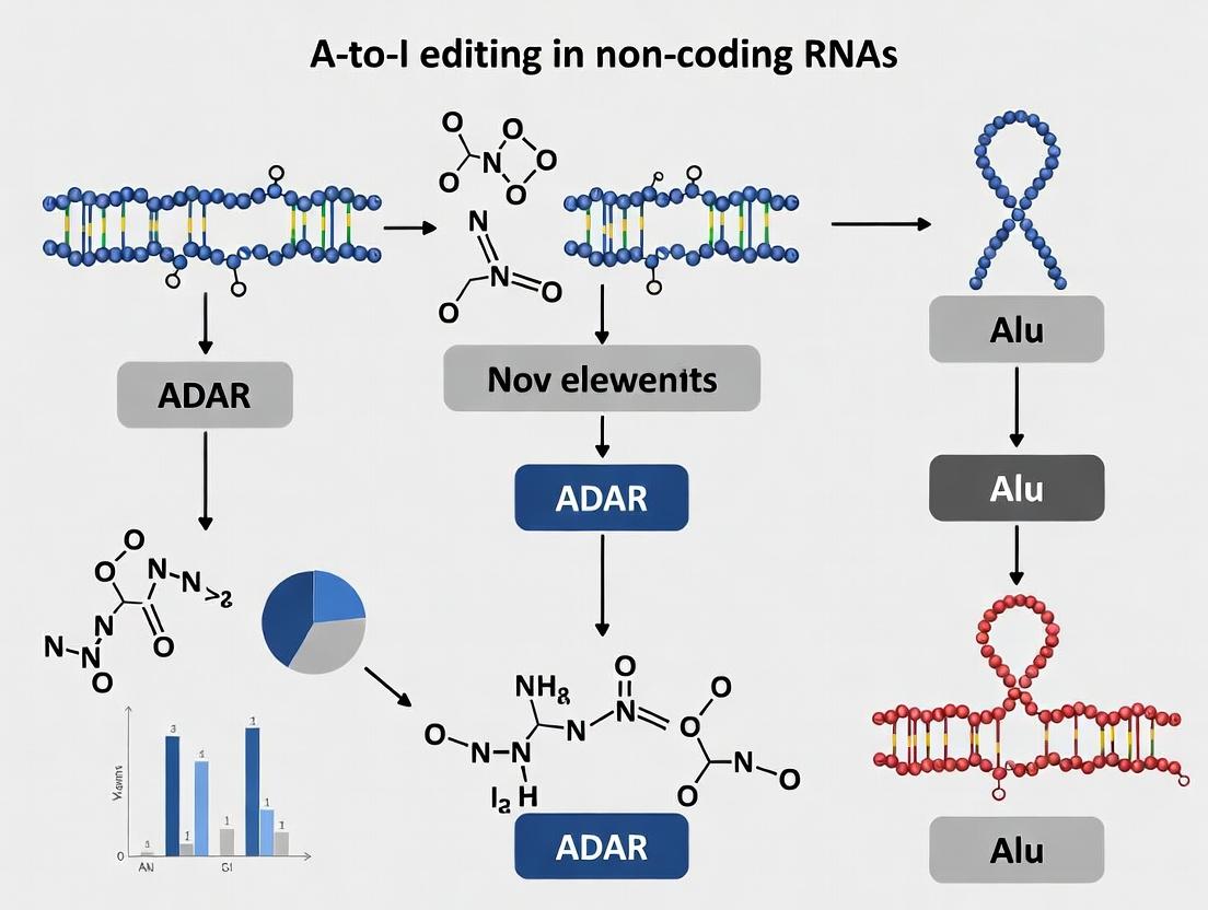
Within the context of a broader thesis on adenosine-to-inosine (A-to-I) editing in non-coding RNAs, this technical guide examines the unique propensity of Alu repetitive elements to undergo extensive RNA editing. This phenomenon is driven by the formation of double-stranded RNA (dsRNA) secondary structures, which serve as ideal substrates for adenosine deaminases acting on RNA (ADARs). The editing within Alus, predominantly located in introns and untranslated regions, has profound implications for transcriptome diversity, regulatory network modulation, and disease pathogenesis, presenting novel targets for therapeutic intervention.
A-to-I RNA editing, catalyzed by ADAR enzymes, is a prevalent post-transcriptional modification in metazoans. In humans, the majority of editing events occur within Alu elements, which are ~300-bp short interspersed nuclear elements (SINEs) numbering over one million copies. Their bi-directional transcription and inherent sequence complementarity allow them to form intramolecular or intermolecular dsRNA structures, creating the requisite context for ADAR recognition. This guide details the mechanistic, genomic, and functional reasons behind this targeting.
Mechanistic Drivers of Editing in Alu Elements
dsRNA Structure Formation
Alu elements are primate-specific retrotransposons characterized by two homologous monomers (left and right arms). When two Alus are inserted in opposite orientations in nearby genomic loci, their transcribed RNAs can form long, nearly perfectly complementary dsRNA stems. Even a single Alu can form intramolecular hairpins due to its internal dimeric structure.
Diagram 1: Alu dsRNA Formation Pathways
ADAR Enzyme Specificity and Catalysis
ADARs (ADAR1, ADAR2) possess dsRNA-binding domains (dsRBDs) that recognize the A-form helix of dsRNA without strict sequence specificity. Editing efficiency is influenced by neighboring nucleotides (preference for 5' U/A and 3' G), local dsRNA stability, and ADAR expression levels. Alu-rich regions provide extensive, if imperfect, dsRNA landscapes, making them genomic "hotspots."
Quantitative Landscape of Alu Editing
Recent high-throughput studies (e.g., from GTEx, TCGA consortiums) quantify the prevalence of Alu editing.
Table 1: Quantitative Profile of A-to-I Editing in Human Transcriptomes
| Metric | Approximate Value / Finding | Primary Source & Method |
|---|---|---|
| Total A-to-I Sites | >4.5 million in non-repetitive regions; >100 million in repetitive (Alu) regions | RNA-seq analysis with rigorous filtering (RADAR, REDIportal databases) |
| Fraction in Repetitive Elements | >95% of all editing events | Whole-transcriptome analysis of human tissues |
| Editing Frequency Range | 1% to >50% (site and tissue-dependent) | Deep sequencing of poly-A+ RNA |
| Tissues with Highest Editing | Brain, lung, heart, adrenal gland | GTEx project analysis |
| Key Influencing Factor | ADAR1 p110 & p150 isoform expression levels | qPCR & Western Blot correlation studies |
Experimental Protocols for Studying Alu Editing
Protocol: Genome-wide Identification of Editing Sites
Objective: To identify in vivo A-to-I editing sites within Alu elements from total RNA.
- RNA Extraction & DNase Treatment: Isolate total RNA using TRIzol reagent. Treat with Turbo DNase (Thermo Fisher) to remove genomic DNA contamination.
- Library Preparation: Use Illumina TruSeq Stranded Total RNA kit with Ribo-Zero Gold to deplete rRNA. Critical: Do not use random hexamers during cDNA synthesis if assessing editing in intronic Alus, as they capture unprocessed RNA. Use oligo(dT) for mature transcript analysis.
- High-Throughput Sequencing: Perform paired-end 150bp sequencing on Illumina NovaSeq platform to achieve >100 million reads per sample for sufficient coverage.
- Bioinformatics Pipeline:
- Alignment: Map reads to the human reference genome (GRCh38) using STAR aligner in 2-pass mode, soft-clipping allowed.
- Variant Calling: Use GATK's SplitNCigarReads and HaplotypeCaller, or specialized tools like REDItools2, with parameters set to retain mismatches in repetitive regions.
- Editing Site Filtering: Filter SNPs (dbSNP), known genomic variants (gnomAD), and sites with low coverage (<10 reads) or low editing frequency (<1%). Retain sites where A-to-G (forward strand) or T-to-C (reverse strand) mismatches predominate.
- Annotation: Annotate sites with respect to Alu elements (RepeatMasker track) and genomic features (Ensembl) using BEDTools.
Protocol: Validating Editing and Measuring Frequency
Objective: To validate candidate sites and quantify precise editing levels.
- cDNA Synthesis: Use gene-specific primers or random hexamers with SuperScript IV Reverse Transcriptase.
- PCR Amplification: Design primers flanking the candidate editing site, ensuring they are unique in the genome to avoid paralogous Alu co-amplification.
- Sanger Sequencing or Pyrosequencing:
- For Sanger: Purify PCR product, sequence, and analyze chromatogram peak heights (A vs G) using QuantPrime software.
- For higher accuracy: Use Pyrosequencing (Qiagen). Design a sequencing primer one base upstream of the editing site. Quantify the ratio of A and G incorporation via light emission intensity.
Functional Consequences & Therapeutic Relevance
Editing within Alus, primarily in introns and 3'UTRs, can alter RNA processing, stability, localization, and translation. Key implications include:
- Alternative Splicing: Edited Alus can create or disrupt splice site recognition motifs.
- miRNA Targeting: Editing in 3'UTRs can create or destroy microRNA binding sites, altering post-transcriptional regulation.
- Immunogenicity: Unedited Alu dsRNA is recognized by cytoplasmic sensors (MDA5, RIG-I) triggering interferon response. ADAR1 editing masks these dsRNAs, preventing autoimmunity.
- Disease Link: Dysregulated Alu editing is implicated in cancer (e.g., glioblastoma, leukemia), neurological disorders (e.g., ALS, epilepsy), and autoimmune diseases like Aicardi-Goutières syndrome.
Diagram 2: Functional Outcomes of Alu Editing
The Scientist's Toolkit: Research Reagent Solutions
Table 2: Essential Reagents for Alu RNA Editing Research
| Reagent / Material | Function & Application | Example Product / Assay |
|---|---|---|
| ADAR1/2-specific Antibodies | Immunoblotting, immunofluorescence to correlate enzyme expression with editing levels. | Rabbit anti-ADAR1 (Abcam, ab126745); Mouse anti-ADAR2 (Santa Cruz, sc-73409) |
| ADAR Chemical Inhibitor | Functional validation of editing-dependent phenotypes in vitro. | 8-Azaadenosine (inhibits ADAR activity) |
| Inosine-specific Chemical Detection | Direct detection and mapping of inosine sites in RNA. | Inosine Chemical Erasing (ICE) assay kit (NEB) |
| dsRNA-specific Antibody | Detection of unedited Alu dsRNA structures in cells. | J2 anti-dsRNA antibody (SCICONS) |
| ADAR Knockout/Knockdown Tools | Establish isogenic lines to study Alu editing loss. | CRISPR-Cas9 knockout kits (Synthego); siRNA pools (Dharmacon) |
| Reporter Plasmids with Alu inserts | Quantify editing efficiency on specific Alu sequences. | Custom pGL3 or pMINI vectors with inverted Alus flanking a reporter gene. |
| High-Fidelity Polymerase | Accurate amplification of GC-rich, repetitive Alu sequences for validation. | Q5 High-Fidelity DNA Polymerase (NEB) |
This whitepaper explores the functional consequences of Adenosine-to-Inosine (A-to-I) RNA editing, catalyzed primarily by ADAR enzymes, on key non-coding RNA (ncRNA) classes. The thesis is positioned within the broader landscape of A-to-I editing research, which recognizes Alu elements—abundant primate-specific retrotransposons—as major hotspots for editing. The formation of long, double-stranded RNA structures by inverted Alu repeats in non-coding regions provides the canonical substrate for ADARs. The editing events within these elements, particularly in introns and untranslated regions (UTRs), are now understood to have profound ripple effects on the biogenesis and function of miRNAs, siRNAs, and lncRNAs, thereby expanding the "functional repertoire" of the transcriptome and proteome with implications for cellular regulation and disease.
Impact on miRNA Biogenesis and Function
A-to-I editing can impact microRNAs at multiple stages, from pri-miRNA processing to target recognition.
2.1 Mechanisms of Intervention:
- Editing within the Seed Region (Positions 2-8): Alters complementarity to target mRNAs, completely redirecting the miRNA's target repertoire. An I is read as a G by the cellular machinery, changing A:U pairings to I:C (effectively G:C) matches.
- Editing in the Pre-miRNA Stem: Can affect processing by Drosha/Dicer enzymes by altering the double-stranded structure's stability or by creating bulges that inhibit cleavage.
- Editing in Flanking Sequences: May influence the efficiency of primary miRNA (pri-miRNA) cleavage by the Microprocessor complex (Drosha-DGCR8).
2.2 Quantitative Data Summary: Table 1: Documented Impacts of A-to-I Editing on Specific miRNAs
| miRNA | Editing Site | Effect on Processing | Effect on Target Recognition | Biological Context |
|---|---|---|---|---|
| pri-miR-142 | Multiple sites in stem | Strong inhibition of Drosha & Dicer processing (~80% reduction) | N/A (miRNA is degraded) | Hematopoietic cells; immune regulation |
| miR-376a-5p | Seed region (pos 4) | Minimal effect | Shift from targeting PRPS1 to AUTS2 | Brain; cancer metabolism |
| miR-200b | 3' flanking region (Alu) | Moderate reduction (~40%) in pri-to-pre conversion | Altered mature levels affect EMT targets | Cancer cell lines |
Diagram 1: A-to-I Editing Pathways in miRNA Biogenesis
Disruption of Endogenous siRNA Silencing
Endogenous siRNAs (endo-siRNAs) often derive from transposable elements like Alus. Their silencing function is tightly linked to perfect complementarity.
3.1 Core Mechanism: A-to-I editing introduces I:U (or I:A) mismatches within the duplex formed by the endo-siRNA and its transposon target mRNA. These mismatches disrupt perfect complementarity, leading to:
- Reduced efficiency of Argonaute 2 (Ago2)-mediated cleavage.
- Potential recruitment of different Argonaute proteins (e.g., Ago1 in flies).
- Overall attenuation of silencing, potentially leading to increased transposable element activity, a hallmark of genomic instability.
3.2 Experimental Protocol: Assessing siRNA Silencing Disruption
- Objective: Quantify the impact of A-to-I editing on the silencing efficacy of a specific endo-siRNA.
- Methodology:
- Construct Design: Create dual-luciferase reporter plasmids. The Firefly luciferase gene is fused to the target sequence (e.g., an Alu element) in its sense or antisense orientation. The Renilla luciferase serves as an internal control.
- Editing Modulation: Co-transfect reporter constructs into HeLa cells with either:
- ADAR1/2 overexpression plasmids.
- siRNA against ADAR1/2 (or use ADAR1-/- cell lines).
- Silencing Trigger: Co-transfect a plasmid expressing the cognate endo-siRNA precursor.
- Measurement: After 48h, perform a dual-luciferase assay. Normalize Firefly luminescence to Renilla.
- Analysis: Compare silencing efficiency (reduction in Firefly signal) in ADAR-high vs. ADAR-low conditions. Deep sequencing of the target site can confirm editing levels.
Modulation of lncRNA Function
lncRNAs are frequently edited due to their enrichment in Alu elements. Editing can alter their function through several mechanisms.
4.1 Functional Consequences:
- Structural Remodeling: I-U mismatches destabilize dsRNA helices, potentially causing large-scale refolding of the lncRNA and altering its interaction surfaces.
- Protein Binding: Creation/disruption of protein binding motifs (e.g., for STAU1, NF90/NF110) affects lncRNA-protein complex (RNP) formation.
- Subcellular Localization: Altered RNP composition can change the lncRNA's trafficking.
- Stability: Edited transcripts may be subject to different degradation pathways.
4.2 Quantitative Data Summary: Table 2: Examples of A-to-I Editing Effects on lncRNAs
| lncRNA | Editing Level (Tissue) | Key Consequence | Functional Outcome |
|---|---|---|---|
| XIST | Moderate (Brain) | Alters interaction with PRC2 complex | Potential modulation of X-chromosome inactivation |
| NEAT1 | High (Multiple) | Affects paraspeckle architecture & protein retention | Modulates stress response & miRNA sequestration |
| MALAT1 | Low (Cancer) | Potential change in protein partners | Linked to alternative splicing regulation |
Diagram 2: Editing-Induced Functional Modulation of lncRNAs
The Scientist's Toolkit: Key Research Reagent Solutions
Table 3: Essential Reagents for Investigating Editing in ncRNAs
| Reagent / Material | Provider Examples | Function in Research |
|---|---|---|
| ADAR1/2 Knockout Cell Lines | ATCC, Academia | Isolate the effect of specific ADAR enzymes on editing events in ncRNAs. |
| Catalytically Dead ADAR Mutants | Plasmid repositories (Addgene) | Used as controls to distinguish between editing-dependent and -independent effects of ADAR proteins. |
| Inosine-Specific RNA Sequencing Kits | GL Sciences, NEB | Methods like ICE-seq or CLEAR-CLIP to precisely map inosine sites at transcriptome-wide scale. |
| Selective ADAR Inhibitors | Medicinal Chemistry Suppliers | Probe the acute functional consequences of loss of editing (e.g., 8-Azaadenosine derivatives). |
| Antibodies: ADAR1 (p150/p110), ADAR2 | Santa Cruz, Abcam, Cell Signaling | Validate protein expression, perform RIP-seq to identify ADAR-bound ncRNAs. |
| Dual-Luciferase Reporter Assay Systems | Promega | Quantify the impact of editing on miRNA/siRNA targeting efficiency or lncRNA regulatory function. |
| Stable Isotope-Labeled Nucleosides | Cambridge Isotope Labs | For metabolic tracing of RNA turnover to assess editing effects on ncRNA stability. |
| High-Fidelity RT Enzymes for I-discrimination | Thermo Fisher, NEB | Enzymes like SuperScript IV for accurate cDNA synthesis from inosine-containing RNA for validation. |
Within the broader thesis on adenosine-to-inosine (A-to-I) RNA editing in non-coding RNAs and repetitive Alu elements, this whitepaper details the profound biological significance of this process. Catalyzed primarily by adenosine deaminases acting on RNA (ADARs), A-to-I editing is a critical post-transcriptional mechanism that directly modulates innate immune responses, prevents pathological auto-inflammation, and safeguards genomic stability. The editing of Alu elements, which are abundant in introns and untranslated regions, is central to these functions, acting as a key distinguisher between self and non-self nucleic acids.
Roles in Innate Immunity and Auto-inflammation Prevention
A-to-I editing of endogenous RNA structures, particularly double-stranded RNA (dsRNA) formed by inverted Alu repeats, is a primary mechanism for preventing aberrant activation of cytosolic innate immune sensors.
Mechanistic Insight: Unedited or minimally edited endogenous dsRNA can be recognized as foreign by cytoplasmic pattern recognition receptors (PRRs) such as MDA5 (IFIH1) and PKR (EIF2AK2). MDA5 activation triggers a type I interferon (IFN) response, while PKR phosphorylation halts global translation. ADAR1, through its deaminase activity, introduces I-U mismatches that disrupt the perfect dsRNA structure, effectively "marking" it as "self" and preventing PRR activation.
Key Experimental Protocol: Assessing ADAR1-KO Immune Activation
- Objective: To demonstrate the essential role of ADAR1 p150 in preventing MDA5-mediated auto-inflammation.
- Methodology:
- Generate Adar1 p150-specific knockout (KO) or Adar1 null mouse embryonic fibroblasts (MEFs) using CRISPR-Cas9.
- Transfert cells with a luciferase reporter plasmid under the control of an interferon-stimulated response element (ISRE).
- Treat cells with a synthetic dsRNA analog (e.g., poly(I:C)) to mimic viral infection or simply assay baseline activation in KO cells.
- Measure luciferase activity as a readout of IFN pathway activation.
- Perform co-treatment with an MDA5-specific inhibitor (e.g., compound C16) or use siRNA knockdown of Mda5. A rescue (reduced luciferase signal) confirms MDA5-dependent signaling.
- Validate by quantifying downstream interferon-stimulated gene (ISG) expression (e.g., Isg15, Oas1a) via qRT-PCR and by immunoblotting for phosphorylated PKR.
Quantitative Data Summary:
Table 1: Innate Immune Activation in ADAR1-Deficient Systems
| Cell Type / Model | Intervention | Key Metric | Result (vs. Wild-Type) | Reference (Example) |
|---|---|---|---|---|
| Human HEK293T | ADAR1 siRNA Knockdown | ISG Transcript Levels (RNA-seq) | 10-50 fold increase | PMID: 28798046 |
| Mouse Adar1 p150-/- MEFs | Baseline (No Treatment) | ISRE-Luciferase Activity | ~8-fold increase | PMID: 28798046 |
| Mouse Adar1 p150-/- MEFs | + MDA5 Inhibitor (C16) | ISRE-Luciferase Activity | ~70% reduction | PMID: 28798046 |
| Patient (AGS-like) | ADAR1 Loss-of-Function Mutation | Serum IFN-α Activity | Consistently Elevated | PMID: 35303430 |
Diagram: ADAR1-Mediated Prevention of dsRNA Immune Sensing
Role in Maintaining Genomic Stability
Beyond immune regulation, A-to-I editing in non-coding regions influences genomic stability through two primary avenues: modulating RNA structure and function, and indirectly influencing DNA integrity.
1. Preventing R-Loop Associated Instability: Unedited dsRNA structures can favor the formation of R-loops (RNA-DNA hybrids with a displaced single-stranded DNA). Persistent R-loops are major sources of DNA double-strand breaks (DSBs) and genomic instability. ADAR1 editing destabilizes dsRNA, reducing R-loop propensity.
2. Editing-Dependent microRNA Regulation: Editing in pri-miRNA or mature miRNA seed regions can alter target specificity, potentially regulating the expression of genes involved in DNA damage repair (e.g., ATM, BRCA1/2 pathways).
Key Experimental Protocol: Quantifying R-Loop Formation in ADAR1-Deficient Cells
- Objective: To measure the increase in R-loops upon loss of ADAR1 function.
- Methodology (DRIP-seq - DNA:RNA Hybrid Immunoprecipitation Sequencing):
- Extract genomic DNA from isogenic wild-type and ADAR1-KO cells under native conditions using gentle lysis to preserve RNA-DNA hybrids.
- Fragment DNA by restriction digest (e.g., with BsrGI, SspI, XbaI).
- Immunoprecipitate R-loop-containing fragments overnight at 4°C using the S9.6 monoclonal antibody (specific for RNA-DNA hybrids).
- Wash beads stringently, elute, and purify the immunoprecipitated DNA.
- Prepare libraries for next-generation sequencing (DRIP-seq) or analyze specific loci of interest (e.g., sites with Alu clusters) via qPCR (DRIP-qPCR).
- Validate by treating a parallel sample with purified RNase H prior to IP, which degrades RNA in hybrids and should abolish S9.6 signal.
Quantitative Data Summary:
Table 2: Genomic Instability Phenotypes Linked to ADAR1 Deficiency
| Phenotype / Assay | ADAR1-WT Cells | ADAR1-KO/Deficient Cells | Measurement Technique |
|---|---|---|---|
| R-Loop Abundance | Baseline Level | 2-4 fold increase | DRIP-qPCR at Alu-rich loci |
| DNA Damage Foci | Low # of γH2AX/53BP1 foci | Significantly Increased # of foci | Immunofluorescence Microscopy |
| Chromosomal Aberrations | Normal Karyotype | Increased breaks, gaps, fusions | Metaphase Spread Analysis |
| Transcription-Replication Conflicts | Minimal | Increased co-localization of RNAPII & PCNA | Proximity Ligation Assay (PLA) |
Diagram: Consequences of ADAR1 Loss on Genomic Stability
The Scientist's Toolkit: Key Research Reagents
Table 3: Essential Reagents for Studying A-to-I Editing in Immunity & Genomics
| Reagent / Material | Provider Examples | Function in Research |
|---|---|---|
| S9.6 Monoclonal Antibody | Kerafast, Sigma-Aldrich, Millipore | Gold-standard for immunoprecipitating or detecting RNA-DNA hybrids (R-loops) in techniques like DRIP-seq and immunofluorescence. |
| Poly(I:C) (HMW) | InvivoGen, Sigma-Aldrich | Synthetic dsRNA analog used to mimic viral infection and stimulate MDA5/RIG-I and PKR pathways in vitro and in vivo. |
| C16 (MDA5 Inhibitor) | Merck Millipore, Cayman Chemical | A selective inhibitor of MDA5 (IFIH1) oligomerization, used to confirm MDA5-dependent signaling in ADAR1-deficient models. |
| RNase H | NEB, Thermo Fisher | Enzyme that specifically degrades the RNA strand of an RNA-DNA hybrid. Critical negative control for R-loop assays (S9.6 based). |
| Anti-phospho-PKR (Thr446) Ab | Abcam, Cell Signaling Tech | Antibody to detect activated (phosphorylated) PKR via immunoblotting, a direct readout of innate immune activation by dsRNA. |
| ADAR1-Specific siRNA/sgRNA | Dharmacon, Sigma, IDT | For targeted knockdown (siRNA) or knockout (sgRNA for CRISPR) of ADAR1 in cell lines to establish functional models. |
| ISRE-Luciferase Reporter | Promega, InvivoGen | Plasmid reporter system to quantify activation of the interferon-stimulated response element pathway. |
| γH2AX (Ser139) Antibody | Millipore, Abcam, CST | Marker for DNA double-strand breaks. Used in immunofluorescence or immunoblotting to assess genomic instability. |
Adenosine-to-inosine (A-to-I) RNA editing, catalyzed primarily by ADAR enzymes, is a critical post-transcriptional modification. This whitepaper examines the evolutionary dynamics of A-to-I editing sites, with a focus on their conservation and diversification across primate lineages. The analysis is framed within the broader thesis that editing in non-coding regions, particularly within Alu repetitive elements, plays a significant regulatory role, influencing transcriptome diversity and potentially contributing to primate-specific adaptations and neurological complexity.
Current Landscape of Primate A-to-I Editing Research
Recent studies leveraging deep sequencing and comparative genomics across primate species—including humans, chimpanzees, gorillas, orangutans, and macaques—have mapped millions of editing sites. Key findings indicate a dual evolutionary trend: a core set of highly conserved, functionally important sites, primarily in coding regions, and a vast, rapidly evolving set of sites within non-coding Alu elements.
Table 1: Quantitative Overview of A-to-I Editing Sites Across Primates
| Primate Species | Total Editing Sites (approx.) | Alu-Associated Sites (%) | Conserved Sites (Pan-Primate) | Species-Specific Sites | Reference (Latest) |
|---|---|---|---|---|---|
| Human (H. sapiens) | ~4.6 million | >97% | ~35,000 | >4 million | PMID: 36703192 (2023) |
| Chimpanzee (P. troglodytes) | ~3.8 million | >96% | ~34,500 | Species-specific expansions | PMID: 36163281 (2022) |
| Rhesus Macaque (M. mulatta) | ~1.2 million | ~92% | ~27,000 | High in 3' UTRs | PMID: 36703192 (2023) |
| Gorilla (G. gorilla) | Data emerging | >95% | Under study | Under study | Preprint: BioRxiv 2024 |
| Evolutionary Insight | Positive correlation with Alu element abundance | Driver of diversification | Enriched in genes for neural & synaptic function | Potential source of regulatory innovation |
Core Hypotheses and Mechanistic Drivers
The conservation and diversification patterns are driven by several interconnected factors:
- Conservation Pressure: Editing sites within coding sequences (e.g., in genes like GRIA2, CYFIP2) are often highly conserved due to their essential role in protein function and neuronal signaling.
- Alu-Driven Diversification: The primate-specific expansion of Alu elements provides a massive substrate for ADARs. Editing within these inverted repeat Alu pairs forms double-stranded RNA (dsRNA) structures. The rapid evolution of Alu sequences and their genomic positions leads to extensive, lineage-specific editing site creation and loss.
- ADAR Enzyme Evolution: While ADAR1 and ADAR2 proteins are themselves conserved, changes in their expression patterns, splicing isoforms, and regulatory networks across primates influence editing site profiles.
- Selection on RNA Structure: Evolutionary selection acts on the underlying dsRNA structure required for editing, not necessarily on the specific edited adenosine itself, allowing for sequence turnover while maintaining editable structures.
Experimental Protocols for Cross-Primate Editing Analysis
Below are detailed methodologies for key experiments generating data in this field.
Protocol: Comparative Editing Site Identification from RNA-Seq
Objective: To identify and compare A-to-I editing sites across multiple primate species from bulk tissue RNA sequencing data.
- Sample Collection & Sequencing: Obtain poly-A+ RNA from matched tissues (e.g., prefrontal cortex, liver) from human, chimpanzee, bonobo, gorilla, orangutan, and macaque. Sequence on an Illumina platform to generate ≥100M paired-end 150bp reads per sample.
- Bioinformatic Processing:
- Alignment: Trim adapters (Trimmomatic). Align reads to the respective reference genome (hg38, panTro6, etc.) using a splice-aware aligner (STAR) in 2-pass mode.
- Variant Calling: Use a specialized RNA editing caller (e.g., REDItools2, JACUSA2) to identify A-to-G mismatches from the reference genome. Critical Parameter: Disable SNP filters from standard DNA variant callers.
- Strand-Specific Filtering: Apply stringent filters: i) Remove known SNPs (dbSNP, species-specific SNP databases). ii) Require minimum read depth (e.g., 10x). iii) Require presence of supporting reads on both strands. iv) Remove sites in simple repeats and homopolymer regions.
- Cross-Species Analysis: LiftOver genomic coordinates of editing sites to a common reference (e.g., hg38). Define "orthologous sites" as those where the genomic adenosine is present in all species. Conservation rate = (# of species with editing at orthologous site) / (total # of species analyzed).
Protocol: Validation and Functional Assessment via Mass Spectrometry
Objective: To validate editing events at the protein level and assess cross-species conservation of recoding events.
- Target Selection: Select candidate conserved editing sites in coding regions (e.g., the Q/R site in GRIA2).
- Sample Preparation: Isolate protein from primate brain tissues. Perform tryptic digestion.
- LC-MS/MS Analysis: Analyze peptides on a high-resolution tandem mass spectrometer (e.g., Orbitrap Fusion). Use a targeted parallel reaction monitoring (PRM) method for peptides spanning the edited site.
- Data Analysis: Search spectra against a custom database containing both the unedited (A, coded as lysine, K) and edited (I, coded as arginine, R) peptide sequences. Quantify the ratio of edited to unedited peptide based on extracted ion chromatograms.
Visualizing Pathways and Workflows
The Scientist's Toolkit: Research Reagent Solutions
Table 2: Essential Reagents for Primate A-to-I Editing Research
| Reagent / Material | Function & Application in Primate Editing Studies | Example Product / Assay |
|---|---|---|
| Species-Specific ADAR Antibodies | For measuring ADAR protein expression and localization via western blot or IHC across primate tissues. Validated cross-reactivity is critical. | Rabbit anti-ADAR1 (p150) antibody (Abcam, cat# ab126745); requires validation for non-human primates. |
| Cross-Reactive RNA Immunoprecipitation (RIP/CLIP) Kits | To identify ADAR-bound RNA targets in primate cell lines or tissue lysates. Optimized buffers for RNase treatment and dsRNA recovery are key. | Magna RIP RNA-Binding Protein Immunoprecipitation Kit (MilliporeSigma). |
| Long-Read RNA Sequencing Kits | To resolve full-length transcripts containing clustered Alu edits and haplotype phasing, crucial for understanding cis-editing relationships. | Oxford Nanopore Technologies cDNA-PCR Sequencing Kit (SQK-PCS111). |
| Synthetic dsRNA Oligo Standards | For creating calibration curves in mass spectrometry validation of recoding events or for in vitro ADAR activity assays with primate enzyme extracts. | Custom RNA oligos with defined I content (e.g., from IDT). |
| Primate Brain Tissue Lysate Arrays | For high-throughput screening of editing levels at conserved sites across multiple individuals and species in a standardized format. | BioChain Primate Brain Tissue Lysate Array (Frontal Cortex). |
| ADAR Activity Reporter Plasmids | To compare the functional activity of ADAR isoforms cloned from different primate species in an isogenic cellular background (e.g., HEK293 ADAR KO). | pEGFP-ADAR reporter with a synthetic editable stop codon (Addgene, #111166). |
| Selective ADAR Inhibitors/Activators | To probe the functional consequences of acute editing modulation in primate-derived neural progenitor cells or organoids. | 8-Azaadenosine (inhibitor); specific small-molecule activators under development. |
From Detection to Function: Methodologies for Studying A-to-I Editing in ncRNAs
Adenosine-to-inosine (A-to-I) RNA editing, catalyzed primarily by ADAR (Adenosine Deaminase Acting on RNA) enzymes, is a crucial post-transcriptional modification. In the human genome, this editing is overwhelmingly concentrated within repetitive Alu elements, especially in non-coding regions like introns and untranslated regions (UTRs). Inosines are interpreted as guanosines by cellular machinery, potentially altering RNA structure, stability, localization, and splicing. Research within this thesis focuses on elucidating the functional impact of A-to-I editing within non-coding RNAs and Alu elements on gene regulatory networks and its implications for human disease and therapeutic targeting. High-throughput RNA sequencing (RNA-Seq) is the principal method for genome-wide detection of editing sites, necessitating robust bioinformatics pipelines.
Core Bioinformatics Tools for A-to-I Editing Detection
The accurate identification of A-to-I editing events from RNA-Seq data presents significant challenges, including distinguishing true editing from single nucleotide polymorphisms (SNPs), sequencing errors, and alignment artifacts. Two specialized tools are central to this field.
REDItools
A comprehensive suite of Python scripts designed for the identification of RNA editing events using aligned RNA-Seq data (BAM files) and reference genome data. It is particularly adept at handling the complexities of repetitive regions like Alu elements.
Key Methodology:
- Data Input: Requires BAM alignment files (RNA-Seq) and a reference genome (FASTA). A database of known SNPs (e.g., dbSNP) is essential for filtration.
- Position Identification: Iterates over all genomic positions covered by RNA-Seq reads.
- Base Counting: For each position, it counts the number of observed A, C, G, T bases from aligned RNA reads, considering mapping quality and base quality scores.
- Statistical Filtering: Employs multiple filters:
- SNP Filter: Removes positions matching known SNPs.
- Strandness Filter: For candidate A-to-G (T-to-C on cDNA) changes, ensures edits are consistent with the strandedness of sequencing.
- Alignment Filter: Uses paired DNA-Seq data (if available) from the same sample to confirm the genomic reference base is adenosine and rule out genomic variants.
- Statistical Test: Applies a binomial test to assess if the observed edited base count is significantly higher than expected from the sequencing error rate.
- Output: Produces detailed tables of candidate editing sites with read coverage, edited read counts, frequency, and p-values.
SPRINT (SNP-free RNA Editing Identification Toolkit)
A highly efficient, alignment-free tool that identifies RNA editing directly from raw RNA-Seq reads (FASTQ), circumventing alignment biases in repetitive regions—a critical advantage for Alu-rich areas.
Key Methodology:
- Reference Preparation: Builds an "editome" reference by converting all annotated adenosines (A) in the reference genome to guanosines (G), creating an "A-to-I altered" reference.
- Alignment-free Read Mapping:
- Raw RNA-Seq reads are separately aligned to the standard reference genome and the "A-to-I altered" reference using ultrafast aligners (e.g., Bowtie, HISAT2).
- A read that aligns uniquely and with higher quality to the altered reference (matching a 'G') than to the standard reference (matching an 'A') provides evidence for an editing event.
- Clustering and Filtering: Candidate sites are clustered based on genomic proximity. Stringent filters are applied, including:
- Removal of sites in simple repeats.
- Filtering against known SNP databases.
- Requiring a minimum number of supporting reads and a minimum editing level.
- Output: A list of high-confidence RNA editing sites.
Table 1: Comparison of REDItools and SPRINT
| Feature | REDItools | SPRINT |
|---|---|---|
| Core Approach | Alignment-based (post-BAM analysis) | Alignment-free (raw read analysis) |
| Input | Aligned BAM files | Raw FASTQ files |
| Handling Repetitive Regions (Alu) | Can be challenging; requires careful alignment and filtering | Excellent; avoids alignment bias in repeats |
| Dependency on DNA-Seq | Highly recommended for high-confidence calls | Not required |
| Speed | Moderate to Slow | Fast |
| Primary Output | Tables of editing sites with statistical metrics | Tables of high-confidence editing sites |
A Standard RNA-Seq Analysis Pipeline for A-to-I Editing Discovery
The following integrated protocol details a comprehensive workflow, incorporating both tools for validation.
Experimental Protocol: From Tissue to Editing Sites
A. Sample Preparation & Sequencing
- Material: Tissue/cell lines of interest (e.g., neuronal tissues, cancer cell lines with high ADAR expression).
- RNA Extraction: Use TRIzol or column-based kits with DNase I treatment to remove genomic DNA contamination. Critical: Preserve RNA integrity (RIN > 8).
- Library Construction: Use stranded, poly-A-selection or rRNA-depletion RNA-Seq library prep kits. Paired-end sequencing (2x150bp) is recommended for better alignment.
- Sequencing: Perform deep sequencing on an Illumina platform. Minimum recommended depth: 50-100 million reads per sample. Optional but powerful: Sequence genomic DNA from the same sample/organism in parallel.
B. Computational Analysis Workflow
Diagram 1: RNA-Seq Analysis Workflow for A-to-I Editing.
Step-by-Step Protocol:
- Quality Control: Use
FastQCto assess read quality. Trim adapter sequences and low-quality bases usingTrimmomaticorcutadapt. - Alignment (for REDItools path): Align cleaned RNA-Seq reads to the human reference genome (e.g., GRCh38) using a splice-aware aligner like
HISAT2orSTAR. Generate a sorted BAM file usingsamtools. - REDItools Execution:
SPRINT Execution:
Integration: Intersect the high-confidence outputs from REDItools (DNA-filtered) and SPRINT using
bedtools intersectto generate a robust, consensus set of editing sites.- Downstream Analysis: Quantify editing levels (edited reads/total reads), perform differential editing analysis between sample groups (using tools like
JACUSA2or custom R scripts), and annotate sites relative to Alu elements, genes, and other genomic features.
The Scientist's Toolkit: Essential Research Reagent Solutions
Table 2: Key Reagents and Materials for A-to-I Editing Research
| Item | Function/Description | Example/Supplier |
|---|---|---|
| High-Fidelity RNA Extraction Kit | Isolates high-integrity, DNA-free total RNA, critical for accurate representation of the transcriptome. | Qiagen RNeasy, Zymo Research Direct-zol |
| Stranded mRNA-Seq Library Prep Kit | Preserves strand information, essential for correctly assigning edits to transcribed strands. | Illumina Stranded mRNA Prep, NEBNext Ultra II Directional |
| rRNA Depletion Kit | Enriches for non-polyadenylated transcripts (e.g., some non-coding RNAs), broadening editing landscape discovery. | Illumina Ribo-Zero Plus, NEBNext rRNA Depletion |
| ADAR-specific Antibodies | For immunoprecipitation (IP) or western blotting to assess ADAR protein expression and activity levels. | Santa Cruz Biotechnology (sc-73408), Abcam (ab126745) |
| SINE/Alu Element Probes | For fluorescence in situ hybridization (FISH) to visualize Alu-rich genomic loci or transcripts. | Custom-designed probes from Biosearch Technologies |
| Inosine-Specific Chemical Reagents | Compounds like inosine-6-azide enable click-chemistry-based labeling and pulldown of inosine-containing RNAs. | Published in Nat. Biotechnol. 2017; available from specialized chemical suppliers. |
| Positive Control RNA Spike-ins | Synthetic RNA oligos with known A-to-I edits to benchmark editing detection sensitivity and specificity of wet-lab & computational pipelines. | Custom synthesized from IDT or Sigma. |
Signaling Pathways Involving ADAR andAluEditing
A-to-I editing in Alu elements within non-coding RNAs can influence critical cellular pathways.
Diagram 2: ADAR-Alu Editing in Innate Immune Regulation.
Adenosine-to-inosine (A-to-I) RNA editing, catalyzed by the ADAR enzyme family, is a critical post-transcriptional modification. Within the broader thesis on A-to-I editing in non-coding RNAs and Alu elements, quantifying editing levels is fundamental. This guide details the computational and experimental frameworks for calculating site-specific editing frequencies and analyzing heterogeneity, which is essential for understanding the regulatory impact of editing in repetitive elements and its potential implications in disease and drug development.
Core Quantitative Metrics and Data Presentation
Accurate quantification relies on specific metrics derived from next-generation sequencing (NGS) data.
Table 1: Core Metrics for Quantifying A-to-I Editing
| Metric | Formula / Description | Interpretation |
|---|---|---|
| Editing Frequency (EF) | EF = (Number of 'G' reads) / (Number of 'A' + 'G' reads) * 100% |
Percentage of edited transcripts at a specific genomic coordinate. |
| Editing Index (EI) | EI = (Total edited adenosines in region) / (Total candidate adenosines) |
Global measure of editing activity across a defined region (e.g., an Alu element). |
| Site-Specific Heterogeneity Index (SHI) | SHI = 1 - (∑(p_i^2)) where p_i is the frequency of each editing pattern (e.g., unedited, single-site edited, multi-site edited). |
Measures the diversity of editing combinations across multiple sites within a single read (0=homogeneous, 1=highly heterogeneous). |
| Read-Support Depth | Total number of sequencing reads covering the locus. | Filters low-confidence calls; typically >10-20 reads for reliable quantification. |
| Binomial P-value | Probability of observing the 'G' count by chance, given sequencing error rate. | Identifies significant editing sites (P < 0.05 after multiple testing correction). |
Table 2: Representative Editing Levels in Human Tissues (Recent Studies)
| Tissue / Cell Type | Alu Element EI Range | High-EF Site Example (Gene/Region) | Typical SHI Value |
|---|---|---|---|
| Brain Cortex | 0.15 - 0.25 | GRIA2 (Q/R site) EF: ~95% | 0.4 - 0.7 |
| Liver | 0.05 - 0.12 | AZIN1 (Antizyme inhibitor) EF: ~50% | 0.3 - 0.6 |
| Primary Neutrophils | < 0.05 | Alu junctions in ncRNAs | 0.1 - 0.3 |
| Cancer Cell Lines | Highly variable (0.02-0.20) | Depends on ADAR1/2 expression | Often elevated |
Detailed Experimental Protocols
Protocol: RNA-Seq Library Preparation for Editing Detection
Goal: Generate strand-specific, ribosomal RNA-depleted RNA-seq libraries.
- RNA Extraction: Use TRIzol or column-based kits with DNase I treatment. Assess integrity (RIN > 7).
- rRNA Depletion: Use riboPOOL or Ribo-Zero kits to enrich for ncRNAs and mRNA.
- Strand-Specific Library Prep: Use kits like Illumina's TruSeq Stranded Total RNA. Fragmentation (200-300 bp), reverse transcription with actinomycin D to prevent spurious second-strand synthesis, and incorporation of dUTP in the second strand.
- High-Depth Sequencing: Perform 150bp paired-end sequencing on Illumina platforms. Target >50 million read pairs per sample to robustly detect editing in repetitive Alu regions.
Protocol: Computational Pipeline for Editing Quantification
Goal: Identify and quantify A-to-I editing sites from RNA-seq data.
- Preprocessing & Alignment:
- Trim adapters using Trimmomatic.
- Map reads to the reference genome (e.g., GRCh38) using a splice-aware aligner like STAR in 2-pass mode. Crucially, disable soft-clipping for better mapping of hyper-edited reads.
- Duplicate Marking: Use Picard Tools to mark PCR duplicates.
- Editing Site Identification:
- Use GATK SplitNCigarReads to handle splice junctions.
- Perform base recalibration and variant calling with GATK HaplotypeCaller in RNA-seq mode.
- Extract A-to-G (T-to-C on cDNA strand) mismatches.
- Filtering & Quantification:
- Filter 1: Remove known SNPs (dbSNP, 1000 Genomes Project).
- Filter 2: Require minimum read depth (e.g., 10) and binomial p-value < 0.05.
- Filter 3: For Alu sites, require editing in opposite-strand overlapping Alu elements.
- Quantification: For each passing site, compute Editing Frequency (EF) using
samtools mpileupor custom scripts.
- Heterogeneity Analysis: Use tools like SAILOR or custom Python/R scripts to analyze co-editing patterns within single reads across multiple sites to calculate the Site-Specific Heterogeneity Index (SHI).
Visualization of Workflows and Pathways
Diagram 1: Computational workflow for quantifying RNA editing.
Diagram 2: ADAR pathway and functional consequences of editing.
The Scientist's Toolkit: Research Reagent Solutions
Table 3: Essential Reagents and Tools for A-to-I Editing Research
| Item | Function & Application | Example Product/Kit |
|---|---|---|
| RiboCOP rRNA Depletion Kit | Depletes cytoplasmic and mitochondrial rRNA, crucial for ncRNA and Alu-transcript analysis. | RiboCOP (Human/Mouse) |
| Strand-Specific RNA Library Prep Kit | Preserves strand information, essential for identifying the edited transcript. | Illumina TruSeq Stranded Total RNA |
| Recombinant Human ADAR Proteins | For in vitro editing assays to validate enzyme specificity and kinetics. | Novoprotein ADAR1 p110 (Cat# CR92) |
| ADAR1/2 siRNA or CRISPRi Kits | For functional knockdown/knockout studies to assess editing dependency. | Dharmacon ON-TARGETplus siRNA |
| Inosine-Specific Chemical Reagent | CMC treatment for biochemical validation of inosine sites. | N-Cyclohexyl-N'-(2-morpholinoethyl)carbodiimide |
| High-Fidelity PCR & Cloning Kit | For amplifying and cloning edited sequences for validation via Sanger sequencing. | NEB Q5 Hot Start Master Mix |
| Editing-Specific Bioinformatics Pipeline | Containerized pipeline for reproducible detection/quantification. | REDItools2 or JACUSA2 Docker Image |
| Long-Read Sequencing Kit | For resolving complex, co-editing patterns within single RNA molecules. | Oxford Nanopore Direct RNA Sequencing Kit |
This technical guide addresses a critical experimental gap in the broader thesis on adenosine-to-inosine (A-to-I) RNA editing in non-coding RNAs and repetitive Alu elements. While bioinformatics can predict millions of editing sites, functional validation is essential to distinguish consequential events from transcriptional noise. This document provides a framework for deploying functional assays that mechanistically connect a specific editing event to an altered RNA structure, a change in protein-RNA interaction, and ultimately, a measurable cellular phenotype. This causal linkage is fundamental for understanding the role of editing in regulation, disease, and as a potential therapeutic target.
Table 1: Common A-to-I Editing Effects and Associated Assay Readouts
| Editing Consequence | Key Measurable Output | Typical Quantitative Readout (Example Range) | Primary Assay Category |
|---|---|---|---|
| Altered RNA Secondary Structure | Free Energy Change (ΔΔG) | -5 to +2 kcal/mol | In-line probing, SHAPE-MaP |
| Altered Protein Binding (RBP) | Binding Affinity (Kd) | 10 nM - 1 µM shift | RIP-seq, CLIP variants, EMSA |
| Altered Protein Binding (dsRNA Sensors) | Immune Pathway Activation | 2- to 20-fold IFN/ISG expression | Luciferase reporter, qPCR |
| Altered microRNA:mRNA Interaction | Gene Silencing Efficiency | 20-80% change in target repression | Dual-luciferase 3'UTR reporter |
| Altered RNA Stability (Half-life) | RNA Decay Rate (t1/2) | 1- to 4-fold change | Transcription arrest (ActD) + qPCR |
| Altered Translation Efficiency | Protein Output | 1.5- to 5-fold change | Ribosome profiling, puromycin labeling |
Table 2: Comparison of High-Throughput Protein Binding Assays
| Assay | Resolution | Input Material | Key Advantage | Throughput |
|---|---|---|---|---|
| CLIP-seq | ~30-60 nt | Native cell lysate | Identifies in vivo binding sites | Medium |
| PAR-CLIP | Single-nucleotide | Crosslinked cells (4SU) | Identifies precise crosslink site | Medium |
| eCLIP | ~30-60 nt | Native cell lysate | Improved signal-to-noise | High |
| RIP-seq | Fragment-level | Native cell lysate | No crosslinking; captures complexes | High |
Experimental Protocols
Protocol: SHAPE-MaP for Editing-Dependent RNA Structural Analysis
Objective: Quantify changes in RNA secondary structure induced by a specific A-to-I editing event. Principle: SHAPE (Selective 2'-Hydroxyl Acylation analyzed by Primer Extension) reagents (e.g., NMIA, 1M7) covalently modify flexible, unpaired nucleotides. Mutational Profiling (MaP) via reverse transcription introduces mutations at modified sites, which are then quantified by deep sequencing.
Detailed Steps:
- RNA Template Preparation: Generate two RNA samples (≥200 ng) by in vitro transcription: one containing the wild-type (A) sequence and one containing the edited (G) sequence, using synthetic DNA templates.
- Folding: Denature RNA at 95°C for 2 min, snap-cool on ice, then fold in appropriate buffer (e.g., 100 mM HEPES, pH 8.0, 100 mM NaCl, 10 mM MgCl₂) at 37°C for 20 min.
- SHAPE Modification: Add 6.5 µL of folded RNA to 2.5 µL of either 100 mM 1M7 in DMSO (experimental) or pure DMSO (control). Incubate at 37°C for 5 min.
- RNA Clean-up: Purify RNA using silica spin columns. Elute in 15 µL nuclease-free water.
- MaP Reverse Transcription: Assemble reaction with SHAPE-modified RNA, random hexamers, and a thermostable group II intron reverse transcriptase (e.g., TGIRT, 55°C for 3 hr). This enzyme promotes mutation incorporation at modified sites.
- cDNA Amplification & Library Prep: Amplify cDNA by PCR with barcoded primers. Purify and pool libraries for Illumina sequencing.
- Data Analysis: Use the ShapeMapper 2 software to calculate SHAPE reactivity (0 = constrained/unpaired, >0.5 = highly flexible) at each nucleotide. Compare profiles between A and G variants.
Protocol: eCLIP for Identifying Editing-Dependent RBP Binding
Objective: Determine if an editing event alters the binding of a specific RNA-binding protein (RBP) in vivo. Principle: Enhanced Crosslinking and Immunoprecipitation (eCLIP) involves UV crosslinking of RBPs to RNA, stringent immunoprecipitation, and sequencing of bound RNA fragments.
Detailed Steps:
- Crosslinking & Lysis: Culture cells (e.g., HEK293T) expressing edited or unedited RNA contexts. Wash with PBS and UV crosslink at 254 nm (400 mJ/cm²). Lyse cells in high-stringency RIPA buffer with RNase inhibitors.
- Partial RNase Digestion: Treat lysate with RNase I to fragment RNA to ~100-200 nt.
- Immunoprecipitation: Incubate lysate with antibody-conjugated magnetic beads against the target RBP (e.g., ADAR1, SND1) or IgG control. Wash extensively with high-salt buffers.
- RNA Ligations & Dephosphorylation: On-bead, dephosphorylate RNA ends, then ligate a pre-adenylated DNA adapter to the 3' end.
- RNA Isolation & Reverse Transcription: Isolve RNA, transfer to a fresh tube, and reverse transcribe using a primer containing a second adapter and a unique molecular identifier (UMI).
- cDNA Ligation & PCR: Ligate the cDNA 3' end to a single-stranded DNA linker. PCR amplify with indexed primers.
- Sequencing & Analysis: Sequence on an Illumina platform. Process with the eCLIP pipeline (https://github.com/YeoLab/eclip). Significant peaks in the edited sample vs. wild-type indicate editing-dependent binding changes.
Protocol: Phenotypic Rescue with Editing-Locked Constructs
Objective: Establish a causal link between an editing event and a cellular phenotype (e.g., proliferation, migration, immune response). Principle: Use CRISPR/Cas9 to knock out ADAR in a relevant cell line, observe phenotype, and rescue by expressing editing-deficient (catalytic dead, E912A) or editing-hyperactive ADAR mutants, or by transfecting "editing-locked" (A or G) minigene constructs.
Detailed Steps:
- Generate ADAR-KO Cell Line: Transfect cells with a plasmid expressing Cas9 and a gRNA targeting ADAR1 exon. Single-cell clone and validate knockout by western blot and Sanger sequencing.
- Characterize Baseline Phenotype: In ADAR-KO and parental cells, measure the phenotype of interest (e.g., using Incucyte for proliferation/wound healing, flow cytometry for apoptosis, ELISA for cytokine secretion).
- Design Rescue Constructs: Clone the genomic locus containing the edit of interest into an expression vector. Create two variants via site-directed mutagenesis: an "A-locked" (unedited) and a "G-locked" (edited) version.
- Transfection & Rescue: Transfect ADAR-KO cells with the A-locked or G-locked construct (or an empty vector control). Include a condition with re-expressed wild-type ADAR1.
- Quantify Phenotype & Editing: 48-72h post-transfection, re-measure the cellular phenotype. In parallel, isolate RNA and validate editing status at the site via RT-PCR and Sanger sequencing or deep sequencing.
- Statistical Analysis: A phenotype that rescues specifically with the G-locked construct, but not the A-locked construct, provides strong evidence for the functional impact of that specific edit.
Visualizations
Title: Functional Validation Workflow for A-to-I Editing Events
Title: Editing in Alu Elements Modulates Innate Immune Sensing
The Scientist's Toolkit: Research Reagent Solutions
Table 3: Essential Reagents for Functional Assays of RNA Editing
| Reagent / Kit | Provider (Example) | Function in Assay |
|---|---|---|
| 1M7 (1-methyl-7-nitroisatoic anhydride) | Sigma-Aldrich | SHAPE chemical probe for RNA structure probing. Modifies flexible nucleotides. |
| TGIRT-III Enzyme | InGex | Thermostable group II intron reverse transcriptase for SHAPE-MaP. Enables high mutation rates at modified sites. |
| RNAclean XP Beads | Beckman Coulter | Solid-phase reversible immobilization (SPRI) beads for consistent RNA/cDNA clean-up and size selection in library prep. |
| Magna RIP Kit | MilliporeSigma | Streamlined protocol for RNA Immunoprecipitation (RIP) to study RBP interactions without crosslinking. |
| Protein A/G Magnetic Beads | Thermo Fisher | Universal beads for antibody coupling in CLIP/RIP experiments. |
| NEBNext Ultra II Directional RNA Library Prep Kit | NEB | Robust kit for converting immunoprecipitated RNA into sequencing libraries. |
| pCRISPR-CG01 ADAR1 gRNA Vector | Sigma-Aldrich (MISSION) | Pre-cloned gRNA for efficient knockout of human ADAR1 via CRISPR/Cas9. |
| Lipofectamine 3000 | Thermo Fisher | High-efficiency transfection reagent for delivering rescue plasmids into ADAR-KO cells. |
| Dual-Luciferase Reporter Assay System | Promega | Quantifies microRNA targeting efficiency or translational effects altered by editing in 3'UTRs. |
| RiboCop rRNA Depletion Kit | Lexogen | Removes ribosomal RNA prior to sequencing of CLIP libraries, enriching for RBP-bound transcripts. |
Adenosine-to-inosine (A-to-I) RNA editing, catalyzed primarily by ADAR enzymes, is a widespread post-transcriptional modification. Within the context of non-coding RNAs and repetitive Alu elements, this editing plays critical roles in transcriptome diversity, cellular function, and immune regulation. The heterogeneity of A-to-I editing across individual cells within complex tissues, however, remains largely unmapped. This whitepaper details how integrating single-cell RNA sequencing (scRNA-seq) and spatial transcriptomics enables the high-resolution dissection of editing landscapes, providing unprecedented insights into cellular heterogeneity, tissue microenvironment, and disease pathogenesis relevant to therapeutic development.
Quantitative Landscape of A-to-I Editing in Non-Coding Regions
Recent studies leveraging bulk and single-cell approaches have quantified the prevalence and impact of A-to-I editing. The following tables summarize key quantitative findings.
Table 1: Global Quantification of A-to-I Editing in Human Tissues (Bulk Sequencing)
| Tissue / Cell Type | Total Editing Sites (Million) | % in Alu Elements | % in Non-Coding RNAs (e.g., introns, lincRNAs) | Median Editing Level (%) | Key Reference (Year) |
|---|---|---|---|---|---|
| Cerebral Cortex | ~2.1 | 98.7% | ~1.0% | 15-25 | Tan et al. (2022) |
| Prefrontal Cortex | ~1.8 | 98.5% | ~1.2% | 10-20 | Breuss et al. (2022) |
| Heart | ~1.4 | 97.9% | ~1.5% | 5-12 | Wang et al. (2023) |
| Liver | ~1.2 | 97.5% | ~1.8% | 3-8 | Wang et al. (2023) |
| HEK293T Cell Line | ~1.6 | 98.2% | ~1.1% | 20-30 | Bazak et al. (2021) |
Table 2: Single-Cell Resolution Reveals Editing Heterogeneity
| Study Focus | Technology | Cell Types Analyzed | Range of Editing Sites per Cell | Coefficient of Variation (CV) in Editing Levels Across Cells | Key Finding |
|---|---|---|---|---|---|
| Neuronal Diversity | snRNA-seq (10x Genomics) | Excitatory/Inhibitory Neurons, Glia | 500 - 5,000 | 0.35 - 0.85 | Editing levels are cell-type-specific and correlate with ADAR expression. |
| Tumor Microenvironment | scRNA-seq (Smart-seq2) | Cancer, T-cell, Myeloid, Stroma | 200 - 3,000 | 0.5 - 1.2 | Immune cell infiltration correlates with hyper-editing in adjacent cancer cells. |
| Brain Development | scRNA-seq (SHARE-seq) | Neural Progenitors, Neurons | 1,000 - 8,000 | 0.25 - 0.7 | Editing dynamics are stage-specific and enrich in 3' UTRs of synaptic genes. |
Core Experimental Protocols
Protocol A: Single-Cell RNA Sequencing for A-to-I Editing Detection
Objective: To profile the transcriptome and identify A-to-I editing events at single-cell resolution. Workflow:
- Tissue Dissociation & Cell Sorting: Fresh or frozen tissue is dissociated into a single-cell suspension using enzymatic cocktails (e.g., Liberase). Live cells are sorted via FACS.
- Library Preparation:
- Use a high-fidelity scRNA-seq platform (e.g., 10x Genomics Chromium, Smart-seq3).
- Critical Step: Perform strand-specific cDNA synthesis to preserve the origin of RNA molecules, crucial for distinguishing genuine A-to-I edits from sequencing errors or SNPs.
- Use a high-accuracy polymerase (e.g., KAPA HiFi) during cDNA amplification and library construction.
- Sequencing: Deep sequencing (≥ 100,000 reads per cell) on an Illumina NovaSeq platform with paired-end 150bp reads is recommended.
- Computational Analysis Pipeline:
a. Preprocessing: Demultiplexing, read alignment to the reference genome (STAR or HISAT2) without removing duplicates, as editing analysis requires them.
b. Variant Calling: Use specialized tools (
SCREAM,REDItools2-singlecell) to call RNA variants, applying rigorous filters for mapping quality, base quality, and strand bias. c. A-to-I Identification: Filter variants to retain only A-to-G (T-to-C on cDNA) mismatches. Use a database of known SNPs (dbSNP) and genomic DNA controls to exclude polymorphisms. d. Cell-type Assignment & Integration: Process gene expression counts withSeuratorScanpyfor clustering and cell-type annotation. e. Editing Quantification: Aggregate editing events per cell type/cluster, calculating editing rate as (G reads) / (A + G reads) at each site.
Protocol B: Spatial Transcriptomics for Editing Localization
Objective: To map the spatial distribution of A-to-I editing events within intact tissue architecture. Workflow:
- Tissue Preparation: Flash-frozen or FFPE tissue sections (5-10 µm) are mounted on barcoded spatial capture slides (Visium, Stereo-seq, or CosMx).
- On-Slide Permeabilization & cDNA Synthesis: Tissue is permeabilized to release RNA, which binds to spatially barcoded oligonucleotides on the slide. Reverse transcription occurs in situ.
- Library Prep & Sequencing: Libraries are constructed from the spatially barcoded cDNA and sequenced.
- Spatial Editing Analysis:
a. Alignment & Spot Deconvolution: Align reads and assign them to spatial barcodes (Space Ranger). Use deconvolution tools (
SPOTlight,RCTD) to infer cell-type composition at each capture spot. b. Spatial Variant Calling: Apply variant callers adapted for spatial data (SPRED,Spatial-RED) that account for lower sequencing depth per spot. c. Integration with Histology: Correlate high-editing "niches" with H&E or immunofluorescence (IF) images to link editing states with tissue morphology (e.g., tumor core vs. invasive margin).
Protocol C: Validation by Targeted Amplicon Sequencing
Objective: To validate candidate cell-type-specific editing sites with ultra-high depth. Workflow:
- Primer Design: Design PCR primers flanking the candidate editing site, ensuring they are within a short amplicon (<200bp) suitable for degraded RNA from sorted cells or microdissected tissue.
- Target Amplification: Perform reverse transcription on RNA from FACS-sorted cell populations, followed by PCR amplification with barcoded primers.
- Library Construction & Sequencing: Pool amplicons and sequence on an Illumina MiSeq (≥10,000x depth per site).
- Analysis: Quantify editing levels directly from the sequencing data. Compare with scRNA-seq-derived levels to confirm accuracy.
Visualizing Workflows and Pathways
Single-Cell Editing Analysis Workflow
ADAR Editing Impacts on Non-Coding RNA
Spatial Transcriptomics Editing Pipeline
The Scientist's Toolkit: Key Research Reagent Solutions
Table 3: Essential Reagents and Kits for sc/snRNA-seq Editing Studies
| Item | Function in Editing Research | Example Product/Catalog |
|---|---|---|
| Tissue Dissociation Kit | Generates high-viability single-cell suspensions from complex tissues for scRNA-seq. | Miltenyi Biotec Adult Brain Dissociation Kit; Worthington Liberase TM. |
| Live Cell Stain | Identifies live cells for FACS sorting, crucial for high-quality RNA input. | Thermo Fisher LIVE/DEAD Fixable Viability Dye. |
| Strand-Specific scRNA-seq Kit | Preserves strand information, essential for accurate A-to-I edit calling. | 10x Genomics Chromium Single Cell 3’ Kit (Strand-Specific); Takara Bio SMART-Seq Stranded Kit. |
| High-Fidelity Polymerase | Minimizes PCR errors during library amplification that can be mistaken for edits. | KAPA HiFi HotStart ReadyMix; Q5 High-Fidelity DNA Polymerase. |
| ADAR1/2 Antibody | For validating protein expression via IF or Western, correlating with editing levels. | Santa Cruz Biotechnology sc-73408 (ADAR1); Abcam ab187260 (ADAR2). |
| RNase Inhibitor | Protects RNA from degradation during lengthy scRNA-seq protocols. | Lucigen RiboSafe RNase Inhibitor. |
| Spatial Transcriptomics Slide | Captures location-specific transcriptome data from intact tissue sections. | 10x Genomics Visium Spatial Tissue Optimization & Gene Expression Slides. |
| Targeted Amplicon Seq Kit | High-sensitivity validation of candidate editing sites from sorted cells. | Illumina AmpliSeq for Illumina Custom DNA Panel. |
| dsRNA-Specific Antibody | Detects immunogenic unedited Alu dsRNA, a key readout of editing loss. | MilliporeSigma J2 anti-dsRNA antibody. |
Adenosine-to-inosine (A-to-I) RNA editing, catalyzed primarily by the ADAR enzyme family, is a widespread post-transcriptional modification. Within the broader thesis of A-to-I editing in non-coding RNAs and repetitive Alu elements, this process is recognized as a critical regulator of transcriptome diversity, RNA stability, and immune response. Dysregulation of these editing profiles, particularly in non-coding regions and Alu-rich areas, is emerging as a hallmark of complex diseases. This whitepaper details the application of these aberrant editing "signatures" or "profiles" as novel biomarkers for disease modeling, early detection, prognosis, and therapeutic monitoring in oncology and neurology.
A-to-I Editing Biomarkers in Cancer
Recent research has identified global hypoediting as a common feature in many cancers, often linked to reduced ADAR1 expression or activity. Conversely, specific hyperedited sites are found in oncogenes or tumor suppressors. Editing profiles can distinguish tumor subtypes, predict metastasis, and indicate therapeutic resistance.
Table 1: Key A-to-I Editing Biomarker Findings in Selected Cancers
| Cancer Type | Editing Alteration | Genomic Location/Target | Clinical Correlation | Potential Utility |
|---|---|---|---|---|
| Glioblastoma | Global reduction | Alu elements, non-coding RNAs | Associated with poor prognosis, tumor aggressiveness | Diagnostic & Prognostic |
| Breast Cancer | Increased editing in AZIN1 | Coding (serine → glycine) | Promotes stemness, correlates with poor survival | Prognostic |
| Liver Cancer | Reduced editing in ATXN2L, FLNB | 3' UTRs, Alu elements | Distinguishes tumor from normal tissue | Diagnostic |
| Leukemia | ADAR1 overexpression | Global | Drives leukemia stem cell survival; resistance to immunotherapy | Predictive of therapy response |
| Esophageal SCC | Hypoediting of Alu elements | Repetitive elements | Correlates with advanced stage and metastasis | Prognostic |
Experimental Protocol: Genome-Wide Editing Site Identification (REDIportal Method)
Objective: To identify differential RNA editing events between diseased and control tissues.
Materials:
- Total RNA from matched tumor/adjacent normal or case/control brain tissue.
- Poly-A Selection or rRNA Depletion Kits for RNA-seq library preparation.
- High-Throughput Sequencer (Illumina NovaSeq, etc.).
- Computational Resources: High-performance computing cluster.
Method:
- Library Prep & Sequencing: Prepare stranded RNA-seq libraries. Sequence to a minimum depth of 50-100 million paired-end reads per sample.
- Quality Control & Preprocessing: Use FastQC and Trimmomatic to assess and trim adapter/low-quality bases.
- Alignment: Align reads to the human reference genome (GRCh38) using a splice-aware aligner (STAR), with BAM file sorting and indexing.
- Variant Calling: Use dedicated RNA editing callers (e.g., REDItools2, JACUSA2) to identify A-to-G (and T-to-C on opposite strand) mismatches from the reference.
- Filtering: Stringently filter to remove SNPs (dbSNP), sequencing errors, and mapping artifacts. Retain sites with significant editing levels.
- Differential Analysis: Compare editing ratios (edited reads/total reads) between groups using statistical tests (Fisher's exact, Mann-Whitney). Correct for multiple testing.
- Annotation & Validation: Annotate sites relative to genes and Alu elements (using RepeatMasker). Validate top hits via Sanger sequencing or targeted amplicon-seq.
Title: Workflow for RNA Editing Biomarker Discovery
A-to-I Editing Biomarkers in Neurological Disorders
In the brain, A-to-I editing is exceptionally abundant, fine-tuning transcripts involved in neurotransmission and neural excitability. Aberrant editing profiles are implicated in Alzheimer's disease (AD), Amyotrophic Lateral Sclerosis (ALS), Parkinson's disease (PD), and neuropsychiatric conditions.
Table 2: A-to-I Editing Alterations in Neurological Disorders
| Disorder | Key Editing Site/Gene | Editing Change | Functional Consequence | Biomarker Potential |
|---|---|---|---|---|
| Alzheimer's | GRIA2 (Q/R site), CYFIP2 | Reduced | Increased Ca²⁺ permeability in AMPA receptors; altered actin dynamics | Disease progression |
| ALS | GRIA2 (Q/R site), NEIL1 | Reduced | Excitotoxicity, impaired DNA repair | Diagnostic/Prognostic |
| Parkinson's | Global editing in Alus | Increased (in brain) | Potential immune activation, unclear | Mechanistic insight |
| Autism Spectrum | 5-HT₂CR serotonin receptor | Altered pattern | Disrupted serotonin signaling | Subtyping |
| Epilepsy | GABRA3 (I/M site) | Increased | Altered GABA receptor function | Therapeutic target |
Experimental Protocol: Targeted Amplicon Sequencing for Validation
Objective: To validate and quantify specific editing sites from discovery pipelines in a large cohort.
Materials:
- cDNA from reverse-transcribed RNA.
- PCR Primers flanking the editing site of interest.
- High-Fidelity DNA Polymerase (e.g., Q5 Hot Start).
- Library Prep Kit for Amplicons (e.g., Illumina Nextera XT).
- MiSeq or iSeq System for deep, targeted sequencing.
Method:
- Primer Design: Design primers to generate amplicons 150-300bp encompassing the editing site.
- PCR Amplification: Perform PCR with high-fidelity polymerase. Include no-template controls.
- Amplicon Purification: Clean PCR products with magnetic beads.
- Library Preparation & Indexing: Use a tagmentation-based amplicon library prep kit. Attach dual indices and sequencing adapters.
- Pooling & Sequencing: Quantify libraries, pool equimolarly, and sequence on a MiSeq with 2x150bp or 2x250bp runs.
- Data Analysis: Demultiplex. Align reads to the amplicon reference sequence. Calculate the editing ratio (percentage) for each sample as (G reads)/(A+G reads) at the site. Perform statistical comparison between cohorts.
The Scientist's Toolkit: Research Reagent Solutions
Table 3: Essential Reagents and Tools for Editing Biomarker Research
| Item Name | Supplier Examples | Function in Experiment |
|---|---|---|
| Ribo-Zero Gold/RiboCop | Illumina, Lexogen | Depletes rRNA for total RNA-seq, enriching for ncRNAs and Alu-containing transcripts. |
| NEBNext Ultra II Directional RNA Kit | New England Biolabs | Prepares strand-specific RNA-seq libraries for accurate editing strand assignment. |
| TRIzol/RNAiso Plus | Thermo Fisher, Takara | Maintains RNA integrity during extraction from complex tissues (tumor, brain). |
| RNase H/RNase A | Thermo Fisher, Sigma | Used in validation assays (e.g., RH-seq) to distinguish DNA polymorphisms from RNA edits. |
| ADAR1/ADAR2 Specific Antibodies | Abcam, Cell Signaling Tech | Validate ADAR protein expression levels via Western blot or IHC in tissue samples. |
| SsoAdvanced Universal SYBR Green | Bio-Rad | qPCR for relative expression of ADARs or editing target genes post-validation. |
| CRISPR/dCas13-ADAR Recruiting Systems | Synthego, ToolGen | Functional validation via directed editing to rescue or mimic disease profiles in models. |
| REDItools2, JACUSA2 Software | Open Source | Core computational pipelines for reliable editing detection from RNA-seq data. |
Pathway Integration and Functional Modeling
Editing alterations in Alu elements within 3'UTRs can impact miRNA binding sites and RNA stability. In coding regions, they can recode proteins, altering signaling cascades critical in disease.
Title: From Editing Dysregulation to Disease Biomarker
Editing profiles, especially those derived from the vast landscape of non-coding RNAs and Alu elements, offer a rich, largely untapped source of disease-specific biomarkers. Their integration into multi-omics disease models enhances our understanding of pathogenesis. Future work requires standardized protocols for clinical-grade detection (e.g., liquid biopsy via exosomal RNA editing profiles) and the development of therapies that modulate ADAR activity to restore physiological editing landscapes.
Navigating Technical Challenges in A-to-I Editing Research
Within the broader investigation of adenosine-to-inosine (A-to-I) editing in non-coding RNAs and Alu elements, the primary technical challenge lies in accurate variant calling. Inosine is read as guanosine by reverse transcriptase and sequencers, making A-to-G mismatches the hallmark of editing. However, these signals are confounded by single nucleotide polymorphisms (SNPs), sequencing errors (e.g., from reverse transcription or base-calling), and mapping artifacts, especially in repetitive Alu regions. This guide details strategies to validate bona fide editing events, a critical step for elucidating the functional impact of editing in regulatory non-coding RNAs.
Core Confounding Factors and Initial Filtering
The first step involves rigorous bioinformatic filtration to generate a high-confidence candidate list.
Table 1: Key Confounding Factors and Initial Bioinformatic Filters
| Confounding Factor | Description | Primary Bioinformatic Filtering Strategy |
|---|---|---|
| Germline SNPs | Inherited genomic A/G variation. | Remove sites matching known SNPs in dbSNP or cohort-matched genomic DNA (gDNA) sequences. |
| Somatic Mutations | Acquired genomic variants in tissues/cells. | Compare RNA-seq data with matched gDNA-seq from the same sample. True editing sites show A in gDNA, G in RNA. |
| Sequencing Errors | Errors during library prep, sequencing, or base-calling. | Apply a minimum sequencing depth threshold (e.g., ≥10 reads) and variant allele frequency (VAF) threshold (e.g., ≥10%). Use high-base-quality scores (Q≥30). |
| Mapping Artifacts | Misalignment of reads, particularly problematic in repetitive Alu elements. | Use spliced aligners (STAR, HISAT2) with soft-clipping; filter out multi-mapping reads; use editors-aware aligners like REDItools2. |
| RNA-DNA Differences (RDDs) | Differences not due to editing (e.g., technical artifacts). | Require multiple reads supporting the edit from both strands (for double-stranded protocols) and replicate samples. |
Gold-Standard Experimental Validation Protocols
Bioinformatic predictions require orthogonal experimental validation.
Protocol 3.1: Sanger Sequencing of Cloned PCR Products
- Objective: To confirm the presence and frequency of an A-to-G change at a specific locus without next-generation sequencing bias.
- Materials: TRIzol (RNA isolation), DNase I, Reverse Transcriptase (e.g., SuperScript IV), High-Fidelity DNA Polymerase (e.g., Q5), TA Cloning Kit, Competent E. coli, Sanger Sequencing primers.
- Steps:
- Isolate total RNA from tissue/cells of interest and treat extensively with DNase I.
- Synthesize cDNA using gene-specific primers (to avoid amplifying residual gDNA) and reverse transcriptase.
- Perform PCR amplification of the target region using high-fidelity polymerase.
- Clone the purified PCR product into a TA vector and transform competent bacteria.
- Pick 20-50 individual bacterial colonies, prepare plasmid DNA, and perform Sanger sequencing.
- Analysis: Calculate the editing percentage as (number of clones with G / total clones sequenced) * 100. Compare to the genomic locus amplified from gDNA (which should show only A).
Protocol 3.2: RNA-seq Validation with Matched gDNA-seq
- Objective: The most definitive method to distinguish true RNA editing from genomic variation.
- Materials: Paired RNA and gDNA from the same biological sample, rRNA depletion kit, Strand-specific RNA-seq kit, Whole-genome sequencing kit, High-throughput sequencer.
- Steps:
- Extract high-quality, intact RNA (RIN > 8) and high-molecular-weight gDNA from the same sample aliquot.
- For RNA: Deplete rRNA and prepare strand-specific RNA-seq libraries. For gDNA: Prepare a standard whole-genome sequencing library.
- Sequence both libraries on the same platform (e.g., Illumina) to adequate depth (≥50M RNA-seq reads, ≥30X gDNA coverage).
- Map RNA-seq and gDNA-seq reads to the reference genome using a consistent pipeline.
- Analysis: Use a tool like REDItools2 or GATK with an RNA-editing specific workflow. A validated editing site must show: (i) A reference allele in >99% of gDNA reads, (ii) Significant A-to-G mismatch in RNA reads, (iii) No nearby splice junctions or SNPs.
Protocol 3.3: Targeted Amplicon Sequencing (Deep Sequencing)
- Objective: High-throughput, quantitative validation of multiple candidate sites across many samples.
- Materials: cDNA, Targeted amplification primers with overhang adapters, High-fidelity polymerase, Next-generation sequencing index kits.
- Steps:
- Design multiplex PCR primers for 100-200bp amplicons covering candidate editing sites.
- Perform a first-round PCR to amplify targets from cDNA.
- Perform a second-round PCR to add Illumina sequencing adapters and sample-specific barcodes.
- Pool and purify libraries, then sequence on a MiSeq or HiSeq platform with 2x150bp or 2x250bp reads for high accuracy.
- Analysis: Map reads, call variants, and quantify VAF. Compare to amplicons from gDNA (which should show near-zero A-to-G VAF).
Visualization of Workflow and Pathway
Title: Candidate RNA Edit Validation Workflow
Title: ADAR Editing Mechanism in Alu Elements
The Scientist's Toolkit: Research Reagent Solutions
Table 2: Essential Reagents for A-to-I Editing Validation
| Reagent / Kit | Primary Function in Validation |
|---|---|
| DNase I (RNase-free) | Critical for complete removal of genomic DNA from RNA preps to prevent false positives from gDNA amplification. |
| High-Fidelity Reverse Transcriptase (e.g., SuperScript IV) | Minimizes mis-incorporation during cDNA synthesis, reducing artifactual base mismatches. |
| High-Fidelity DNA Polymerase (e.g., Q5, Phusion) | Essential for error-free PCR amplification in cloning and amplicon-seq protocols to avoid polymerase-induced mutations. |
| Strand-Specific RNA-seq Kit | Preserves strand information, crucial for identifying editing in antisense transcripts and Alu elements. |
| rRNA Depletion Kit | Enriches for non-coding and messenger RNA, increasing sequencing coverage of target ncRNA regions. |
| Targeted Amplicon Sequencing Kit (e.g., Illumina Nextera XT) | Enables high-throughput, quantitative validation of multiple candidate sites across many samples. |
| TA Cloning Kit | Allows for ligation of PCR products into vectors for Sanger sequencing of individual cDNA molecules. |
| ADAR-specific Antibodies (for IP) | For RIP-seq or CLIP-seq experiments to directly identify ADAR-bound RNAs, providing functional evidence of editing potential. |
Adenosine-to-inosine (A-to-I) RNA editing, catalyzed primarily by ADAR enzymes, is a widespread post-transcriptional modification with critical implications for transcriptome diversity, immune response modulation, and neurological function. Within the context of research on non-coding RNAs and Alu elements, accurate detection of these editing events is paramount. Alu elements, which are abundant in primate genomes, form double-stranded RNA structures that are prime substrates for ADARs. Editing within these repetitive elements and non-coding regions can alter RNA stability, localization, and interaction networks. This technical guide focuses on optimizing RNA sequencing (RNA-Seq) library preparation—specifically the critical parameters of library strandedness and sequencing depth—to maximize the sensitivity and specificity of A-to-I editing detection in these complex genomic contexts.
The Impact of Library Strandedness on Editing Identification
Standard, non-stranded RNA-Seq protocols lose information about the transcriptional origin of reads, leading to ambiguous mapping, especially in regions where genes overlap or in antisense transcription common near Alu elements. This ambiguity is detrimental for editing detection, as it can:
- Mistakenly assign reads to the wrong strand, corrupting the apparent base (e.g., an A-to-G change on the cDNA level could be a true edit on the transcript or a T-to-C polymorphism on the genomic DNA strand).
- Reduce mapping accuracy and yield in dense, repetitive regions.
Stranded library protocols preserve the strand information of the original RNA molecule. For A-to-I editing research, this is non-negotiable. It allows for unambiguous assignment of reads to the transcribed strand, ensuring that observed A-to-G (or T-to-C in cDNA) discrepancies are interpreted correctly as genuine RNA editing events rather than DNA polymorphisms or mapping artifacts.
Table 1: Stranded vs. Non-stranded RNA-Seq for A-to-I Editing Detection
| Feature | Non-stranded Library | Stranded Library | Implication for A-to-I Editing |
|---|---|---|---|
| Strand Information | Lost | Preserved | Unambiguous assignment of A-to-G changes to the transcript. |
| Mapping in Repetitive Regions | Poor, ambiguous | Significantly improved | Critical for analyzing Alu elements and other repeats. |
| Antisense Transcription | Cannot be resolved | Clearly resolved | Essential for studying editing in antisense ncRNAs. |
| Base Disambiguation | Low (A-G vs. T-C) | High | Directly increases specificity of editing calls. |
| Cost & Protocol Complexity | Lower | Higher (~20-30% cost increase) | Necessary investment for accurate detection. |
Determining Optimal Sequencing Depth
Sequencing depth requirements are dramatically elevated for editing detection compared to standard differential gene expression analysis. Editing events can be highly sub-stoichiometric, with editing fractions varying from <1% to nearly 100% at a given site. Insufficient depth leads to false negatives for low-level editing, which may be biologically significant.
The required depth depends on:
- Expected editing fraction: Detecting low-frequency events requires more reads.
- Transcript abundance: Lowly expressed transcripts require deeper sequencing to capture enough covering reads.
- Analysis stringency: Common pipelines (e.g., REDItools, SPRINT) require a minimum number of reads covering a site (e.g., 10-20x) to make a reliable call.
Table 2: Recommended Sequencing Depth for Editing Detection Scenarios
| Research Focus | Minimum Recommended Depth (Mapped Reads) | Rationale |
|---|---|---|
| Global discovery in highly expressed regions | 80 - 100 million paired-end reads | Balances cost with ability to detect moderate-frequency events. |
| Detection of low-frequency (<10%) editing | 150 - 200 million paired-end reads | Increases probability of sampling rare edited molecules. |
| Editing in low-abundance ncRNAs or single-cell | 200+ million paired-end reads | Compensates for low starting molecule count. |
| Differential editing analysis between conditions | 100+ million reads per sample | Provides power for statistical comparison of editing levels. |
A Recommended Experimental Protocol for Stranded RNA-Seq Library Prep
The following protocol is adapted for optimal editing detection, using a ribodepletion approach (preferable for ncRNA analysis) and a stranded, paired-end design.
Protocol: Strand-Specific Total RNA-Seq Library Preparation for Editing Detection
Principle: Use dUTP incorporation during second-strand synthesis to mark and subsequently degrade the second strand, preserving strand orientation.
Key Materials (The Scientist's Toolkit):
| Reagent/Material | Function in Editing Detection Context |
|---|---|
| Ribo-depletion Kit (e.g., rRNA removal) | Removes abundant ribosomal RNA, enriching for mRNA, lncRNA, and other ncRNAs containing Alu elements and editing sites. |
| Fragmentation Buffer (Mg²⁺-based) | Generates appropriately sized RNA fragments (200-300 nt) for sequencing, avoiding bias from GC-rich or structured regions. |
| Reverse Transcriptase (High-fidelity) | Synthesizes first-strand cDNA from RNA template with minimal error to distinguish sequencing errors from true editing. |
| dUTP (instead of dTTP) | Incorporated during second-strand synthesis. Serves as a specific marker for enzymatic degradation prior to PCR, ensuring strand specificity. |
| Uracil-Specific Excision Enzyme (USER) | Enzymatically removes the dUTP-containing second strand, ensuring only the first strand is amplified. |
| High-Fidelity DNA Polymerase | Amplifies the final library with minimal PCR errors and duplicates. Use minimal PCR cycles. |
| Dual-indexed Adapters | Allows for multiplexing of many samples to achieve required depth cost-effectively. |
| Size Selection Beads (SPRI) | Cleans up reactions and selects for optimal library insert size, improving sequencing uniformity. |
Workflow:
- RNA Quality Control: Verify RNA Integrity Number (RIN) > 8.5 (Agilent Bioanalyzer).
- Ribosomal RNA Depletion: Treat 500ng - 1μg of total RNA using a ribodepletion kit. Do not use poly-A selection, as it depletes non-polyadenylated ncRNAs.
- RNA Fragmentation: Fragment purified RNA using divalent cations at 94°C for specific time (e.g., 5-7 min) to achieve desired fragment size.
- First-Strand cDNA Synthesis: Use random hexamer priming and high-fidelity reverse transcriptase.
- Second-Strand Synthesis: Use DNA Polymerase I and RNase H in a buffer containing dUTP (not dTTP).
- End Repair, A-tailing, and Adapter Ligation: Prepare dsDNA ends for ligation to dual-indexed adapters.
- Strand Degradation: Treat with Uracil-Specific Excision Enzyme (USER) to degrade the dUTP-marked second strand.
- Library Amplification: Perform 8-12 cycles of PCR using a high-fidelity polymerase and index primers.
- Size Selection and QC: Perform double-sided SPRI bead cleanup (e.g., 0.7x / 0.2x ratio) to select ~300 bp inserts. Quantify by qPCR and check profile on Bioanalyzer.
- Sequencing: Pool libraries and sequence on an Illumina platform using 2x150 bp paired-end chemistry to a minimum depth of 100 million read pairs per sample.
Data Analysis Considerations
Primary Alignment: Use a splice-aware aligner (e.g., STAR or HISAT2) with options to account for mismatches, but set a low threshold for soft-clipping to preserve potentially edited bases. Use a genome reference that includes common polymorphic sites (e.g., dbSNP) to aid in filtering. Editing Detection: Employ specialized tools like REDItools2, SPRINT, or JACUSA2, which are designed to handle the high noise level in RNA-Seq data. Critical filtering steps include:
- Removing known SNPs (using dbSNP and in-house genomic DNA data if available).
- Requiring a minimum depth of coverage (e.g., 10-20x).
- Applying a statistical threshold (e.g., Fisher's exact test p-value < 0.05).
- For Alu editing, requiring the site to be within an annotated Alu element and often focusing on hyper-edited regions.
Visualizations
Title: Stranded RNA-Seq Library Prep Workflow for Editing Detection
Title: Impact of Sequencing Depth on Editing Detection Accuracy
Title: Strandedness Resolves Mapping Ambiguity for A-to-I Calls
Accurate detection of A-to-I RNA editing, particularly within the complex landscape of non-coding RNAs and repetitive Alu elements, requires a tailored RNA-Seq approach. This guide underscores that adopting a stranded library preparation protocol is essential to eliminate strand ambiguity, a major source of false positives. Furthermore, committing to significantly higher sequencing depths (typically >100 million paired-end reads) than standard transcriptome profiling is necessary to capture the full spectrum of editing, including low-frequency, biologically regulated events. By optimizing these two core parameters—strandedness and depth—researchers can generate data that reliably supports the discovery and quantification of RNA editing, thereby advancing our understanding of its role in gene regulation, disease mechanisms, and potential therapeutic interventions.
Adenosine-to-inosine (A-to-I) RNA editing, catalyzed by ADAR enzymes, is a widespread post-transcriptional modification with critical roles in cellular function and disease. A predominant fraction of these events occurs within primate-specific Alu repetitive elements, which are densely packed in non-coding RNAs (ncRNAs) and intronic regions. This concentration presents a formidable bioinformatic challenge: standard short-read alignment algorithms routinely fail in Alu-rich regions due to multi-mapping reads, high sequence similarity, and complex genomic architecture. Accurate mapping is the foundational step for quantifying editing levels, understanding ncRNA regulation, and exploring therapeutic targets. This guide addresses the core alignment issues and provides methodologies for robust analysis in the context of A-to-I editing research.
Core Challenges in Alu Alignment
The table below summarizes the primary computational challenges and their impacts on A-to-I editing analysis.
Table 1: Key Challenges in Mapping Reads to Alu-Rich Regions
| Challenge | Description | Impact on A-to-I Editing Analysis |
|---|---|---|
| Multi-Mapping Reads | A short read derived from one Alu copy can align perfectly to hundreds or thousands of other Alu copies in the genome. | Ambiguous assignment inflates or obscures true editing signal, leading to false positives/negatives in editing site quantification. |
| Sequence Identity | Individual Alu subfamilies (e.g., AluY, AluS) share >85% sequence similarity. | Reduces mapping quality (MAPQ) scores, complicating the filtering of reliable alignments. |
| RNA Secondary Structure | Alu elements form double-stranded RNA (dsRNA) structures, the substrate for ADARs. | Standard aligners are structure-agnostic; editing events can alter the alignment itself, creating a circular problem. |
| Genetic Variation | SNPs, indels, and structural variants within Alus differentiate copies. | Unexplored variation can be mis-identified as A-to-I editing events (G/A mismatches in RNA-DNA comparisons). |
| Transcriptional Complexity | Reads from ncRNAs, antisense transcription, and intronic retention. | Difficult to assign reads to a specific transcriptional unit, confounding the study of editing in specific ncRNA contexts. |
Experimental Protocols for Foundational Data Generation
Protocol 1: Library Preparation for Alu-Rich Transcriptome Sequencing
- Aim: Generate RNA-seq data optimized for detecting editing in repetitive regions.
- Key Reagents: Ribo-Zero Gold Kit (depletion of cytoplasmic and mitochondrial rRNA); RNase R (for circular RNA and linear ncRNA enrichment); DUPLICseq or similar duplex sequencing adapters (for ultra-high-fidelity sequencing).
- Steps:
- Isolate total RNA from tissue/cells using a phenol-free method to preserve dsRNA.
- Treat RNA with RNase R (1 U/µg, 37°C, 15 min) to enrich for non-polyadenylated and circular RNAs rich in Alu elements.
- Deplete ribosomal RNA using a probe-based kit (e.g., Ribo-Zero Gold).
- Construct sequencing libraries using a strand-specific protocol. For definitive variant calling, employ a duplex sequencing protocol that tags original RNA molecules.
- Sequence on a platform providing long reads (PacBio HiFi, Oxford Nanopore) or very deep, paired-end short reads (Illumina, 2x150bp).
Protocol 2: Validating A-to-I Editing Sites in Alu Regions
- Aim: Orthogonal validation of computationally predicted editing sites.
- Key Reagents: Specific primers flanking unique genomic loci; RNase H; ADAR1/2 knockout cell lines; Sanger sequencing reagents.
- Steps:
- Target Selection: Identify candidate editing sites from RNA-seq data. Design PCR primers in the unique genomic regions flanking the Alu element of interest to ensure specificity.
- cDNA Synthesis: Perform reverse transcription on DNase I-treated RNA using gene-specific primers and a high-fidelity reverse transcriptase.
- PCR Amplification: Amplify the target region from cDNA and, separately, from genomic DNA (gDNA) of the same sample.
- Sequence Analysis: Purify PCR products and perform Sanger sequencing. Compare the cDNA and gDNA chromatograms. A consistent A/G peak in cDNA at an adenosine in gDNA confirms an A-to-I editing event.
- ADAR Dependency: Repeat in ADAR1/2 knockdown or knockout cell lines; true editing signals should be abolished or significantly reduced.
Bioinformatic Workflow for Improved Mapping
A specialized workflow is required to handle Alu-derived reads.
Diagram 1: Alu-Rich Read Mapping Workflow
Workflow Steps:
- Initial Alignment: Use a splice-aware aligner (STAR, HISAT2) with relaxed thresholds (
--score-min L,0,0). Map to a reference genome augmented with a decoy sequence containing all Alu consensus sequences to "trap" repetitive reads. - Multi-Map Resolution: Process alignments with tools designed for multi-mapping reads:
- Unique Alignment: Use
WASPorGREATto leverage known genetic variation (SNPs) near Alus to disambiguate reads. - Probabilistic Assignment: Use
Salmonorkallistoin alignment-based mode, which probabilistically assigns multi-mapping reads to loci of origin, weighted by local unique coverage.
- Unique Alignment: Use
- Editing Site Calling: Use specialized variant callers (
REDItools2,JACUSA2) that are aware of RNA-DNA differences. Crucially, filter against databases of known genomic SNPs (dbSNP) and perform within-sample DNA-seq comparison if available. - Genomic Context Assignment: Annotate high-confidence editing sites with genomic features (intronic, ncRNA, antisense) using
BEDToolsand comprehensive annotations (GENCODE).
Table 2: Performance Comparison of Mapping Strategies for Alu Reads
| Strategy / Tool | Core Principle | Advantage for Alu Regions | Key Limitation |
|---|---|---|---|
| Standard Alignment (STAR) | Best unique alignment. | Fast, standard. | Discards or randomly assigns multi-mappers; loses most Alu signal. |
| WASP/GATK AS-MQD | Uses known SNPs to filter. | Reduces false positives from mapping bias. | Requires a high-quality SNP set; ineffective for Alu copies without SNPs. |
| Probabilistic (Salmon) | Quasi-mapping & EM algorithm. | Quantifies expression/editing at both unique and multi-mapped loci. | Results are estimated counts, not direct alignments; complex interpretation. |
| Long-Read (Iso-seq) | Sequences full-length transcripts. | Resolves specific Alu copy within its full transcript context. | Lower throughput, higher error rate (though improving); cost. |
The Scientist's Toolkit: Research Reagent Solutions
Table 3: Essential Reagents and Resources for Alu & A-to-I Editing Research
| Item | Function in Research | Example/Supplier |
|---|---|---|
| RNase R | Degrades linear RNA to enrich for circular RNAs (circRNAs) and other structured ncRNAs, which are highly enriched in Alu elements and editing targets. | Epicentre, Lucigen |
| Ribo-Zero Gold Kit | Removes cytoplasmic and mitochondrial ribosomal RNA, increasing sequencing depth on non-coding and intronic Alu-rich transcripts. | Illumina |
| ADAR1/2 Knockout Cell Lines | Isogenic controls (e.g., via CRISPR-Cas9) to definitively establish the ADAR-dependency of an observed editing event, distinguishing it from SNPs or other artifacts. | Available from academic repositories (e.g., ATCC, Sigma). |
| Duplex Sequencing Adapters | Molecular barcoding that allows identification of PCR duplicates derived from the original RNA molecule, enabling ultra-high-fidelity variant calling critical for low-abundance editing. | DUPLEXseq, IDT |
| Alu-Specific PCR Primers | Primers designed to unique flanking sequences for unambiguous amplification of a single Alu copy from genomic DNA or cDNA for validation (Sanger sequencing, cloning). | Custom design required (e.g., Primer-BLAST). |
| Curated Alu Annotation Database | A BED file of Alu element locations and subfamilies (e.g., from Dfam, RepeatMasker) is essential for intersect analyses and understanding editing landscape. | UCSC Genome Browser, RepeatMasker |
| dbSNP Database | A critical filter to remove common (and rare) genetic variants that manifest as G/A mismatches in RNA-DNA comparisons, preventing their mis-annotation as A-to-I editing sites. | NCBI dbSNP |
Adenosine-to-inosine (A-to-I) RNA editing, catalyzed primarily by ADAR enzymes, is a widespread post-transcriptional modification. While historically studied in protein-coding transcripts and repetitive Alu elements, its functional impact in low-abundance non-coding RNAs (ncRNAs) remains a frontier. This whitepaper addresses the central technical challenge: detecting and quantifying A-to-I editing events within rare ncRNA species (e.g., specific piRNAs, snoRNAs, low-expression lncRNAs) against a background of abundant unedited transcripts. The broader thesis posits that editing in these rare ncRNAs, particularly those embedded within or regulated by Alu elements, represents a critical, understudied layer of epitranscriptomic regulation with implications for cellular homeostasis and disease, offering novel targets for therapeutic intervention.
Core Technical Challenges & Quantitative Landscape
The detection of editing in rare ncRNAs is constrained by several factors, summarized in the table below.
Table 1: Key Challenges in Detecting Editing in Rare ncRNAs
| Challenge | Typical Quantitative Range | Impact on Detection |
|---|---|---|
| Low Absolute Abundance | 1-100 copies per cell | Signal is buried within sequencing noise. |
| High Background of Genomic DNA & Total RNA | ncRNA may be <0.01% of total RNA input. | Requires exquisite specificity during capture and library prep. |
| Editing Frequency Heterogeneity | Editing efficiency can range from <1% to >90% per site. | Must distinguish true low-frequency editing from technical artifacts (typically >0.1% required). |
| Sequence Homology (esp. with Alus) | Many ncRNAs are embedded in repetitive Alu elements. | Mapping ambiguity leads to loss of rare species data. |
Experimental Protocols for Sensitive Detection
Protocol: Targeted Enrichment Followed by Ultra-Deep Sequencing
This protocol maximizes the signal from specific rare ncRNAs prior to sequencing.
- Design and Synthesis of LNA/DNA Mixmer Probes: Design 80-120 nt biotinylated DNA or Locked Nucleic Acid (LNA) oligonucleotides complementary to the target ncRNA region(s), tiling across the transcript. Critical: Avoid probe binding to the edited adenosine itself to capture both edited and unedited variants.
- Sample Preparation: Isolate total RNA using a column-based method with DNase I treatment. Assess integrity (RIN > 7). For small ncRNAs (<200 nt), use specific isolation protocols (e.g., MirVana kit).
- Hybridization Capture:
- Fragment total RNA (for longer ncRNAs) to ~200 nt using controlled RNase III or metal-ion hydrolysis.
- Hybridize fragmented RNA with the probe pool (0.5-1.0 pmol each) in 4x SSC, 0.1% SDS, 10% PEG-8000 at 65°C for 16-24 hours.
- Bind biotinylated RNA-DNA hybrids to streptavidin-coated magnetic beads. Wash stringently (e.g., 2x SSC/0.1% SDS at 65°C, then 0.1x SSC at room temperature).
- Elute captured RNA in nuclease-free water at 80°C.
- Library Preparation and Sequencing:
- Use a strand-specific, ultra-low-input (<10 ng) RNA library kit (e.g., SMARTer smRNA Seq or TGIRT-based protocols for small RNAs).
- Incorporate unique molecular identifiers (UMIs) during reverse transcription to correct for PCR duplicates and sequencing errors.
- Perform PCR amplification (≤18 cycles). Purify library.
- Sequence on a platform capable of >100M reads per sample (e.g., Illumina NovaSeq) with paired-end 150 bp reads to ensure sufficient coverage (>100,000x on-target) for low-frequency variant calling.
Protocol: CIRCLE-seq for ncRNA-Specific Editing Analysis
Adapted for ncRNAs, this method circularizes RNAs to eliminate false positives from mispriming or genomic DNA.
- RNA 3'-End Dephosphorylation and Repair: Treat total RNA with T4 Polynucleotide Kinase (PNK) without ATP to remove 3'-phosphates.
- Adapter Ligation and Circularization:
- Ligate a pre-adenylated DNA adapter to the RNA 3'-end using T4 Rnl2(tr).
- Reverse transcribe using a primer complementary to the adapter.
- Ligate the cDNA 3'-end to its 5'-end using CircLigase, forming a single-stranded DNA circle.
- Rolling Circle Amplification (RCA): Use Phi29 DNA polymerase to amplify the circular template, generating long concatemeric products.
- Fragmentation and Library Prep: Shear the RCA product, ligate sequencing adapters, and amplify with primers containing sample indexes.
- Bioinformatic Processing: Map reads, identify back-splice junctions confirming circularization, and call A-to-G (I) variants. The circularization step ensures only original RNA molecules are sequenced, dramatically reducing false positives.
The Scientist's Toolkit: Research Reagent Solutions
Table 2: Essential Reagents for Sensitive Editing Detection
| Reagent/Tool | Function & Rationale |
|---|---|
| LNA/DNA Mixmer Capture Probes | Provide high binding affinity and specificity for targeted enrichment of rare sequences from complex RNA pools. |
| Streptavidin Magnetic Beads (MyOne C1/T1) | Enable efficient pull-down of biotinylated probe-RNA hybrids with low non-specific binding. |
| UMI-Adapters (e.g., from SMARTer kit) | Uniquely tag each original RNA molecule to control for PCR bias and sequencing errors in variant calling. |
| T4 RNA Ligase 2, truncated (Rnl2(tr)) | Specifically ligates pre-adenylated adapters to RNA 3'-ends, crucial for circRNA and CIRCLE-seq protocols. |
| Phi29 DNA Polymerase | Used in Rolling Circle Amplification (RCA) for isothermal, high-fidelity amplification of circularized templates. |
| ADAR-specific Antibodies (for RIP-seq) | Immunoprecipitate ADAR1/2-bound RNAs to focus sequencing effort on likely editing substrates. |
| RiboZero/GloV2 Kits | Deplete abundant ribosomal RNAs, increasing the proportion of sequencing reads from ncRNAs. |
| High-Fidelity PCR Enzyme (e.g., Q5, KAPA HiFi) | Minimizes polymerase-introduced mutations during library amplification that could be mistaken for editing events. |
Visualization of Workflows & Pathways
Targeted Sequencing Workflow for Rare ncRNA Editing
Logical Framework: From Thesis to Technical Solution
Adenosine-to-inosine (A-to-I) RNA editing, catalyzed primarily by ADAR enzymes, is a critical post-transcriptional modification with profound implications in gene regulation, immune response, and neurological function. Research focusing on its role in non-coding RNAs and repetitive Alu elements presents unique methodological challenges. The hyper-editing within Alu sequences and the often low-abundance or cell-type-specific expression of non-coding RNAs necessitate exceptionally robust experimental design. This guide details the essential controls and replication strategies required to ensure the validity and reproducibility of findings in this complex field, which is foundational for understanding its therapeutic potential in diseases like cancer and neurodegeneration.
Core Principles of Control Design for Editing Studies
Negative Controls
These are designed to detect false-positive signals arising from technical artifacts.
- No-Editing Controls: Use of RNA or cDNA from ADAR1/ADAR2 knockout cell lines, or from tissues/organisms with minimal editing activity (e.g., some yeast species).
- No-Reverse-Transcriptase (No-RT) Controls: Essential for PCR-based assays to rule out amplification from genomic DNA contamination.
- Mock Treatment Controls: For intervention studies (e.g., ADAR knockdown/overexpression), include samples treated with empty vectors or scrambled siRNAs.
Positive Controls
These verify that the experimental system is capable of detecting an editing event.
- Synthetic RNA Spike-ins: Commercially synthesized RNA oligonucleotides with known editing levels at specific sites. These allow for absolute quantification and assay calibration.
- Endogenous High-Editing Sites: Known, highly edited sites within housekeeping genes or specific Alu elements that can be monitored across experiments.
Technical vs. Biological Replicates
A clear distinction and appropriate application are non-negotiable.
- Technical Replicates: Multiple measurements from the same biological sample (e.g., running the same cDNA library on three different sequencing lanes). They assess measurement precision.
- Biological Replicates: Measurements from independently derived biological samples (e.g., RNA extracted from three different cell cultures grown from separate passages). They assess experimental reproducibility and biological variability.
The table below summarizes key quantitative benchmarks for ensuring robust data in A-to-I editing studies.
Table 1: Minimum Standards for Experimental Design in A-to-I Editing Studies
| Parameter | Recommended Minimum | Purpose & Rationale |
|---|---|---|
| Biological Replicates | 3 per condition (≥5 for in vivo studies) | To account for biological variability and enable meaningful statistical analysis. |
| Technical Replicates | 2-3 per assay (e.g., PCR) | To identify technical outliers and ensure measurement consistency. |
| Sequencing Depth | ≥50x for whole transcriptome; ≥500x for targeted validation | To confidently call low-frequency editing events prevalent in non-coding regions. |
| Editing Level Threshold | Typically ≥1% with statistical support (p<0.05) | To distinguish true editing from sequencing/base-calling errors. |
| Variant Read Support | ≥10 reads per site for NGS data | To ensure the edited allele is reliably detected and not an artifact. |
| Knockdown/Efficiency | ≥70% for genetic interventions (si/shRNA) | To ensure a phenotypic effect is due to the intended manipulation. |
Detailed Experimental Protocols
Protocol 1: Validating A-to-I Editing Sites via Sanger Sequencing and RNA-seq
Objective: To confirm and quantify an A-to-I editing candidate identified in silico.
Materials: High-quality total RNA (RIN > 8), DNase I, reverse transcription kit with proofreading polymerase, gene-specific primers, PCR purification kit, Sanger sequencing service, NGS library prep kit.
Procedure:
- DNase Treatment & Reverse Transcription: Treat 1 µg total RNA with DNase I. Perform reverse transcription using a strand-specific primer and a high-fidelity RT enzyme.
- PCR Amplification: Design primers flanking the candidate site. Use a high-fidelity DNA polymerase (e.g., Phusion) for amplification. Include a No-RT control.
- Product Purification: Clean PCR product using magnetic beads or column purification.
- Sanger Sequencing: Submit purified amplicon for bidirectional sequencing. Analyze chromatograms for adenosine-to-guanosine peaks (A-to-I read as A-to-G on cDNA).
- RNA-seq Validation: Prepare an independent RNA-seq library (e.g., using poly-A selection or rRNA depletion). Sequence at sufficient depth (≥50M paired-end reads). Map reads to the genome using a splice-aware aligner (e.g., STAR) while allowing soft-clipping to capture hyper-edited reads. Use specialized tools like REDItools2 or JACUSA2 to call editing events, requiring the site to be covered in all biological replicates.
Protocol 2: Functional Validation via ADAR Knockdown and Rescue
Objective: To establish a causal link between ADAR enzyme activity and an observed editing phenotype in a non-coding RNA.
Materials: siRNA targeting ADAR1 and/or ADAR2, non-targeting siRNA control, transfection reagent, expression plasmid for wild-type ADAR (rescue construct), plasmid for catalytically dead ADAR mutant (E-to-A mutation in deaminase domain), qPCR reagents, editing quantification assay (e.g., RNP-sequencing or targeted PCR-seq).
Procedure:
- Knockdown: Seed cells in triplicate (biological replicates). Transfect with ADAR-targeting siRNA and non-targeting control. Incubate for 48-72 hours.
- Rescue: In a parallel triplicate set, co-transfect ADAR-targeting siRNA with either the wild-type rescue plasmid or the catalytically dead mutant plasmid.
- Harvest: Collect RNA and protein fractions.
- Efficiency Check: Confirm knockdown at RNA (qPCR) and protein (western blot) levels.
- Phenotype Assessment: Quantify editing levels at the target site(s) using a targeted method. Assess the functional downstream consequence (e.g., changes in miRNA processing, RNA stability via qPCR, or protein binding via CLIP).
- Analysis: Editing levels in the knockdown should decrease significantly vs. control. This decrease should be restored by the wild-type, but not the mutant, rescue construct, confirming the phenotype is due to ADAR's catalytic activity.
Signaling Pathways and Workflow Visualizations
Title: Experimental Workflow for Validating A-to-I Editing
Title: A-to-I Editing in ncRNAs: Molecular Pathways
The Scientist's Toolkit: Research Reagent Solutions
Table 2: Essential Reagents for A-to-I Editing Studies
| Reagent / Material | Function & Application in Editing Studies | Example/Note |
|---|---|---|
| RNase Inhibitors | Protects RNA integrity during extraction and handling; critical for preserving labile editing signatures. | Recombinant RNase Inhibitors. Use at every step. |
| High-Fidelity Reverse Transcriptase | Minimizes misincorporation during cDNA synthesis, preventing false-positive A-to-G calls. | SuperScript IV, PrimeScript RT. |
| ADAR-Specific siRNAs/shRNAs | For targeted knockdown of ADAR1 or ADAR2 to establish functional dependency of an editing event. | Validated pools from Dharmacon or Sigma. |
| ADAR Knockout Cell Lines | Definitive negative control for editing studies; confirms antibody specificity and editing origin. | Commercially available (e.g., from Horizon). |
| Synthetic Edited RNA Spike-ins | Absolute quantitation positive controls; calibrate editing level measurements across platforms. | Spike-in RNA variants (SIRVs), custom oligos. |
| Selective ADAR Inhibitors | Pharmacological tools for acute, reversible inhibition of ADAR activity (chemical rescue). | 8-Azaadenosine derivatives (research use). |
| Anti-ADAR Antibodies (CLIP-grade) | For protein detection (western) and identifying direct RNA targets via CLIP-seq experiments. | Validate for specific isoforms (e.g., ADAR1 p150). |
| Inosine-Specific Chemical Reagents | For selective detection/enrichment of inosine-containing RNAs (e.g., acrylonitrile treatment). | Used in protocols like ICE-seq or CLE-seq. |
| Ribo-depletion Kits | For RNA-seq of non-coding and nuclear RNAs where poly-A selection would discard key targets. | rRNA depletion kits (Illumina, NEB). |
| Specialized Bioinformatics Pipelines | For accurate calling of A-to-I edits from NGS data, especially in repetitive Alu regions. | REDItools2, JACUSA2, SPRINT. |
Validating Impact and Comparative Insights in A-to-I Editing Biology
In the study of adenosine-to-inosine (A-to-I) RNA editing within non-coding RNAs and repetitive Alu elements, validation of editing sites is paramount. A-to-I editing, catalyzed by ADAR enzymes, is a prevalent post-transcriptional modification that alters transcript sequences, impacting stability, splicing, and miRNA targeting. Given the high sequence similarity of Alu elements and the potential for next-generation sequencing (NGS) artifacts, orthogonal gold-standard validation methods are critical for distinguishing true editing events from technical noise. This guide details three cornerstone validation techniques, contextualized within A-to-I editing research.
Core Validation Methodologies
Sanger Sequencing
Sanger sequencing remains the definitive method for validating specific editing sites identified via RNA-seq.
Experimental Protocol:
- Target Amplification: Design primers flanking the candidate editing site (typically within an Alu or non-coding region). Perform reverse transcription (RT) of total RNA using a gene-specific primer or random hexamers, followed by PCR with high-fidelity polymerase.
- Purification: Clean the PCR product using spin columns or enzymatic cleanup.
- Sequencing Reaction: Set up a cycle sequencing reaction with a primer close to the site of interest, fluorescently labeled dideoxynucleotides (ddNTPs), and purified PCR product.
- Capillary Electrophoresis: Analyze the reaction products on a capillary sequencer.
- Data Analysis: Examine chromatograms. An A-to-I edit (read as A-to-G due to inosine pairing with cytosine) will show a double peak (A and G) at the genomic adenosine position in the cDNA trace.
Limitations: Low sensitivity (~15-20% allele frequency threshold); not ideal for quantifying low-level editing.
PCR-Based Cloning and Sequencing
This method provides quantitative data on editing frequency and allele distribution within a sample.
Experimental Protocol:
- RT-PCR: Amplify the target region as described for Sanger sequencing.
- Cloning: Ligate the purified, blunt-ended PCR product into a linearized plasmid vector (e.g., pCR-Blunt). Transform competent E. coli.
- Colony Screening: Pick 20-50 individual bacterial colonies, perform colony PCR, and prepare plasmid DNA.
- Sequencing: Sanger sequence individual plasmid clones using a universal primer (e.g., M13 forward).
- Quantification: Calculate the editing percentage as (Number of clones with G at the site / Total clones sequenced) * 100. This reveals the proportion of edited transcripts.
Limitations: Labor-intensive; potential PCR and cloning biases.
Mass Spectrometry (MS) Approaches
MS directly detects the mass difference between adenosine and inosine, offering orthogonal, sequence-agnostic validation.
Experimental Protocol:
- Oligonucleotide Selection: Design probes to capture the target non-coding RNA or Alu-containing transcript.
- Digestion: Isolate the RNA and digest it with RNase T1 (cuts after G residues) or another ribonuclease to generate short oligonucleotides.
- LC Separation: Separate the digestion products via liquid chromatography (LC).
- MS Analysis: Analyze eluted fractions by tandem mass spectrometry (MS/MS). The mass shift of +0.984 Da (A to I) in the precursor ion and characteristic fragmentation patterns confirm the edit.
- Data Analysis: Use software (e.g., Ariadne, RNAModMapper) to match MS/MS spectra to theoretical spectra of edited and unedited sequences.
Limitations: Requires substantial RNA input; complex data analysis; lower throughput.
Table 1: Quantitative Comparison of Gold-Standard Validation Methods
| Method | Primary Application | Sensitivity | Throughput | Quantitative Output | Key Advantage |
|---|---|---|---|---|---|
| Sanger Sequencing | Site-specific confirmation | Low (~15-20%) | Low-Medium | No (qualitative) | Simple, cost-effective, definitive for high-frequency sites |
| PCR Cloning + Seq | Allele frequency & distribution | Medium (~5%) | Low | Yes (digital count) | Provides clonal resolution and precise frequency |
| Mass Spectrometry | Orthogonal, direct detection | Medium-High (~1-5%) | Low | Yes (spectral intensity) | Direct detection of modification, no sequence bias |
Table 2: Typical Workflow Outcomes for A-to-I Editing Validation in Alu Elements
| Method | Input (Total RNA) | Time to Result | Key Metric for Positives | Common Artifact Control |
|---|---|---|---|---|
| Sanger Sequencing | 100 ng - 1 µg | 1-2 days | Mixed A/G peak at genomic A site | Treat with glyoxal to prevent RNA secondary structure |
| PCR Cloning + Seq | 500 ng - 2 µg | 3-5 days | >5% of clones show G at position | Use high-fidelity polymerase; sequence ≥20 clones |
| Mass Spectrometry | 5 - 20 µg | 2-4 days | MS/MS spectrum matching I-containing fragment | Compare +/- ADAR overexpression/knockdown samples |
The Scientist's Toolkit
Table 3: Key Research Reagent Solutions for A-to-I Editing Validation
| Item | Function | Example/Catalog Consideration |
|---|---|---|
| High-Fidelity Polymerase | Minimizes PCR errors during target amplification for cloning/Sanger. | Platinum SuperFi II, Q5 Hot Start. |
| Blunt-End Cloning Kit | Efficient cloning of PCR products for clonal analysis. | Zero Blunt TOPO, pJET1.2/blunt. |
| RNA Capture Probes | Enrich specific non-coding RNAs or transcripts with Alu elements for MS. | xGen Lockdown Probes, SureSelectXT. |
| Ribonuclease T1 | Specific digestion of RNA after G residues for MS sample prep. | Thermo Scientific EN0541. |
| ADAR-Specific Antibodies | Confirm ADAR protein presence/level in samples via western blot (context control). | Abcam ab126745 (ADAR1), Santa Cruz sc-73408 (ADAR2). |
| dNTP/ddNTP Mixes | For Sanger sequencing reactions and PCR. | BigDye Terminator v3.1 kit. |
| SPRI Beads | For rapid purification and size selection of PCR products. | AMPure XP Beads. |
| Stable Cell Lines | ADAR1/2 overexpression or knockdown lines to confirm editing dependence. | Generated via lentiviral transduction. |
Experimental Workflow and Pathway Diagrams
Title: Validation Strategy Workflow for A-to-I RNA Editing
Title: A-to-I Editing Context & Validation Trigger
Title: PCR Cloning Validation Protocol Steps
Within the broader thesis investigating the role of Adenosine-to-Inosine (A-to-I) RNA editing in non-coding RNAs and repetitive Alu elements, a critical methodological challenge emerges: the reproducibility of editing catalogs across different platforms and studies. A-to-I editing, catalyzed by ADAR enzymes, is pervasive in the human transcriptome, particularly within Alu elements, and influences RNA structure, stability, and function. Discrepancies in bioinformatic pipelines, sequencing technologies, and analysis parameters significantly impact the identification and quantification of editing sites, complicating meta-analyses and validation. This whitepaper provides an in-depth technical guide for ensuring robust, reproducible editing catalog generation, essential for research and therapeutic discovery in neurobiology, cancer, and autoimmune diseases.
Core Challenges in Reproducibility
The reproducibility of A-to-I editing catalogs is confounded by multiple variables:
- Sequencing Platform Biases: Differences in library preparation (e.g., poly-A selection vs. rRNA depletion), read length, and error profiles between Illumina, PacBio, and Oxford Nanopore technologies.
- Bioinformatic Pipeline Divergence: Variability in read alignment (splice-aware aligners), duplicate marking, base quality recalibration, and, crucially, editing caller algorithms (e.g., GATK SplitNCigarReads, REDItools, JACUSA2).
- Annotation and Filtering Heterogeneity: Inconsistent use of genomic databases (GENCODE, Repbase for Alu), filters for SNP removal (dbSNP, 1000 Genomes), and thresholds for editing frequency and read depth.
- Sample & Study Design: Differences in tissue type, cell state, and cohort demographics profoundly affect the observed editome.
Quantitative Comparison of Platforms and Tools
Table 1: Performance Metrics of Common Sequencing Platforms for Editome Discovery
| Platform | Typical Read Length | Key Strength for A-to-I Editing | Primary Limitation | Estimated False Positive Rate (A-to-I) |
|---|---|---|---|---|
| Illumina Short-Read (NovaSeq) | 150-300 bp | High accuracy, depth; cost-effective for large cohorts | Cannot resolve complex Alu-Alu regions | 0.1-1% (post-filtering) |
| PacBio HiFi (Long-Read) | 10-25 kb | Phases edits, resolves repetitive Alu elements | Lower throughput, higher cost per sample | <0.5% |
| Oxford Nanopore | 10s-100s kb | Direct RNA sequencing, detects modifications | Higher raw error rate requires specialized basecallers | 1-5% (requires robust models) |
Table 2: Comparison of Widely-Used A-to-I Editing Detection Tools
| Software (Algorithm) | Core Methodology | Best For | Key Filtering Parameters | Inter-Study Concordance Rate* |
|---|---|---|---|---|
| REDItools2 | Statistical comparison of RNA-seq vs. DNA-seq (or reference) | DNA-RNA paired studies; Alu regions | Editing frequency > 0.1; Read depth > 10; p-value < 0.05 | ~65-75% |
| JACUSA2 | Site-specific and combinatorial variant calling from RNA-seq alone | Studies without matched DNA | Read depth > 20; Base quality > 30; Fisher's exact p-value | ~70-80% |
| GATK ASEReadCounter | Adapted for RNA after Splitting N cigars | Integration within broad variant discovery pipelines | MAPQ > 255; Depth > 10; Strand bias filter | ~60-70% |
| SPRINT | High-performance mapping to repetitive regions | Genome-wide Alu editing discovery | Quality score > 30; Frequency > 0.1; Unique mapping | ~75-85% |
*Approximate pairwise overlap of high-confidence sites under standardized conditions.
Detailed Experimental Protocols for Reproducible Catalogs
Protocol 4.1: Cross-Platform Validation Workflow
Objective: To generate a consensus A-to-I editing catalog from matched samples sequenced on short-read (Illumina) and long-read (PacBio) platforms.
- Sample Preparation: Isolate total RNA from the same tissue aliquot using a column-based kit with DNase I treatment. Assess integrity (RIN > 8).
- Library Construction & Sequencing:
- Illumina: Prepare stranded, paired-end (150bp) libraries using poly-A selection. Sequence on a NovaSeq 6000 to a minimum depth of 100 million reads per sample.
- PacBio: Generate Iso-Seq libraries following the SMRTbell protocol. Sequence on a Sequel II system to target >5 million HiFi reads per sample.
- Independent Processing:
- Illumina Data: Align to human reference (GRCh38) using STAR (2-pass mode). Perform base recalibration with GATK. Call editing sites using REDItools2 in DNA-seq mode (if matched genomic DNA is available) or JACUSA2 in paired-sample mode.
- PacBio Data: Process CCS reads (>99% accuracy) with the Iso-Seq3 pipeline. Align transcripts to GRCh38 using minimap2. Identify editing sites via variant calling on aligned transcripts using GATK HaplotypeCaller in RNA mode.
- Catalog Intersection: Merge calls from both pipelines using BEDTools. Retain only sites identified by both callers (union requires stringent post-filtering). Annotate final sites with ANNOVAR, overlaying with Alu genomic coordinates from RepeatMasker.
Protocol 4.2: In Vitro Validation by Sanger Amplicon Sequencing
Objective: Orthogonal validation of high-priority candidate editing sites.
- Primer Design: Design primers (150-200 bp amplicon) flanking the candidate site using Primer-BLAST, ensuring specificity outside repetitive regions.
- cDNA Synthesis & PCR: Synthesize cDNA from 500ng total RNA using a high-fidelity reverse transcriptase. Perform PCR with a proofreading polymerase.
- Purification & Sequencing: Gel-purify the PCR product. Clone into a TA-cloning vector. Transform competent E. coli. Pick 10-12 colonies per site for Sanger sequencing.
- Analysis: Manually inspect chromatograms or use a tool like ICE (Inferential Chimeric Editing) to quantify editing frequency from the pooled chromatogram. A site is validated if editing is observed in >50% of cloned sequences.
Visualized Workflows and Relationships
Title: Multi-Platform Consensus Editing Identification
Title: A-to-I Editing in ncRNAs: Functional Pathway
The Scientist's Toolkit: Research Reagent Solutions
Table 3: Essential Reagents and Tools for Editome Research
| Item | Function & Application in A-to-I Editing Research | Example Product/Resource |
|---|---|---|
| DNase I, RNase-free | Critical for removing genomic DNA during RNA isolation to prevent false-positive editing calls from genomic variants. | Thermo Fisher Scientific, #EN0521 |
| RiboCop rRNA Depletion Kit | For total RNA-seq, preserves non-polyadenylated ncRNAs and improves coverage in intronic Alu regions. | Lexogen, #108.24 |
| SMARTer cDNA Synthesis Kit | Generates high-yield, full-length cDNA for long-read sequencing, ideal for capturing complete edited isoforms. | Takara Bio, #634925 |
| ADAR1/RB1 Validated Antibody | For Western blot or IP to correlate ADAR protein expression levels with editing catalogs across samples. | Cell Signaling Tech, #14175 |
| Splice-Aware Aligner (STAR) | Essential software for accurate RNA-seq read alignment across exon-intron boundaries, affecting editing site identification. | GitHub, Dobinlab/STAR |
| Editing-Specific Caller (JACUSA2) | Specialized software for detecting RNA-DNA differences and editing sites from RNA-seq data alone. | GitHub, fresna/JACUSA2 |
| Alu Element Annotation File | BED file of genomic coordinates for Alu repeats, required for annotating and filtering editing sites. | UCSC Table Browser, RepeatMasker track |
| Sanger Sequencing Primers | Custom oligos designed to flank candidate sites for orthogonal validation via amplicon sequencing. | IDT DNA, Standard Desalting |
Achieving reproducible A-to-I editing catalogs across platforms and studies demands rigorous standardization of wet-lab protocols, transparent bioinformatic pipelines with shared parameters, and orthogonal validation. This guide provides a framework for such standardization, directly supporting the broader thesis goal of elucidating the consistent and biologically significant roles of RNA editing in non-coding RNAs and Alu elements. Robust catalogs are the foundation for discovering editing-based biomarkers and therapeutic targets in human disease.
Adenosine-to-inosine (A-to-I) RNA editing, catalyzed by ADAR enzymes, is a critical post-transcriptional modification. Within the context of a broader thesis on A-to-I editing in non-coding RNAs and Alu elements, this analysis compares the editing landscapes in healthy tissues versus pathological states such as cancer, amyotrophic lateral sclerosis (ALS), and Aicardi-Goutières Syndrome (AGS). Editing in repetitive Alu elements, prevalent in non-coding regions, plays a key role in immune signaling, transcript stability, and cellular homeostasis. Dysregulation of this finely tuned system contributes to oncogenesis, neurotoxicity, and autoinflammation.
Quantitative Comparison of Editing Landscapes
Table 1: Global A-to-I Editing Metrics Across Conditions
| Condition/Tissue | Avg. Editing Rate in Alu Elements | ADAR1 p110/p150 Ratio | Key Dysregulated Targets | Primary Consequence |
|---|---|---|---|---|
| Healthy Brain | ~0.85 (highly tissue-specific) | Balanced | GRIA2 (GluR2), AZIN1 | Normal neural function, immune tolerance |
| Glioblastoma | ~0.45 (global hypoediting) | p150 dominant | miR-376a*, IGFBP7 | Tumor proliferation, invasion |
| Colorectal Cancer | ~1.2 (focal hyperediting) | p110 decreased | ANTXR2, COPA | Genomic instability, immune evasion |
| ALS (C9orf72) | ~0.60 (site-specific loss) | p150 nuclear mislocalization | CYFIP2, FLNA | Neuroinflammation, TDP-43 pathology |
| AGS (ADAR1 loss-of-function) | ~0.15 (severe hypoediting) | p150 absent/defective | Alu dsRNA accumulation | MDA5 activation, interferonopathy |
Table 2: Disease-Specific Editing Site Examples
| Gene/Region | Healthy Editing Level (%) | Disease State & Level (%) | Functional Impact |
|---|---|---|---|
| GRIA2 (Q/R site) | ~100 | ALS: ~60 | Increased Ca2+ permeability, excitotoxicity |
| AZIN1 (S/G site) | 50-70 | Hepatocellular Carcinoma: >90 | Stabilized protein, promotes polyamine synthesis |
| BLCAP (Y/C site) | 20-40 | Bladder Cancer: <5 | Loss of tumor suppressor function |
| Alu in 3' UTR of PKR | High | AGS: Very Low | PKR activation, translational shutdown |
Experimental Protocols for Editing Landscape Analysis
Protocol: Genome-Wide RNA Editing Identification (Illumina Sequencing)
Objective: To identify and quantify A-to-I editing sites from total RNA.
- RNA Extraction & QC: Isolate total RNA using TRIzol, assess integrity (RIN > 8).
- rRNA Depletion: Use Ribozero or equivalent kit to enrich for non-coding and mRNA.
- Library Prep: Fragment RNA, synthesize cDNA (random priming). Use UDG treatment to minimize false positives from DNA contamination.
- Sequencing: Perform 150bp paired-end sequencing on Illumina NovaSeq to >80M reads per sample.
- Bioinformatic Pipeline:
- Alignment: Map reads to reference genome (hg38) using STAR in 2-pass mode.
- Variant Calling: Use GATK best practices for RNA-seq. Retain A-to-G/T-to-C (antisense) mismatches.
- Filtering: Remove known SNPs (dbSNP), genomic DNA variants (compare to WGS if available). Require site coverage ≥10 reads, editing level ≥1%.
- Alu Annotation: Intersect sites with RepeatMasker Alu annotations.
- Analysis: Calculate editing levels (edited reads/total reads). Perform differential editing analysis (EDITR, REDItools).
Protocol: Validation of Editing Sites by Sanger Sequencing (PCR-Amplified cDNA)
Objective: Orthogonal validation of candidate editing sites.
- Reverse Transcription: Use gene-specific primers or random hexamers on DNase-treated RNA.
- PCR Amplification: Design primers flanking the editing site (~200-300bp product). Use high-fidelity polymerase.
- Purification: Clean PCR amplicon with spin columns.
- Sequencing Reaction: Perform cycle sequencing with one PCR primer using BigDye Terminator v3.1.
- Capillary Electrophoresis: Run on ABI 3730xl. Analyze chromatograms for A/G peaks at the site.
Protocol: Measuring dsRNA & Innate Immune Activation (ELISA)
Objective: Quantify interferon response due to Alu dsRNA accumulation (e.g., in AGS models).
- Cell Lysate/Serum Collection: From patient iPSC-derived neurons or patient serum.
- dsRNA Capture: Coat ELISA plate with J2 anti-dsRNA antibody (SCICONS). Block with 5% BSA.
- Sample Incubation: Add lysate/serum. dsRNA binds to capture antibody.
- Detection: Add biotinylated J2 antibody, then streptavidin-HRP.
- Signal Development: Add TMB substrate, measure absorbance at 450nm.
- Interferon-beta Parallel Assay: Use human IFN-β ELISA kit (VeriKine) per manufacturer's protocol.
Visualizations
Title: ADAR Editing Balance in Immune Tolerance
Title: RNA Editing Discovery Workflow
The Scientist's Toolkit: Research Reagent Solutions
Table 3: Essential Reagents for A-to-I Editing Research
| Reagent/Catalog # | Vendor | Function in Experiments |
|---|---|---|
| TRIzol Reagent | Thermo Fisher | Simultaneous RNA/DNA/protein isolation from cells/tissues for downstream editing analysis. |
| NEBNext rRNA Depletion Kit v2 | NEB | Removes ribosomal RNA to enrich for non-coding RNAs and mRNAs containing Alu elements. |
| RiboCop rRNA Depletion Kit | Lexogen | Alternative for human/mouse rRNA depletion with high efficiency for sequencing. |
| KAPA HyperPrep Kit with UDG | Roche | Library prep kit incorporating Uracil-DNA Glycosylase to reduce false positives from DNA. |
| J2 Anti-dsRNA Antibody (IgG2a) | SCICONS | Gold-standard monoclonal for detecting and capturing dsRNA via ELISA or immunofluorescence. |
| VeriKine Human IFN-β ELISA Kit | PBL Assay Science | Quantifies interferon-beta protein levels in cell supernatants or serum. |
| ADAR1 (D8E9E) Rabbit mAb | Cell Signaling Tech | Western blot detection of both p150 and p110 ADAR1 isoforms. |
| HiScript II Reverse Transcriptase | Vazyme | High-efficiency cDNA synthesis with low error rate for editing site validation. |
| Q5 High-Fidelity DNA Polymerase | NEB | High-accuracy PCR amplification of cDNA for Sanger sequencing validation. |
| REDItools2 / EDITR Software | Open Source | Bioinformatics suites for differential RNA editing detection from RNA-seq BAM files. |
This whitepaper, framed within a broader thesis on adenosine-to-inosine (A-to-I) editing in non-coding RNAs and repetitive Alu elements, examines the critical role of mouse models in elucidating the mechanisms and functions of RNA editing. A primary focus is the comparative analysis of editing landscapes between species, highlighting the insights gained from murine systems and the significant limitations they present for modeling human-specific Alu-mediated editing events, which are central to primate neurodevelopment and disease.
The Landscape of A-to-I Editing: Murine vs. Primate Systems
A-to-I RNA editing, catalyzed by adenosine deaminase acting on RNA (ADAR) enzymes, is a conserved post-transcriptional modification. Its scope and genomic context, however, diverge dramatically between mice and humans, largely due to the primate-specific expansion of Alu repetitive elements.
Table 1: Comparative Landscape of A-to-I RNA Editing in Mouse and Human
| Feature | Mouse Model | Human System | Implications for Modeling |
|---|---|---|---|
| Primary Genomic Locus | Predominantly in coding regions, 3' UTRs, and intronic non-repetitive sequences. | Over 95% of editing occurs within Alu elements in non-coding regions (introns, 3' UTRs, lncRNAs). | Mouse models poorly replicate the Alu-dense editing environment. |
| Total Editing Sites | ~1 million (C57BL/6J brain, predominantly non-repetitive). | ~4.5 million (predominantly in Alu elements). | Murine editing repertoire is quantitatively and qualitatively different. |
| ADAR1 Dependency | p150 isoform essential for embryonic survival; edits both repetitive and non-repetitive sites. p110 function less clear. | p150 essential for self/non-self RNA discrimination and preventing autoimmunity (MDA5 sensing). | Core immune function is conserved, but substrate spectrum differs. |
| Key Tissue | Central nervous system (highest editing levels). | Central nervous system; also significant in immune, cardiovascular tissues. | Neural focus is conserved, but human editing has broader systemic roles. |
| Exemplar Disease Link | Gria2 (GluA2) Q/R site editing: 99% in mouse; knock-in unedited allele causes epilepsy, death. | Imbalanced editing linked to ALS, epilepsy, autism, schizophrenia, and cancer (often via Alu-containing transcripts). | Recapitulating human Alu-linked neuropsychiatric diseases is challenging. |
Experimental Protocols for Comparative Editing Analysis
Protocol: Cross-Species Editingome Profiling by RNA-seq
Objective: To identify and quantify A-to-I editing sites in matched tissues (e.g., prefrontal cortex) from mouse and human.
- Sample Prep: Isolate total RNA (RIN > 8) from flash-frozen tissue using a column-based kit with DNase I treatment.
- Library Construction: Perform ribosomal RNA depletion (not poly-A selection, to capture non-coding transcripts). Use strand-specific, paired-end (150bp) library prep kits. Aim for >50 million read pairs per sample.
- Sequencing: Run on an Illumina NovaSeq platform.
- Bioinformatic Analysis:
- Alignment: Map reads to respective reference genomes (mm39, GRCh38) using STAR with
--twopassMode Basic. - Editing Site Calling: Use REDItools2 or SPRINT with stringent filters: minimum read depth (20), editing frequency (>1%), and exclude known SNPs (dbSNP). For human, use the Alu annotation track (RepeatMasker) to classify sites.
- Conservation Analysis: Use liftover tools and multiple sequence alignment to identify orthologous genomic regions. Distinguish between conserved editing sites (same genomic position) and species-specific sites.
- Alignment: Map reads to respective reference genomes (mm39, GRCh38) using STAR with
Protocol: Functional Validation of anAlu-Edited Human Transcript in a Mouse Model
Objective: To test the in vivo impact of a human Alu-edited isoform (e.g., in AZIN1 or NOVA1) in a murine background.
- Construct Design: Synthesize a human BAC transgene containing the entire genomic locus (including intronic Alus) of the target gene. Introduce a point mutation (A>G) in the specific Alu adenosine to mimic the edited "I" state using CRISPR/Cas9-mediated base editing in E. coli.
- Transgenic Mouse Generation: Microinject the purified, sequence-verified BAC into FVB/N mouse zygotes. Genotype founders by tail-PCR and southern blot for copy number. Establish homozygous transgenic lines.
- Phenotypic Characterization:
- Molecular: Perform RT-PCR and Sanger sequencing on brain RNA to confirm the edited transcript is expressed. Assess proteomic changes via mass spectrometry.
- Behavioral: Subject age-matched cohorts to a standardized test battery (e.g., open field, elevated plus maze, social interaction, Morris water maze) to identify neurological or cognitive phenotypes.
- Histological: Analyze brain sections for neuronal morphology, synapse density (via immunofluorescence for PSD95, Synapsin I), and any signs of gliosis.
Key Diagrams
Title: Species Divergence in A-to-I Editing Substrates and Outcomes
Title: Workflow for Modeling Human Alu Editing in Mice
The Scientist's Toolkit: Research Reagent Solutions
Table 2: Essential Reagents for Comparative Alu Editing Research
| Reagent / Material | Function & Application | Key Considerations |
|---|---|---|
| Ribo-depletion Kits (e.g., Illumina Ribo-Zero Plus, NEBNext rRNA Depletion) | Removal of ribosomal RNA prior to RNA-seq library prep. Essential for capturing non-polyadenylated ncRNAs and intronic Alu-containing transcripts. | More effective than poly-A selection for full editingome analysis. Verify compatibility with low-input samples. |
| ADAR1-p150 Specific Antibodies (e.g., Sigma D5440, Abcam ab126745) | Immunoprecipitation (RIP-seq), western blot, and immunohistochemistry to quantify ADAR1 expression, localization, and protein interactions. | Must distinguish between p150 and p110 isoforms. Validate in knockout cell lines for specificity. |
| CRISPR Base Editors (BE3, BE4max) | For introducing precise A•T to G•C mutations in cellular or animal models to mimic A-to-I edited "I" bases (read as G) in genomic DNA. | Used to create stable cell lines or transgenic animals expressing "hyper-edited" transcript isoforms. Off-target effects require careful assessment. |
| Inosine-Specific Chemical Sequencing (Ic-Seq) | Direct biochemical detection of inosines in RNA via cyanoethylation and reverse transcription truncation. Gold standard for validating editing sites. | Low-throughput but highly specific. Complements computational predictions from RNA-seq data. |
| Human BAC Transgenes (e.g., from CHORI, BACPAC) | Large-insert genomic clones (~150-200 kb) containing the entire human gene locus with native regulatory elements and intronic Alu clusters. | Provides a more physiological genomic context for transgenic expression compared to cDNA minigenes. Sequence verification is critical. |
| MDA5 (IFIH1) Antibodies / Knockout Cell Lines | To study the immune signaling pathway triggered by unedited Alu dsRNA. IP for bound RNA, or use knockout lines to isolate editing's role in gene regulation from its role in innate immune suppression. | Central to investigating the link between ADAR1 deficiency, Alu sensing, and autoinflammation (e.g., Aicardi-Goutières syndrome). |
1. Introduction Within the broader thesis on adenosine-to-inosine (A-to-I) editing in non-coding RNAs and repetitive Alu elements, a critical challenge is moving beyond cataloging edit sites to understanding their functional consequences. A-to-I editing, catalyzed by ADAR enzymes, is not an isolated event but is embedded within a complex cellular milieu. Its functional impact—particularly for editing events in non-coding regions—may be mediated through interactions with the epigenetic landscape, chromatin architecture, and ultimately, the proteome. This technical guide outlines an integrative multi-omics framework to systematically correlate RNA editing landscapes with epigenetic marks, chromatin states, and proteomic output, thereby elucidating the regulatory cascade from DNA accessibility to protein variation.
2. Quantitative Data Summary: Key Correlations in A-to-I Editing Research Table 1: Documented Correlations Between A-to-I Editing, Chromatin, and Proteomic Features
| Multi-Omics Layer | Observed Correlation with A-to-I Editing | Reported Quantitative Measure/Effect Size | Key References (Recent Examples) |
|---|---|---|---|
| Epigenetic Marks | H3K9ac, H3K27ac (active marks) positively correlate with editing in Alu elements. | Editing levels 2-5x higher in regions with high vs. low H3K9ac. | [1, 2] |
| H3K9me3 (heterochromatin mark) negatively correlates with editing. | Editing reduced by ~60-80% in H3K9me3-enriched regions. | [1, 3] | |
| Chromatin State & Accessibility | Open chromatin (ATAC-seq peaks, DNase I hypersensitive sites) strongly associates with hyper-editing clusters. | Odds ratio of 3.2 for editing sites overlapping ATAC-seq peaks. | [2, 4] |
| Long-range chromatin interactions (Hi-C) link editing-rich Alu clusters with active promoters. | Significant enrichment (p < 10⁻¹⁵) in interacting regions. | [5] | |
| Proteomic Output | Editing in 3' UTR Alu elements can alter miRNA binding sites, impacting protein expression. | Up to ~40% change in protein levels for specific targets. | [6] |
| Recoding events can lead to protein isoforms with altered function (e.g., AZIN1, COPA). | Site-specific editing efficiency ranging from 1% to >80% in tumors. | [7] |
3. Detailed Experimental Methodologies
3.1. Protocol: Integrated Profiling of Editing and Chromatin State
- Cell Preparation: Crosslink cells (e.g., 1% formaldehyde for 10 min). Quench with glycine.
- Chromatin Immunoprecipitation Sequencing (ChIP-seq): Sonicate chromatin to 200-500 bp fragments. Immunoprecipitate with antibodies against specific histone marks (e.g., H3K9ac, H3K27ac, H3K9me3). Reverse crosslinks, purify DNA, and prepare libraries for NGS.
- Assay for Transposase-Accessible Chromatin Sequencing (ATAC-seq): Using viable nuclei, perform transposition with loaded Tn5 transposase (37°C, 30 min). Purify DNA and amplify with indexed primers for NGS.
- RNA Extraction & Sequencing: In parallel, extract total RNA from the same cell population. Use ribodepletion to capture non-coding RNAs. Perform 150bp paired-end sequencing on a platform like Illumina NovaSeq.
- Bioinformatic Integration:
- Editing Detection: Map RNA-seq reads to genome (STAR). Use REDItools2 or JACUSA2 to call A-to-G (I) mismatches, requiring depth >10 and frequency >1%.
- Chromatin Feature Calling: Call peaks for ChIP-seq (MACS2) and ATAC-seq (MACS2). Define chromatin states (e.g., active, repressed) using segmentation tools (ChromHMM).
- Overlap & Correlation: Use bedtools to intersect editing sites with chromatin features. Perform statistical tests (Fisher's exact, regression) to correlate editing levels (from RNA-seq) with histone mark signal intensity or chromatin accessibility score.
3.2. Protocol: Linking RNA Editing to Proteomic Alterations
- Sample Preparation for Proteomics: From the same biological sample, lyse cells in strong denaturant (8M urea).
- Liquid Chromatography-Tandem Mass Spectrometry (LC-MS/MS): Digest lysates with trypsin/Lys-C. Desalt peptides. Fractionate using high-pH reverse-phase chromatography. Analyze fractions on a high-resolution LC-MS/MS system (e.g., Q Exactive HF-X) with data-dependent acquisition (DDA) or data-independent acquisition (DIA).
- Proteomic Data Analysis: For DDA, search spectra (MaxQuant, Spectronaut) against a reference proteome. For recoding events, include alternate amino acids (I to M, T, V, etc.) in the search database. Quantify label-free protein abundance (MaxLFQ).
- Multi-Omic Integration: Correlate site-specific editing ratios (from RNA-seq) with: a) abundance changes of the corresponding protein, and b) relative abundance of peptide spectra containing the edited vs. unedited amino acid sequence. Use linear mixed-effects models to account for confounding factors.
4. Visualization of Integrated Workflows and Pathways
Title: Integrative Multi-Omics Experimental Workflow
Title: Regulatory Pathway from Chromatin to Proteome via Editing
5. The Scientist's Toolkit: Research Reagent Solutions
Table 2: Essential Reagents and Tools for Integrative A-to-I Multi-Omics Studies
| Item | Function/Application | Example Vendor/Product |
|---|---|---|
| Triple-Modality Crosslinker | Simultaneous fixation of protein-DNA-RNA interactions for concurrent ChIP, CLIP, and chromatin assays. | ProteoGenix, TempO-Seq kits |
| RiboMAX Ribodepletion Kit | Efficient removal of rRNA from total RNA to enrich for non-coding and mRNA for RNA-seq. | Promega |
| Hyperactive Tn5 Transposase | For robust ATAC-seq library preparation from low-input or frozen cell samples. | Illumina (Tagment Enzyme) |
| Histone Modification Specific Antibodies | High-specificity antibodies for ChIP-seq of marks like H3K9ac, H3K27ac, H3K9me3. | Cell Signaling Technology, Active Motif |
| ADAR1/2 Monoclonal Antibodies | For immunoprecipitation (CLIP-seq) or western blot to quantify ADAR protein levels. | Santa Cruz Biotechnology, Abcam |
| S-trap Micro Spin Columns | Universal protein digestion for MS, compatible with strong detergents for membrane protein recovery. | ProtiFi |
| TMTpro 16plex Label Reagent | Tandem mass tag for multiplexed quantitative proteomics of up to 16 samples simultaneously. | Thermo Fisher Scientific |
| REDItools2 / JACUSA2 | Bioinformatics software for precise A-to-I editing detection from RNA-seq data. | Open Source (Bioconda) |
| MaxQuant / Spectronaut | Industry-standard software for LC-MS/MS data analysis, including search for recoding variants. | Max Planck Institute, Biognosys |
Conclusion
A-to-I editing in non-coding RNAs and Alu elements represents a vast, dynamic layer of epitranscriptomic regulation with profound implications for cellular function and disease. This synthesis underscores that foundational understanding of ADAR specificity, coupled with robust methodological pipelines and rigorous validation, is essential to decipher its complex roles. The field is moving from cataloging editing sites towards functional mechanism and therapeutic exploitation. Key future directions include developing small molecule modulators of ADAR activity, engineering precise RNA editing for therapy, and leveraging tissue-specific editing signatures as diagnostic and prognostic biomarkers. For drug development professionals, the dysregulation of this pathway offers novel targets, particularly in immuno-oncology and interferonopathies, where modulating ADAR1 activity or its downstream effects could yield transformative treatments. Ultimately, mastering this 'hidden transcriptome' will be crucial for advancing personalized medicine and next-generation nucleic acid therapeutics.