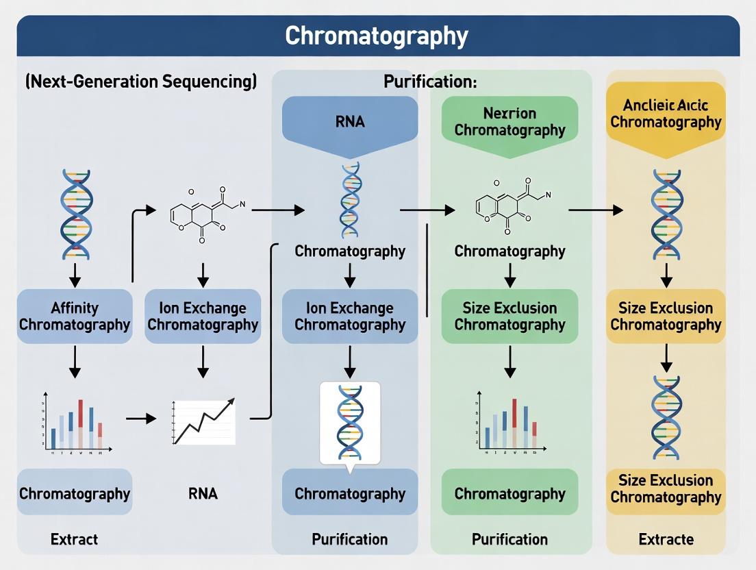
Chromatography in NGS: A Complete Guide to Nucleic Acid Extraction Methods for High-Quality Sequencing
This comprehensive guide explores the critical role of chromatography-based methods in extracting and purifying nucleic acids for Next-Generation Sequencing (NGS).
Chromatography in NGS: A Complete Guide to Nucleic Acid Extraction Methods for High-Quality Sequencing
Abstract
This comprehensive guide explores the critical role of chromatography-based methods in extracting and purifying nucleic acids for Next-Generation Sequencing (NGS). We detail foundational principles of key chromatographic techniques (affinity, ion-exchange, reversed-phase, and size-exclusion), their practical application in NGS workflows for DNA and RNA, and optimization strategies to maximize yield and purity. The article provides troubleshooting guidance for common challenges and a comparative analysis of chromatography versus alternative methods like magnetic beads and SPRI. Designed for researchers, scientists, and drug development professionals, this resource synthesizes current methodologies to enable informed protocol selection and ensure successful, reproducible NGS library preparation.
Understanding the Basics: How Chromatography Purifies DNA and RNA for Sequencing
Within the framework of advancing Chromatography methods for nucleic acid extraction in NGS workflow research, the initial extraction step is the critical foundation upon which all subsequent sequencing data rests. The purity of extracted nucleic acids (NA)—specifically the absence of contaminants like salts, proteins, organic solvents, and enzymatic inhibitors—directly impacts library preparation efficiency, sequencing performance, and data fidelity. This application note details the quantitative impact of common contaminants, provides validated protocols for assessing purity, and outlines advanced chromatographic solutions.
The Impact of Impurities on NGS Workflows
Contaminants co-purified with nucleic acids can inhibit or alter enzymatic reactions critical to NGS library preparation, leading to biased or failed runs.
Table 1: Impact of Common Contaminants on Key NGS Enzymatic Reactions
| Contaminant | Typical Source | Affected NGS Step | Observed Effect (Quantitative Impact) |
|---|---|---|---|
| Ethanol | Incomplete drying post-precipitation | Adapter Ligation | >5% v/v residue can reduce ligation efficiency by 50-70%. |
| Guanidinium Salts | Chaotropic lysis buffers | PCR Amplification | 10 mM residue can inhibit Taq polymerase by up to 90%. |
| Phenolic Compounds | Organic extraction (TRIzol) | Reverse Transcription | 0.1% v/v residue can reduce cDNA yield by >60%. |
| Carrier RNA | Certain viral NA kits | Quantitative Assays (qPCR) | Can co-purify and interfere with accurate quantification. |
| Proteinase K | Incomplete inactivation | Fragmentation & End-Repair | Residual activity degrades essential enzymes. |
| Humic Acids | Environmental/plant samples | PCR & Polymerases | 0.5 µg/µL can completely inhibit amplification. |
| Polysaccharides | Bacterial/plant tissues | Library Normalization | Increase viscosity, leading to pipetting inaccuracies. |
Assessing Nucleic Acid Purity: Essential QC Protocols
Protocol 1: Spectrophotometric Purity Assessment (A260/A280 & A260/A230 Ratios)
Principle: Absorbance at 260 nm (A260) quantifies nucleic acids, while ratios at 280 nm and 230 nm indicate protein/phenol and solvent/salt contamination, respectively. Procedure:
- Blank the spectrophotometer with the elution buffer used (e.g., 10 mM Tris-HCl, pH 8.5).
- Dilute 2 µL of the extracted NA in 98 µL of nuclease-free water (1:50 dilution) in a UV-transparent microcuvette.
- Measure absorbance at 230 nm, 260 nm, and 280 nm.
- Calculate ratios: Purity Ratio = A260 / A280; Purity Ratio = A260 / A230. Acceptance Criteria for NGS: For DNA, A260/A280 ~1.8; A260/A230 >2.0. For RNA, A260/A280 ~2.0; A260/A230 >2.0. Significant deviations suggest contamination requiring clean-up.
Protocol 2: Fluorometric Quantification and Inhibitor Detection
Principle: Fluorescent dyes bind NA specifically, offering robust quantification even in the presence of common contaminants that affect absorbance. Materials: Fluorometer, dsDNA/RNA-specific assay dye, standards, black-walled assay plates. Procedure:
- Prepare a standard curve from the provided standard (e.g., 0-200 ng/µL).
- Mix 1-2 µL of sample with 198-199 µL of diluted dye in assay tubes/wells.
- Incubate for 5 minutes protected from light.
- Read fluorescence and interpolate sample concentration from the standard curve.
- Perform a spike-in recovery test: Spike a known quantity of standard into a diluted sample. Recovery of <90% indicates presence of inhibitors.
Advanced Chromatographic Extraction for High-Purity NGS
Silica-membrane column chromatography remains the gold standard. Modern iterations utilize modified silica or magnetic particles in bind-wash-elute workflows optimized for specific sample types and downstream NGS.
Diagram 1: Silica-Membrane Column Chromatography Workflow
Table 2: Chromatographic Solutions for Challenging Sample Types
| Sample Type | Key Challenge | Chromatographic Solution | Outcome for NGS |
|---|---|---|---|
| FFPE Tissue | Cross-linking, fragmentation | Specialized high-proteinase K digestion + post-extraction bead-based clean-up | Improved library complexity, reduced duplicates. |
| Plasma (cfDNA) | Low abundance, high frag. | High-salt binding with small-volume elution & carrier RNA | Higher yield of target fragments, improved detection. |
| Microbiome (Stool) | Polysaccharides, humics | Inhibitor removal technology (IRT) wash buffers in columns | Restored polymerase activity, successful 16S amplification. |
| Whole Blood | Hemoglobin, heparin | White cell lysis + protein precipitation prior to column binding | High-molecular-weight gDNA, optimal for WGS. |
The Scientist's Toolkit: Research Reagent Solutions
Table 3: Essential Materials for High-Purity Nucleic Acid Extraction
| Item | Function in Extraction | Key Consideration for NGS Purity |
|---|---|---|
| Silica-Membrane Spin Columns | Selective binding of nucleic acids via chaotropic salts. | Pore size impacts fragment retention. Use columns with >20 µg binding capacity. |
| Magnetic Beads (e.g., SPRI) | Size-selective binding and clean-up via PEG/NaCl. | Bead-to-sample ratio is critical for fragment size selection during library prep. |
| Chaotropic Salt Binding Buffer (e.g., GuHCl) | Denatures proteins, promotes NA binding to silica. | Complete removal via washes is essential to prevent enzyme inhibition. |
| Wash Buffer with Ethanol | Removes salts, metabolites, and other impurities. | Ensure complete ethanol evaporation in drying step to prevent inhibition. |
| Nuclease-Free Elution Buffer (e.g., 10 mM Tris-HCl) | Hydrates and releases NA from membrane/beads. | Low EDTA (0.1 mM) prevents Mg²⁺ chelation in downstream enzymes. |
| RNase/DNase Inhibitors | Protects target NA from degradation during extraction. | Verify compatibility with downstream enzymatic steps. |
| Inhibitor Removal Technology (IRT) Wash | Specifically removes humics, polyphenols, polysaccharides. | Critical for non-standard samples (soil, plants, stool). |
| Fluorometric Assay Kit (e.g., Qubit dsDNA HS) | Accurate, contaminant-resistant quantification. | Essential for normalizing input into library prep. |
The pursuit of optimal NGS data begins at the first bench step: extraction. Integrating rigorous purity assessment via the described protocols and selecting chromatographic methods tailored to the sample matrix are non-negotiable practices for ensuring sequencing success. This focus on foundational quality directly supports the broader thesis that continuous refinement of chromatographic extraction principles is paramount to the evolution of robust, reproducible NGS workflows in research and drug development.
In Next-Generation Sequencing (NGS) workflows, the purity and integrity of nucleic acids are paramount. Chromatography, a cornerstone of biomolecular separation, offers diverse principles to achieve high-quality nucleic acid extraction essential for library preparation and sequencing. This article details the application of affinity, charge (ion exchange), size (size exclusion), and hydrophobicity (reversed-phase) chromatography within the context of NGS research, providing specific protocols and comparative data.
Principles & Applications in NGS Nucleic Acid Extraction
1. Affinity Chromatography
- Principle: Exploits specific, reversible interactions between a target molecule (e.g., poly-A RNA, His-tagged proteins) and an immobilized ligand (e.g., oligo-dT, metal ions).
- NGS Application: Primarily for mRNA isolation from total RNA using oligo(dT) matrices for cDNA library construction. Critical for transcriptome sequencing (RNA-Seq).
2. Ion Exchange Chromatography (IEC)
- Principle: Separates molecules based on net surface charge. Cation exchangers bind positively charged molecules; anion exchangers (commonly used for nucleic acids) bind negatively charged molecules like DNA/RNA.
- NGS Application: Removal of contaminants (proteins, nucleotides, salts) from nucleic acid preparations. Can separate different nucleic acid species (e.g., dsDNA vs. ssDNA) based on charge density.
3. Size Exclusion Chromatography (SEC)
- Principle: Separates molecules by hydrodynamic size/radius as they pass through a porous matrix. Larger molecules elute first, while smaller molecules enter pores and elute later.
- NGS Application: Desalting, buffer exchange of purified nucleic acids, and removal of short primers, adapter dimers, or enzyme inhibitors post-amplification.
4. Hydrophobic Interaction / Reversed-Phase Chromatography (RPC)
- Principle: Separates molecules based on hydrophobicity. HIC uses high-salt buffers to promote binding, while RPC uses polar mobile phases. Nucleic acids are weakly retained unless highly modified.
- NGS Application: Primarily for purification of labeled or modified nucleotides, oligonucleotides, and removal of hydrophobic contaminants (phenol, dyes).
Comparative Quantitative Data
Table 1: Performance Metrics of Chromatographic Methods in NGS Sample Prep
| Principle | Typical Yield (µg) | Purity (A260/A280) | Key Contaminant Removed | Processing Time (min) | Scalability |
|---|---|---|---|---|---|
| Affinity (Oligo-dT) | 1-10 (from 100µg total RNA) | 1.9-2.1 | rRNA, tRNA, genomic DNA | 45-60 | Moderate |
| Ion Exchange (Anion) | >90% recovery | 1.8-2.0 | Proteins, polysaccharides, dyes | 30-45 | High |
| Size Exclusion | >95% recovery | 1.8-1.9 | Salts, primers, dimers (<100 bp) | 20-30 | Low-Moderate |
| Reversed-Phase | Varies by target | N/A | Phenol, organic solvents, hydrophobic impurities | 15-25 | Moderate |
Table 2: Suitability for Nucleic Acid Types in NGS
| Principle | gDNA | ssRNA | mRNA | FFPE-DNA | cDNA | Library Fragments |
|---|---|---|---|---|---|---|
| Affinity | - | - | +++ | - | - (Unless tagged) | - (Unless tagged) |
| Ion Exchange | +++ | ++ | ++ | ++ | +++ | +++ |
| Size Exclusion | ++ (for cleanup) | ++ (for cleanup) | ++ (for cleanup) | + (for cleanup) | +++ (for cleanup) | +++ (for dimer removal) |
| Reversed-Phase | - | - | - | + (Contaminant removal) | - | + (Oligo purification) |
Experimental Protocols
Protocol 1: mRNA Isolation via Oligo(dT) Affinity Chromatography for RNA-Seq Objective: Isolate polyadenylated mRNA from total RNA.
- Equilibration: Load 1 mL of oligo(dT) cellulose slurry into a microcolumn. Wash with 5 column volumes (CV) of binding buffer (20 mM Tris-HCl, pH 7.5, 1 M NaCl, 0.5 mM EDTA).
- Sample Binding: Heat 100 µg of total RNA at 65°C for 5 min, snap-cool on ice. Mix with equal volume of 2X binding buffer. Apply sample to the column at room temperature, collecting flow-through.
- Washing: Wash column with 10 CV of binding buffer, followed by 5 CV of medium-salt wash buffer (20 mM Tris-HCl, pH 7.5, 0.1 M NaCl, 0.5 mM EDTA).
- Elution: Elute mRNA with 3 CV of elution buffer (10 mM Tris-HCl, pH 7.5, 0.5 mM EDTA). Pre-heat buffer to 65°C for higher yield. Collect fractions.
- Precipitation: Pool mRNA-containing fractions, add 1/10 volume 3M NaOAc (pH 5.2) and 2.5 volumes ethanol. Precipitate at -20°C for 1 hour. Centrifuge, wash pellet with 70% ethanol, and resuspend in nuclease-free water.
- QC: Analyze yield by spectrophotometry (A260) and integrity by Bioanalyzer (RIN > 8.0).
Protocol 2: Purification of NGS Library Fragments by Anion Exchange Chromatography Objective: Remove enzymatic inhibitors and salts post-library amplification.
- Column Setup: Use a pre-packed anion exchange spin column (e.g., quaternary ammonium functional group). Equilibrate with 3 CV of equilibration buffer (25 mM Tris-HCl, pH 8.0, 50 mM NaCl).
- Sample Binding: Dilute the PCR-amplified NGS library 1:1 with equilibration buffer. Load onto the column. Centrifuge at 3000 x g for 1 min. Discard flow-through.
- Washing: Wash with 5 CV of low-salt wash buffer (25 mM Tris-HCl, pH 8.0, 150 mM NaCl). Centrifuge and discard flow-through.
- Elution: Place column in a clean collection tube. Elute DNA with 2 CV of high-salt elution buffer (25 mM Tris-HCl, pH 8.0, 1 M NaCl). Centrifuge.
- Concentration: Desalt and concentrate the eluate using a size-exclusion spin column or ethanol precipitation.
- QC: Quantify by Qubit dsDNA HS Assay. Verify size distribution by TapeStation.
Protocol 3: Removal of Adapter Dimers by Size Exclusion Chromatography (Spin Column) Objective: Purify final NGS library from short adapter dimers (<100 bp).
- Column Preparation: Resuspend SEC resin gel slurry. Load into a disposable microcolumn, let settle. Centrifuge at 800 x g for 2 min to pack.
- Equilibration: Add 200 µL of TE buffer (pH 8.0). Centrifuge at 800 x g for 2 min. Repeat twice.
- Sample Loading & Elution: Carefully load 25 µL of library sample onto the center of the resin bed. Place column in a clean 1.5 mL tube. Centrifuge at 800 x g for 2 min. The purified library is collected in the flow-through.
- QC: Analyze on a high-sensitivity Bioanalyzer chip or TapeStation to confirm dimer removal.
Visualizations
Workflow for mRNA Affinity Purification
Chromatography Principles & NGS Applications Map
The Scientist's Toolkit: Research Reagent Solutions
Table 3: Essential Materials for Chromatographic Nucleic Acid Purification
| Item | Function in NGS Workflow | Example/Notes |
|---|---|---|
| Oligo(dT) Cellulose/Magnetic Beads | Solid-phase ligand for affinity capture of poly-A+ mRNA from total RNA. | Critical for RNA-Seq library prep. Magnetic beads enable high-throughput automation. |
| Anion Exchange Spin Columns (Q Sepharose) | Bind negatively charged nucleic acids; separate from proteins, salts, and inhibitors based on ionic strength. | Used for post-PCR cleanup of NGS libraries. Quaternary ammonium (Q) groups are common. |
| Size Exclusion Resin (Sephadex G-50, G-100) | Porous matrix for separating molecules by size. Removes short oligonucleotides and salts. | Fast desalting columns (spin or gravity) for final library cleanup. |
| Chaotropic Salt Buffers (e.g., Guanidine HCl) | Denature proteins, inhibit RNases, and promote nucleic acid binding to silica or certain matrices. | Used in combined lysis/binding steps for nucleic acid isolation from complex samples. |
| RNase/DNase Inactivation Reagents | Protect target nucleic acids from degradation during purification. | Often included in lysis or binding buffers. |
| Nuclease-Free Water & Elution Buffers | Final resuspension of purified nucleic acids to ensure stability and compatibility with downstream enzymatic steps (e.g., fragmentation, ligation). | Low EDTA or TE buffers are common. |
| Magnetic Stand (for bead-based protocols) | Physical separation of magnetic bead-nucleic acid complexes from solution during wash/elution steps. | Enables rapid, multi-sample processing essential for high-throughput NGS. |
| High-Sensitivity Assay Kits (Qubit, Bioanalyzer) | Accurate quantification and quality assessment of purified nucleic acids before library construction. | Fluorometric assays are preferred over absorbance for NGS library QC. |
This application note details four key chromatographic techniques for nucleic acid purification and analysis, contextualized within Next-Generation Sequencing (NGS) workflow research. Efficient nucleic acid extraction and fractionation are critical for obtaining high-quality sequencing libraries, impacting data fidelity and downstream analysis. Here, we provide a comparative analysis, structured protocols, and essential toolkits for researchers and development professionals.
Table 1: Comparison of Key Nucleic Acid Chromatography Techniques
| Technique | Principle | Primary Application in NGS Workflow | Typical Scale | Key Advantages | Key Limitations |
|---|---|---|---|---|---|
| Affinity Chromatography | Bioselective interaction (e.g., oligo-dT for mRNA, antigen-antibody) | Capture of specific nucleic acid types (e.g., poly-A+ mRNA isolation) | Micro to preparative | High specificity, excellent purity from complex lysates | High cost, ligand stability, requires specific binding moiety |
| Ion-Exchange (IEX) | Electrostatic attraction between charged solutes and oppositely charged matrix | Separation of nucleic acids by length/charge, removal of contaminants (proteins, metabolites) | Analytical to preparative | High capacity, good resolution for similar sizes, effective for desalting | Sensitivity to pH and ionic strength, may require sample desalting |
| Reversed-Phase (RP) | Hydrophobic partitioning between nonpolar stationary phase and polar mobile phase | Purification of synthetic oligonucleotides, desalting, removal of organic compounds | Analytical to semi-prep | Excellent for hydrophobic impurities, robust for small oligonucleotides | Can denature dsDNA/RNA, not ideal for large nucleic acids |
| Size-Exclusion (SEC) | Physical sieving based on hydrodynamic volume | Buffer exchange, desalting, removal of primers/dNTPs/NGS reaction cleanup | Analytical to preparative | Mild conditions, fast, no sample binding | Low capacity, limited resolution, requires narrow sample volume |
Application Notes & Detailed Protocols
Affinity Chromatography for mRNA Capture
Application Note: Critical for transcriptome sequencing (RNA-Seq). Poly(A)+ mRNA is selectively captured from total RNA using oligo(dT) ligands immobilized on a resin (e.g., magnetic beads or column). This protocol yields mRNA devoid of ribosomal RNA, significantly improving sequencing efficiency and data quality.
Protocol: Magnetic Oligo(dT) Bead-Based mRNA Isolation Objective: Isolate poly-adenylated mRNA from total RNA derived from human cell lines. Materials: Total RNA sample (≥50 µg), magnetic rack, oligo(dT) magnetic beads, binding buffer (20 mM Tris-HCl, pH 7.5, 1.0 M LiCl, 2 mM EDTA), wash buffer (10 mM Tris-HCl, pH 7.5, 0.15 M LiCl, 1 mM EDTA), nuclease-free water.
- Bind: Mix 50 µg total RNA with 50 µL bead slurry and 200 µL binding buffer. Incubate at 65°C for 5 min, then 5 min at room temperature with gentle mixing.
- Wash: Place tube on magnetic rack. Discard supernatant. Wash beads twice with 200 µL wash buffer.
- Elute: Resuspend beads in 20 µL nuclease-free water. Heat to 80°C for 2 min, immediately place on magnet, and transfer the eluate (mRNA) to a new tube.
- Quality Control: Assess yield via spectrophotometry (A260) and integrity via Bioanalyzer (RINe > 8.5).
Anion-Exchange Chromatography for Plasmid DNA Purification
Application Note: Used for high-purity plasmid DNA preparation for NGS library amplification. Separates supercoiled plasmid DNA from RNA, genomic DNA fragments, and endotoxins based on charge density.
Protocol: Fast-Performance Liquid Chromatography (FPLC) for Plasmid Purification Objective: Purify supercoiled plasmid DNA from alkaline lysate of E. coli culture. Materials: Clarified lysate, FPLC system with anion-exchange column (e.g., quaternary ammonium resin), Buffer A (50 mM Tris-HCl, pH 8.0), Buffer B (50 mM Tris-HCl, pH 8.0, 1 M NaCl), 0.22 µm filter.
- Equilibration: Filter lysate. Equilibrate column with 5 column volumes (CV) of 25% Buffer B (0.25 M NaCl) at 1 mL/min.
- Load & Wash: Load filtered lysate. Wash with 10 CV of 25% Buffer B until A260 baseline stabilizes.
- Elute: Apply a linear gradient from 25% to 60% Buffer B over 20 CV. Collect 1 mL fractions. Supercoiled plasmid typically elutes at ~0.55-0.65 M NaCl.
- Desalt & Concentrate: Pool plasmid-rich fractions and desalt using a SEC spin column or ethanol precipitation.
Reversed-Phase Chromatography for Oligonucleotide Purification
Application Note: Essential for purifying synthetic primers and probes used in NGS library preparation (e.g., adapters, barcodes, PCR primers). Separates full-length product from failure sequences.
Protocol: HPLC Purification of Synthetic Oligonucleotides Objective: Purify a 25-mer DNA oligonucleotide from synthesis failure sequences. Materials: Crude oligonucleotide, HPLC system with C18 column, Buffer A (0.1 M TEAA, pH 7.0), Buffer B (Acetonitrile), syringe filter (0.45 µm).
- Sample Prep: Dilute crude oligo in nuclease-free water. Filter through 0.45 µm syringe filter.
- Run Conditions: Set flow rate to 1 mL/min. Use gradient: 5% to 25% Buffer B over 30 minutes. Detect at A260 nm.
- Collection: Collect the peak corresponding to the full-length product (typically the latest major peak).
- Drying & Resuspension: Lyophilize the collected fraction to remove acetonitrile and TEAA. Resuspend in nuclease-free water.
Size-Exclusion Chromatography for NGS Reaction Cleanup
Application Note: Used for buffer exchange, desalting, and removal of excess primers, dNTPs, and small fragments post-PCR amplification of NGS libraries. A fast, spin-column format is standard.
Protocol: Spin Column SEC for PCR Cleanup Objective: Purify a 300 bp NGS library fragment from PCR reagents and primers. Materials: PCR reaction mix, SEC spin columns (e.g., with Sephadex G-50 resin), collection tube, microcentrifuge.
- Column Preparation: Resuspend resin. Let column settle, then spin at 750 x g for 2 minutes to remove storage buffer.
- Sample Application: Carefully apply the entire PCR reaction (up to 100 µL) to the center of the compacted resin bed.
- Elution: Place column in a clean collection tube. Centrifuge at 750 x g for 2 minutes. The eluate contains the purified library DNA.
- Assessment: Quantify DNA by fluorescence assay (e.g., Qubit).
Visualized Workflows
Title: Affinity mRNA Isolation Workflow
Title: SEC Spin Column Cleanup Process
The Scientist's Toolkit: Research Reagent Solutions
Table 2: Essential Materials for Nucleic Acid Chromatography in NGS
| Item | Function & Application |
|---|---|
| Oligo(dT) Magnetic Beads | Poly(T) sequences covalently bound to magnetic particles for selective poly(A)+ mRNA capture via affinity. |
| Anion-Exchange Resin (Q Sepharose) | Quaternary ammonium functional groups for strong anion-exchange (SAX) purification of plasmid DNA and larger nucleic acids. |
| Reversed-Phase C18 Column | Hydrophobic stationary phase for HPLC purification of synthetic oligonucleotides based on hydrophobicity. |
| Size-Exclusion Resin (Sephadex G-50) | Cross-linked dextran gel for rapid desalting and removal of sub-100 nt contaminants from DNA samples. |
| Chaotropic Salt Binding Buffer | High-salt buffer (e.g., with guanidine HCl) used in silica-based and some affinity methods to promote nucleic acid binding. |
| Nuclease-Free Water & Buffers | Essential for preventing degradation of RNA and DNA during all chromatographic steps. |
| Magnetic Separation Rack | Enables efficient phase separation for magnetic bead-based affinity protocols. |
| High-Purity Elution Buffers | Low-salt buffers or nuclease-free water used to elute purified nucleic acids from various media without inhibiting downstream enzymes. |
The Role of Solid-Phase Extraction (SPE) in Modern Chromatography Workflows
1. Introduction and Thesis Context
Within the comprehensive investigation of chromatography methods for nucleic acid extraction in Next-Generation Sequencing (NGS) workflow research, Solid-Phase Extraction (SPE) emerges as a fundamental pre-chromatographic and sample preparation cornerstone. This document details its critical application notes and protocols, underscoring how SPE enhances the performance of downstream analytical chromatography (e.g., HPLC, LC-MS) by providing purified, concentrated analytes free of PCR inhibitors, salts, and contaminants.
2. Application Notes: SPE for NGS Library Prep QC
SPE is extensively used to purify and desalt sequencing libraries prior to qualitative and quantitative chromatographic analysis. Key applications include:
- Adapter Dimer Removal: Purifying libraries to remove short, adapter-ligated fragments that contaminate pools and reduce sequencing efficiency.
- Buffer Exchange: Exchanging library storage buffers into chromatographically compatible solvents (e.g., from EDTA-containing buffers to water or Tris-HCl).
- Concentration: Concentrating dilute libraries to meet the minimum loading requirements for analytical size-exclusion or ion-pair reversed-phase chromatography.
Table 1: Performance Comparison of SPE Sorbents for NGS Library Clean-Up
| Sorbent Type (Chemistry) | Primary Application in NGS Workflow | Average Recovery Yield (dsDNA > 100 bp) | Key Contaminant Removal | Compatible Downstream Chromatography |
|---|---|---|---|---|
| Silica-Based (Bridged Ethylene Glycol) | Size-selective clean-up, adapter dimer removal. | 85-95% | Primers, adapter dimers, salts, proteins. | IP-RP-HPLC, SEC-HPLC |
| Carboxylated Magnetic Beads (SPRI) | High-throughput bead-based clean-up and size selection. | 80-90% (varies with bead:sample ratio) | Salts, dNTPs, enzymes, short fragments. | Compatible with most after elution. |
| Anion-Exchange (DEAE, Q) | Purification of large, high-integrity DNA fragments. | 70-85% | Proteins, RNA, organic contaminants. | IEC-HPLC, SEC-HPLC |
| C18 Reversed-Phase | Desalting and concentration of oligonucleotides. | >90% (for short oligos) | Salts, polar impurities. | IP-RP-HPLC, LC-MS |
3. Experimental Protocols
Protocol 3.1: Silica-Based SPE for Post-PCR NGS Library Purification
Objective: To purify and concentrate a double-stranded DNA NGS library post-amplification for downstream QC via HPLC.
Materials:
- Amplified NGS library in PCR buffer.
- Silica-membrane spin column (e.g., with bridged ethylene glycol coating).
- Binding Buffer (e.g., high-concentration GuHCl or NaI).
- Wash Buffer (e.g., 70-80% ethanol in Tris-HCl, pH 7.5).
- Elution Buffer (10 mM Tris-HCl, pH 8.5, or nuclease-free water).
- Microcentrifuge, pipettes, collection tubes.
Procedure:
- Binding: Add 5 volumes of Binding Buffer to 1 volume of the library sample. Mix thoroughly and transfer the entire volume to the silica spin column. Centrifuge at ≥10,000 x g for 30-60 seconds. Discard flow-through.
- Washing: Add 700 µL of Wash Buffer to the column. Centrifuge at ≥10,000 x g for 30-60 seconds. Discard flow-through. Repeat wash step once. Centrifuge the empty column for an additional 60 seconds to dry the membrane.
- Elution: Place the column in a clean 1.5 mL microcentrifuge tube. Apply 15-30 µL of pre-warmed (50°C) Elution Buffer directly to the center of the membrane. Incubate at room temperature for 2 minutes. Centrifuge at ≥10,000 x g for 60 seconds to elute the purified DNA. The eluate is now ready for concentration measurement and chromatographic analysis.
Protocol 3.2: Magnetic Bead-Based (SPRI) Size Selection
Objective: To perform a dual-sided size selection to remove both adapter dimers and excessively large fragments.
Materials:
- PEG/NaCl-based SPRI magnetic beads.
- Magnetic separation rack.
- 80% Freshly prepared ethanol.
- Elution Buffer.
- Thermoshaker (optional).
Procedure:
- First Binding (Remove Large Fragments): Bring sample to 100 µL with water. Add a volume of bead suspension calculated for the upper size cut-off (e.g., 0.5X sample volume). Mix thoroughly and incubate for 5 minutes. Place on magnet until supernatant is clear. Transfer supernatant (containing fragments smaller than target) to a new tube. Discard beads.
- Second Binding (Remove Small Fragments): To the supernatant, add beads calculated for the lower size cut-off (e.g., 0.8X original sample volume). Mix and incubate for 5 minutes. Place on magnet. Discard supernatant.
- Wash: With beads on the magnet, add 500 µL of 80% ethanol without disturbing the pellet. Incubate 30 seconds, then remove ethanol. Repeat once. Air-dry beads for 5 minutes.
- Elution: Remove from magnet, resuspend beads in Elution Buffer, incubate for 2 minutes, place on magnet, and transfer purified eluate to a new tube.
4. Visualization of Workflows
Title: SPE Integration in NGS QC Workflow
Title: Four Core Steps of SPE
5. The Scientist's Toolkit: Key Research Reagent Solutions
| Item | Function in SPE for NGS Chromatography |
|---|---|
| Silica Spin Columns (Bridged Ethylene Glycol) | Provides a hydrophilic, negatively charged surface for selective binding of DNA vs. contaminants in high-salt conditions. |
| Magnetic Beads (SPRI - PEG/NaCl) | Carboxyl-coated beads for reversible DNA binding via PEG-induced crowding; enable high-throughput, automatable size selection. |
| Guanidine Hydrochloride (GuHCl) Binding Buffer | Chaotropic salt that disrupts water structure, facilitating DNA adsorption onto silica surfaces. |
| Size-Selective PEG/NaCl Solutions | Polyethylene glycol and salt mixtures used with SPRI beads to precisely control the fragment size range retained. |
| Mass Spectrometry-Grade Water/Eluents | Ultra-pure solvents for elution to prevent ion suppression and background noise in downstream LC-MS analysis. |
| IP-RP HPLC Columns (e.g., C18 with Ion-Pairing Agent) | Downstream analytical columns used to separate and quantify nucleic acid fragments after SPE clean-up. |
Next-Generation Sequencing (NGS) sample preparation requires high-purity, high-integrity nucleic acids. The choice of extraction method directly impacts library complexity, sequencing accuracy, and cost. This application note positions chromatography-based methods against alternative techniques within the broader thesis of optimizing nucleic acid extraction for NGS research.
Quantitative Comparison of Extraction Methods
Table 1: Performance Metrics of Nucleic Acid Extraction Methods for NGS
| Method | Principle | Avg. Yield (ng/µL) | Avg. Purity (A260/A280) | Process Time (Hands-on, mins) | Cost per Sample (USD) | Suitability for Challenging Samples | NGS Read Quality Impact (Post-Library) |
|---|---|---|---|---|---|---|---|
| Silica Spin-Column (Chromatography) | Selective adsorption/desorption | 50-150 | 1.8-2.0 | 20-30 | 5-15 | Moderate | Low risk of inhibitors; consistent. |
| Magnetic Bead (Chromatography) | Magnetic particle binding/wash | 60-200 | 1.8-2.1 | 15-25 | 4-12 | Good | Very low inhibitor carryover. |
| Liquid-Liquid Extraction | Phenol-chloroform partition | 100-300 | 1.6-1.9 | 45-60 | 1-3 | Excellent (e.g., tissue) | High inhibitor risk; requires cleanup. |
| Anion-Exchange Chromatography | Charge-based binding to resin | 80-250 | 1.9-2.1 | 30-40 | 10-20 | Good for high-volume | Low PCR inhibitors. |
| Salting-Out Precipitation | Protein precipitation & DNA recovery | 80-200 | 1.7-1.9 | 30-50 | <1 | Moderate | Moderate inhibitor risk. |
Data synthesized from recent vendor whitepapers (2023-2024) and peer-reviewed method comparisons. Costs are approximate for reagents.
Table 2: NGS-Specific Output Metrics by Extraction Method
| Method | Library Preparation Success Rate (%) | GC Bias (Deviation from Expected) | Mean Insert Size Consistency | Automation Compatibility | Throughput (Samples per 8-hr shift) |
|---|---|---|---|---|---|
| Silica Spin-Column | 98 | Low-Medium | High | High (96-well) | 96-384 |
| Magnetic Bead | 99+ | Low | Very High | Very High (96/384-well) | 192-1536 |
| Liquid-Liquid | 85-90 | Medium-High | Medium | Low | 24-48 |
| Anion-Exchange | 97 | Low | High | Medium | 48-96 |
| Salting-Out | 88-92 | Medium | Low-Medium | Low-Medium | 48-96 |
Detailed Experimental Protocols
Protocol 3.1: High-Throughput Genomic DNA Extraction using Magnetic Bead Chromatography for Whole Genome Sequencing (WGS)
Objective: Isolate high-molecular-weight gDNA from human whole blood for WGS library prep. Materials: See "The Scientist's Toolkit" (Section 6). Procedure:
- Lysis: Mix 200 µL whole blood with 20 µL Proteinase K and 400 µL Lysis Buffer BL. Vortex vigorously. Incubate at 56°C for 10 min.
- Binding: Add 400 µL of room-temperature isopropanol to the lysate. Mix by pipetting. Transfer 600 µL of the mixture to a deep-well plate containing 20 µL of pre-dispensed magnetic beads. Seal and mix on a plate shaker (1200 rpm) for 5 min.
- Capture & Washes: Place plate on a magnetic stand for 2 min until clear. Aspirate and discard supernatant.
- Wash 1: With plate on magnet, add 500 µL Wash Buffer 1 (with ethanol). Incubate 30 sec. Aspirate fully.
- Wash 2: Remove plate from magnet. Add 500 µL Wash Buffer 2. Resuspend beads by pipetting. Return to magnet for 2 min. Aspirate fully. Perform a second Wash Buffer 2 step identically.
- Elution: Air-dry beads on magnet for 5-7 min. Remove from magnet. Add 52 µL of pre-heated (70°C) Elution Buffer TE. Resuspend thoroughly. Incubate at 70°C for 5 min. Place on magnet for 2 min. Transfer 50 µL of clear eluate to a clean plate.
- QC: Quantify by fluorometry. Check integrity by agarose gel electrophoresis or genomic tape assay. Proceed to shearing and library construction.
Protocol 3.2: Comparative Analysis: Silica Column vs. Salting-Out for FFPE RNA Extraction in Transcriptomics
Objective: Compare RNA yield and quality from Formalin-Fixed Paraffin-Embedded (FFPE) tissue sections for RNA-Seq. A. Silica Spin-Column Protocol (Commercial Kit):
- Deparaffinization & Lysis: Cut 2 x 10 µm FFPE sections into a tube. Add 1 mL xylene. Vortex. Centrifuge. Remove supernatant. Repeat with 100% ethanol. Air-dry.
- Digestion: Add 200 µL Digestion Buffer and 10 µL Proteinase K. Incubate at 56°C for 15 min, then 80°C for 15 min.
- Binding: Add 200 µL binding buffer and 300 µL ethanol. Mix. Load onto column. Centrifuge at 11,000 x g for 30 sec.
- Washes: Wash with 500 µL Wash Buffer 1 (centrifuge). Wash twice with 500 µL Wash Buffer 2/ethanol.
- Elution: Centrifuge dry column for 1 min. Elute in 30 µL RNase-free water. B. Salting-Out Protocol:
- Deparaffinization & Lysis: Perform as in Step A1. Add 600 µL Lysis Buffer (4M guanidinium thiocyanate, 0.1M Tris-HCl pH7.5) and 10 µL β-mercaptoethanol. Homogenize.
- Precipitation: Add 60 µL 3M sodium acetate (pH 5.2) and 600 µL acid phenol:chloroform. Vortex. Centrifuge. Transfer aqueous phase.
- RNA Precipitation: Add equal volume isopropanol. Incubate -20°C for 1 hr. Centrifuge 12,000 x g, 30 min, 4°C. Wash pellet with 75% ethanol.
- Resuspension: Air-dry pellet. Dissolve in 30 µL RNase-free water. Analysis: Quantify yield (Qubit), assess purity (Nanodrop), and analyze integrity (RIN on Bioanalyzer). Proceed with ribosomal RNA depletion and library prep, tracking DV200 and final library yield.
Signaling Pathway & Workflow Diagrams
Title: NGS Sample Prep: Extraction Method Workflow
Title: Decision Logic for NGS Nucleic Acid Extraction Method
The Scientist's Toolkit: Key Research Reagent Solutions
Table 3: Essential Materials for Chromatography-Based NGS Extraction Protocols
| Item Name | Function in Protocol | Key Characteristics for NGS |
|---|---|---|
| Magnetic Silica Beads | Solid-phase for nucleic acid binding. | Uniform size (1-3 µm), superparamagnetic, high binding capacity (>50 µg DNA/mg), surface-coated for minimal carryover. |
| Chaotropic Lysis Buffer | Denatures proteins, releases nucleic acids, promotes binding to silica. | Contains guanidine hydrochloride/thiocyanate; RNase-free for RNA work; optimized pH. |
| Selective Wash Buffers | Removes contaminants (proteins, salts, organics) while retaining nucleic acids on solid phase. | Ethanol-based for salt removal; may contain proprietary detergents; inhibitor removal formulations available. |
| Low-Salt Elution Buffer | Releases pure nucleic acids from solid phase. | Typically TE buffer or nuclease-free water; low EDTA for compatibility with downstream enzymatic steps. |
| RNase/DNase Inactivation Reagents | Protects target nucleic acid integrity. | Included in lysis buffer or as separate additive (e.g., RNase inhibitor for RNA). |
| Carrier RNA | Improves yield of low-concentration targets (e.g., viral RNA, cfDNA). | Poly-A RNA or glycogen; must be inert and not interfere with sequencing. |
| Solid-Phase Reversible Immobilization (SPRI) Beads | Used for post-extraction size selection and clean-up prior to library prep. | Polyethylene glycol (PEG)-based binding; critical for insert size selection and adapter-dimer removal. |
| Automation-Compatible Plates/Strips | Enables high-throughput processing. | Deep-well 96/384-well plates, low-binding, suitable for magnetic stands and liquid handlers. |
Step-by-Step Protocols: Applying Chromatography for DNA and RNA Extraction in NGS
Within a thesis on chromatography methods for nucleic acid extraction in NGS workflows, selecting the appropriate purification strategy is foundational. The choice is dictated by the nucleic acid type (DNA vs. RNA) and sample preservation method (FFPE vs. fresh/frozen). Solid-phase extraction (SPE) chromatography, predominantly using silica matrices, remains the core technology, but binding conditions, lysis protocols, and nuclease treatments vary significantly. This application note details the critical parameters and provides validated protocols for high-quality nucleic acid isolation suitable for next-generation sequencing (NGS).
Table 1: Key Chromatographic Binding & Elution Conditions by Sample Type
| Parameter | Genomic DNA (gDNA) | Total RNA | microRNA / Small RNA | FFPE-Derived Nucleic Acids |
|---|---|---|---|---|
| Optimal Binding pH | High Salt, Chaotropic Agent (pH ≤7.5) | High Salt, Chaotropic Agent (pH ≤7.5) | High Salt, Chaotropic Agent, High % Ethanol | High Salt, Chaotropic Agent, Extended Proteolysis |
| Binding Matrix | Silica Membrane/Glass Fiber | Silica Membrane/Glass Fiber | Silica Membrane with Enhanced Small RNA Retention | Silica Membrane, Paramagnetic Beads |
| Critical Wash Buffer | Ethanol-Based (70-80%) with Mild Chaotrope | Ethanol-Based (70-80%) with Mild Chaotrope | Ethanol-Based (70-80%) with Mild Chaotrope | Aggressive Ethanol Wash (often >80%) |
| Elution Buffer | Low-Salt Buffer (TE or Tris) or Nuclease-Free Water | Nuclease-Free Water or TE (low EDTA) | Nuclease-Free Water or TE (low EDTA) | Low-Salt Buffer, Optional Rehydration Step |
| Key Inhibitor Challenge | Protein, Polysaccharides | RNases, Organic Solvents | Large RNA (competition), Organic Solvents | Formalin Adducts, Degradation, Dyes |
| Typical Yield (Varies by input) | 1-20 µg from 1-5 mg tissue | 2-15 µg from 1-5 mg tissue | 0.5-5 µg from 1-5 mg tissue | 0.5-10 µg (highly variable) from 5-10 µm section |
| Integrity Metric (Qubit/Bioanalyzer) | DIN (DNA Integrity Number) >7.0 | RIN (RNA Integrity Number) >8.0 | Smear analysis, miRNA peak | DV200 (%) >30% for FFPE-RNA |
Table 2: Recommended Primary Lysis and Pre-Chromatography Steps
| Sample Type | Primary Lysis Method | Mandatory Pre-Cleaning / Digestion | Critical Nuclease Step |
|---|---|---|---|
| Fresh/Frozen Tissue (DNA) | Proteinase K + Mechanical Homogenization | Centrifugation to pellet debris | RNase A (if DNA-only is desired) |
| Fresh/Frozen Tissue (RNA) | Guanidinium Isothiocyanate + Mechanical Homogenization | Centrifugation, Optional Organic Extraction | DNase I (on-column post-wash) |
| Cultured Cells (DNA/RNA) | Chaotropic Lysis Buffer (e.g., RLT) | Not typically required | See above for DNA vs. RNA |
| FFPE Tissue Sections (DNA) | Xylene Deparaffinization, Proteinase K (overnight, 56°C) | Centrifugation, possible rehydration | RNase A |
| FFPE Tissue Sections (RNA) | Xylene Deparaffinization, Proteinase K + Specialized Buffer (e.g., PKD) | Centrifugation, possible rehydration | DNase I (on-column post-wash) |
Detailed Experimental Protocols
Protocol 1: Silica-Membrane Column-Based DNA & RNA Co-Purification from Fresh/Frozen Tissue
- Principle: Chaotropic salts (guanidine HCl) denature proteins and facilitate nucleic acid binding to silica. Sequential elution separates RNA and DNA.
- Materials: Fresh tissue (≤30 mg), Liquid Nitrogen, Mortar & Pestle, QIAzol Lysis Reagent, Chloroform, BCP (1-bromo-3-chloropropane), Silica-membrane spin columns (e.g., RNeasy with gDNA eliminator), Collection tubes, 70% Ethanol, RNase-free Water, 96-100% Ethanol, 3M Sodium Acetate (pH 5.2).
- Method:
- Snap-freeze tissue in liquid N₂, pulverize.
- Homogenize in 600 µL QIAzol with a rotor-stator homogenizer.
- Incubate 5 min, RT.
- Add 120 µL chloroform/BCP, shake vigorously, incubate 3 min, RT.
- Centrifuge at 12,000 x g, 15 min, 4°C. Result: Three phases form.
- Transfer upper aqueous phase (contains RNA) to a new tube. Add 1.5 vols 100% ethanol. Mix.
- Transfer lower organic phase and interphase to a new tube for DNA (back-extraction optional).
- For RNA: Apply mix from step 6 to silica column. Centrifuge, discard flow-through. Wash with buffer RW1 and RPE (provided). Dry column. Elute RNA in 30-50 µL water.
- For DNA: Add 100% ethanol to organic phase mix from step 7. Mix. Apply to a separate silica column (specific for DNA). Centrifuge, discard flow-through. Wash with buffer containing ethanol. Dry column. Elute DNA in 30-50 µL Tris-EDTA buffer.
- Quantify via fluorometry (Qubit), assess integrity (Bioanalyzer/TapeStation).
Protocol 2: Paramagnetic Bead-Based RNA Isolation from FFPE Tissue Sections for NGS
- Principle: Paramagnetic beads with a silica coating bind nucleic acids in high-salt, high-EtOH conditions. Beads are magnetically captured, enabling efficient washing of FFPE inhibitors.
- Materials: FFPE curls/sections (5-20 µm), Xylene, 100% & 70% Ethanol, Proteinase K, FFPE-RNA Lysis Buffer (e.g., with β-mercaptoethanol), RNase-free DNase I, Paramagnetic Silica Beads, 80% Ethanol Wash Buffer, Magnetic Stand, Nuclease-free Water.
- Method:
- Deparaffinization: Add 1 mL xylene to sample, vortex, incubate 10 min, RT. Centrifuge max speed, 2 min. Discard supernatant. Repeat once.
- Ethanol Wash: Add 1 mL 100% ethanol to pellet, vortex. Centrifuge max speed, 2 min. Discard supernatant. Air-dry pellet 5-10 min.
- Lysis: Resuspend pellet in 200 µL FFPE Lysis Buffer + 10 µL Proteinase K. Incubate at 56°C for 15 min, then 80°C for 15-30 min.
- Binding: Add 200 µL binding buffer (high-salt/chaotrope) and 280 µL 100% ethanol to lysate. Mix thoroughly.
- Add pre-washed paramagnetic beads. Incubate 10 min, RT, with mixing.
- Place on magnetic stand for 5 min until clear. Discard supernatant.
- Wash: Wash beads twice with 500 µL 80% ethanol (on magnet). Remove all residual ethanol.
- DNase Digestion (on-bead): Prepare DNase I mix (10 µL DNase I + 70 µL digestion buffer). Resuspend beads in mix. Incubate 15 min, RT.
- Final Wash: Add 200 µL high-salt binding buffer and 400 µL 100% ethanol. Mix. Place on magnet, discard supernatant. Wash once with 80% ethanol. Dry beads.
- Elution: Elute RNA in 20-30 µL nuclease-free water (pre-warmed to 55°C). Place on magnet, transfer eluate to clean tube.
- Quantify (Qubit RNA HS Assay) and assess DV200 on Bioanalyzer.
Visualizations
Diagram 1: Decision Logic for Nucleic Acid Chromatography
Diagram 2: FFPE-RNA Workflow with Bead-Based Chromatography
The Scientist's Toolkit: Research Reagent Solutions
Table 3: Essential Materials for Nucleic Acid Chromatography in NGS Prep
| Item | Function & Rationale | Key Considerations for Selection |
|---|---|---|
| Chaotropic Salt Lysis Buffer | Denatures proteins, inhibits RNases, enables nucleic acid binding to silica. | Contains guanidinium salts (thiocyanate or HCl). Must be RNase-free for RNA work. |
| Silica-Based Binding Matrix | The solid phase for selective nucleic acid adsorption. | Choice between spin-column membranes (convenience) and paramagnetic beads (scalability, automation). |
| Proteinase K | Digests histones and crosslinked proteins, critical for tissue and FFPE lysis. | Requires verification of RNase-free activity for RNA protocols. |
| DNase I (RNase-free) | Removes contaminating genomic DNA from RNA preps to ensure NGS library accuracy. | Must be effective in high-salt conditions if used on-column/on-bead. |
| RNase A (DNase-free) | Removes RNA from DNA preps for applications like whole-genome sequencing. | Not needed for total nucleic acid preps. |
| High-Percentage Ethanol Wash Buffers | Removes salts, metabolites, and residual FFPE contaminants while retaining nucleic acids. | 80%+ ethanol often required for FFPE washes versus 70% for fresh tissue. |
| Low-EDTA or EDTA-Free Elution Buffers | Elutes purified nucleic acid; compatible with downstream enzymatic NGS steps. | TE buffer with low EDTA (0.1 mM) or Tris/H₂O. Avoid high EDTA with enzymatic steps. |
| Solid-Phase Reversible Immobilization (SPRI) Beads | Size-selective purification beads for post-extraction NGS library cleanup. | Different bead:buffer ratios select for different fragment sizes (e.g., cDNA, adapter-ligated fragments). |
| Fluorometric Quantitation Assays | Accurately quantifies dilute nucleic acids (Qubit). More specific than A260. | Use dsDNA HS, RNA HS, or miRNA assays as appropriate. Critical for NGS input normalization. |
| Fragment Analyzer / Bioanalyzer | Assesses nucleic acid integrity and size distribution (RIN, DIN, DV200). | DV200 is key QC metric for degraded FFPE-RNA suitability for NGS. |
Within the broader thesis on chromatography methods for nucleic acid extraction in NGS workflows, affinity chromatography stands as a cornerstone technique. This application note details two dominant solid-phase extraction methodologies: silica-membrane columns and cellulose-based matrix columns. Both leverage affinity principles—silica for nucleic acids under chaotropic conditions, and cellulose for specific molecular interactions—to purify high-quality DNA and RNA for downstream sequencing applications.
Key Protocol Comparison
Table 1: Comparative Overview of Silica-Membrane vs. Cellulose-Based Affinity Chromatography
| Parameter | Silica-Membrane Column Protocol | Cellulose-Based Column Protocol |
|---|---|---|
| Binding Principle | Chaotropic salt-induced hydration layer disruption; DNA adsorption to silica. | High-salt binding of DNA to cellulose, often via electrostatic & hydrophobic interactions. |
| Typical Binding Capacity | 20–100 µg per minicolumn, depending on format. | Often higher, 50–150 µg per column for some modified celluloses. |
| Optimal Nucleic Acid Size | Optimal for fragments >100 bp. Can lose very small fragments (<70 bp). | Effective for a broad range, including large genomic DNA and small fragments. |
| Primary Elution Buffer | Low-ionic-strength buffer or nuclease-free water (e.g., 10 mM Tris-HCl, pH 8.5). | Low-ionic-strength buffer or water; sometimes requires pre-warmed elution buffer (e.g., 55°C). |
| Typical Processed Sample Volume | 200 µL to 1 mL lysate per spin column. | Can handle larger volumes (1–10 mL) in batch or column format. |
| Key Chaotropic Agent | Guanidine hydrochloride (GuHCl) or guanidine thiocyanate (GuSCN). | Not always required; binding often uses ammonium acetate or sulfate. |
| Common Applications in NGS | Plasmid, genomic DNA, and total RNA extraction from cells/tissues; library clean-up. | PCR product purification, ssDNA binding for phage display, isolation of specific protein-DNA complexes. |
| Average Processing Time | 15–30 minutes (including centrifugation steps). | 30–60 minutes (may include longer incubation/batch binding). |
| Typical Elution Volume | 30–100 µL. | 100–500 µL. |
| Cost per Sample (Estimate) | $2–$10. | $1–$8 (cellulose matrix can be cheaper). |
Detailed Experimental Protocols
Protocol 1: Silica-Membrane Column DNA Extraction from Cultured Cells for NGS
Purpose: To isolate high-molecular-weight genomic DNA suitable for next-generation sequencing library preparation.
The Scientist's Toolkit: Research Reagent Solutions
| Item | Function |
|---|---|
| Lysis Buffer (GuHCl-based) | Denatures proteins, releases nucleic acids, and provides chaotropic conditions for silica binding. |
| Wash Buffer 1 (GuHCl in Ethanol) | Removes contaminants while keeping DNA bound to the silica membrane. |
| Wash Buffer 2 (Ethanol/Salt) | Further removes salts, solvents, and other impurities. |
| Silica-Membrane Spin Column | The affinity matrix; nucleic acids bind selectively under high-salt conditions. |
| RNase A (optional) | Degrades RNA to increase DNA purity. |
| Elution Buffer (10 mM Tris, pH 8.5) | Low-ionic-strength solution disrupts DNA-silica interaction, eluting pure DNA. |
| Proteinase K | Digests proteins and nucleases, improving yield and quality. |
Procedure:
- Cell Lysis: Pellet 1–5 x 10^6 cells. Resuspend in 200 µL phosphate-buffered saline. Add 20 µL Proteinase K and 200 µL Lysis Buffer. Mix thoroughly and incubate at 56°C for 10 minutes.
- Ethanol Addition: Add 200 µL of 96–100% ethanol to the lysate and mix immediately by vortexing.
- Binding: Apply the entire mixture to the silica-membrane spin column placed in a 2 mL collection tube. Centrifuge at 8,000 x g for 1 minute. Discard flow-through.
- Washing: a. Add 500 µL Wash Buffer 1 to the column. Centrifuge at 8,000 x g for 1 minute. Discard flow-through. b. Add 500 µL Wash Buffer 2 to the column. Centrifuge at 12,000 x g for 1 minute. Discard flow-through. c. Perform an additional empty centrifugation at 12,000 x g for 2 minutes to dry the membrane completely.
- Elution: Place the column in a clean 1.5 mL microcentrifuge tube. Apply 30–100 µL of pre-warmed (55°C) Elution Buffer directly to the center of the membrane. Let it stand for 2 minutes. Centrifuge at 12,000 x g for 1 minute. The eluate contains purified DNA. Store at -20°C.
Protocol 2: Cellulose-Based Column Purification of PCR Amplicons for NGS Library Construction
Purpose: To purify and concentrate double-stranded PCR products from amplification reactions, removing primers, enzymes, and nucleotides prior to sequencing.
The Scientist's Toolkit: Research Reagent Solutions
| Item | Function |
|---|---|
| Binding Buffer (High-Salt, e.g., 2M NaCl) | Creates conditions for DNA to bind to the cellulose matrix. |
| Cellulose Suspension/Column | The affinity matrix; binds DNA efficiently in high salt. |
| Wash Buffer (Ethanol/Salt) | Removes contaminants like dNTPs and proteins without eluting DNA. |
| Elution Buffer (Low Salt, e.g., TE) | Disrupts DNA-cellulose interaction by lowering ionic strength. |
Procedure:
- Binding Condition Setup: Combine the PCR reaction (50 µL) with 200 µL of Binding Buffer in a fresh tube. Mix thoroughly.
- Batch Binding: Add 20 µL of well-resuspended cellulose matrix slurry to the mixture. Incubate at room temperature for 5 minutes with gentle agitation every minute.
- Pellet Matrix: Centrifuge at 3,000 x g for 1 minute. Carefully aspirate and discard the supernatant.
- Washing: Add 500 µL of 70% ethanol to the pellet. Vortex briefly to resuspend. Centrifuge at 3,000 x g for 1 minute. Aspirate the supernatant completely. Repeat this wash step once.
- Drying: Air-dry the pellet for 5–10 minutes at room temperature to evaporate residual ethanol.
- Elution: Add 30–50 µL of Elution Buffer (or nuclease-free water) to the dried cellulose pellet. Vortex to resuspend. Incubate at 55°C for 5 minutes to enhance elution.
- Pellet Separation: Centrifuge at 12,000 x g for 2 minutes. Carefully transfer the supernatant, which contains the purified DNA, to a new tube. Store at -20°C.
Workflow and Context Visualization
Title: Nucleic Acid Affinity Chromatography Workflow for NGS
Title: Thesis Context of Affinity Chromatography Methods
Ion-Exchange Chromatography for High-Purity Plasmid and Viral DNA Isolation
This document provides detailed application notes and protocols for the use of anion-exchange chromatography in the isolation of plasmid and viral DNA. Within the broader thesis on chromatography methods for nucleic acid extraction in Next-Generation Sequencing (NGS) workflows, this technique is positioned as a high-resolution, scalable, and automation-compatible alternative to silica-membrane and magnetic bead-based methods. Its principal advantage lies in the separation mechanism based on the interaction between the negatively charged phosphate backbone of nucleic acids and positively charged functional groups on the chromatographic resin, enabling high purity isolation critical for downstream NGS applications, including plasmid verification, viral vector production for gene therapy, and viral genome sequencing.
Principle of Anion-Exchange for DNA Isolation
Anion-exchange chromatography separates molecules based on their net negative surface charge. Under optimized buffer conditions (pH ~7.5-8.5), DNA molecules are strongly anionic. The stationary phase is functionalized with positively charged groups (e.g., quaternary ammonium). When a crude lysate is applied, nucleic acids bind while proteins, RNAs, and other contaminants are washed away. Elution is achieved by increasing the ionic strength (e.g., with a chloride ion gradient), which competes for binding sites. Larger DNA molecules like plasmids and viral genomes, with higher charge density, typically elute at higher salt concentrations than RNA or small nucleotide fragments.
Key Research Reagent Solutions & Materials
Table: Essential Materials for Ion-Exchange Chromatography of DNA
| Item | Function/Description |
|---|---|
| Anion-Exchange Resin | Porous beads with quaternary ammonium (Q) or diethylaminoethyl (DEAE) groups. Provides high-binding capacity for nucleic acids. |
| Lysis Buffer (Alkaline) | Contains NaOH and SDS. Denatures proteins and linearizes chromosomal DNA; critical for initial sample preparation. |
| Neutralization Buffer | Potassium acetate, pH ~5.5. Precipitates proteins, SDS, and chromosomal DNA, leaving plasmid/viral DNA in solution. |
| Equilibration Buffer (Low Salt) | 20-50 mM Tris-HCl, pH 8.0. Prepares the column for sample binding under low ionic strength conditions. |
| Wash Buffer (Medium Salt) | ~0.3-0.5 M NaCl in Tris buffer. Removes weakly bound contaminants (e.g., proteins, short RNA, cellular metabolites). |
| Elution Buffer (High Salt) | 1.0-2.0 M NaCl in Tris buffer. Competitively displaces pure plasmid or viral DNA from the resin. |
| Ethanol or Isopropanol | For precipitation and concentration of eluted DNA. |
| Nuclease-Free Water | Final resuspension of purified DNA for downstream applications. |
| Spin Columns or FPLC System | Format for housing the resin, from manual spin columns to automated Fast Protein Liquid Chromatography systems. |
Detailed Experimental Protocols
Protocol 4.1: High-Purity Plasmid DNA Isolation fromE. coliusing Spin-Column Anion-Exchange
Objective: Isolate transfection-grade plasmid DNA from a bacterial culture. Materials: Anion-exchange spin column kit, microcentrifuge, buffers (see Table above). Procedure:
- Harvest & Lysis: Pellet 1-5 mL of overnight bacterial culture. Resuspend pellet in 250 µL Resuspension Buffer. Add 250 µL Lysis Buffer, mix gently by inversion (do not vortex). Incubate for 2-5 minutes at room temperature.
- Neutralization: Add 350 µL chilled Neutralization Buffer. Mix immediately by gentle inversion until a fluffy white precipitate forms. Centrifuge at ≥12,000 × g for 10 minutes.
- Column Binding: Transfer the clear supernatant to an anion-exchange spin column pre-equilibrated with 500 µL Equilibration Buffer. Centrifuge at 12,000 × g for 1 minute. Discard flow-through.
- Washing: Wash column with 700 µL Wash Buffer. Centrifuge for 1 minute. Discard flow-through. Repeat with 500 µL Wash Buffer. Centrifuge for an additional 2 minutes to dry the resin.
- Elution: Place column in a clean 1.5 mL tube. Apply 50-100 µL pre-warmed (65°C) Elution Buffer or nuclease-free water to the center of the resin. Let stand for 2 minutes. Centrifuge for 1 minute to collect purified plasmid DNA.
- Quantification: Measure DNA concentration via UV spectrophotometry (A260/A280 ratio ~1.8).
Protocol 4.2: Viral DNA Isolation from Cell Culture Supernatant using FPLC-Based Anion-Exchange
Objective: Purify viral genomic DNA (e.g., from herpesviruses, adenoviruses) for NGS library prep. Materials: FPLC system, anion-exchange column (e.g., Mono Q, HiTrap Q), 0.22 µm filter, buffers. Procedure:
- Sample Clarification & Concentration: Clear virus-containing supernatant by centrifugation (2,000 × g, 10 min) and 0.22 µm filtration. Concentrate virus particles by ultrafiltration (100 kDa MWCO) or PEG precipitation.
- Viral Lysis: Treat concentrate with Lysis Buffer containing proteinase K and SDS (final 0.5%) at 56°C for 1 hour.
- System & Column Setup: Equilibrate FPLC system and anion-exchange column with 5 column volumes (CV) of Low-Salt Buffer (e.g., 20 mM Tris, pH 8.0).
- Sample Application & Gradient Elution: Inject the lysed sample. Run a linear salt gradient (e.g., 0 to 1 M NaCl over 20 CV) at a flow rate of 1 mL/min. Monitor UV absorbance at 260 nm.
- Peak Collection: Collect the major A260 peak eluting at ~0.6-0.8 M NaCl (verified for specific virus).
- Desalting & Concentration: Desalt the pooled fraction using a centrifugal filter unit (e.g., 30 kDa MWCO) or by ethanol precipitation. Resuspend in nuclease-free water.
Performance Data & Comparison
Table: Representative Performance Metrics of Ion-Exchange vs. Silica-Membrane Methods
| Parameter | Anion-Exchange (Spin Column) | Anion-Exchange (FPLC) | Silica-Membrane (Mini-Prep Kit) |
|---|---|---|---|
| Typical Yield (from 5 mL culture) | 15-30 µg | Scalable (mg scale) | 5-15 µg |
| A260/A280 Purity Ratio | 1.8-2.0 | 1.8-2.0 | 1.7-1.9 |
| Host Genomic DNA Contamination | <1% | <0.1% | 1-5% |
| Endotoxin Level | <5 EU/µg | <1 EU/µg | <10 EU/µg |
| Process Time (Hands-on) | ~30 minutes | ~2 hours (setup + run) | ~25 minutes |
| Suitability for NGS | Excellent for amplicon-seq | Excellent for viral genome sequencing | Good for routine checks |
| Automation Potential | Medium (96-well plates) | High (system-integrated) | Low |
Visualized Workflows
Diagram Title: Spin-Column Plasmid DNA Isolation Workflow
Diagram Title: FPLC Workflow for Viral DNA Purification
Diagram Title: Ion-Exchange Elution Order of Nucleic Acids
Optimizing Binding, Washing, and Elution Conditions for Maximum Yield and Purity
This application note details the systematic optimization of silica-magnetic bead-based chromatography for nucleic acid extraction, a critical upstream step in Next-Generation Sequencing (NGS) workflows. The efficiency and purity of nucleic acid binding, washing, and elution directly impact library preparation quality, sequencing accuracy, and overall research outcomes in genomics and drug development.
Key Parameters for Optimization
Binding Conditions
Binding efficiency is governed by the concentration and type of chaotropic salt, pH, ethanol concentration, and incubation time with magnetic beads.
Table 1: Optimization of Binding Buffer Composition for DNA Yield and Purity
| Parameter | Tested Range | Optimal Condition (gDNA) | Optimal Condition (cfDNA) | Impact on Yield (A260) | Impact on Purity (A260/A280) |
|---|---|---|---|---|---|
| GuHCl Concentration | 2M - 6M | 4.5M | 4.0M | Peak at 4.5M (±15%) | Best (1.85-1.9) at 4.0-4.5M |
| Ethanol % (v/v) | 30% - 80% | 65% | 55% | Max at 60-65% for gDNA | Optimal (1.88) at 55-65% |
| pH | 4.0 - 7.5 | 5.5 | 6.0 | >90% yield at pH 5.0-6.0 | Most consistent at pH 5.5-6.2 |
| Incubation Time | 1 - 10 min | 5 min | 8 min | 95% yield at 5 min | No significant effect |
Washing Conditions
Washing removes contaminants (proteins, salts, inhibitors) without compromising nucleic acid retention.
Table 2: Wash Buffer Optimization for Contaminant Removal
| Wash Step | Buffer Composition | Volume (x bead pellet) | Number of Washes | Residual Protein (ng/µL) | Residual Salt (Conductivity) |
|---|---|---|---|---|---|
| Wash 1 | 80% EtOH, 10mM Tris-HCl pH 7.5 | 2x | 1 | < 5 | High |
| Wash 1 (Optimal) | 80% EtOH, 20mM NaCl, 2mM EDTA pH 8.0 | 3x | 1 | < 2 | Reduced by 40% |
| Wash 2 | 80% EtOH | 2x | 1 or 2 | < 1 | Moderate |
| Wash 2 (Optimal) | 70% EtOH | 3x | 2 | < 0.5 | Low (< 5 µS/cm) |
Elution Conditions
Elution efficiency depends on temperature, time, buffer ionic strength, and pH.
Table 3: Elution Buffer Condition Optimization
| Elution Parameter | Tested Range | Optimal Condition | Elution Yield | Eluate Purity (A260/A280) | Suitability for NGS |
|---|---|---|---|---|---|
| Temperature | 20°C - 70°C | 55°C | 98% ± 2% | 1.88 ± 0.02 | High |
| Time | 1 - 15 min | 5 min | 97% ± 3% | 1.87 ± 0.03 | High |
| Buffer | TE, nuclease-free H₂O, 10mM Tris-HCl | 10mM Tris-HCl pH 8.5 | 100% (ref) | 1.90 ± 0.02 | Optimal |
| Pre-heat Buffer | No / Yes | Yes (55°C) | +12% vs. RT | No negative impact | Recommended |
Detailed Experimental Protocols
Protocol 1: Systematic Optimization of Binding Conditions
Objective: To determine the optimal binding buffer composition for maximum high-quality yield from human plasma. Materials: See "The Scientist's Toolkit" below. Procedure:
- Sample Preparation: Spike 1 mL of human plasma with 10 ng of sheared human genomic DNA (gDNA) and 5 ng of synthetic cfDNA fragments (170bp).
- Lysis: Add 1 mL of Lysis Buffer (4M GuHCl, 10mM Tris, 30mM EDTA, 2% Triton X-100, pH 5.5). Vortex thoroughly.
- Parametric Binding: For each tested condition in Table 1, prepare a separate binding mixture: a. Combine 1 mL of lysate with 20 µL of magnetic silica bead suspension. b. Add the variable component (e.g., Ethanol to final % v/v, adjust pH with HCl/NaOH). c. Mix by inversion for the specified incubation time (1-10 min) at room temperature.
- Capture: Place tubes on a magnetic stand for 2 min until supernatant clears. Discard supernatant.
- Proceed to standardized wash and elution steps (Protocol 3) for consistent downstream analysis.
- Quantification: Elute in 50 µL pre-heated elution buffer. Measure yield via fluorometry and purity via spectrophotometry (A260/A280).
Protocol 2: Wash Stringency Assessment
Objective: To minimize contaminants while retaining >95% of bound nucleic acids. Procedure:
- Bind nucleic acids from a standardized lysate using optimal conditions from Protocol 1.
- First Wash: Resuspend bead pellet in Wash Buffer I (variable composition/volume from Table 2). Mix by pipetting. Capture on magnet and discard supernatant.
- Second Wash: Repeat Step 2 with Wash Buffer II.
- Dry Beads: After removing final wash supernatant, air-dry pellet for 5-10 min to evaporate residual ethanol. Do not over-dry.
- Elute and quantify yield as in Protocol 1.
- Assay for Contaminants: Use a commercial fluorescence-based protein assay on the eluate. Measure solution conductivity of a 1:10 diluted eluate.
Protocol 3: Optimized End-to-End Extraction
Objective: Execute the full extraction using optimized parameters for NGS-ready nucleic acids. Procedure:
- Bind: Combine 1 mL lysate (prepared as in Protocol 1) with 65% v/v molecular-grade ethanol and 20 µL beads in 4.5M GuHCl, pH 5.5. Mix for 5 min.
- Capture & Wash: Capture beads. Wash once with 3 bead-volumes of Wash Buffer I (80% EtOH, 20mM NaCl, 2mM EDTA pH 8.0). Wash twice with 3 bead-volumes of Wash Buffer II (70% EtOH).
- Dry: Air-dry beads for 7 min.
- Elute: Resuspend beads in 50 µL of pre-heated (55°C) 10mM Tris-HCl, pH 8.5. Incubate at 55°C for 5 min with occasional mixing.
- Capture & Recover: Place on magnet, transfer eluate to a clean tube.
- Quality Control: Quantify via fluorometer. Check purity (A260/A280 target 1.8-2.0). Analyze fragment size distribution (e.g., Bioanalyzer) and PCR amplification efficiency for NGS library prep.
Visualizations
Diagram Title: Key Parameters for Binding Optimization
Diagram Title: Optimized NA Extraction Workflow for NGS
The Scientist's Toolkit: Research Reagent Solutions
| Item | Function & Rationale |
|---|---|
| Magnetic Silica Beads (e.g., carboxyl-coated) | Solid-phase matrix for reversible nucleic acid binding via chaotropic salt-mediated adsorption. Magnetic core enables easy separation. |
| Guanidine Hydrochloride (GuHCl) | Chaotropic agent. Disrupts hydrogen bonding, denatures proteins, and promotes nucleic acid binding to silica. Preferred over guanidine thiocyanate for reduced inhibition in downstream enzymes. |
| Molecular Grade Ethanol (96-100%) | Modifies solution polarity to facilitate nucleic acid adsorption onto silica surface during binding and removes salts during washing. |
| RNAse A/T1 Cocktail | Critical for DNA extraction, degrades contaminating RNA which can inflate yield measurements and interfere with NGS library quantification. |
| Proteinase K | Broad-spectrum serine protease. Digests nucleases and structural proteins during lysis, improving nucleic acid release and purity. |
| Carrier RNA (e.g., poly-A) | Enhances recovery of low-concentration nucleic acids (e.g., cfDNA, viral RNA) by providing backbone for silica bead binding, especially in dilute samples. |
| Nuclease-Free Water | Used in elution buffer preparation. Free of nucleases that could degrade the extracted product. |
| Pre-heated Elution Buffer (10mM Tris-HCl, pH 8.5) | Low-ionic-strength, slightly alkaline buffer promotes desorption from silica. Pre-heating to 55°C increases elution efficiency, especially for high-fragment-length DNA. |
| Wash Buffer with EDTA | Contains EDTA (2mM) to chelate Mg2+ ions, inactivating residual nucleases that may have survived lysis. |
| Fluorometric DNA/RNA Binding Dye (e.g., Qubit dye) | For specific, accurate quantification of nucleic acid yield, unaffected by common contaminants that interfere with UV spectrophotometry. |
Integrating Chromatography Extraction into the Downstream NGS Library Prep Workflow
Within the broader thesis on chromatography methods for nucleic acid extraction for NGS workflows, integrating solid-phase extraction (SPE) or magnetic bead-based chromatography directly into library preparation represents a significant innovation. This integration aims to reduce sample loss, contamination risk, and hands-on time by creating a seamless process from extracted nucleic acids to sequencing-ready libraries. Traditional workflows involve discrete, often manual steps for purification between enzymatic reactions (e.g., end-repair, adapter ligation, PCR). Integrated chromatography allows for the use of a single-bead chemistry or spin-column platform to perform all clean-up steps without sample transfers, improving yield and reproducibility—critical factors for researchers, scientists, and drug development professionals working with precious clinical or low-input samples.
Recent advancements (2023-2024) demonstrate a shift towards automated, cartridge-based systems where chromatography membranes or magnetic beads are used in a sequential, on-deck manner. Key performance metrics include recovery efficiency (>90% for fragments >100 bp), effective removal of enzymes, primers, and adapter dimers, and compatibility with both DNA and RNA inputs for whole genome, exome, and transcriptome sequencing.
Table 1: Comparison of Integrated Chromatography Clean-Up vs. Traditional Methods in NGS Library Prep
| Performance Metric | Traditional Ethanol Precipitation | Stand-Alone Column Clean-Up | Integrated Bead/SPE Clean-Up |
|---|---|---|---|
| Average Hands-On Time (per sample) | 45-60 minutes | 20-30 minutes | 5-15 minutes |
| Mean Library Yield Recovery | 60-75% | 75-85% | 85-95% |
| Adapter Dimer Rate | Variable, often high | <5% | <2% |
| Process Contamination Risk | High (tube transfers) | Moderate | Low (closed or on-bead) |
| Automation Compatibility | Low | Moderate | High |
| Typical Cost per Sample | Low | Medium | Medium to High |
Table 2: Performance of Selected Integrated Kits (2024 Data)
| Commercial Solution | Input DNA Range | Avg. Fragment Retention (>100 bp) | Key Integrated Step |
|---|---|---|---|
| Kit A (Bead-based) | 1 ng - 1 µg | 98% | End-repair/A-tailing to Ligation |
| Kit B (SPE Cartridge) | 10 ng - 500 ng | 95% | Post-ligation & Post-PCR combined |
| Kit C (Magnetic Plate) | 0.1 ng - 100 ng | 90% | All enzymatic clean-ups |
Experimental Protocols
Protocol 3.1: Integrated Magnetic Bead Clean-Up for Post-Ligation Purification
Principle: This protocol replaces traditional column-based purification after adapter ligation. Paramagnetic beads with specific binding properties (e.g., size-selective PEG/NaCl solutions) are used in a "bind-wash-elute" cycle directly in the PCR plate or tube, without sample transfer.
Detailed Methodology:
- Reagent Setup: Prepare fresh 80% ethanol. Equilibrate SPRI (Solid Phase Reversible Immobilization) magnetic beads to room temperature. Ensure bead suspension is homogeneous.
- Binding: To the 50 µL adapter ligation reaction, add 50 µL (1.0x ratio) of room-temperature SPRI beads. Mix thoroughly by pipetting 10-15 times. Incubate at room temperature for 5 minutes.
- Capture: Place the tube/plate on a magnetic stand for 5 minutes or until the supernatant is clear. Carefully remove and discard the supernatant.
- Wash: With the tube on the magnet, add 200 µL of freshly prepared 80% ethanol without disturbing the bead pellet. Incubate for 30 seconds. Remove and discard all ethanol. Repeat this wash step a second time.
- Dry: Air-dry the bead pellet on the magnet for 5-7 minutes until it appears matte and begins to crack. Critical: Do not over-dry.
- Elute: Remove the tube from the magnet. Add 22 µL of nuclease-free water or low-EDTA TE buffer to the bead pellet. Mix thoroughly to resuspend. Incubate at room temperature for 2 minutes.
- Final Capture: Place the tube back on the magnet for 2 minutes. Transfer 20 µL of the clear supernatant containing the purified ligated library to a new tube. Proceed directly to PCR amplification.
Protocol 3.2: On-Cartridge SPE Purification for Automated Library Build
Principle: This protocol is designed for automated liquid handlers utilizing disposable SPE cartridge strips. The cartridge contains a silica or polymer membrane that binds nucleic acids under high-salt conditions.
Detailed Methodology:
- System Priming: Load the SPE cartridge strip onto the deck of the liquid handler. The system primes all lines and equilibrates the cartridge with 200 µL of conditioning buffer (e.g., guanidine HCl-based).
- Load and Bind: The robotic arm transfers the entire end-repair/a-tailing reaction (≈60 µL) to the cartridge reservoir. It then aspirates and dispenses the mixture through the membrane 3-5 times to promote binding in the presence of high-salt binding buffer.
- Wash: The system performs two wash steps: first with 200 µL of a salt/ethanol wash buffer, followed by 200 µL of an 80% ethanol wash. Vacuum or positive pressure is applied to dry the membrane briefly.
- Elute: The purified nucleic acids are eluted from the dry membrane by applying 25 µL of low-ionic-strength elution buffer (pre-heated to 55°C) and collecting the flow-through into a fresh plate.
- Direct Transfer: The eluate is then robotically transferred and combined with the subsequent ligation master mix in the next plate, completing the integrated clean-up and reaction setup.
Visualizations
Integrated NGS Library Prep with On-Bead Clean-Up
Chromatography Clean-Up Core Cycle
The Scientist's Toolkit: Research Reagent Solutions
Table 3: Essential Materials for Integrated Chromatography NGS Workflows
| Item | Function in Workflow | Key Consideration |
|---|---|---|
| Size-Selective SPRI Beads | Binds nucleic acids based on size (PEG/NaCl concentration); enables all clean-up steps in a single tube. | Ratio optimization (e.g., 0.6x to 1.8x) is critical for fragment selection and yield. |
| Magnetic Bead-Compatible Plates | High-recovery, low-binding PCR plates for performing on-bead reactions and clean-ups without transfer. | Must have minimal bead adhesion and withstand thermal cycling. |
| Automation-Compatible SPE Cartridges | Disposable chromatography columns for automated bind-wash-elute on liquid handlers. | Must interface precisely with robotic pipetting tips and have low dead volume. |
| Universal Binding/Wash Buffer | A single solution for binding nucleic acids to beads/membrane after each enzymatic step. | Typically contains PEG and high-concentration salt (e.g., NaCl). |
| Low-EDTA Elution Buffer | Elutes purified DNA without inhibiting subsequent enzymatic steps (e.g., ligase, polymerase). | 10 mM Tris-HCl, pH 8.0-8.5 is common. EDTA is minimized. |
| Non-Template Control (NTC) Reagents | Water and master mixes used to monitor adapter dimer and cross-contamination. | Essential for validating the stringency of integrated clean-ups. |
| Automated Liquid Handler | Platform to execute sequential chromatography clean-ups and reagent additions. | Must be programmable for precise magnetic separation and bead handling. |
Solving Common Problems: Optimizing Chromatography for Peak NGS Performance
In Next-Generation Sequencing (NGS) workflows, efficient nucleic acid extraction is a critical initial step that profoundly impacts downstream results. This application note, framed within a broader thesis on chromatography methods for nucleic acid purification, addresses the common challenge of low yield. We detail targeted diagnostic experiments and optimization protocols for three key parameters: sample input, lysis efficiency, and elution. These protocols are designed for researchers, scientists, and drug development professionals seeking to maximize recovery from precious or low-concentration samples.
Diagnostic Framework for Low Yield
A systematic approach is required to isolate the primary cause of suboptimal nucleic acid yield. The following workflow outlines the logical diagnostic pathway.
Title: Diagnostic Decision Tree for Low Nucleic Acid Yield
Key Research Reagent Solutions
Table 1: Essential Reagents for Nucleic Acid Extraction Optimization
| Reagent/Material | Function in Optimization | Key Considerations |
|---|---|---|
| Proteinase K | Degrades nucleases & cellular proteins, enhancing lysis. | Activity varies by vendor/buffer; requires optimal temperature (56°C). |
| RNase A | Degrades RNA in DNA extraction, reducing column clogging. | Essential for "DNA-only" preps; verify it is DNase-free. |
| Magnetic Beads (Silica) | Solid-phase reversible immobilization (SPRI) for binding. | Bead size/polymer ratio critical for fragment size selection. |
| Chaotropic Salt (GuHCl) | Denatures proteins, promotes NA binding to silica. | Concentration is critical for efficient binding in high-volume lysates. |
| Carrier RNA | Improves recovery of low-concentration NA from large volumes. | Co-precipitates with target NA, enhancing binding efficiency. |
| Ethanol (Molecular Grade) | Adjusts binding/ wash buffer stringency. | Concentration must be precise (±5%); impurities inhibit elution. |
| Low TE Buffer (pH 8.0-8.5) | Elution buffer; low EDTA prevents enzyme inhibition in NGS. | Pre-heating to 55-60°C significantly increases elution efficiency. |
| Spin Columns (Silica Membrane) | Chromatography medium for bind-wash-elute. | Membrane pore size and silica purity affect capacity and yield. |
Experimental Protocols & Data
Protocol 4.1: Sample Input & Homogenization Benchmarking
Objective: Determine the optimal input mass/volume for a given extraction system without exceeding binding capacity. Method:
- Prepare a homogenized tissue sample (e.g., mouse liver) in PBS.
- Perform a series of 6 extractions using a standardized silica-membrane column kit, varying the input volume: 10 µL, 25 µL, 50 µL, 100 µL, 200 µL, and 300 µL of homogenate.
- Spike each sample with 5 ng of a known exogenous DNA control (e.g., lambda phage DNA) prior to lysis to monitor recovery efficiency.
- Follow the manufacturer’s lysis and binding protocol precisely.
- Elute all samples in an identical, fixed volume (e.g., 50 µL) of pre-heated Low TE Buffer.
- Quantify total DNA yield (ng/µL) via fluorometry and calculate percent recovery of the spike-in control via qPCR.
Table 2: Sample Input Optimization Results
| Input Volume (µL) | Avg. Total DNA Yield (ng) | Spike-in Recovery (%) | A260/A280 | Notes |
|---|---|---|---|---|
| 10 | 125 ± 15 | 98 ± 5 | 1.85 | Low total yield, high purity. |
| 25 | 310 ± 22 | 97 ± 3 | 1.83 | Optimal recovery efficiency. |
| 50 | 580 ± 45 | 96 ± 4 | 1.82 | Recommended Input. |
| 100 | 950 ± 80 | 94 ± 3 | 1.81 | High yield, good recovery. |
| 200 | 1100 ± 120 | 82 ± 6 | 1.78 | Near column capacity; recovery drops. |
| 300 | 1150 ± 150 | 65 ± 8 | 1.70 | Column overload; poor recovery/purity. |
Protocol 4.2: Lysis Efficiency Evaluation
Objective: Systematically test lysis buffer composition and incubation conditions. Method:
- Aliquot 50 µL of a standardized cell pellet (e.g., 1x10^6 cultured HEK293 cells) into 6 tubes.
- Tube 1 (Control): Use standard lysis buffer (w/ Proteinase K), incubate 10 min @ 56°C.
- Tube 2: Add 1 µL RNase A (100 mg/mL) to standard lysis, incubate 10 min @ 56°C.
- Tube 3: Increase Proteinase K concentration by 2x, incubate 10 min @ 56°C.
- Tube 4: Standard lysis buffer, but increase incubation to 30 min @ 56°C.
- Tube 5: Standard lysis buffer, incubate 10 min @ 70°C.
- Tube 6: Use a specialized, commercially available "tough lysis" buffer.
- Following lysis, centrifuge all samples at 12,000 x g for 2 min to pellet debris.
- Transfer supernatant to fresh tubes and proceed with identical binding, wash, and elution steps.
- Measure total DNA yield and assess fragment size distribution via TapeStation/Bioanalyzer.
Table 3: Lysis Condition Optimization Results
| Condition Tested | Total DNA Yield (ng) | % Yield vs. Control | Avg. Fragment Size (kb) | Visual Debris |
|---|---|---|---|---|
| 1. Standard Lysis (Control) | 520 ± 30 | 100% | 23.5 | Low |
| 2. + RNase A | 535 ± 25 | 103% | 23.8 | Low |
| 3. 2x Proteinase K | 610 ± 40 | 117% | 22.1 | Very Low |
| 4. 30 min Incubation | 580 ± 35 | 112% | 23.0 | Low |
| 5. 70°C Incubation | 480 ± 50 | 92% | 18.5 | High |
| 6. Tough Lysis Buffer | 650 ± 45 | 125% | 20.4 | Moderate |
Protocol 4.3: Elution Buffer Optimization
Objective: Maximize the release of bound nucleic acid from the silica matrix. Method:
- Bind a standardized lysate (from 50 µL of HEK293 cell pellet) to silica-membrane columns in duplicate.
- Perform identical wash steps.
- Apply 50 µL of the following elution buffers to the center of the dry membrane:
- E1: Nuclease-free water, room temp (RT).
- E2: Low TE Buffer (10 mM Tris, 0.1 mM EDTA, pH 8.0), RT.
- E3: Low TE Buffer, pH 8.0, pre-heated to 55°C.
- E4: Low TE Buffer, pH 9.0, pre-heated to 55°C.
- E5: Two-step elution: 30 µL of pre-heated (55°C) Low TE (pH 8.0), incubate 2 min, spin; then apply a second 30 µL of the same buffer.
- Allow a 2-minute incubation at room temperature (or as per heated buffer protocol) before centrifugation.
- Quantify eluate concentration. Perform a second identical elution on the same column to quantify residual DNA.
Table 4: Elution Buffer Optimization Results
| Elution Condition | 1st Elution Yield (ng) | 2nd Elution Yield (ng) | Total Recovery (%) | A260/A280 |
|---|---|---|---|---|
| E1: H2O, RT | 310 ± 20 | 95 ± 10 | 65% | 1.70 |
| E2: TE pH8.0, RT | 380 ± 25 | 80 ± 8 | 75% | 1.82 |
| E3: TE pH8.0, 55°C | 460 ± 30 | 25 ± 5 | 97% | 1.84 |
| E4: TE pH9.0, 55°C | 470 ± 35 | 20 ± 4 | 98% | 1.83 |
| E5: 2x 30µL, 55°C | 435 ± 20 (1st) 40 ± 5 (2nd) | 5 ± 2 | 99% | 1.85 |
Integrated Optimization Workflow
The following diagram synthesizes the key optimized steps from the diagnostic protocols into a recommended workflow.
Title: Optimized Nucleic Acid Extraction Workflow for NGS
Methodical diagnosis of sample input, lysis, and elution parameters is fundamental to overcoming low yield in nucleic acid extraction for NGS. The data presented confirm that exceeding column binding capacity is a primary failure point, that lysis can be enhanced by increased protease concentration and time, and that elution efficiency is highly dependent on buffer pH and temperature. Integrating these optimized steps into the chromatography-based extraction workflow ensures maximal recovery of high-quality nucleic acids, providing a robust foundation for sensitive downstream sequencing applications.
Chromatographic methods, particularly silica-membrane and magnetic bead-based solid-phase extraction, are the cornerstone of modern nucleic acid purification for Next-Generation Sequencing (NGS). Within this thesis on Chromatography for NGS workflows, a critical challenge is the co-elution of impurities that severely impact downstream applications. The A260/A280 and A260/A230 ratios are key spectrophotometric metrics for assessing these purity issues. An ideal A260/A280 ratio (~1.8-2.0) indicates protein contamination, while the A260/A230 ratio (~2.0-2.2) reflects the presence of chaotropic salts, organic solvents, and other PCR inhibitors. This application note details protocols to diagnose, troubleshoot, and resolve these common post-chromatographic purity challenges.
Quantitative Data: Purity Ratios and Implications
Table 1: Interpretation of Nucleic Acid Purity Ratios and Common Contaminants
| Ratio | Optimal Value | Low Value Indicates | Common Source in Chromatography | Impact on NGS Workflow |
|---|---|---|---|---|
| A260/A280 | 1.8 (DNA), 2.0 (RNA) | <1.8 = Protein/phenol contamination | Incomplete lysis or protein wash; carryover of binding buffer components | Inhibits enzymatic steps (ligation, PCR); causes library prep failure |
| A260/A230 | 2.0 - 2.2 | <2.0 = Salt/carbohydrate/organic solvent contamination | Incomplete ethanol wash; carryover of chaotropic salts (guanidine) or wash buffers | Inhibits polymerase activity in PCR and sequencing; reduces library yield |
| Absorbance ~230nm | Should be minimal | High absorbance = Phenol, chaotropes, thiocyanates | Residual lysis or wash buffer | Strong PCR inhibition; inaccurate quantification |
Experimental Protocols for Troubleshooting and Repurification
Protocol 3.1: Diagnostic Re-measurement with Dilution
Purpose: To confirm that low ratios are not an artifact of high sample concentration.
- Dilute the eluted nucleic acid sample 1:10 in the same elution buffer used (e.g., TE buffer, nuclease-free water).
- Measure absorbance from 230nm to 320nm using a spectrophotometer.
- Recalculate ratios. If ratios normalize, the initial measurement was skewed by high concentration. If they remain low, proceed to repurification.
Protocol 3.2: Ethanol Reprecipitation for Salt and Inhibitor Removal
Purpose: To remove salts, organic compounds, and small molecule inhibitors.
- To the aqueous nucleic acid sample, add 0.1 volumes of 3M sodium acetate (pH 5.2) and mix.
- Add 2.5 volumes of ice-cold 100% ethanol. Mix thoroughly and incubate at -20°C for 30+ minutes.
- Centrifuge at >12,000 x g for 15 minutes at 4°C. Carefully decant the supernatant.
- Wash pellet with 500 µL of ice-cold 70% ethanol. Centrifuge for 5 minutes and decant.
- Air-dry the pellet for 5-10 minutes (do not over-dry).
- Resuspend in an appropriate volume of TE buffer (pH 8.0) or nuclease-free water. The Tris and EDTA in TE help chelate residual ions and stabilize pH.
Protocol 3.3: Silica Column Clean-up for Broad-Spectrum Contaminant Removal
Purpose: To remove proteins, salts, and inhibitors via a second, optimized chromatographic step.
- Adjust the sample to the binding conditions of your commercial silica column kit (typically by adding a chaotropic salt/binding buffer).
- Load onto the column. Centrifuge as per manufacturer's instructions.
- Perform two separate wash steps with the provided wash buffer (usually ethanol-based). Ensure the column is spun dry after the final wash to remove residual ethanol.
- Critical Step for A260/A230: Perform an additional wash with 80% ethanol (prepared with nuclease-free water) after the standard wash. Centrifuge and spin dry.
- Elute in a low-salt buffer (e.g., TE, 10 mM Tris-HCl) or nuclease-free water pre-warmed to 55-65°C to increase elution efficiency.
Diagrams of Workflows and Relationships
Title: Diagnostic & Repurification Workflow for Nucleic Acid Purity
Title: Source of Contaminants and Their Impact on NGS
The Scientist's Toolkit: Research Reagent Solutions
Table 2: Essential Materials for Addressing Purity Issues
| Reagent/Material | Function & Role in Troubleshooting |
|---|---|
| TE Buffer (pH 8.0) | Preferred resuspension buffer post-cleanup. Tris maintains pH; EDTA chelates Mg²⁺, inhibiting nucleases and helping solubilize impurities. |
| Nuclease-Free Water (Low EDTA) | For elution when minimizing salt carryover is critical. Essential for making dilution and ethanol solutions. |
| Molecular Biology Grade Ethanol (100% & 70%) | For reprecipitation and making wash buffers. Removes salts and organic contaminants. Must be nuclease-free. |
| 3M Sodium Acetate (pH 5.2) | Provides counter-ions for efficient ethanol precipitation of nucleic acids. |
| Commercial Silica-Membrane Clean-up Kit | Provides optimized buffers for binding, washing, and eluting nucleic acids. The core tool for repurification. |
| Glycogen or Carrier RNA | Aid in precipitation of low-concentration nucleic acid samples (e.g., cfDNA, sRNA) to improve recovery during Protocol 3.2. |
| Spectrophotometer/Nanodrop | Essential for initial diagnosis and post-cleanup validation of purity ratios. |
| Fluorometric Assay (e.g., Qubit) | For accurate quantification post-cleanup, as absorbance may be skewed by residual impurities. |
Within the broader thesis on Chromatography methods for nucleic acid extraction in NGS workflows, this application note addresses critical pre-analytical challenges. Successful Next-Generation Sequencing (NGS) is predicated on the quality of extracted nucleic acids. Formalin-Fixed Paraffin-Embedded (FFPE) tissues, cell-free DNA (cfDNA), and low-input samples present unique obstacles that require tailored chromatography-based extraction and purification protocols to ensure integrity, yield, and compatibility with downstream sequencing.
FFPE Tissue Nucleic Acid Extraction
FFPE specimens are invaluable for retrospective studies but suffer from nucleic acid fragmentation, cross-linking, and chemical damage from formalin fixation.
Key Challenges & Solutions Table
| Challenge | Impact on NGS | Chromatographic Solution | Typical Yield Improvement |
|---|---|---|---|
| Formalin-induced cross-links | Low yield, biased amplification | Enhanced de-crosslinking incubation (e.g., 80°C with specialized buffer) | 30-50% increase vs. standard protocols |
| Nucleic acid fragmentation | Short library fragments, poor coverage | Size-selection chromatography post-extraction (e.g., bead-based cleanup) | Enriches >100bp fragments by ~70% |
| Paraffin contamination | Inhibits enzyme reactions in library prep | Initial xylene/ethanol wash followed by silica-membrane cleanup | Reduces PCR inhibition by >90% |
| Degradation & low yield | High sequencing failure rate | Use of carrier RNA in binding buffer during silica-column extraction | Improves recovery of <200bp fragments by 40% |
Detailed Protocol: FFPE DNA Extraction Using Silica-Membrane Chromatography
Materials: FFPE tissue sections (10-20 µm), microtome, xylene, ethanol (100%, 70%), proteinase K, de-crosslinking buffer, silica-column extraction kit (with optimized binding buffers), carrier RNA, elution buffer. Procedure:
- Deparaffinization: Add 1 ml xylene to 5-10 sections in a microcentrifuge tube. Vortex, incubate at 55°C for 10 min. Centrifuge at full speed for 2 min. Remove supernatant.
- Rehydration: Wash pellet with 1 ml 100% ethanol, vortex, centrifuge. Repeat with 70% ethanol. Air-dry pellet for 5-10 min.
- Digestion & De-crosslinking: Resuspend pellet in 180 µl digestion buffer with 20 µl proteinase K (20 mg/ml). Incubate at 56°C for 3 hours, then at 80°C for 2 hours in de-crosslinking buffer.
- Nucleic Acid Binding: Add carrier RNA (1 µg) to lysate. Add 5 volumes of binding buffer optimized for high-salt conditions. Transfer to a silica-membrane column. Centrifuge at 11,000 x g for 1 min.
- Washes: Perform two washes with wash buffer containing ethanol. Centrifuge after each.
- Elution: Elute DNA in 30-50 µl of low-EDTA TE buffer or nuclease-free water pre-heated to 70°C. Centrifuge at 11,000 x g for 1 min.
- Quality Assessment: Quantify using fluorometry (e.g., Qubit dsDNA HS Assay). Assess fragment size distribution using a Bioanalyzer/TapeStation.
Cell-Free DNA (cfDNA) Isolation
cfDNA, notably from liquid biopsies, is short (≈170 bp) and low in concentration, requiring high-sensitivity isolation.
Performance Metrics for cfDNA Isolation Methods
| Method | Principle | Typical Input Volume | Average Yield (from 1 ml plasma) | Key Contaminant Removed |
|---|---|---|---|---|
| Silica-Membrane Spin Column | Chaotropic salt binding to silica | 1-4 ml plasma | 5-20 ng | Proteins, high molecular weight genomic DNA |
| Magnetic Bead (SPRI) | Size-selective binding to carboxylated beads | 1-10 ml plasma | 5-30 ng | Efficient removal of >500bp contaminants |
| Anion-Exchange Chromatography | Binding to positively charged matrix | 2-5 ml plasma | 10-25 ng | Hemoglobin, lactoferrin |
Detailed Protocol: cfDNA Extraction Using Magnetic Bead-Based Chromatography
Materials: Blood plasma (collected in EDTA or cfDNA-specific tubes), magnetic stand, cfDNA-specific magnetic beads, binding buffer, wash buffers, elution buffer. Procedure:
- Plasma Preparation: Centrifuge whole blood twice at 1600 x g for 10 min at 4°C to generate platelet-poor plasma. Aliquot 1-4 ml.
- Lysis & Binding: Mix plasma with an equal volume of binding buffer containing chaotropic salts. Add a defined volume of magnetic beads. Incubate for 10 min at room temperature with mixing.
- Capture & Washes: Place on a magnetic stand for 5 min until clear. Discard supernatant. Wash beads twice with 80% ethanol while on the magnet.
- Drying & Elution: Air-dry bead pellet for 5-10 min to remove residual ethanol. Elute cfDNA in 20-30 µl of low-EDTA TE buffer (pH 8.5). Incubate at 55°C for 5 min, then capture beads and transfer eluate.
- QC: Use a high-sensitivity fluorometric assay. Analyze fragment profile using a High Sensitivity Bioanalyzer chip.
Low-Input Sample Strategies
Samples with limited starting material (e.g., single cells, fine-needle aspirates) demand maximum recovery and minimal loss.
Low-Input Recovery Enhancement Techniques
| Technique | Application | Principle | Estimated Recovery Gain |
|---|---|---|---|
| Carrier Materials (e.g., RNA) | Low-concentration DNA/RNA | Provides bulk for silica binding, co-precipitated | Up to 100% improvement for <1 ng inputs |
| Reduced Elution Volume | All low-input extractions | Minimizes dilution post-extraction | Increases concentration by 3-5x |
| Post-Extraction Concentration | Dilute eluates | Vacuum centrifugation or small-volume concentrators | Up to 10x concentration |
| Direct Library Prep Kits | Bypasses extraction | Transposase-based tagmentation directly in lysate | Reduces pre-PCR steps, minimizes loss |
Detailed Protocol: Nucleic Acid Extraction from Low-Input Cells via Silica Column
Materials: Limited cell suspension (<10,000 cells), carrier RNA (10 µg/ml), lysis buffer with guanidine hydrochloride, silica spin columns, reduced-volume elution tubes. Procedure:
- Lysis: Transfer cells to a tube. Add lysis buffer with carrier RNA. Mix thoroughly.
- Binding: Add ethanol, mix, and apply entire lysate to silica column. Centrifuge. Repeat flow-through application a second time to increase binding.
- Washes: Perform two stringent washes with wash buffer.
- Elution: Place column in a 1.5 ml tube. Apply 15 µl of pre-heated (70°C) elution buffer directly to the membrane center. Incubate for 2 min. Centrifuge at maximum speed for 1 min.
- Post-Extraction Concentration (Optional): Use a vacuum concentrator to reduce volume to 5-10 µl if necessary.
The Scientist's Toolkit: Research Reagent Solutions
| Item | Function in Challenging Sample Prep |
|---|---|
| Silica-Membrane Spin Columns | Selective binding of nucleic acids in high-salt conditions; effective for fragmented DNA. |
| Magnetic Beads (Carboxylated) | Size-selective binding for cfDNA; scalable and automatable. |
| Carrier RNA (e.g., poly-A, glycogen) | Increases effective binding mass to silica/magnetic surfaces, preventing adsorption losses in low-input samples. |
| De-Crosslinking Buffer (with high pH) | Reverses formalin-induced methylene bridges in FFPE samples, critical for yield. |
| Size-Selective Beads (SPRI) | Enables precise selection of desired fragment ranges (e.g., cfDNA). |
| High-Sensitivity DNA Assay Kits (Fluorometric) | Accurate quantitation of dilute, fragmented DNA without overestimation from contaminants. |
| Nuclease-Free Water (Low-EDTA TE Buffer) | Optimal elution/storage medium to prevent chelation of enzymes in downstream steps. |
| Proteinase K (Molecular Grade) | Efficient digestion of proteins cross-linked to nucleic acids in FFPE samples. |
Visualizations
Title: FFPE DNA Extraction & QC Workflow
Title: cfDNA Isolation with Magnetic Beads
Title: Chromatography Solutions for Challenging NGS Samples
Application Notes Within the context of advancing chromatography methods for nucleic acid extraction in Next-Generation Sequencing (NGS) workflows, the integrity of the input genetic material is paramount. The pursuit of long-range genomic information and accurate transcriptomic profiles is fundamentally constrained by the physical shearing and enzymatic degradation of DNA and RNA during sample handling and extraction. Silica-based and magnetic bead-based chromatography methods, while efficient, can involve binding and wash steps that exert significant shear forces on high-molecular-weight (HMW) DNA and are often conducted in conditions that fail to fully inulate RNases.
Recent studies emphasize that gentle lysis, minimization of vortexing and pipetting, and the use of optimized, low-binding surfaces and tips are critical for preserving fragment length. For RNA, the immediate inhibition of RNases via potent denaturants or specific inhibitors during the initial lysis phase, prior to any chromatography step, is non-negotiable. The integration of these principles into automated liquid handling systems for chromatography protocols presents both a challenge and an opportunity for standardization.
The quantitative impact of aggressive versus gentle handling on downstream NGS metrics is substantial, as summarized in Table 1.
Table 1: Impact of Handling Methods on Nucleic Acid Integrity and NGS Outcomes
| Handling Parameter | Aggressive Method | Gentle Method | Measured Outcome (Gentle vs. Aggressive) |
|---|---|---|---|
| DNA Pipetting | High-speed, repetitive pipetting | Wide-bore tips, slow aspiration/dispense | Average fragment length: +152% |
| Vortexing Post-Lysis | Vigorous, continuous | Inversion or gentle rocking | HMW DNA yield (>50 kb): +80% |
| RNA Homogenization | Mechanical rotor-stator | Gentle chemical lysis + β-mercaptoethanol | RNA Integrity Number (RIN): 9.5 vs. 6.2 |
| Sample Temperature | Room temperature processing | Consistently maintained at 4°C | DV200 for FFPE RNA: +35% |
| Magnetic Bead Separation | Vigorous shaking during binding | Stationary or gentle tilt-rotation | Long-read NGS library concentration: +2.3x |
Experimental Protocols
Protocol 1: Gentle Extraction of High-Molecular-Weight (HMW) DNA for Long-Read Sequencing Objective: To isolate ultra-long DNA fragments (>100 kb) suitable for PacBio or Oxford Nanopore sequencing using a modified magnetic bead chromatography protocol.
- Gentle Lysis: Incubate tissue/cells in a proteinase K lysis buffer with 0.5 M EDTA at 50°C for 2 hours with no agitation.
- Precipitation & Binding: Without shearing, add 0.7x volume of room-temperature isopropanol by slowly inverting the tube 10 times. Add functionalized magnetic beads and incubate for 5 minutes with gentle end-over-end rotation.
- Washing: Place tube on a magnetic stand. Remove supernatant. Wash beads twice with 80% ethanol by carefully dripping ethanol down the tube wall. Let beads air-dry for 5 minutes (do not overdry).
- Elution: Resuspend beads in a low-EDTA elution buffer or nuclease-free water. Incubate at 37°C for 5 minutes, then allow elution to occur by diffusion for 15 minutes at room temperature without shaking. Pellet beads and carefully transfer the eluate to a new low-binding tube.
Protocol 2: Maintaining RNA Integrity During Silica-Column Extraction Objective: To extract high-quality, intact total RNA with minimal degradation for RNA-seq applications.
- Immediate RNase Inactivation: Homogenize samples in a guanidine-thiocyanate-based lysis buffer (e.g., QIAzol or similar) supplemented with 1% β-mercaptoethanol within 30 seconds of tissue disruption.
- Phase Separation: Add chloroform and shake tubes manually by vigorous inversion for 15 seconds. Incubate at room temperature for 3 minutes.
- Column Loading & Washing: Centrifuge for phase separation. Transfer the aqueous phase to a silica membrane column using wide-bore tips. Perform all subsequent centrifugation steps at or below 10,000 x g as per manufacturer instructions.
- DNase Treatment & Final Elution: Perform on-column DNase I digestion. For the final elution, apply 30-50 µL of pre-heated (65°C) nuclease-free water to the center of the membrane, incubate for 2 minutes, then centrifuge. Repeat the elution step with a fresh aliquot to maximize yield without compromising integrity.
The Scientist's Toolkit: Essential Research Reagent Solutions
| Item | Function in Gentle Handling |
|---|---|
| Wide-Bore/Low-Binding Pipette Tips | Minimizes shear forces during aspiration and dispensing of viscous HMW DNA or lysates. |
| Guanidine-Thiocyanate Lysis Buffer | Powerful protein denaturant that instantly inactivates RNases upon cell lysis. |
| β-Mercaptoethanol | Reducing agent added to lysis buffers to disrupt RNases by breaking disulfide bonds. |
| Magnetic Beads for Large Fragments | Surface-functionalized beads optimized for increased salt concentration binding, reducing shear from precipitation steps. |
| RNase Inhibitor Proteins | Added to cell lysates and elution buffers to provide ongoing protection against residual RNase activity. |
| Low-EDTA TE Buffer | Gentle elution buffer for HMW DNA; low EDTA prevents inhibition of downstream enzymes while stabilizing DNA. |
| Pre-Cast Pulsed-Field Gels | For quantitative assessment of HMW DNA fragment size distribution post-extraction. |
| Bioanalyzer/RNA ScreenTape | Microfluidics-based system for precise quantification of RNA Integrity Number (RIN) or DNA size. |
Visualizations
Title: HMW DNA Gentle Extraction Workflow
Title: RNA Degradation Pathway and Prevention
Within the broader thesis on Chromatography methods for nucleic acid extraction in Next-Generation Sequencing (NGS) workflow research, a critical challenge is the transition from manual, low-throughput silica-column purification to automated, high-throughput systems. Manual column protocols, while reliable for small sample numbers, create bottlenecks in sample preparation, leading to inter-operator variability and limiting scalability for large-scale genomic studies or drug development pipelines. This application note details the methodology for adapting manual column-based nucleic acid extraction protocols for automated liquid handling platforms, focusing on maintaining yield and purity while achieving scalability and reproducibility essential for robust NGS library preparation.
Comparative Data: Manual vs. Automated Column-Based Extraction
A key experiment compared the performance of a manual silica-membrane column kit against its adaptation on a standard 96-channel liquid handler. Performance was assessed using fragmented human genomic DNA (200-500 bp) at two input amounts, relevant for challenging NGS samples like FFPE-derived or cfDNA.
Table 1: Performance Metrics: Manual vs. Automated Protocol
| Metric | Manual Protocol (50ng input) | Automated Protocol (50ng input) | Manual Protocol (10ng input) | Automated Protocol (10ng input) |
|---|---|---|---|---|
| Average Yield (ng) | 45.2 ± 2.1 | 43.8 ± 1.5 | 8.7 ± 0.9 | 8.5 ± 0.6 |
| A260/A280 Purity | 1.89 ± 0.03 | 1.88 ± 0.02 | 1.86 ± 0.05 | 1.87 ± 0.03 |
| A260/A230 Purity | 2.12 ± 0.10 | 2.08 ± 0.08 | 2.01 ± 0.15 | 2.00 ± 0.12 |
| CV of Yield (%) | 4.6 | 3.4 | 10.3 | 7.1 |
| Hands-on Time (min) | 45 | 8 | 45 | 8 |
| Throughput (samples/8hr) | 96 | 960 | 96 | 960 |
Detailed Protocol: Adaptation for Liquid Handling Automation
Protocol Title: Automated Purification of Fragmented DNA using Silica-Magnetic Beads on a 96-Channel Liquid Handler.
I. Principle: This protocol replaces manual centrifugal column handling with magnetic bead-based binding, washing, and elution in a 96-well plate format. The process leverages the precision of a liquid handler for consistent bead handling and buffer dispensing, critical for scalable nucleic acid isolation.
II. Key Reagents and Equipment:
- Liquid Handler: 96-channel head (e.g., Hamilton STARlet, Beckman Coulter Biomek i7).
- Magnetic Module: On-deck 96-well magnetic separation stand.
- Reagent Reservoir(s): 1 x 250mL, 4 x 100mL.
- Consumables: 96-well deep-well plate (2mL), 96-well skirted PCR plate (0.2mL), adhesive seals.
III. The Scientist's Toolkit: Research Reagent Solutions
| Item | Function in Protocol |
|---|---|
| Silica-Coated Magnetic Beads | Solid-phase reversible immobilization (SPRI) matrix for selective binding of nucleic acids by size in the presence of PEG/NaCl. |
| Binding Buffer (PEG 8000, NaCl) | Creates conditions for nucleic acid adsorption to the silica bead surface. |
| Ethanol-Based Wash Buffer (80%) | Removes salts, solvents, and other impurities while keeping nucleic acids bound. |
| Nuclease-Free Elution Buffer (10mM Tris-HCl, pH 8.5) | Low-salt, slightly alkaline buffer that disrupts bead-DNA interaction, releasing purified nucleic acids. |
| Proteinase K (Optional) | Added to lysis buffer for complex samples (e.g., FFPE) to digest proteins and improve yield. |
| Carrier RNA (Optional) | Enhances recovery of low-concentration nucleic acids (e.g., viral RNA, cfDNA) by providing bulk for bead binding. |
IV. Step-by-Step Workflow:
- Plate Setup: The liquid handler dispenses 50µL of sample (pre-lysed as per original manual protocol) into each well of a deep-well plate.
- Binding: 100µL of well-mixed magnetic beads and 100µL of binding buffer are added to each sample. The mix is aspirated and dispensed 10 times to homogenize. Incubation: 5 minutes at room temperature.
- Capture: The plate is transferred by the robot arm to the on-deck magnetic stand. After a 2-minute separation, the supernatant is aspirated and discarded without disturbing the bead pellet.
- Wash (2x): With the plate on the magnet, 200µL of freshly prepared 80% ethanol is added to each well. After a 30-second contact time, the ethanol is fully aspirated. This step is repeated once. The plate is removed from the magnet, and a final trace ethanol removal step is performed.
- Drying: The bead pellet is air-dried for 5 minutes with the plate off-magnet to ensure complete ethanol evaporation.
- Elution: The plate is removed from the magnet. 52µL of pre-heated (55°C) elution buffer is added to each well. The beads are fully resuspended by pipette mixing. Incubation: 2 minutes at 55°C.
- Final Capture: The plate is returned to the magnetic stand for 2 minutes. 50µL of the purified eluate is transferred to a new 96-well PCR plate.
- Quality Control: The eluate plate is quantified via fluorescent dsDNA assay (e.g., Qubit) and purity checked by spectrophotometry (e.g., Take3 plate). A fragment analyzer run is recommended pre-NGS.
Visualizing the Automated Workflow
Diagram Title: Automated Magnetic Bead Nucleic Acid Extraction Workflow
Critical Considerations for Scalable Adaptation
- Liquid Class Optimization: Precisely calibrate liquid classes for viscous buffers (PEG-based binding buffer) and volatile solvents (ethanol) to ensure accurate dispensing and avoid droplet retention.
- Bead Handling: Implement slow, wide-bore tip aspirations for bead resuspension to prevent shear forces. Maintain consistent bead suspension during dispensing via onboard mixing.
- Cross-Contamination: Employ a robust tip-washing routine between reagent additions and a fresh-tip strategy for final eluate transfer.
- Process Validation: Implement routine monitoring using calibrators and internal controls across the plate to identify edge effects or pipetting drift.
Making the Right Choice: Validating and Comparing Chromatography to Other Extraction Methods
Within the broader thesis on chromatography methods for nucleic acid extraction for Next-Generation Sequencing (NGS), validating the resulting nucleic acids is paramount. The transition from conventional silica-column methods to advanced chromatographic techniques (e.g., anion-exchange, magnetic bead-based, and monolithic chromatography) necessitates rigorous assessment using four cardinal metrics: Yield, Purity, Integrity, and ultimately, Downstream NGS Success. This protocol details standardized methods for quantifying these metrics, ensuring extracted nucleic acids are fit for purpose in research and drug development pipelines.
Key Metrics and Assessment Protocols
Yield: Quantification of Nucleic Acid Amount
Yield, typically measured in nanograms (ng) or micrograms (µg), is the total amount of nucleic acid recovered. Accurate quantification is critical for normalizing downstream applications.
Protocol 1.1: Fluorometric Quantification using dsDNA/RNA Assay Kits
- Principle: Fluorescent dyes bind specifically to dsDNA or RNA, providing a highly accurate measure of concentration compared to absorbance methods.
- Materials: Fluorometer, assay-specific tubes, Qubit dsDNA HS/BR Assay Kit or RNA HS Assay Kit, TE buffer.
- Procedure:
- Prepare working solution by diluting fluorescent dye 1:200 in the provided buffer.
- Prepare standards (0 ng/µL, 10 ng/µL, 100 ng/µL, etc.) as per kit instructions.
- Mix 1-20 µL of sample (depending on expected yield) with 200 µL of working solution in an assay tube.
- Vortex briefly and incubate at room temperature for 2 minutes, protected from light.
- Read fluorescence in the fluorometer using the appropriate assay setting.
- Calculate concentration based on the standard curve.
Table 1: Expected Yield Ranges from Different Chromatographic Methods
| Chromatography Method | Typical DNA Yield (per 10^6 cells) | Typical RNA Yield (per 10^6 cells) | Key Influencing Factor |
|---|---|---|---|
| Silica-based (Column) | 1-5 µg | 5-10 µg | Binding capacity of membrane |
| Anion-Exchange | 3-8 µg | 8-15 µg | Salt gradient elution efficiency |
| Magnetic Bead (SPRI) | 2-6 µg | 6-12 µg | Bead-to-sample ratio |
| Monolithic (Disk) | 4-10 µg | 10-20 µg | Flow rate and pore structure |
Purity: Assessment of Contaminants
Purity indicates the presence of contaminants like proteins, phenolic compounds, or salts that can inhibit enzymatic reactions in NGS library prep.
Protocol 2.1: Spectrophotometric Purity Assessment (A260/A280 & A260/A230)
- Principle: Nucleic acids absorb maximally at 260 nm. The ratios of absorbance at 260/280 nm and 260/230 nm indicate protein/organic and salt/organic contamination, respectively.
- Materials: UV-Vis spectrophotometer with microvolume capability, pipettes, nuclease-free water.
- Procedure:
- Blank the spectrophotometer with the same elution buffer used for sample storage.
- Apply 1-2 µL of the nucleic acid sample to the measurement pedestal.
- Record the absorbance values at 230 nm, 260 nm, and 280 nm.
- Calculate ratios: Purity Ratio 1 = A260/A280; Purity Ratio 2 = A260/A230.
- Interpretation: For pure DNA, A260/A280 ~1.8; for pure RNA, ~2.0. A260/A230 ratios should typically be >2.0. Deviations indicate contamination.
Integrity: Evaluation of Molecular Size and Degradation
Integrity assesses the fragmentation level of the nucleic acid population, crucial for NGS insert size selection.
Protocol 3.1: Microfluidic Electrophoresis for Integrity Number (RIN/DIN)
- Principle: Samples are electrophoresed through microfluidic channels with a sieving polymer and fluorescent dye. Software algorithms generate an Integrity Number.
- Materials: Bioanalyzer or TapeStation system, appropriate assay kit (e.g., DNA HS, RNA Nano), ladder, electrodes.
- Procedure:
- Prepare gel-dye mix, prime the chip, and load ladder and samples as per manufacturer's instructions.
- Run the assay on the instrument.
- Analyze the electropherogram. For RNA, the software calculates an RNA Integrity Number (RIN, 1-10). For DNA, a DNA Integrity Number (DIN, 1-10) is provided.
- Interpretation: A RIN/DIN ≥ 7 is generally recommended for most NGS applications. Chromatography methods that minimize shear stress are superior for high-integrity yields.
Downstream NGS Success: Functional Validation
The ultimate validation is performance in the NGS workflow, measured by library conversion efficiency and sequencing metrics.
Protocol 4.1: End-to-End NGS Library Preparation and QC
- Principle: A standardized aliquot of extracted nucleic acid is processed through a representative NGS library prep workflow, followed by quantitative and qualitative QC.
- Materials: NGS library preparation kit (e.g., Illumina DNA Prep), size selection beads, real-time PCR system for qPCR quantification, Bioanalyzer.
- Procedure:
- Using 100 ng of input nucleic acid (quantified fluorometrically), perform library construction as per kit protocol.
- Post-library amplification, quantify the final library yield using a Qubit dsDNA HS Assay.
- Assess the library fragment size distribution using a Bioanalyzer High Sensitivity DNA assay.
- Quantify the "amplifiable" library concentration via qPCR using a library quantification kit (this metric is critical for cluster generation).
- Calculate the Library Conversion Efficiency = (qPCR concentration * library volume) / (input mass in ng * 1000) * 100%.
Table 2: Benchmark Metrics for Downstream NGS Success
| Validation Metric | Ideal/Passing Value | Impact on NGS Workflow |
|---|---|---|
| Library Conversion Efficiency | > 50% of theoretical max | Indicates minimal inhibition from carryover contaminants. |
| Average Library Fragment Size | As expected for protocol (e.g., 350-450 bp) | Correct size selection ensures optimal cluster density. |
| Final Library Yield by qPCR | ≥ 10 nM for most platforms | Ensures sufficient material for sequencing. |
| Post-Sequence Data: %Q30 | > 75% (varies by platform) | High purity/integrity reduces sequencing errors. |
The Scientist's Toolkit: Essential Research Reagent Solutions
| Item | Function in Validation Workflow |
|---|---|
| Fluorometric Assay Kits (e.g., Qubit) | Provide highly specific, accurate quantification of dsDNA/RNA in solution, unaffected by common contaminants. |
| Microvolume UV Spectrophotometer | Rapidly assesses sample concentration and purity (A260/A280/A230 ratios). |
| Automated Electrophoresis System (e.g., Agilent Bioanalyzer) | Provides digital electrophoregrams and numerical integrity scores (RIN/DIN) for RNA and DNA. |
| NGS Library Quantification Kit (qPCR-based) | Quantifies the concentration of amplifiable, adapter-ligated library fragments, essential for accurate sequencing loading. |
| SPRI (Magnetic Bead) Size Selection Beads | Enable precise size selection of NGS libraries post-amplification, critical for insert size distribution. |
| Nuclease-Free Water and TE Buffer | Used for sample dilution and as a blanking solution to prevent RNase/DNase contamination during QC steps. |
| High-Sensitivity DNA/RNA Assay Chips/Ladders | Consumables for automated electrophoresis systems, containing the gel-dye matrix and size standards. |
Workflow and Relationship Diagrams
Nucleic Acid QC to NGS Success Workflow
Sequential Validation Decision Pathway
Within a comprehensive thesis on chromatography methods for nucleic acid extraction in NGS workflows, this application note provides a critical comparison between silica-based column chromatography and magnetic bead-based solid-phase extraction. Both are dominant solid-phase extraction (SPE) methods for purifying DNA/RNA from complex biological samples, directly impacting NGS library quality, yield, and reproducibility.
Table 1: Performance & Throughput Comparison
| Parameter | Silica Column Chromatography | Magnetic Bead-Based Extraction |
|---|---|---|
| Binding Principle | Silica membrane in column | Silica-coated paramagnetic beads |
| Throughput (Manual) | 1-24 samples per run (batch) | 1-96+ samples per run (parallel) |
| Processing Time (for 24 samples) | ~90-120 minutes | ~45-60 minutes |
| Average Elution Volume | 30-100 µL | 20-50 µL |
| NGS Suitability (DNA) | High-quality, high-molecular-weight DNA; ideal for WGS, large-insert libraries. | Consistent, fragment-size-friendly; ideal for high-throughput, automated applications. |
| Automation Compatibility | Moderate (requires liquid handlers for column handling). | High (easily adapted to magnetic plate handlers). |
| Relative Cost per Sample | Moderate to High | Low to Moderate |
| Inhibitor Removal | Good, but can carryover if washed improperly. | Excellent, as beads are transferred between washes. |
Table 2: NGS-Quality Nucleic Acid Output
| Metric | Silica Column Result (Human Blood gDNA) | Magnetic Bead Result (Human Blood gDNA) |
|---|---|---|
| Yield (per 200 µL whole blood) | 1.5 - 4 µg | 1.2 - 3.5 µg |
| A260/A280 Purity | 1.8 - 2.0 | 1.7 - 2.0 |
| A260/A230 Purity | 2.0 - 2.2 | 1.8 - 2.2 |
| Fragment Size Integrity | >20 kbp (genomic) | 500 bp - 50 kbp (adjustable) |
| PCR Inhibitor Carryover (qPCR Cq delay) | ≤ 1 cycle delay | ≤ 0.5 cycle delay |
Detailed Experimental Protocols
Protocol 1: Silica Column-Based Genomic DNA Extraction from Blood for WGS
Objective: To purify high-integrity genomic DNA from human whole blood suitable for whole genome sequencing (WGS).
Materials: See "Scientist's Toolkit" below. Procedure:
- Lysis: Mix 200 µL of whole blood with 20 µL Proteinase K and 400 µL of GB Buffer. Vortex thoroughly and incubate at 56°C for 10 minutes.
- Column Binding: Add 400 µL of absolute ethanol to the lysate, mix, and apply the entire mixture to a GD Column placed in a Collection Tube.
- Wash 1: Centrifuge at 10,000 x g for 30 seconds. Discard flow-through. Add 500 µL of W1 Buffer. Centrifuge at 10,000 x g for 30 seconds. Discard flow-through.
- Wash 2: Add 700 µL of Wash Buffer (ethanol-added). Centrifuge at 10,000 x g for 30 seconds. Discard flow-through. Perform an additional empty spin at 16,000 x g for 2 minutes to dry the membrane.
- Elution: Transfer the column to a clean 1.5 mL microcentrifuge tube. Apply 50-100 µL of pre-heated (70°C) Elution Buffer (10 mM Tris-HCl, pH 9.0) directly onto the membrane center. Incubate at room temperature for 2 minutes. Centrifuge at 16,000 x g for 2 minutes to elute DNA.
- Quantification & QC: Measure DNA concentration via fluorometry (Qubit). Assess purity by Nanodrop (A260/A280, A260/A230) and integrity by agarose gel electrophoresis or FEMTO Pulse system.
Protocol 2: Magnetic Bead-Based Total Nucleic Acid Extraction for Viral NGS
Objective: To purify total nucleic acid (including viral RNA/DNA) from human plasma for metagenomic NGS.
Materials: See "Scientist's Toolkit" below. Procedure:
- Lysis: Combine 200 µL of plasma with 25 µL of Proteinase K and 200 µL of Lysis/Binding Buffer. Vortex and incubate at 56°C for 15 minutes.
- Binding: Add 250 µL of isopropanol and 50 µL of thoroughly resuspended Magnetic Bead Suspension to the lysate. Mix by pipetting or vortexing for 5 minutes at room temperature.
- Capture: Place the tube on a Magnetic Stand for 5 minutes or until the supernatant clears. Carefully aspirate and discard the supernatant without disturbing the bead pellet.
- Wash 1 (with bead immobilization): Remove from the magnet. Add 500 µL of Wash Buffer 1 (high-salt). Resuspend beads by vortexing or pipetting. Capture beads on the magnet. Aspirate and discard supernatant.
- Wash 2: Repeat Wash 1 step using 500 µL of Wash Buffer 2 (low-salt/ethanol).
- Dry: Air-dry the bead pellet on the magnet with lids open for 5-10 minutes to evaporate residual ethanol.
- Elution: Remove the tube from the magnet. Add 50 µL of Nuclease-Free Water or low-EDTA TE Buffer. Resuspend beads thoroughly and incubate at 65°C for 5 minutes. Immediately place on the magnetic stand for 2 minutes. Transfer the clear eluate containing nucleic acids to a new tube.
- Quantification & QC: Quantify using a broad-range RNA/DNA fluorometric assay. Analyze fragment distribution via Bioanalyzer.
Visualization: Workflow Diagrams
The Scientist's Toolkit: Key Research Reagent Solutions
| Item | Function in Extraction | Example Vendor/Catalog |
|---|---|---|
| Silica Spin Column | Contains a silica membrane that binds nucleic acids in the presence of chaotropic salts and alcohol. | Thermo Scientific GeneJET, Qiagen DNeasy Blood & Tissue Kit. |
| Chaotropic Lysis/Binding Buffer | Denatures proteins, inactivates nucleases, and provides high-ionic-strength conditions for nucleic acid adsorption to silica. | Guanidine HCl or Thiocyanate-based buffers. |
| Silica-Coated Magnetic Beads | Paramagnetic particles with silica surface for binding nucleic acids; enable liquid-phase separation via a magnet. | Beckman Coulter SPRIselect, MagMAX beads. |
| Magnetic Stand | Holds tubes/plates to immobilize magnetic beads against the wall for supernatant removal. | 96-well plate magnetic separator. |
| Wash Buffer (Ethanol-added) | Removes salts, proteins, and other contaminants while keeping nucleic acids bound to the silica matrix. | Typically Tris-HCl, EDTA, NaCl, and >70% ethanol. |
| Low-Ionic Elution Buffer | Low-salt solution (e.g., TE, Tris-HCl, water) that disrupts the silica-nucleic acid interaction, releasing purified nucleic acids. | 10 mM Tris-HCl, pH 8.5, 0.1 mM EDTA. |
| RNase A / Proteinase K | Enzymatic degradation of unwanted cellular components (RNA or proteins) to enhance purity. | Molecular biology-grade enzymes. |
| Nucleic Acid-Specific Dye | Fluorometric quantitation dye (e.g., for dsDNA or RNA) providing accurate concentration without contaminant interference. | Qubit dsDNA HS Assay. |
Within the context of optimizing chromatography methods for nucleic acid extraction in Next-Generation Sequencing (NGS) workflows, a rigorous cost-benefit analysis is paramount. This analysis must evaluate throughput, hands-on time, and reagent costs to determine the most efficient and economical method for high-quality nucleic acid isolation, a critical step influencing downstream sequencing success in research and drug development.
Application Notes: Comparative Analysis of Chromatography Methods
Recent studies and market analyses highlight significant performance and cost variations among the primary chromatography-based nucleic acid extraction methods: silica-membrane columns, magnetic beads, and cellulose-based papers.
Table 1: Comparative Performance and Cost Metrics for Nucleic Acid Extraction Methods
| Parameter | Silica-Membrane Columns | Magnetic Bead Systems | Cellulose-Based Paper |
|---|---|---|---|
| Max Samples per Run | 12-24 (manual) | 96 (automated platforms) | 96 (manual format) |
| Hands-on Time per 96 samples | 180-240 minutes | 30-60 minutes | 120-150 minutes |
| Reagent Cost per Sample (USD) | $1.50 - $3.00 | $0.80 - $2.00 | $0.30 - $0.80 |
| Elution Volume (µL) | 50-100 | 50-100 | 30-50 |
| Potential for Automation | Moderate | High | Low |
| Typical Yield (ng/µL) | High | High | Moderate |
Key Insight: Magnetic bead systems offer the best balance of high throughput and low hands-on time, especially when paired with automation, making them dominant in high-volume NGS labs. Cellulose paper methods offer the lowest reagent cost but may have trade-offs in yield and ease of integration. Silica columns remain a reliable standard but are labor-intensive at scale.
Detailed Experimental Protocols
Protocol 1: Magnetic Bead-Based Genomic DNA Extraction from Blood (Manual 96-Well)
Objective: To isolate high-molecular-weight gDNA for whole genome sequencing.
Materials:
- Lysis/Binding Buffer: (e.g., Guanidine HCl-based) Disrupts cells and nucleoproteins, exposes nucleic acids for binding.
- Magnetic Silica Beads: Paramagnetic particles with a silica coating that bind nucleic acids in high-salt conditions.
- Wash Buffers (1 & 2): Ethanol-containing buffers remove salts, proteins, and other contaminants.
- Nuclease-Free Water (Elution): Low-ionic-strength solution disrupts bead-DNA interaction.
- 96-Well Magnetic Separation Plate/Rack: For immobilizing beads during wash steps.
- Fresh Whole Blood (with anticoagulant, e.g., EDTA).
- Proteinase K
- 96-Well Deep-Well and Standard Plates
- Multichannel Pipettes
Procedure:
- Lysis: Aliquot 200 µL of whole blood into a deep-well plate. Add 20 µL Proteinase K and 400 µL Lysis/Binding Buffer. Mix thoroughly and incubate at 56°C for 10 minutes.
- Binding: Add 50 µL of resuspended magnetic beads to each well. Mix by pipetting or plate shaking for 5 minutes at room temperature to allow DNA binding.
- Magnetic Separation: Place the plate on a magnetic stand for 2 minutes or until the supernatant is clear. Carefully aspirate and discard the supernatant.
- Washes: With the plate on the magnet: a. Add 500 µL of Wash Buffer 1. Resuspend beads by moving the plate off and on the magnet. Separate for 1 minute and aspirate supernatant. b. Repeat with 500 µL of Wash Buffer 2. c. Perform a final wash with 200 µL of 80% ethanol.
- Drying: Air-dry the bead pellet for 5-10 minutes to ensure complete ethanol evaporation.
- Elution: Remove the plate from the magnet. Add 100 µL of pre-warmed (65°C) nuclease-free water. Resuspend beads thoroughly and incubate at 65°C for 5 minutes. Place back on the magnet, then transfer the eluted DNA to a clean plate.
Protocol 2: Comparative Cost-Per-Run Analysis
Objective: To calculate total cost per sample for different extraction methods.
Procedure:
- Define Scope: Select a batch size (e.g., 96 samples).
- Catalog Inputs: For each method (Column, Bead, Paper), list all consumables: extraction kits, plates, tips, waste disposal costs.
- Include Labor Cost: Use the hands-on time from Table 1 and apply an institutional hourly rate for a research technician (e.g., $45/hour including overhead).
- Depreciate Capital: For automated bead systems, amortize the cost of the liquid handler over expected usage (e.g., 5 years, 5000 samples/year). Add a per-sample instrument cost.
- Calculate: Total Cost/Sample = (Reagent & Consumable Cost/Sample) + (Labor Cost/Sample) + (Capital Depreciation/Sample).
- Sensitivity Analysis: Model how changes in sample volume (e.g., from 96 to 192 per day) affect the per-sample cost, particularly for automated systems.
Visualization of Workflow and Cost-Benefit Logic
Title: Nucleic Acid Extraction Method Decision Logic
The Scientist's Toolkit: Research Reagent Solutions
Table 2: Essential Reagents & Materials for Chromatographic Nucleic Acid Extraction
| Item | Function in NGS Extraction Workflow |
|---|---|
| Magnetic Silica Beads | Solid-phase matrix for selective binding of nucleic acids under high-salt conditions; enables automation. |
| Guanidinium Thiocyanate Lysis Buffer | Chaotropic salt that denatures proteins, inhibits nucleases, and promotes nucleic acid binding to silica. |
| Proteinase K | Broad-spectrum protease that digests histones and other cellular proteins, improving yield and purity. |
| SPRI (Solid Phase Reversible Immobilization) Beads | Size-selective magnetic beads for DNA cleanup and size selection, critical for NGS library prep. |
| Ethanol (80-100%) | Wash solution to remove salts and residual contaminants while keeping nucleic acids bound. |
| Nuclease-Free Water (Low TE Buffer) | Elution solution; low ionic strength releases purified nucleic acids from the silica matrix. |
| 96-Well Magnetic Plate | Plasticware designed for high-throughput magnetic separation in automated or manual workflows. |
| Automated Liquid Handler | Robotic platform to perform bead-based extractions, standardizing process and minimizing hands-on time. |
Application Notes
Within the broader thesis investigating chromatography-based nucleic acid extraction methods for Next-Generation Sequencing (NGS) workflows, the purity and integrity of the isolated nucleic acids are critical determinants of downstream data quality. This analysis compares NGS data outcomes from libraries prepared using DNA extracted via three distinct chromatography methods: Silica-Membrane Spin Columns (Method A), Magnetic Bead-Based Paramagnetic Technology (Method B), and Anion-Exchange Cartridge Chromatography (Method C). The impact on coverage uniformity, GC bias, and subsequent variant calling accuracy is quantified.
Table 1: Quantitative Comparison of NGS Performance Metrics
| Metric | Method A (Silica-Membrane) | Method B (Magnetic Bead) | Method C (Anion-Exchange) | Ideal/Goal |
|---|---|---|---|---|
| Mean Fold-80 Base Penalty | 1.85 | 1.45 | 1.62 | 1.00 |
| % of Target Bases ≥20x | 95.2% | 98.7% | 96.8% | 100% |
| GC Bias (Slope of GC vs. Coverage) | 0.65 | 0.28 | 0.41 | 0.00 |
| PCR Duplicate Rate | 12.5% | 8.2% | 9.7% | <10% |
| False Positive SNV Rate | 2.1 per Mb | 1.2 per Mb | 1.6 per Mb | 0 per Mb |
| False Negative SNV Rate | 3.8% | 1.5% | 2.4% | 0% |
| Average DNA Fragment Size (bp) | 315 | 285 | 350 | >250 |
| A260/A280 Ratio | 1.85 | 1.92 | 1.88 | 1.8-2.0 |
| A260/A230 Ratio | 2.05 | 2.20 | 1.95 | >2.0 |
Interpretation: Method B (Magnetic Bead) demonstrated superior performance in uniformity (lowest Fold-80 penalty), minimal GC bias, and the highest variant calling accuracy, correlating with optimal removal of enzymatic inhibitors like salts and organics. Method C yielded longer fragments but showed moderate GC bias, potentially due to co-purification of certain polysaccharides. Method A exhibited the highest bias and error rates, consistent with residual chaotropic salts impacting early library preparation steps.
Experimental Protocols
Protocol 1: Nucleic Acid Extraction via Three Chromatographic Methods
- Sample: 200 µL of human whole blood, preserved with EDTA.
- Common Lysis: Add 400 µL of commercial lysis buffer (containing guanidine hydrochloride and detergent). Vortex thoroughly. Incubate at 65°C for 10 minutes.
- Differential Purification:
- Method A (Silica-Membrane): Apply lysate to a spin column. Centrifuge at 12,000 x g for 30s. Wash twice with 700 µL of wash buffer (ethanol-based). Elute DNA in 50 µL of 10 mM Tris-HCl, pH 8.5.
- Method B (Magnetic Bead): Combine lysate with 30 µL of carboxyl-modified paramagnetic beads in 500 µL of binding buffer (PEG/NaCl). Incubate 5 min, separate on magnet, discard supernatant. Wash beads twice with 80% ethanol. Elute in 50 µL of TE buffer.
- Method C (Anion-Exchange): Dilute lysate with 400 µL of binding buffer (low salt, high pH). Load onto anion-exchange mini-column. Wash with intermediate salt buffer. Elute DNA with 50 µL of high-salt elution buffer, followed by desalting.
- QC: Quantify using fluorometry. Assess integrity via agarose gel electrophoresis or Fragment Analyzer.
Protocol 2: NGS Library Preparation & Sequencing for Comparative Analysis
- Input: 100 ng of DNA from each extraction method, normalized by concentration.
- Fragmentation: Use a standardized enzymatic fragmentation kit for 15 minutes at 37°C to target 250 bp insert size.
- Library Construction: Employ a dual-indexed, adapter-ligation based library prep kit. Perform 8 cycles of PCR amplification.
- Target Enrichment: Hybridize libraries to a pan-cancer gene panel (1 Mb target) using a standard capture protocol.
- Sequencing: Pool libraries in equimolar ratios. Sequence on an Illumina NovaSeq 6000 platform using a 2x150 bp paired-end run, targeting a mean coverage of 150x per sample.
Protocol 3: Bioinformatic Analysis for Key Metrics
- Primary Analysis: Demultiplexing with
bcl2fastq. Adapter trimming withCutadapt. - Alignment: Map reads to reference genome (GRCh38) using
BWA-MEM. - Coverage Analysis: Calculate depth with
mosdepth. Derive Fold-80 base penalty and coverage uniformity plots. - GC Bias Analysis: Compute GC content of 100 bp read windows and correlate with coverage depth. Report slope of linear regression.
- Variant Calling: Call SNVs and small indels using
GATK Best Practices(HaplotypeCaller). Use a validated truth set (e.g., GIAB) to calculate false positive and false negative rates.
Mandatory Visualizations
Diagram 1: Experimental Workflow for Comparative NGS Analysis
Diagram 2: Impact of Extraction Purity on NGS Data
The Scientist's Toolkit: Research Reagent Solutions
| Item | Function in Context |
|---|---|
| Chaotropic Salt-Based Lysis Buffer | Denatures proteins, inactivates nucleases, and prepares samples for silica-binding in Methods A and B. |
| Carboxyl-Modified Paramagnetic Beads | Solid phase for nucleic acid binding in Method B; enables automation and efficient wash steps. |
| Anion-Exchange Resin Cartridge | Binds nucleic acids via phosphate backbone at low salt for Method C; separates from organic contaminants. |
| PCR-Inhibitor Removal Wash Buffers | Critical ethanol or alcohol-based washes for all methods; residual inhibitors directly cause GC bias. |
| High-Sensitivity DNA Fluorometric Assay | Accurate quantitation of low-yield samples post-extraction to ensure equal NGS input. |
| Standardized Enzymatic Fragmentation Mix | Replaces mechanical shearing for consistent fragment size distribution across comparative samples. |
| Dual-Indexed Adapter Kit | Allows multiplexing of samples from different extraction methods for identical sequencing conditions. |
| Pan-Cancer Target Capture Probe Set | Provides defined genomic regions for calculating coverage uniformity and variant calling accuracy. |
| Benchmarked Reference DNA (e.g., GIAB) | Essential truth set for calculating false positive/negative variant rates in the final analysis. |
Application Notes
Introduction Within the broader thesis investigating chromatography methods for nucleic acid extraction in NGS workflow research, the evolution toward integrated, automated systems and advanced media is paramount. These developments aim to address critical bottlenecks: scalability, reproducibility, and the efficient purification of challenging sample types (e.g., cfDNA, long fragments) for modern sequencing applications.
Quantitative Comparison of Novel Chromatography Media for NGS Library Prep
Table 1: Performance Metrics of Selected Novel Chromatography Media in NGS Workflows
| Media Type (Core Chemistry) | Target Application | Binding Capacity (µg/mL resin) | Elution Volume (µL) | Average Recovery Yield (%) | Key Demonstrated Advantage |
|---|---|---|---|---|---|
| Cationic Polymer-Coated Silica | dsDNA cleanup, size selection | 15-20 | 15-25 | >95 | Superior recovery of >500 bp fragments |
| Hydroxyapatite (Ceramic Form) | dsDNA, RNA separation, inhibitor removal | 10-15 (DNA) | 30-50 | 90-98 | Excellent removal of humic acids, salts |
| Magnetic Anionic Exchange | PCR cleanup, viral nucleic acids | 5-10 (per mg beads) | 20-30 | >90 | Rapid processing, automatable |
| Size-Exclusion Monolith | Plasmid purification, buffer exchange | High Flow-Through | 50-100 | >85 | Very fast processing, low shear force |
| Carboxylated Magnetic Beads | Fragment selection, SPRI | 3-5 (per mg beads) | 15-25 | 85-95 | Tunable binding for precise size selection |
Protocols
Protocol 1: High-Recovery Size Selection Using Cationic Polymer-Coated Silica Columns
Objective: To perform high-efficiency size selection for NGS libraries, optimizing for recovery of long (>500 bp) fragments.
Research Reagent Solutions & Materials:
- Cationic Polymer-Coated Silica Spin Column (e.g., "HighYield" type): Novel media with a positively charged polymer layer for enhanced DNA backbone interaction.
- Binding Buffer BX: High-salt, pH-stabilized buffer with isopropanol.
- Wash Buffer WX: Ethanol-based wash with optimized salt concentration.
- Low-Salt Elution Buffer (10 mM Tris-HCl, pH 8.5): Minimizes co-elution of small fragments.
- Magnetic Stand (for 1.5 mL tubes): For pre- and post-column cleanup steps.
- SPRI Beads: For final cleanup and concentration.
Methodology:
- Sample Preparation: Adjust your NGS library or DNA sample to a volume of 100 µL with nuclease-free water.
- Binding: Add 200 µL of Binding Buffer BX to the sample. Mix thoroughly by pipetting. Transfer the entire mixture to the center of the cationic polymer-coated silica column. Centrifuge at 12,000 × g for 1 minute. Discard flow-through.
- Washing: Add 500 µL of Wash Buffer WX to the column. Centrifuge at 12,000 × g for 1 minute. Discard flow-through. Repeat wash step once. Centrifuge the empty column for an additional 2 minutes to dry the membrane completely.
- Elution: Place the column in a clean 1.5 mL microfuge tube. Apply 25 µL of pre-warmed (55°C) Low-Salt Elution Buffer directly to the center of the membrane. Let it incubate at room temperature for 2 minutes. Centrifuge at 12,000 × g for 1 minute to elute.
- Post-Elution Cleanup: Perform a 1X SPRI bead cleanup on the eluate to concentrate and exchange into TE buffer or water. Quantify using fluorometry.
Protocol 2: Integrated Automated Purification Using Magnetic Anionic Exchange Beads on a Liquid Handler
Objective: To automate the purification of PCR-amplified NGS libraries using novel magnetic anionic exchange beads.
Research Reagent Solutions & Materials:
- Magnetic Anionic Exchange Bead Suspension: Beads with quaternary ammonium groups for binding under low-salt conditions.
- Binding Buffer MA (Low Salt): Contains chaotropic salts for enhanced binding specificity.
- Wash Buffer MW (High Salt/EtOH): Removes proteins and salts while retaining beads.
- Nuclease-Free Water (for elution): Elutes purified nucleic acids.
- 96-Well Magnetic Plate: Compatible with liquid handler.
- Automated Liquid Handling System (e.g., Integra, Hamilton, Beckman): Configured with a magnetic deck module.
Methodology:
- System Setup: Prime the liquid handler and load reagents (Beads, MA, MW, Water) into designated reservoirs. Place a 96-well PCR plate containing up to 96 PCR-amplified NGS library reactions on the deck.
- Binding: To each well, the system adds 30 µL of well-resuspended Magnetic Anionic Exchange Beads and 50 µL of Binding Buffer MA. It mixes by aspirating/dispensing 10 times. The plate is then moved to the magnetic module for a 3-minute separation.
- Washing: While on the magnet, the system aspirates and discards the supernatant. It then adds 150 µL of Wash Buffer MW to each well, moves the plate off the magnet, mixes, and returns it to the magnet for separation. This wash is repeated for a total of two washes. After the final aspiration, the system air-dries the beads for 5 minutes.
- Elution: The plate is moved off the magnet. The system adds 25 µL of Nuclease-Free Water to each well, mixes thoroughly, and incubates for 2 minutes. The plate is returned to the magnet for 2 minutes of separation.
- Collection: The system transfers 22 µL of the clear eluate (containing the purified library) to a new output plate. The plate is sealed and ready for quantification and pooling.
Visualizations
Title: High-Recovery Silica Column Workflow
Title: Integrated Automated Purification Workflow
Conclusion
Chromatography remains a cornerstone of reliable nucleic acid extraction, offering robust, scalable, and highly pure preparations essential for accurate NGS data. From foundational principles to advanced troubleshooting, understanding the nuances of affinity, ion-exchange, and other chromatographic methods empowers researchers to tailor their extraction protocols to specific sample types and sequencing goals. While magnetic beads offer compelling advantages for automation, chromatography provides critical benefits in purity and handling of challenging samples. The optimal choice hinges on a balanced consideration of yield, purity, throughput, and cost. As NGS applications expand into clinical diagnostics and liquid biopsy, continued innovation in chromatography media and integrated, automated platforms will be crucial for standardizing sample preparation, improving reproducibility, and unlocking the full potential of precision medicine.