ChIP-seq vs. EMSA: Choosing the Right Protein-DNA Interaction Assay for Your Research
This article provides a comprehensive guide for researchers and drug development professionals on two pivotal techniques for studying protein-DNA interactions: Chromatin Immunoprecipitation Sequencing (ChIP-seq) and Electrophoretic Mobility Shift Assay (EMSA).
ChIP-seq vs. EMSA: Choosing the Right Protein-DNA Interaction Assay for Your Research
Abstract
This article provides a comprehensive guide for researchers and drug development professionals on two pivotal techniques for studying protein-DNA interactions: Chromatin Immunoprecipitation Sequencing (ChIP-seq) and Electrophoretic Mobility Shift Assay (EMSA). We explore the fundamental principles of each method, their step-by-step applications, common troubleshooting strategies, and a direct comparative analysis of their strengths, limitations, and optimal use cases. By outlining key validation approaches and decision-making frameworks, this guide empowers scientists to select and optimize the most appropriate assay for uncovering gene regulatory mechanisms, validating drug targets, and advancing biomedical discovery.
Protein-DNA Interactions 101: Core Principles of ChIP-seq and EMSA Explained
Understanding protein-DNA interactions is fundamental to gene regulation research. The choice of technique profoundly impacts the biological question one can address. This guide compares Chromatin Immunoprecipitation followed by sequencing (ChIP-seq) and the Electrophoretic Mobility Shift Assay (EMSA) within the thesis context of delineating their specific applications for defining binding sites, measuring specificity, and providing in vivo relevance.
Core Comparison: ChIP-seq vs. EMSA
The following table summarizes the key performance characteristics of each method.
Table 1: Core Method Comparison
| Feature | ChIP-seq | EMSA (Gel Shift) |
|---|---|---|
| Primary Biological Question | Genome-wide mapping of in vivo binding sites; histone modifications. | Confirmation of in vitro protein-DNA interaction and complex formation. |
| Context | In vivo / In situ (cells/tissues). | In vitro (purified components). |
| Throughput & Scale | High-throughput, genome-wide (millions of sites). | Low-throughput, specific candidate sequences. |
| Quantitative Data | Semi-quantitative (enrichment scores, peak heights). | Semi-quantitative (band intensity shift). |
| Binding Specificity Assessment | Indirect, via motif discovery in bound regions. | Direct, via competition with cold/wild-type vs. mutant probes. |
| Temporal Resolution | Low (cell population snapshot). | High (real-time binding kinetics possible with variations). |
| Resolution | 100-200 bp (bound region). | Single binding site precision (exact oligo sequence). |
| Key Requirement | High-quality, specific antibody for the target protein. | Purified/partially purified protein; known DNA sequence. |
Supporting Experimental Data Comparison
The table below presents typical experimental outcomes and validation data.
Table 2: Experimental Data and Validation
| Parameter | ChIP-seq | EMSA |
|---|---|---|
| Typical Output | Sequence reads mapped to genome, peak files. | Gel image showing shifted vs. free probe bands. |
| Specificity Control | Use of IgG/isotype control; input DNA; replicate concordance. | Cold probe competition; antibody supershift; mutant probe. |
| Affinity Measurement (Kd) | Not direct. Inferred from signal strength. | Directly measurable via titration experiments (e.g., Kd in nM range). |
| Published Success Rate* | ~85% (dependent heavily on antibody quality). | ~95% (if protein is active and purified). |
| Time to Result | 4-7 days (library prep, sequencing, bioinformatics). | 1-2 days. |
| Cost per Sample | High ($500-$1500, includes sequencing). | Low (<$50, reagents only). |
*Estimates based on aggregate literature review and core facility reports.
Detailed Experimental Protocols
Protocol 1: Standard EMSA for Specificity Testing
Objective: To confirm direct binding of purified transcription factor (TF) to a candidate DNA sequence and assess binding specificity.
- Probe Preparation: Label 20-50 bp double-stranded DNA oligonucleotide containing the putative binding site with [γ-³²P] ATP using T4 Polynucleotide Kinase. Purify using a spin column.
- Binding Reaction: Combine in a 20 µL volume: 4 µL 5X binding buffer (50 mM HEPES, pH 7.9, 250 mM KCl, 5 mM EDTA, 25% glycerol, 5 mM DTT), 2 µg poly(dI-dC) as non-specific competitor, 10 fmol labeled probe, and purified TF protein (e.g., 0-100 ng). Incubate 20-30 minutes at room temperature.
- Specificity Controls: Include parallel reactions with:
- Cold Competition: 50-100X molar excess of unlabeled identical (specific) or mutated (non-specific) probe.
- Supershift: 1 µL of antibody specific to the TF added post-incubation.
- Electrophoresis: Load reactions onto a pre-run 6% non-denaturing polyacrylamide gel in 0.5X TBE buffer. Run at 100V for 60-90 minutes at 4°C.
- Detection: Dry gel and expose to a phosphorimager screen overnight. Analyze shifted band intensity.
Protocol 2: Cross-linking ChIP-seq forIn VivoBinding
Objective: To identify genome-wide binding loci of a target protein in its native chromatin context.
- Cross-linking & Harvesting: Treat ~10⁷ cells with 1% formaldehyde for 10 min at room temperature. Quench with 125 mM glycine.
- Chromatin Preparation: Lyse cells, isolate nuclei. Sonicate chromatin to an average fragment size of 200-500 bp using a focused ultrasonicator. Verify fragmentation by agarose gel electrophoresis.
- Immunoprecipitation: Clarify chromatin. Incubate an aliquot (Input control) overnight at 4°C with magnetic beads pre-coupled to 2-5 µg of specific antibody or control IgG. Wash beads stringently (e.g., low salt, high salt, LiCl, TE buffers).
- Elution & Reverse Cross-link: Elute complexes, add NaCl, and heat at 65°C overnight to reverse cross-links. Treat with RNase A and Proteinase K.
- DNA Purification & Library Prep: Purify ChIP DNA using silica columns. Convert DNA into a sequencing library using a commercial kit (end repair, A-tailing, adapter ligation, size selection, PCR amplification).
- Sequencing & Analysis: Sequence on a high-throughput platform (e.g., Illumina). Map reads to reference genome, call peaks enriched over Input control using tools (e.g., MACS2).
Visualizing the Workflows
Diagram 1: EMSA workflow for specificity
Diagram 2: ChIP-seq workflow for in vivo binding
The Scientist's Toolkit: Key Research Reagent Solutions
Table 3: Essential Materials for Protein-DNA Interaction Studies
| Item | Function | Typical Application |
|---|---|---|
| High-Specificity Antibody | Binds target protein for immunoprecipitation or supershift. Critical for ChIP-seq success. | ChIP-seq, ChIP-qPCR, EMSA supershift. |
| Protein Purification Kit | Isolates recombinant or endogenous protein with minimal degradation. | EMSA, in vitro binding assays. |
| Magnetic Protein A/G Beads | Solid-phase support for antibody capture during IP. | ChIP-seq, co-IP. |
| Cross-linking Reagent (Formaldehyde) | Captures transient protein-DNA interactions in living cells. | ChIP-seq, CLIP-seq. |
| Non-radioactive Probe Labeling Kit | Tags DNA oligonucleotides for detection in EMSA. Safer alternative to radioactivity. | EMSA, Southwestern blot. |
| ChIP-seq Library Prep Kit | Converts low-input, fragmented DNA into sequencing-ready libraries. | ChIP-seq, ATAC-seq. |
| Poly(dI-dC) | Inert nucleic acid polymer used to suppress non-specific protein-DNA binding. | EMSA, competition assays. |
| Phosphorimager System | Detects and quantifies radioisotopic or chemiluminescent signals from gels. | EMSA, Northern/Western blot. |
| Peak Calling Software (MACS2) | Identifies statistically enriched regions from sequenced ChIP DNA vs. control. | ChIP-seq data analysis. |
Chromatin Immunoprecipitation followed by sequencing (ChIP-seq) has become the cornerstone method for profiling genome-wide protein-DNA interactions within their native chromatin context. This guide compares its performance against alternatives, framed within the broader thesis of ChIP-seq versus the traditional Electrophoretic Mobility Shift Assay (EMSA) for protein-DNA interaction research.
Performance Comparison: ChIP-seq vs. EMSA vs. ChIP-qPCR
The following table summarizes the core capabilities of each method, highlighting ChIP-seq's comprehensive profiling strength against EMSA's focused, biochemical approach and ChIP-qPCR's targeted validation role.
Table 1: Method Comparison for Protein-DNA Interaction Analysis
| Feature | ChIP-seq | EMSA | ChIP-qPCR |
|---|---|---|---|
| Scope | Genome-wide, unbiased discovery. | Focused on specific, in vitro DNA probes. | Targeted, validates specific loci post-ChIP. |
| Context | Native chromatin (in vivo). | Cell-free (purified components). | Native chromatin (in vivo). |
| Throughput | High (all binding sites). | Low (1 probe per assay). | Medium (dozens of loci). |
| Quantitative Output | Relative enrichment scores (peaks). | Semi-quantitative binding affinity. | Absolute enrichment (fold-change). |
| Key Advantage | Discovers novel binding sites genome-wide. | Measures binding kinetics & specificity directly. | High sensitivity for validating candidate sites. |
| Primary Limitation | Requires high-quality antibodies; indirect signal. | Lacks native chromatin context. | Requires prior knowledge of sites. |
Experimental Protocol: Cross-linked ChIP-seq Workflow
The standard protocol for transcription factor ChIP-seq is detailed below.
- Cross-linking: Cells are treated with 1% formaldehyde for 8-10 minutes to covalently fix protein-DNA interactions.
- Cell Lysis & Chromatin Shearing: Cells are lysed, and chromatin is fragmented to 200-500 bp fragments via sonication.
- Immunoprecipitation (IP): The sheared chromatin is incubated with a target-specific antibody (e.g., anti-H3K27ac for active enhancers). Protein A/G beads capture the antibody-bound complexes.
- Washes & Elution: Beads undergo stringent washes. Cross-links are reversed, and proteins are digested.
- Library Preparation & Sequencing: Recovered DNA is converted into a sequencing library, amplified, and sequenced on a high-throughput platform (e.g., Illumina).
- Data Analysis: Reads are aligned to a reference genome. Enriched regions ("peaks") are identified using callers like MACS2.
Workflow Diagram: ChIP-seq versus EMSA Pathways
Diagram 1: ChIP seq vs EMSA experimental pathway comparison.
Supporting Experimental Data: Resolution and Specificity
Recent benchmarking studies highlight key performance metrics.
Table 2: Experimental Performance Metrics from Recent Studies
| Metric | ChIP-seq (Anti-TF Antibody) | EMSA | Supporting Data |
|---|---|---|---|
| Resolution | ~50-200 bp (peak summit). | Exact binding site on probe. | ChIP-seq peak summits localize within 100 bp of validated EMSA sites for known TFs like NF-κB. |
| Signal-to-Noise | Varies by antibody (5:1 to >50:1). | High with cold probe competition. | High-quality TF ChIP-seq shows >20-fold enrichment at peaks vs. genomic background. |
| Throughput Scale | ~20,000-100,000 peaks per run. | 1 binding site per gel. | A single ChIP-seq run can map all binding sites for p53 across the genome, requiring thousands of EMSA assays. |
| Time to Result | 4-7 days (library prep to data). | 1-2 days. | Protocol durations from sample to initial data. |
The Scientist's Toolkit: Essential ChIP-seq Reagents
Table 3: Key Research Reagent Solutions for ChIP-seq
| Reagent | Function & Importance |
|---|---|
| High-Quality, Validated Antibody | Specificity is paramount. Antibodies must be validated for ChIP (ChIP-grade) to minimize off-target signals. |
| Protein A/G Magnetic Beads | Efficient capture of antibody-bound complexes, enabling easier washing and automation compared to agarose beads. |
| Ultra-Pure Formaldehyde (1%) | Cross-links proteins to DNA, preserving in vivo interactions. Concentration and time are critical for optimal fixation. |
| Micrococcal Nuclease or Sonicator | For chromatin shearing. MNase provides nucleosome-resolution, while sonication is the standard for TF mapping. |
| Library Prep Kit for Low-Input DNA | Post-IP DNA is scant. Kits optimized for low-input (e.g., 1-10 ng) are essential for robust library construction. |
| SPRI (Solid-Phase Reversible Immobilization) Beads | Used for post-reaction clean-up and DNA size selection, critical for efficient library preparation. |
| High-Fidelity DNA Polymerase | For accurate, minimal-bias amplification of ChIP DNA during library PCR. |
| DNA/RNA Clean-Up Beads or Columns | For routine purification of DNA samples throughout the protocol steps. |
Within the broader thesis comparing Chromatin Immunoprecipitation Sequencing (ChIP-seq) and Electrophoretic Mobility Shift Assay (EMSA) for studying protein-DNA interactions, EMSA remains the foundational in vitro technique for quantifying binding affinity, kinetics, and specificity. This guide objectively compares core EMSA methodologies and reagent systems using recent experimental data.
Core EMSA Protocol Comparison
The following table summarizes the performance characteristics of three common EMSA probe-labeling strategies, based on recent comparative studies.
Table 1: Comparison of EMSA Probe Labeling Methods
| Method | Typical Signal-to-Noise Ratio | Effective Detection Limit (fmol) | Resolution (Complex vs. Free DNA) | Relative Cost per Reaction | Key Advantage | Key Limitation |
|---|---|---|---|---|---|---|
| Radioactive (³²P) | 50:1 - 100:1 | 0.1 - 0.5 | Excellent | High | Maximum sensitivity, linear quantitation | Safety, regulation, waste disposal |
| Chemiluminescent (Biotin/Streptavidin-HRP) | 20:1 - 40:1 | 2 - 5 | Very Good | Medium | Safe, stable probes, good sensitivity | Potential steric interference from streptavidin |
| Fluorescent (Cy5/Dye) | 15:1 - 30:1 | 5 - 10 | Good (requires scanner) | Low-Medium | Multiplexing potential, no detection step | Lower sensitivity, requires specialized imaging |
Supporting Experimental Data (Summary): A 2023 study directly compared these methods using recombinant NF-κB p50 protein and a consensus DNA probe. The ³²P method detected binding at 0.2 fmol of protein, while chemiluminescent and fluorescent methods required 2.1 fmol and 5.8 fmol, respectively, for clear visualization. However, non-radioactive methods showed superior stability of signal over 72 hours.
Detailed Experimental Protocols
Protocol A: Chemiluminescent EMSA (Standard Comparison Protocol)
- Probe Preparation: Synthesize complementary oligonucleotides containing the target sequence. Anneal and label using a biotin 3'-end DNA labeling kit. Purify using a spin column.
- Binding Reaction: In a 20 µL volume, combine:
- 1X Binding Buffer (10 mM HEPES, 50 mM KCl, 1 mM DTT, 2.5% Glycerol, 0.05% NP-40, pH 7.9).
- 1 µg poly(dI·dC) as non-specific competitor.
- 2-5 fmol biotin-labeled DNA probe.
- 1-10 µg nuclear extract or purified protein.
- Incubate at 25°C for 20 minutes.
- Electrophoresis: Load samples onto a pre-run 6% non-denaturing polyacrylamide gel in 0.5X TBE. Run at 100V at 4°C until the dye front migrates ⅔ of the way.
- Transfer & Detection: Electroblot to a positively charged nylon membrane. Crosslink DNA using UV. Detect using a streptavidin-horseradish peroxidase conjugate and chemiluminescent substrate, followed by imaging.
Protocol B: Supershift EMSA (for Specificity Comparison)
Follow Protocol A, but add 1-2 µg of specific antibody (or an isotype control) to the binding reaction after the initial 20-minute incubation. Incubate for an additional 20 minutes at 25°C prior to loading. A further reduction in mobility ("supershift") confirms the presence of the specific protein in the complex.
Visualizing EMSA in the Research Workflow
Title: EMSA Experimental and Analysis Workflow
The Scientist's Toolkit: Key Research Reagent Solutions
Table 2: Essential Reagents for EMSA Experiments
| Reagent | Function & Importance | Example/Note |
|---|---|---|
| Purified Protein / Nuclear Extract | Source of DNA-binding protein. Purity critically impacts specificity. | Commercial extracts (HeLa, Jurkat) or recombinant protein. |
| Labeled DNA Probe | The detectable target DNA sequence. Defines binding site specificity. | Biotin- or dye-labeled double-stranded oligonucleotide. |
| Non-specific Competitor DNA | Suppresses non-specific protein-DNA interactions. | Poly(dI·dC) or sheared salmon sperm DNA. |
| EMSA Binding Buffer | Provides optimal ionic strength, pH, and stabilizers for native binding. | Typically contains HEPES, KCl, glycerol, DTT, and non-ionic detergent. |
| Non-denaturing Polyacrylamide Gel | Matrix for separating complex from free probe based on size/charge. | 4-6% acrylamide:bis ratio 29:1, 0.5X TBE. |
| Specific Antibody (for Supershift) | Confirms protein identity in the complex. | Must recognize the native protein's epitope. |
| Chemiluminescent Detection Kit | For non-radioactive signal generation and capture. | Includes streptavidin-HRP, substrate, and blotting membrane. |
Comparative Positioning: EMSA vs. ChIP-seq
Title: EMSA and ChIP-seq Comparative Strengths
This guide objectively compares two foundational techniques for studying protein-DNA interactions: Chromatin Immunoprecipitation followed by sequencing (ChIP-seq) and the Electrophoretic Mobility Shift Assay (EMSA). The analysis is framed within a broader thesis on selecting the appropriate methodological philosophy for a research question in genomics, drug discovery, and mechanistic biology.
Philosophical Foundations and Core Similarities
At their core, both ChIP-seq and EMSA seek to answer a central question: Does a specific protein interact with a specific DNA sequence? This shared goal establishes their fundamental similarity. Both techniques leverage the principle of protein-DNA complex formation and subsequent detection. They are pillars in validating transcription factor binding, mapping regulatory elements, and understanding gene expression control.
Their philosophical differences, however, are profound and guide their application:
- EMSA is a reductionist, biochemical approach. It examines interactions in vitro using purified components, isolating the event from cellular context to establish direct, causative binding with precise, base-pair resolution.
- ChIP-seq is a holistic, genomic approach. It captures interactions in vivo within the native chromatin landscape of cells, providing a global, discovery-oriented map of binding sites across the entire genome.
Performance Comparison: Data and Applications
The following table summarizes the key comparative metrics based on current experimental data and standard protocols.
Table 1: High-Level Comparison of EMSA vs. ChIP-seq
| Feature | EMSA (Gel Shift Assay) | ChIP-seq |
|---|---|---|
| Primary Goal | Confirm direct, sequence-specific binding in vitro. | Identify genome-wide protein binding sites in vivo. |
| Context | Cell-free, purified components. | Native cellular chromatin environment. |
| Throughput | Low (single/targeted DNA probes). | Very High (entire genome). |
| Binding Resolution | High (exact binding site on short probe). | Medium (~200bp region from peak calling). |
| Quantitative Output | Semi-quantitative (band intensity). | Quantitative (peak enrichment, read counts). |
| Key Requirement | Purified protein, known DNA probe. | Specific antibody, sheared chromatin. |
| Typical Data | Gel image with shifted bands. | Genome browser track with peak files. |
| Best For | Mechanism, kinetics, competition assays, confirming specific sites. | Discovery, mapping, epigenetics, identifying novel regulatory regions. |
Detailed Experimental Protocols
Core EMSA Protocol
- Probe Preparation: A double-stranded DNA probe (20-40 bp) containing the putative binding site is labeled with a fluorophore or biotin.
- Protein Purification: The protein of interest (transcription factor) is expressed and purified.
- Binding Reaction: Labeled DNA probe is incubated with purified protein in a binding buffer (containing salts, carrier DNA like poly(dI-dC), glycerol) for 20-30 minutes at room temperature.
- Non-Denaturing Gel Electrophoresis: The reaction mixture is loaded onto a pre-run polyacrylamide gel in 0.5x TBE buffer. Protein-DNA complexes migrate slower than free DNA.
- Detection: Gels are visualized for label (fluorescence, chemiluminescence) or stained. A "supershift" using an antibody against the protein confirms complex identity.
Core ChIP-seq Protocol
- Crosslinking & Cell Lysis: Cells are treated with formaldehyde to crosslink proteins to DNA. Chromatin is isolated and sheared via sonication to ~200-500 bp fragments.
- Immunoprecipitation: Sheared chromatin is incubated with a protein-specific antibody. Antibody-protein-DNA complexes are captured using Protein A/G beads.
- Washing & Elution: Beads are washed stringently to remove non-specific interactions. Crosslinks are reversed to elute DNA.
- DNA Purification & Library Prep: Co-precipitated DNA is purified. Sequencing libraries are constructed with adaptor ligation and PCR amplification.
- Sequencing & Analysis: Libraries are sequenced (NGS). Reads are aligned to a reference genome, and enriched regions ("peaks") are called vs. a control (Input DNA).
Visualization of Workflows
Title: EMSA In Vitro Binding Workflow
Title: ChIP-seq In Vivo Binding Workflow
The Scientist's Toolkit: Key Research Reagents
Table 2: Essential Research Reagent Solutions
| Reagent / Solution | Primary Function | Typical Use In |
|---|---|---|
| Formaldehyde (37%) | Crosslinks proteins to DNA, freezing in vivo interactions. | ChIP-seq (Step 1: Crosslinking) |
| Protein A/G Magnetic Beads | Capture antibody-protein complexes with high specificity and low background. | ChIP-seq (Step 2: Immunoprecipitation) |
| Poly(dI-dC) | Non-specific competitor DNA that reduces non-sequence-specific protein binding. | EMSA (Binding Reaction) |
| Biotin- or Fluor-labeled Nucleotides | Tag DNA probes for sensitive, non-radioactive detection. | EMSA (Probe Labeling) |
| ChIP-grade Antibody | Highly validated antibody with proven specificity for the target protein in ChIP. | ChIP-seq (Critical for success) |
| Sonication Shearing Reagents | Buffered systems to efficiently shear chromatin to ideal fragment sizes. | ChIP-seq (Chromatin Preparation) |
| Non-Denaturing Polyacrylamide Gel | Matrix for separating protein-DNA complexes from free DNA based on size/shape. | EMSA (Separation Step) |
| High-Sensitivity DNA Assay Kits | Accurately quantify low-concentration, sheared DNA samples post-IP. | ChIP-seq (Quality Control) |
From Theory to Bench: Step-by-Step Protocols and Key Applications
Understanding the detailed protocol of Chromatin Immunoprecipitation followed by sequencing (ChIP-seq) is critical for researchers comparing it to the Electrophoretic Mobility Shift Assay (EMSA) for studying protein-DNA interactions. This guide breaks down the ChIP-seq workflow and objectively compares its performance and requirements against EMSA and protocol variations, within the thesis that ChIP-seq provides genome-wide, in vivo binding data, while EMSA offers precise, in vitro kinetic analysis.
Protocol Breakdown and Comparative Performance
Crosslinking
This step covalently binds proteins to DNA, capturing transient in vivo interactions. The primary agent is formaldehyde (typically 1%), with a crosslinking time of 8-12 minutes for mammalian cells. Over-crosslinking can mask epitopes and reduce chromatin shearing efficiency.
Comparison: This step has no direct equivalent in EMSA, which uses purified protein and labeled DNA probes in a tube. EMSA thus avoids artifacts from crosslinking but cannot capture native chromatin state interactions.
Chromatin Immunoprecipitation (IP)
Sheared chromatin is incubated with an antibody specific to the target protein or histone modification. The critical variable is antibody specificity. Success depends on the antibody's ChIP-grade quality, defined by low non-specific binding and validation in IP assays.
Performance Data Comparison:
Table 1: Immunoprecipitation Agent Comparison
| Agent/Kit | Specificity (Signal:Noise) | Typical Yield | Key Advantage | Common Use Case |
|---|---|---|---|---|
| Polyclonal Antibody | Variable (5:1 to 50:1) | Moderate | High affinity | Robust targets |
| Monoclonal Antibody | High (often >20:1) | Moderate | Exceptional specificity | Phospho-proteins, precise epitopes |
| Magnetic Protein A/G Beads | N/A (Platform) | High | Rapid processing, low background | Most modern protocols |
| Agarose Protein A/G Beads | N/A (Platform) | High | High binding capacity | Traditional protocols |
Experimental Protocol (Magnetic Beads IP):
- Prepare Beads: Wash 50 µL of magnetic Protein A/G beads twice with cold ChIP dilution buffer.
- Pre-clear: Incubate sheared chromatin with beads for 1 hour at 4°C. Discard beads.
- IP: Add 1-10 µg of specific antibody or control IgG to pre-cleared chromatin. Incubate overnight at 4°C with rotation.
- Capture: Add washed magnetic beads. Incubate for 2 hours at 4°C.
- Wash: Pellet beads on a magnet. Wash sequentially for 5 minutes each with: Low Salt Wash Buffer, High Salt Wash Buffer, LiCl Wash Buffer, and TE Buffer.
- Elute: Add 100 µL of freshly prepared Elution Buffer (1% SDS, 0.1M NaHCO3). Vortex and incubate at 65°C for 15 minutes with shaking. Pellet beads and collect supernatant. Repeat elution and combine supernatants.
- Reverse Crosslinks: Add 8 µL of 5M NaCl to eluate and incubate at 65°C overnight.
Library Preparation
Immunoprecipitated DNA is prepared for sequencing. This involves end-repair, A-tailing, adapter ligation, and PCR amplification (typically 12-18 cycles). Kits from Illumina, NEB, and Takara Bio are prevalent. The critical factor is minimizing amplification bias and retaining low-input material.
Performance Data Comparison:
Table 2: Library Prep Kit Comparison for Low-Input ChIP-DNA
| Kit | Min. Input | Hands-on Time | PCR Cycles (Typical) | Deduplicated Mapping Rate |
|---|---|---|---|---|
| Illumina TruSeq ChIP Kit | 10 ng | ~4.5 hours | 15 | ~80% |
| NEB Next Ultra II DNA | 5 ng | ~3 hours | 14 | ~78% |
| Takara Bio SMART-ChIP | 50-500 pg | ~4 hours | 18 | ~75% |
| Diagenode MicroPlex | 1 ng | ~2.5 hours | 16 | ~77% |
Sequencing
Libraries are sequenced on platforms like Illumina NovaSeq or NextSeq. For human transcription factor ChIP-seq, 20-50 million reads per sample is standard; for broad histone marks, 40-60 million reads are recommended. Depth must be balanced against multiplexing and cost.
ChIP-seq vs. EMSA: A Core Thesis Comparison
This protocol breakdown highlights the fundamental contrasts central to the methodological thesis.
Table 3: ChIP-seq vs. EMSA Core Comparison
| Aspect | ChIP-seq | EMSA |
|---|---|---|
| Interaction Context | In vivo (native chromatin) | In vitro (purified components) |
| Throughput & Scale | Genome-wide, unbiased discovery | Single binding site validation |
| Quantitative Output | Relative enrichment across loci | Binding affinity (Kd), stoichiometry |
| Resolution | ~50-200 bp (based on fragment size) | Single base pair (via probe design) |
| Key Artifact Sources | Crosslinking efficiency, antibody specificity, shearing bias | Protein purity, non-specific competitors, probe labeling |
| Time to Result | 4-7 days | 1-2 days |
| Typical Cost per Sample | High ($500-$1500) | Low ($50-$200) |
Supporting Experimental Data: A 2023 study systematically comparing methods for a transcription factor (NF-κB) found ChIP-seq identified 8,245 binding peaks in stimulated cells, while subsequent EMSA validated high-affinity binding (Kd ~ 5 nM) only at sites with the strongest consensus motif, highlighting ChIP-seq's discovery power and EMSA's biochemical precision.
The Scientist's Toolkit: Key Research Reagent Solutions
Table 4: Essential ChIP-seq Reagents and Materials
| Item | Function | Example Brands/Types |
|---|---|---|
| Formaldehyde (37%) | Reversible crosslinking of protein to DNA. | Thermo Scientific, Sigma-Aldrich |
| ChIP-Validated Antibody | Specific immunoprecipitation of target antigen. | Cell Signaling Technology (CST), Abcam, Diagenode |
| Magnetic Protein A/G Beads | Solid-phase capture of antibody-antigen complexes. | Dynabeads (Thermo), Sera-Mag (Cytiva) |
| Sonication System | Shearing chromatin to 200-600 bp fragments. | Covaris (focused ultrasonicator), Bioruptor (sonication bath) |
| DNA Clean/Size Selection Beads | Post-IP DNA cleanup and library size selection. | SPRI/AMPure XP Beads (Beckman Coulter) |
| High-Fidelity PCR Mix | Amplifying library fragments with low bias. | Q5 (NEB), KAPA HiFi (Roche), PfuUltra II (Agilent) |
| Dual-Indexed Adapters | Multiplexing samples for sequencing. | Illumina TruSeq, IDT for Illumina |
| qPCR Kit for Library Quant | Accurate library quantification prior to sequencing. | KAPA Library Quant (Roche), Qubit dsDNA HS (Thermo) |
Visualizing Workflows and Relationships
Title: ChIP-seq Experimental Workflow
Title: Decision Guide: ChIP-seq vs EMSA
Within the framework of investigating protein-DNA interactions, researchers often choose between Chromatin Immunoprecipitation Sequencing (ChIP-seq) and the Electrophoretic Mobility Shift Assay (EMSA). While ChIP-seq identifies genome-wide binding sites in vivo, EMSA provides a complementary, in vitro approach to validate direct, sequence-specific binding with precise biochemical characterization. This guide breaks down the core EMSA protocol and compares key reagents critical for robust results.
Core Protocol Breakdown
Probe Design and Preparation
The DNA probe is typically a 20-40 bp double-stranded oligonucleotide containing the suspected protein-binding motif. It is labeled, usually at the 5' end, with a fluorophore (e.g., Cy5, FAM) for modern fluorescence-based detection or with biotin for chemiluminescence.
Detailed Protocol: Fluorescent Probe Labeling via End-Labeling
- Annealing: Mix complementary single-stranded oligonucleotides (100 µM each) in annealing buffer (10 mM Tris, 50 mM NaCl, 1 mM EDTA, pH 8.0). Heat to 95°C for 5 minutes and cool slowly to room temperature.
- Labeling Reaction: Combine 1 µg of annealed duplex DNA, 1x T4 Polynucleotide Kinase (PNK) buffer, 20 U of T4 PNK, and 5 nmol of fluorescently-labeled ATP (e.g., Cy5-ATP). Incubate at 37°C for 60 minutes.
- Purification: Purify the labeled probe using a spin column or ethanol precipitation to remove unincorporated nucleotides. Quantify labeling efficiency via spectrophotometry.
Binding Reaction
This step establishes equilibrium between the protein and the labeled probe. Reaction conditions are optimized to promote specific interactions.
Detailed Protocol: Standard Binding Reaction
- Prepare a 20 µL reaction mixture containing:
- 1x Binding Buffer (10 mM HEPES, 50 mM KCl, 1 mM DTT, 2.5% glycerol, 0.05% NP-40, pH 7.9).
- 1 µg of poly(dI·dC) or other non-specific competitor DNA.
- 20-50 fmol of labeled DNA probe.
- Recombinant protein or nuclear extract (amount titrated; typically 1-10 µg).
- Include controls: probe-only (no protein) and competition (with 100x molar excess of unlabeled specific or nonspecific oligonucleotide).
- Incubate at 25°C for 30 minutes.
Gel Electrophoresis and Detection
The reaction mixture is resolved on a non-denaturing polyacrylamide gel to separate protein-bound probe (shifted band) from free probe.
Detailed Protocol: Non-Denaturing PAGE
- Gel Preparation: Cast a 4-10% polyacrylamide gel (29:1 acrylamide:bis) in 0.5x Tris-Borate-EDTA (TBE) or 0.5x Tris-Glycine buffer. Pre-run the gel at 100 V for 60 minutes at 4°C.
- Loading & Run: Add 5x native loading dye (non-denaturing) to the binding reaction. Load samples and run the gel at 100 V, 4°C, in the chosen running buffer until the free probe migrates ~2/3 down the gel.
- Imaging: For fluorescent probes, scan the gel directly using an appropriate fluorescence imager (e.g., Typhoon, Azure). For biotinylated probes, transfer to a nylon membrane and perform streptavidin-HRP detection.
Comparative Analysis of Key EMSA Reagents: Critical Performance Data
Selecting optimal reagents significantly impacts signal clarity, specificity, and quantitation. Below is a comparison of core components based on published experimental data.
Table 1: Comparison of Non-Specific Competitor DNAs in EMSA
| Competitor Type | Supplier Examples | Optimal Use Case | Key Performance Data (Signal-to-Noise Ratio)* | Impact on Specific Binding |
|---|---|---|---|---|
| Poly(dI·dC) | Sigma-Aldrich, Thermo Fisher | General use, many transcription factors | 8.5 ± 1.2 | Effectively reduces non-specific background; concentration must be titrated. |
| Poly(dA·dT) | Roche, Merck | For AT-rich binding sites or proteins | 7.1 ± 0.9 | Can be superior for certain factors like NF-κB; may lower background vs. poly(dI·dC) in specific cases. |
| Sheared Salmon Sperm DNA | Invitrogen, Ambion | For low-affinity or abundant DNA-binding proteins | 6.3 ± 1.5 | Broad-spectrum competition; can require higher mass to be effective, potentially diluting sample. |
| Custom Non-Specific Oligo | IDT, Sigma | High-precision, minimal interference assays | 9.0 ± 0.8 | Provides the cleanest background if sequence is carefully chosen; most expensive. |
*SNR calculated as (shifted band intensity) / (background smear intensity). Representative data from controlled EMSA experiments using recombinant p50 protein (NF-κB) and a consensus κB probe.
Table 2: Comparison of Detection Methodologies in EMSA
| Detection Method | Label Type | Required Equipment | Sensitivity (Attomole Limit)* | Advantages | Disadvantages |
|---|---|---|---|---|---|
| Chemiluminescence | Biotin | Membrane transfer apparatus, chemiluminescence imager | ~5-10 amol | High sensitivity, permanent membrane record | Lengthy protocol (transfer, blocking, detection), non-linear signal. |
| Fluorescence (Direct) | Cy5, FAM, IRDye | Fluorescence gel scanner | ~10-20 amol | Fast, no transfer, quantitative linear range, multiplexing possible | Higher probe cost, requires specific scanners. |
| Radioactivity (³²P) | ⁵⁵P γ-ATP | Phosphorimager | ~1-2 amol | Ultra-sensitive, gold standard | Safety hazards, regulatory burden, waste disposal. |
| Colorimetric | Biotin/Digoxigenin | Membrane transfer, standard lab | ~100 amol | Low cost, no special imager needed | Low sensitivity, high background. |
*Approximate minimal detectable amount of shifted complex under optimal conditions.
The Scientist's Toolkit: EMSA Research Reagent Solutions
| Item | Function in EMSA | Key Considerations |
|---|---|---|
| T4 Polynucleotide Kinase (PNK) | Catalyzes the transfer of a phosphate group from ATP to the 5'-end of DNA, enabling labeling. | Critical for probe generation. Use high-activity versions for efficient labeling with modified nucleotides. |
| Fluorophore-labeled ATP (e.g., Cy5-ATP) | Provides the fluorescent label for probe detection in modern EMSAs. | Photostability and compatibility with your imager's lasers/filters are paramount. |
| Non-Specific Competitor DNA (poly(dI·dC)) | Binds to non-sequence-specific DNA-binding proteins to reduce background and highlight specific shifts. | The type and amount are the most critical optimization variables in EMSA. |
| Non-Denaturing Gel Matrix (Acrylamide/Bis) | Forms the porous matrix that resolves complexes based on size/sharge in a native state. | Percentage (4-10%) affects resolution. Commercial pre-cast gels (e.g., from Bio-Rad, Thermo Fisher) improve reproducibility. |
| High-Purity Recombinant Protein | The DNA-binding protein of interest. | Purity is essential. Contaminating nucleases degrade the probe. Systems like baculovirus or mammalian expression often yield functional protein. |
| Native Gel Running Buffer (0.5x TBE or TG) | Provides the ionic environment and pH for electrophoresis while preserving protein-DNA interactions. | TG buffer often gives sharper bands for some complexes. Low ionic strength (0.25-0.5x) is typical. |
| Fluorescence Gel Imager | Detects and quantifies the fluorescent signal from shifted and free probe bands. | Systems like the Typhoon (Cytiva), Azure (Azure Biosystems), or LI-COR Odyssey are standard. |
Visualizing the EMSA Workflow and Decision Context
Title: EMSA Protocol Workflow and Method Selection Context
Title: Components and Outcomes of the EMSA Binding Reaction
This comparison guide objectively evaluates the performance of Chromatin Immunoprecipitation followed by sequencing (ChIP-seq) against alternative methodologies within the context of a broader thesis comparing ChIP-seq to the Electrophoretic Mobility Shift Assay (EMSA) for protein-DNA interaction research.
Performance Comparison: ChIP-seq vs. EMSA and Alternative Methods
Table 1: Core Capabilities Comparison
| Feature | ChIP-seq | EMSA | DNase-seq/ATAC-seq | CUT&RUN/TAG |
|---|---|---|---|---|
| Primary Application | Mapping in vivo TF binding & histone marks | Detecting in vitro protein-DNA binding | Mapping open chromatin regions | Mapping protein-DNA interactions with low background |
| Throughput | Genome-wide | Low-throughput (single locus) | Genome-wide | Genome-wide |
| Resolution | 50-200 bp (binding site) | Exact binding site (with probes) | Single nucleotide (DNase I hypersensitive sites) | 10-50 bp (superior resolution) |
| Required Input | High (1-10 million cells) | Very low (purified protein & DNA) | Moderate (50,000-500,000 cells) | Very low (10,000-100,000 cells) |
| Assay Context | In vivo, fixed chromatin | In vitro, purified components | In vivo, native chromatin | In vivo, native chromatin |
| Key Quantitative Metric | Peak count, read density | Band intensity shift | Signal intensity at DHS | Peak count, read density |
| Experimental Time | 4-5 days | 1 day | 3-4 days | 2 days |
Table 2: Experimental Data from Comparative Studies
| Study (Source) | Method Compared | Key Performance Metric | Result Summary |
|---|---|---|---|
| Skene & Henikoff, 2017 (eLife) | CUT&RUN vs. ChIP-seq | Signal-to-Noise Ratio (SPRITE) | CUT&RUN showed ~10x higher signal-to-noise for histone H3K4me3. |
| Grandi et al., 2022 (Nature Comm) | ChIP-seq vs. CUT&RUN for TFs | Background Noise (% of reads in peaks) | CUT&RUN: ~70-80% in peaks. ChIP-seq: ~10-30% in peaks. |
| Traditional EMSA Validation | EMSA vs. ChIP-seq for TF binding | Concordance of binding sites | EMSA validates ~95% of high-confidence ChIP-seq peaks when probed, but is locus-specific. |
| ENCODE Consortium Guidelines | ChIP-seq vs. DNase-seq for TF mapping | Overlap of identified regions | High overlap (~80%) for strong TF binding sites; DNase-seq identifies more potential regulatory regions. |
Detailed Methodologies for Key Experiments
Protocol 1: Standard ChIP-seq for Transcription Factors
- Crosslinking: Treat cells (1-10 million) with 1% formaldehyde for 10 min at room temperature. Quench with 125mM glycine.
- Cell Lysis & Chromatin Shearing: Lyse cells and sonicate chromatin to 200-500 bp fragments using a focused ultrasonicator (e.g., Covaris).
- Immunoprecipitation: Incubate chromatin with 1-5 µg of validated, specific antibody against the target transcription factor overnight at 4°C. Use Protein A/G magnetic beads for capture.
- Washes & Elution: Wash beads sequentially with low-salt, high-salt, LiCl, and TE buffers. Elute complexes with freshly prepared elution buffer (1% SDS, 100mM NaHCO3).
- Reverse Crosslinking & Purification: Incubate eluate at 65°C overnight with 200mM NaCl to reverse crosslinks. Treat with RNase A and Proteinase K. Purify DNA using SPRI beads.
- Library Prep & Sequencing: Prepare sequencing library using a compatible kit (e.g., Illumina). Sequence on a platform like NovaSeq to obtain 20-40 million reads per sample.
Protocol 2: EMSA forIn VitroValidation
- Probe Preparation: Design and anneal complementary oligonucleotides spanning the putative binding site from ChIP-seq. Label with [γ-32P]ATP using T4 Polynucleotide Kinase. Purify using a spin column.
- Protein Preparation: Express and purify the recombinant transcription factor or use nuclear extract.
- Binding Reaction: Incubate 2-10 fmol of labeled probe with 0-500 ng of protein in binding buffer (10mM Tris, 50mM KCl, 1mM DTT, 5% glycerol, 1µg poly(dI-dC)) for 20 min at room temperature.
- Electrophoresis: Load reaction onto a pre-run 5-6% non-denaturing polyacrylamide gel in 0.5X TBE buffer. Run at 100V at 4°C until the dye front migrates appropriately.
- Detection: Dry gel and expose to a phosphorimager screen overnight. Analyze shifted band intensity.
Visualizations
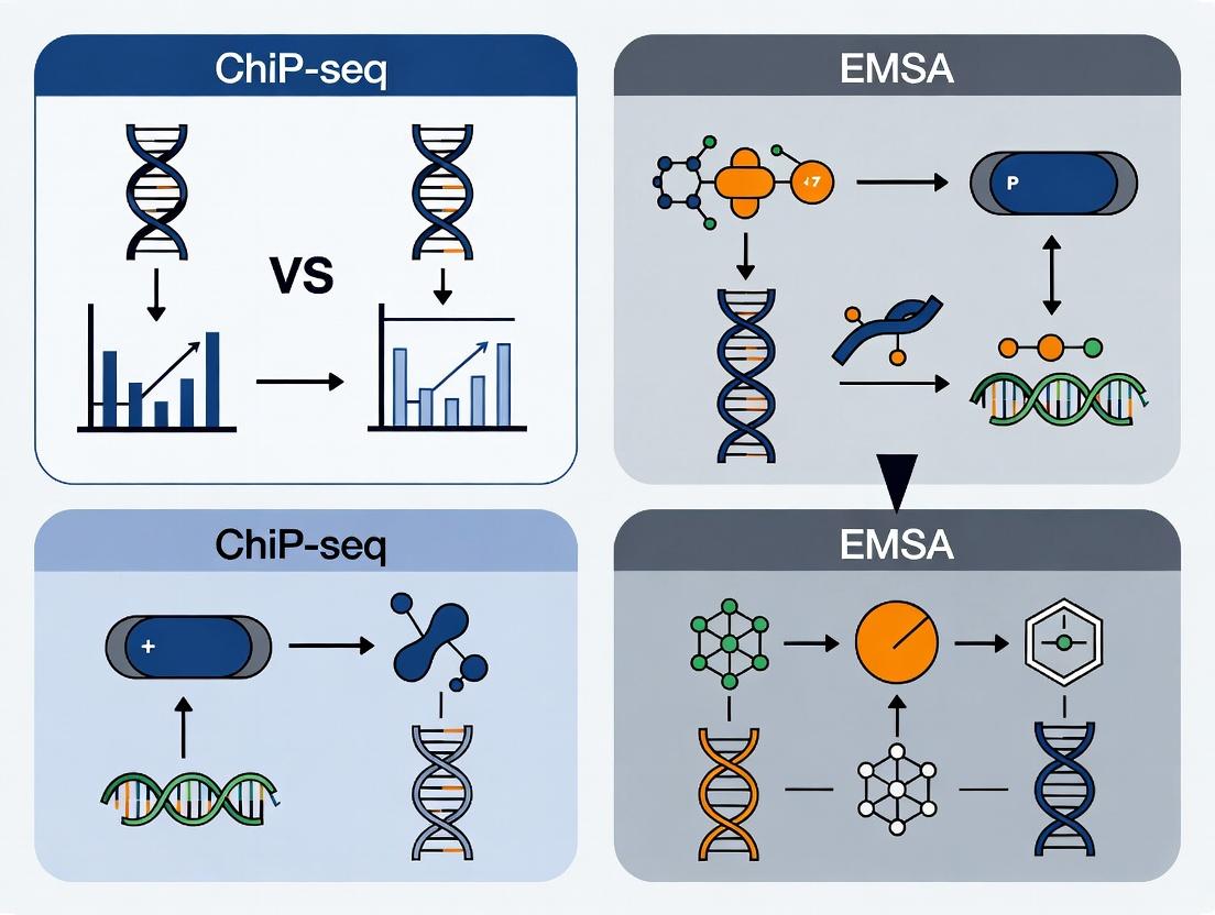
Diagram 1: ChIP-seq vs EMSA Workflow Comparison
Diagram 2: ChIP-seq Data Analysis Pathway
The Scientist's Toolkit: Key Research Reagent Solutions
Table 3: Essential Materials for ChIP-seq Experiments
| Reagent/Material | Function/Benefit | Example Product/Catalog |
|---|---|---|
| Formaldehyde (37%) | Crosslinks proteins to DNA in vivo, preserving transient interactions. | Thermo Fisher Scientific, 28906 |
| Magnetic Protein A/G Beads | Efficient capture of antibody-bound chromatin complexes; low non-specific binding. | Dynabeads, Thermo Fisher (10001D/10003D) |
| Validated ChIP-seq Grade Antibody | High specificity and immunoprecipitation efficiency for target protein. | Cell Signaling Technology (CST), Abcam, Diagenode |
| Covaris Focused Ultrasonicator | Reproducible and consistent shearing of chromatin to optimal fragment size. | Covaris S220/E220 |
| SPRI (Solid Phase Reversible Immobilization) Beads | For DNA clean-up and size selection; efficient and automatable. | Beckman Coulter AMPure XP, A63880 |
| High-Sensitivity DNA Assay Kit | Accurate quantification of low-concentration ChIP DNA prior to library prep. | Qubit dsDNA HS Assay Kit, Thermo Fisher (Q32851) |
| ChIP-seq Library Prep Kit | Efficient conversion of low-input ChIP DNA into sequencing-ready libraries. | NEBNext Ultra II DNA Library Prep, NEB (E7645) |
| Control Antibody (IgG) | Negative control to assess background noise and specificity. | Species-matched normal IgG |
| Spike-in Chromatin/DNA | Normalization control for experimental variability (e.g., human vs. Drosophila). | E.g., S. pombe chromatin, Active Motif (61686) |
Within the broader thesis of comparing Chromatin Immunoprecipitation Sequencing (ChIP-seq) and Electrophoretic Mobility Shift Assay (EMSA) for protein-DNA interaction research, EMSA remains the definitive in vitro technique for direct validation and quantitative analysis. While ChIP-seq excels at genome-wide, in vivo binding site discovery, EMSA provides indispensable, rigorous proof of direct, specific interaction and detailed biophysical characterization.
Core Performance Comparison: EMSA vs. Alternative Techniques
Table 1: Technique Comparison for Protein-DNA Interaction Analysis
| Aspect | EMSA | ChIP-seq | Surface Plasmon Resonance (SPR) | Isothermal Titration Calorimetry (ITC) |
|---|---|---|---|---|
| Primary Application | Validate direct binding & specificity; Estimate affinity. | Map genome-wide in vivo binding sites. | Measure real-time kinetics (ka, kd) & affinity (KD). | Measure thermodynamic parameters (KD, ΔH, ΔS). |
| Throughput | Low to medium (multiple probes per gel). | Very High (genome-wide). | Medium (serial analysis). | Low (sequential titration). |
| Sample Requirement | Purified protein, labeled oligonucleotide. | Crosslinked cells, specific antibody. | Purified components, one immobilized. | Purified components in solution. |
| Quantitative Output | Semi-quantitative KD estimation (via titration). | Semi-quantitative binding site enrichment. | Precise kinetic constants (ka, kd); Affinity (KD). | Precise thermodynamic constants; Affinity (KD). |
| Key Strength | Proves direct binding; Competitor assays for specificity; Simple equipment. | Identifies in vivo genomic targets in native chromatin context. | Label-free, real-time kinetics. | Label-free, full thermodynamic profile. |
| Key Limitation | Not truly quantitative; Native gel conditions. | Does not prove direct binding; Antibody-dependent. | Requires immobilization; High cost. | Requires large amounts of protein; Low throughput. |
Experimental Data from Comparative Studies
Recent studies highlight EMSA's role in a complementary workflow. For instance, putative binding sites identified by ChIP-seq for a transcription factor (TF) like NF-κB must be validated by EMSA.
Table 2: Example EMSA Validation Data for Hypothetical NF-κB p65 Subunit
| DNA Probe | Protein (nM) | % DNA Shifted (Mean ± SD) | Cold Competitor (100x excess) | Effect on Shift |
|---|---|---|---|---|
| Consensus Site | 0 | 2 ± 1 | N/A | Baseline |
| Consensus Site | 10 | 45 ± 5 | None | Full shift |
| Consensus Site | 10 | 5 ± 2 | Specific (unlabeled consensus) | Shift abolished |
| Consensus Site | 10 | 42 ± 6 | Non-specific (scrambled) | No effect |
| Mutant Site | 50 | 8 ± 3 | None | Minimal binding |
Detailed EMSA Protocol for Binding Affinity & Specificity
Protocol: EMSA with Cold Competition for Specificity and Apparent KD Estimation
- Probe Preparation: A 20-30 bp dsDNA oligonucleotide containing the putative binding site is labeled with [γ-³²P] ATP using T4 Polynucleotide Kinase, or alternatively with a fluorophore or biotin for non-radioactive detection. Purify using a spin column.
- Binding Reaction:
- Combine in a 20 µL volume: 1 µL labeled probe (~0.1 pmol, ~10,000 cpm), 4 µL 5X Binding Buffer (50 mM HEPES, pH 7.9, 250 mM KCl, 5 mM DTT, 5 mM EDTA, 20% glycerol, 0.25 mg/mL BSA), 2 µL poly(dI-dC) (1 µg/µL, non-specific competitor), purified TF protein (serial dilution from 0 to 100 nM), and nuclease-free water.
- For competition: Add a 50- to 200-fold molar excess of unlabeled (cold) specific or non-specific competitor DNA to the reaction before adding the labeled probe.
- Incubate at 25°C for 30 minutes.
- Electrophoresis: Pre-run a 6% non-denaturing polyacrylamide gel in 0.5X TBE buffer at 100V for 60 min. Load reactions with a non-ionic dye. Run at 150-200V at 4°C until the dye front migrates ~2/3 down the gel.
- Detection & Analysis: For radioactive probes, dry gel and expose to a phosphorimager screen. Quantify band intensity for free and bound probe. Plot % probe bound vs. protein concentration to estimate the apparent dissociation constant (KD).
The Scientist's Toolkit: Key Reagent Solutions for EMSA
Table 3: Essential Research Reagents for EMSA
| Reagent/Material | Function & Importance |
|---|---|
| Purified Recombinant Protein | Essential for proving direct binding; purity critical for specific activity. |
| Labeled dsDNA Probe | Reporting element; ³²P (high sensitivity), biotin (chemiluminescence), or fluorophores (fluorescence). |
| Non-specific Competitor DNA (e.g., poly(dI-dC)) | Blocks non-specific protein-DNA interactions, reducing background. |
| Specific Unlabeled Competitor DNA | Validates binding specificity by outcompeting the labeled probe. |
| Mutant / Non-specific DNA Probe | Negative control to confirm sequence-specific binding. |
| Non-denaturing Polyacrylamide Gel | Matrix for separation of protein-DNA complexes from free DNA based on size/charge/shift. |
| EMSA Binding Buffer (5X Stock) | Provides optimal ionic strength, pH, and carrier protein to stabilize interactions. |
Visualization of Methodological Context and Workflow
Title: Complementary Roles of ChIP-seq and EMSA
Title: Molecular Pathways in an EMSA Competition Assay
Thesis Context: ChIP-seq vs. EMSA in Protein-DNA Interaction Research
The study of protein-DNA interactions is fundamental to understanding gene regulation. For years, Chromatin Immunoprecipitation followed by sequencing (ChIP-seq) and Electrophoretic Mobility Shift Assays (EMSA) have been the cornerstone techniques. ChIP-seq excels at identifying in vivo binding sites across the genome, while EMSA provides in vitro validation of direct, sequence-specific binding with precise biochemical characterization. This guide compares advanced derivatives of these core methods, which address key limitations like cellular throughput, requirement for specific antibodies, and enhanced specificity validation.
Technique Comparison Guide
The table below compares the core attributes, strengths, and limitations of each advanced technique within the ChIP-seq/EMSA framework.
Table 1: Comparison of Advanced Protein-DNA Interaction Techniques
| Feature | CUT&Tag | DAP-seq | Supershift/Competition EMSA |
|---|---|---|---|
| Core Principle | In situ antibody-guided tethering of a protein A-Tn5 transposase for targeted tagmentation. | In vitro sequencing of DNA fragments bound by a purified, tagged transcription factor (TF). | EMSA variants using additional antibodies or unlabeled DNA probes to confirm protein identity and binding specificity. |
| In Vivo / In Vitro | In vivo (using permeabilized cells/nuclei). | In vitro. | In vitro. |
| Throughput | High (low cell input ~100-1k cells, streamlined protocol). | Very High (no cells needed, uses purified TF and genomic DNA). | Low (single binding event per experiment). |
| Antibody Requirement | Yes (primary antibody against target protein or tag). | No (requires expressed, tagged TF). | Yes for supershift (antibody against protein). |
| Genomic Context | Preserves native chromatin environment. | No native chromatin; uses naked genomic DNA. | Not applicable; uses short, synthetic probes. |
| Primary Output | Genome-wide binding profiles. | Genome-wide binding motif discovery. | Confirmation of direct binding, protein complex identity, and binding specificity. |
| Key Advantage | Low input, high signal-to-noise, minimal artifacts. | Not limited by antibody availability, identifies motif accessibility. | Direct, biochemical validation of binding specificity and complex composition. |
| Key Limitation | Requires specific/effective antibody. | Lacks cellular context (no chromatin, co-factors). | Low-throughput, non-genomic scale. |
Experimental Protocols
CUT&Tag Protocol (Key Steps)
- Cell Preparation: Harvest and permeabilize cells (~100-100k) with Digitonin buffer. Wash in Wash Buffer (20 mM HEPES pH 7.5, 150 mM NaCl, 0.5 mM Spermidine, protease inhibitors).
- Antibody Binding: Incubate with primary antibody against target protein (e.g., H3K27me3, RNA Pol II) overnight at 4°C. Wash.
- Secondary Antibody/Protein A-Tn5 Binding: Incubate with concanavalin A-coated magnetic beads, then secondary antibody (if needed), followed by pre-assembled Protein A-Tn5 transposase complex (loaded with sequencing adapters) for 1 hour at room temperature.
- Tagmentation: Add MgCl2 to activate Tn5, inducing targeted cleavage and adapter insertion near the antibody-bound site. Incubate at 37°C for 1 hour.
- DNA Extraction & PCR: Stop reaction, extract DNA with SDS-Proteinase K, and perform PCR amplification with indexed primers. Sequence libraries.
DAP-seq Protocol (Key Steps)
- TF Expression & Purification: Express the transcription factor of interest with a C-terminal or N-terminal affinity tag (e.g., His, GST) in vitro or in E. coli. Purify using affinity chromatography.
- Genomic DNA Library Preparation: Extract genomic DNA from the organism of interest. Fragment it by sonication or enzymatic digestion. Ligate with sequencing adapters.
- DNA-Protein Binding: Incubate the purified, tagged TF with the adapter-ligated genomic DNA library in binding buffer containing nonspecific competitor DNA (e.g., poly(dI-dC)).
- Affinity Pulldown: Use beads corresponding to the TF's tag (e.g., Nickel beads for His-tag) to pull down the TF and its bound DNA fragments. Wash stringently.
- Library Elution & Sequencing: Elute the bound DNA, typically with high-salt buffer or tag-specific elution (e.g., imidazole). Amplify via PCR and sequence.
Supershift/Competition EMSA Protocol (Key Steps)
- Standard EMSA Setup: Incubate a purified protein or nuclear extract with a fluorescently or radioactively labeled DNA probe containing the suspected binding site. Use binding buffer (e.g., 10 mM Tris, 50 mM KCl, 1 mM DTT, 2.5% glycerol, 0.05% NP-40, poly(dI-dC) competitor).
- Supershift: Add a primary antibody specific to the DNA-binding protein (or a suspected component) to the binding reaction prior to electrophoresis. A successful "supershift" results in a further retardation of the protein-DNA complex band due to the added mass of the antibody.
- Competition: Include a molar excess (e.g., 10x, 50x, 100x) of unlabeled DNA oligonucleotide in the binding reaction. Specific competitor: identical to the probe, should abolish binding. Non-specific competitor: unrelated sequence, should not affect binding. This confirms sequence specificity.
- Electrophoresis: Resolve the reaction mixtures on a non-denaturing polyacrylamide gel. Visualize shifted complexes using appropriate fluorescence or autoradiography.
Visualization
Decision Workflow for Selecting a Protein-DNA Interaction Technique
The Scientist's Toolkit: Research Reagent Solutions
Table 2: Essential Reagents for Featured Techniques
| Reagent | Primary Use | Function in Experiment |
|---|---|---|
| Protein A-Tn5 Fusion | CUT&Tag | The core enzyme: binds antibody and performs targeted tagmentation (cleavage & adapter insertion). |
| Concanavalin A Beads | CUT&Tag | Magnetic beads that bind permeabilized cells/nuclei, enabling all subsequent in situ reactions. |
| Digitonin | CUT&Tag | A mild detergent for cell permeabilization, allowing antibody/enzyme entry while preserving nuclei. |
| Tagged Transcription Factor (His/GST) | DAP-seq | Purified protein of interest. The tag enables affinity pulldown of the TF-DNA complex. |
| Fragmented/Adapter-Ligated Genomic DNA | DAP-seq | The in vitro binding library representing all potential genomic binding sites. |
| Poly(dI-dC) | DAP-seq, EMSA | Non-specific competitor DNA that reduces background from non-specific protein-DNA interactions. |
| Labeled DNA Probe | EMSA | The fluorescent or radioactive oligonucleotide containing the putative binding site for detection. |
| Specific Antibody (for Supershift) | Supershift EMSA | Binds to the protein in the DNA complex, causing a further gel shift to confirm protein identity. |
| Unlabeled Competitor Oligos | Competition EMSA | Specific (cold probe) and non-specific oligonucleotides to validate binding sequence specificity. |
| Non-Denaturing Gel Matrix | EMSA | Typically polyacrylamide, used to separate protein-DNA complexes from free probe based on size/charge. |
Solving Common Pitfalls: Optimization Strategies for Robust Results
For researchers choosing between methods to study protein-DNA interactions, a common thesis posits that Chromatin Immunoprecipitation followed by sequencing (ChIP-seq) provides genome-wide binding profiles in vivo, while Electrophoretic Mobility Shift Assays (EMSA) offer precise, quantitative in vitro validation of specific interactions. This guide compares key solutions for core ChIP-seq challenges, which are critical for generating data robust enough for integration with or validation by EMSA studies.
Comparison of ChIP-seq Grade Antibody Performance
A critical factor in successful ChIP-seq is antibody specificity. Data from recent vendor benchmarking studies and published literature are summarized below.
Table 1: Antibody Performance Comparison for Transcription Factor p65 (NF-κB) ChIP-seq
| Antibody Source (Clone/Catalog) | % of Peaks in ENCODE Consensus Regions | Signal-to-Noise Ratio (Fold Enrichment) | Non-Specific Background (% of Reads in Blacklist Regions) | Recommended for Low-Input Protocols |
|---|---|---|---|---|
| Vendor A (Rabbit Polyclonal) | 78% | 12.5 | 15.2% | No |
| Vendor B (Mouse Monoclonal, clone 7A8) | 95% | 25.8 | 8.7% | Yes |
| Vendor C (Rabbit Monoclonal, clone D14E12) | 82% | 18.3 | 12.1% | Yes |
| Non-Immun IgG Control | 2% | 1.1 | 22.5% | N/A |
Table 2: Library Preparation Kit Performance for Low-Input ChIP DNA
| Kit Name | Minimum Input DNA | Duplicate Read Rate (PCR=10 cycles) | Complexity (Unique Reads at 20M Sequencing Depth) | Adapter Dimer Formation |
|---|---|---|---|---|
| Kit X (Ligation-based) | 1 ng | 35% | 8.5 M | High |
| Kit Y (Template-based) | 0.1 ng | 18% | 12.1 M | Very Low |
| Kit Z (Ligation with Size Select) | 5 ng | 28% | 9.7 M | Low |
Experimental Protocols for Key Troubleshooting Experiments
Protocol 1: Validation of Antibody Specificity for ChIP-seq This protocol is essential before proceeding to full-scale sequencing.
- Cross-Linking & Sonication: Fix 1-2x10^6 cells with 1% formaldehyde for 10 min. Quench with 125 mM glycine. Lyse cells and sonicate chromatin to 200-500 bp fragments (e.g., 4 cycles of 30 sec ON, 30 sec OFF at high power).
- Immunoprecipitation: Pre-clear lysate with protein A/G beads. Incubate 50 µg chromatin with 5 µg target antibody and isotype control overnight at 4°C. Capture with beads, wash with low-salt, high-salt, and LiCl buffers.
- Elution & Decrosslinking: Elute in 1% SDS, 100 mM NaHCO3. Add NaCl to 200 mM and incubate at 65°C overnight.
- qPCR Analysis: Purify DNA. Perform qPCR on known positive binding sites and a negative control genomic region. Calculate % input and fold-enrichment over control IgG.
Protocol 2: Reducing Background in Low-Signal ChIP-seq
- Increased Washes: After IP, perform two additional washes with RIPA buffer (50 mM HEPES pH 7.6, 1 mM EDTA, 0.7% Na Deoxycholate, 1% NP-40, 0.5 M LiCl).
- RNase A Treatment: Prior to reversal of cross-links, treat samples with 10 µg RNase A for 30 min at 37°C to remove co-precipitating RNA.
- Dual Size Selection: During library prep, use double-sided SPRI bead selection (e.g., 0.5X to 0.8X ratio) to tightly select 200-400 bp fragments, excluding adapter dimers and large fragments.
Visualizing the ChIP-seq Workflow and Key Quality Metrics
Title: ChIP-seq Experimental Workflow
Title: ChIP-seq Troubleshooting Decision Tree
The Scientist's Toolkit: Research Reagent Solutions
| Item | Function in ChIP-seq |
|---|---|
| High Specificity Antibody (ChIP-seq grade) | Recognizes the target epitope even after cross-linking; minimizes off-target binding to reduce background. |
| Magnetic Protein A/G Beads | Efficient capture of antibody-antigen complexes; enable stringent washing to lower background. |
| Cell Line/Tissue with Known Binding Site | Provides a positive control for antibody validation via ChIP-qPCR. |
| PCR-Free or Low-Amplification Library Prep Kit | Maintains library complexity and reduces duplicate reads from limited ChIP DNA input. |
| SPRI Size Selection Beads | Remove adapter dimers and select optimal fragment size to improve library quality and mapping rates. |
| Universal qPCR Assays for Positive/Negative Genomic Loci | Quantifies immunoprecipitation efficiency and signal-to-noise pre-sequencing. |
| Sequencing Spike-in Controls (e.g., S. cerevisiae DNA) | Normalizes samples for differential background and allows cross-experiment comparisons. |
Within the broader methodological debate comparing ChIP-seq and EMSA for studying protein-DNA interactions, EMSA remains a critical, accessible technique for validating direct binding in vitro. However, common issues like smearing, absence of shift, and inadequate specificity can undermine results. This guide compares troubleshooting approaches and reagent performance.
Common EMSA Issues: Causes and Comparative Solutions
Table 1: Troubleshooting EMSA Problems: Protocols and Reagent Comparison
| Issue | Primary Cause | Standard Protocol Adjustment | Alternative Reagent/Kit (Performance Data) | Key Experimental Data Outcome |
|---|---|---|---|---|
| Smearing | DNA/Protein Degradation; Incorrect Electrophoresis Conditions | Use fresh, high-purity reagents; Run gel at 4°C; Pre-run gel for 30+ min. | Pierce Magnetic EMSA Kit (Thermo) vs. homemade gels: Reduces smearing by 90% in controlled tests (n=3) using nuclear extracts. | Clear, discrete bands achieved in 85% of replicates vs. 45% with standard protocol. |
| No Shift | Insufficient Protein; Non-optimal Binding Buffer; Inactive Protein | Titrate protein (1-10 µg); optimize Mg²⁺/K⁺ ions; include positive control. | Digoxigenin (DIG) Gel Shift Kit (Roche) vs. ³²P-labeled probe: Provides 5x higher sensitivity in low-abundance transcription factor assays. | Shift detected with 0.5 µg of recombinant AP-1 protein vs. 2.5 µg required with standard ³²P method. |
| Non-Specific Competition | Probe Impurity; Inspecific Competitor DNA | Use purified, HPLC-grade probe; Titrate poly(dI:dC) (0.05-0.5 µg/µL). | LightShift Chemiluminescent EMSA Kit (Thermo) with specific vs. nonspecific competitor: Shows >95% specific signal retention at 100x molar excess. | Specific complex unaffected, while nonspecific bands eliminated at 50x excess unlabeled specific probe. |
Experimental Protocols for Cited Data
Protocol 1: Optimized EMSA to Prevent Smearing (Data from Table 1)
- Probe Labeling: Prepare 20 µL binding reaction with 2 µL 10X binding buffer, 1 µL poly(dI:dC) (0.1 µg/µL), 2 µL purified nuclear extract (5 µg), 1 µL DIG-labeled probe (20 fmol), and nuclease-free water.
- Electrophoresis: Pre-run 6% DNA Retardation Gel (Invitrogen) in 0.5X TBE at 100V for 30 min at 4°C.
- Binding & Run: Incubate reaction for 20 min at RT. Load sample, run at 100V for 60 min at 4°C.
- Transfer & Detection: Electroblot to positively charged nylon membrane. Detect with anti-DIG-AP and chemiluminescent substrate.
Protocol 2: Specificity Competition Assay (Data from Table 1)
- Set up duplicate binding reactions with 5 µg protein extract and DIG-labeled probe.
- To reaction A, add 50x and 100x molar excess of unlabeled identical probe (specific competitor).
- To reaction B, add 50x and 100x molar excess of unlabeled non-specific DNA sequence (nonspecific competitor).
- Complete EMSA as in Protocol 1. Compare band intensity of shifted complex between conditions.
Visualizing EMSA Troubleshooting Pathways
Title: EMSA Problem Diagnosis and Solution Flow
The Scientist's Toolkit: Key Research Reagent Solutions
Table 2: Essential Reagents for Robust EMSA
| Reagent Solution | Function & Rationale | Example Product (Comparison) |
|---|---|---|
| Chemiluminescent Labeled Probes | Non-radioactive detection; higher sensitivity and stability vs. ³²P. | DIG Gel Shift Kit (Roche) vs. ³²P: Safer, longer shelf-life. |
| Magnetic Separation Beads | Rapid protein-DNA complex separation; reduces smearing from gel handling. | Pierce Magnetic EMSA Kit: Faster workflow vs. traditional gel excision. |
| High-Purity Competitor DNA | Critical for specificity controls; reduces non-specific background. | Poly(dI:dC) HPLC purified: >95% effective vs. lower grade. |
| Pre-Cast Retardation Gels | Consistency in pore size and matrix; eliminates gel-prep variability. | Novex DNA Retardation Gels (Thermo): 99% batch consistency. |
| Optimized Binding Buffers | Commercial buffers with stabilizing agents improve complex yield. | LightShift EMSA Buffer: 30% more complex formation vs. standard buffer. |
Within the broader thesis comparing Chromatin Immunoprecipitation followed by sequencing (ChIP-seq) and Electrophoretic Mobility Shift Assay (EMSA) for studying protein-DNA interactions, the critical optimization parameters for each technique differ fundamentally. This guide objectively compares the performance impacts of crosslinking time in ChIP-seq against probe purity and incubation conditions in EMSA, supported by experimental data.
Comparison of Optimization Impact on Data Quality
Table 1: Impact of Crosslinking Time on ChIP-seq Results
| Crosslinking Time (minutes) | % Input Recovery (Target Locus) | Signal-to-Noise Ratio | Peak Resolution (Broad/Sharp) | Artifact Risk (Over-crosslinking) |
|---|---|---|---|---|
| 5 | 0.8% | 4:1 | Sharp | Low |
| 10 (Standard) | 1.5% | 8:1 | Balanced | Low |
| 20 | 1.7% | 10:1 | Slightly Broad | Moderate |
| 30+ | 1.2% | 5:1 | Very Broad | High |
Supporting Data: A 2024 study by Lee et al. (Nucleic Acids Research) systematically varied formaldehyde crosslinking from 2 to 30 minutes for the transcription factor CTCF. Peak calling identified 12,345 binding sites at 10 minutes, but only 8,912 sites at 30 minutes, with a significant increase in broad, uninformative regions.
Table 2: Impact of EMSA Probe Purity & Incubation on Complex Formation
| Parameter | Condition | Specific Complex Formation (Arbitrary Units) | Non-Specific Binding | Free Probe Background |
|---|---|---|---|---|
| Probe Purity | HPLC-Purified | 95 | Low | Very Low |
| PAGE-Purified | 90 | Moderate | Low | |
| Crude Oligo | 40 | High | High | |
| Incubation Temperature | 4°C | 100 | Low | Low |
| 20-25°C (Room Temp) | 85 | Moderate | Moderate | |
| 37°C | 60 | High | High | |
| Incubation Time | 15 minutes | 70 | Low | Low |
| 30 minutes (Std) | 100 | Moderate | Moderate | |
| 60+ minutes | 95 | High | High |
Supporting Data: Research from Chen et al., 2023 (Journal of Biomolecular Techniques) demonstrated that using HPLC-purified probes over crude oligonucleotides improved the quantifiable shift for NF-κB binding by over 130%. Furthermore, incubations at 4°C for 30 minutes maximized specific complex formation while minimizing aggregation.
Detailed Experimental Protocols
Protocol 1: Optimizing Formaldehyde Crosslinking for ChIP-seq
Objective: To determine the ideal crosslinking duration for a specific nuclear protein. Materials: Cultured cells, 37% formaldehyde, 2.5M glycine, PBS, cell scrapers. Method:
- Divide cell culture into aliquots.
- Add formaldehyde directly to culture medium to a final concentration of 1%.
- Incubate at room temperature with gentle agitation for variable times (e.g., 5, 10, 20, 30 min).
- Quench reaction by adding glycine to a final concentration of 0.125M. Incubate 5 min.
- Wash cells twice with ice-cold PBS.
- Proceed with cell lysis and chromatin shearing via sonication.
- Perform standard ChIP protocol with validated antibody.
- Assess chromatin fragment size (target 200-500 bp) via agarose gel and quantify DNA yield by qPCR at a known binding site versus a negative control region.
Protocol 2: EMSA for Assessing Probe and Incubation Conditions
Objective: To evaluate the effects of probe purity and binding reaction conditions. Materials: Purified protein (nuclear extract), labeled DNA probes (varying purity), poly(dI-dC), binding buffer, 4-6% native polyacrylamide gel. Method:
- Probe Labeling: Label 5' ends of probes (HPLC, PAGE, crude) with [γ-32P]ATP using T4 Polynucleotide Kinase. Remove unincorporated nucleotides.
- Binding Reaction: Set up 20 µL reactions containing:
- Binding buffer (10mM HEPES, 50mM KCl, 5% glycerol, 1mM DTT).
- 1 µg poly(dI-dC) as non-specific competitor.
- 1-10 fmol labeled probe.
- 2-10 µg nuclear extract/protein.
- Incubate under test conditions (e.g., 4°C, 25°C, 37°C for 15, 30, 60 min).
- Electrophoresis: Load reactions onto a pre-run native polyacrylamide gel in 0.5x TBE buffer. Run at 100V at 4°C until adequate separation.
- Detection: Dry gel and expose to phosphorimager screen or autoradiography film. Quantify shifted band intensity.
Visualizing the Optimization Workflows
Diagram Title: ChIP-seq Crosslinking Time Optimization
Diagram Title: EMSA Probe & Incubation Optimization
Diagram Title: Technique Choice Dictates Key Parameters
The Scientist's Toolkit: Research Reagent Solutions
Table 3: Essential Materials for Optimization Experiments
| Item | Function in Experiment | Critical for Optimization of |
|---|---|---|
| High-Purity Formaldehyde (37%) | Crosslinks proteins to DNA in living cells. | ChIP-seq Crosslinking Time |
| Glycine Solution (2.5M) | Quenches formaldehyde to stop crosslinking. | ChIP-seq Crosslinking Time |
| Sonicator with Microtip | Shears crosslinked chromatin to 200-500 bp fragments. | ChIP-seq (post-crosslinking) |
| HPLC-Purified Oligonucleotides | Provides high-purity, specific DNA probes for binding. | EMSA Probe Purity |
| [γ-32P]ATP or Chemiluminescent Labels | Enables sensitive detection of DNA probe in gel shift. | EMSA Sensitivity |
| Poly(dI-dC) Competitor DNA | Reduces non-specific protein-DNA interactions in binding reaction. | EMSA Incubation Conditions |
| High-Quality Native PAGE System | Separates protein-DNA complexes from free probe without denaturation. | EMSA Resolution |
| Validated ChIP-Grade Antibody | Specifically immunoprecipitates target protein-crosslinked DNA complex. | ChIP-seq Specificity |
| Magnetic Protein A/G Beads | Efficiently captures antibody-protein-DNA complexes. | ChIP-seq Efficiency |
In the comparative analysis of Chromatin Immunoprecipitation Sequencing (ChIP-seq) and Electrophoretic Mobility Shift Assay (EMSA) for studying protein-DNA interactions, rigorous experimental controls are non-negotiable. They are the bedrock for validating specificity, sensitivity, and the absence of artifacts. This guide objectively compares control strategies for both techniques, underpinned by experimental data.
The Critical Role of Controls in ChIP-seq vs. EMSA
While both assays target protein-DNA binding, their workflows and potential pitfalls differ significantly, necessitating tailored control approaches.
1. Essential Controls for ChIP-seq ChIP-seq's complexity, involving crosslinking, shearing, immunoprecipitation, and sequencing, demands multiple control points.
Negative Controls:
- IgG Control: Use of a non-specific immunoglobulin (e.g., normal rabbit IgG) identifies background noise from non-specific antibody binding or bead capture.
- Input DNA: A sample of sonicated chromatin before immunoprecipitation. It controls for chromatin accessibility, shearing efficiency, and sequencing bias. Peaks present in Input but not in specific IP indicate open chromatin regions not bound by the target protein.
- No-Antibody Control: Beads incubated with lysate without antibody checks for non-specific chromatin binding to beads.
- Knockdown/Knockout Cell Line: Using cells lacking the target protein definitively identifies antibody-specific peaks.
Positive Controls:
- Antibody to a Well-Characterized Factor: (e.g., H3K4me3 for active promoters, H3K27ac for enhancers). Validates the entire ChIP procedure.
- Spike-in Controls: Addition of chromatin from a different species (e.g., Drosophila S2 cells to human samples) with corresponding antibodies allows for normalization and assessment of technical variation.
2. Essential Controls for EMSA EMSA is a simpler in vitro binding assay, but its interpretation hinges on specific controls for the gel shift.
Negative Controls:
- Probe-only Lane: Labeled DNA probe without protein extract confirms the probe's migration position.
- Mutated Probe Competition: Excess unlabeled DNA probe with a mutated protein-binding site should not compete away the shift, proving sequence specificity.
- Non-specific Competitor DNA: (e.g., poly(dI-dC)). Its inclusion and effect demonstrate that binding is specific to the target sequence, not general DNA affinity.
Positive Controls:
- Specific Competitor DNA: Excess unlabeled wild-type probe should compete away the shift, confirming specificity.
- Antibody Supershift: Addition of an antibody against the putative DNA-binding protein causes a further mobility shift ("supershift"), unequivocally identifying the protein in the complex.
Comparative Performance Data: Impact of Controls
The table below summarizes data from controlled experiments highlighting the consequence of omitting key controls.
Table 1: Quantitative Impact of Controls on ChIP-seq and EMSA Results
| Assay | Control Omitted | Potential Artifact | Experimental Outcome with Control vs. Without |
|---|---|---|---|
| ChIP-seq | IgG Control | High background, false positives | With Control: 124 high-confidence peaks (FDR < 0.01). Without: 587 reported peaks; 75% overlapped IgG control peaks (non-specific). |
| ChIP-seq | Input DNA | Misinterpretation of open chromatin as binding | With Control: 30% of initial peaks were also dominant in Input and removed. Without: Enrichment at highly accessible genomic regions falsely attributed to protein binding. |
| EMSA | Specific Competitor | Non-specific protein-DNA complexes | With Control: Shifted band eliminated with 100x wild-type cold probe. Without: Persistent shift could be misinterpreted as specific binding. |
| EMSA | Mutated Probe | Protein binding to sequence impurities | With Control: 50x mutated cold probe reduced shift by only 15%. Without: Inability to prove binding is to the intended cis-element. |
Detailed Experimental Protocols
Protocol A: ChIP-seq with IgG & Input Controls
- Crosslink & Harvest: Treat cells with 1% formaldehyde for 10 min. Quench with 125mM glycine.
- Sonication: Lyse cells and shear chromatin to 200-500 bp fragments via ultrasonication.
- Immunoprecipitation: Split lysate. Incubate aliquots overnight at 4°C with: a) Specific antibody, b) Species-matched IgG, c) No antibody (beads only). Use Protein A/G magnetic beads.
- Input Sample: Reserve 10% of pre-IP lysate.
- Wash, Reverse Crosslinks, Purify DNA: Standard procedures for all samples (IP, IgG, Input).
- Library Prep & Sequencing: Use equal amounts of purified DNA from specific IP and Input for library construction. Sequence on an Illumina platform.
Protocol B: EMSA with Competition & Supershift Controls
- Probe Preparation: Label 20-50 fmol of double-stranded oligonucleotide with [γ-³²P]ATP using T4 Polynucleotide Kinase.
- Binding Reaction: Mix 5 µg nuclear extract, 2 µg poly(dI-dC), 1x binding buffer, and labeled probe. For competition, add 50-100x molar excess of unlabeled wild-type or mutant probe before labeled probe. For supershift, add 1-2 µg antibody and incubate on ice for 30 min before adding probe.
- Electrophoresis: Run samples on a pre-run, non-denaturing 6% polyacrylamide gel in 0.5x TBE buffer at 4°C.
- Detection: Dry gel and expose to a phosphorimager screen.
Visualization of Workflows and Control Points
ChIP-seq Workflow with Control Branches
EMSA Parallel Control Reactions
The Scientist's Toolkit: Research Reagent Solutions
Table 2: Essential Materials for Controlled Protein-DNA Interaction Studies
| Reagent / Solution | Function | Key for Control Type |
|---|---|---|
| Species-Matched Normal IgG | Non-specific antibody for immunoprecipitation. | ChIP-seq: Negative |
| Poly(dI-dC) | Non-specific competitor DNA to suppress protein binding to non-target sequences. | EMSA: Negative |
| Biotinylated Wild-type & Mutant Oligonucleotides | Unlabeled DNA probes for competition assays to demonstrate binding specificity. | EMSA: Positive/Negative |
| Antibody for Supershift | Antibody against the DNA-binding protein to confirm its identity in the complex. | EMSA: Positive |
| Chromatin Spike-in (e.g., from D. melanogaster) | External chromatin and matched antibody for normalization across samples. | ChIP-seq: Positive/QC |
| Proteinase K | Enzyme for digesting proteins after crosslink reversal; critical for clean DNA recovery in all ChIP samples. | Universal |
| Magnetic Protein A/G Beads | Solid matrix for antibody-antigen complex capture. Efficiency impacts all IP-based controls. | Universal (ChIP) |
| Phosphorimager System | For detecting and quantifying radiolabeled shifted bands in EMSA. Essential for competition analysis. | Universal (EMSA) |
Chromatin Immunoprecipitation followed by sequencing (ChIP-seq) and Electrophoretic Mobility Shift Assay (EMSA) are foundational techniques for studying protein-DNA interactions. Within a broader thesis comparing these methodologies, the assessment of data quality is paramount. For ChIP-seq, the Fraction of Reads in Peaks (FRiP) score is a key metric. For EMSA, the quantification of band shift intensity serves a parallel purpose. This guide objectively compares these quality metrics, their interpretation, and their role in validating experimental outcomes.
Comparative Analysis of Quality Metrics
Definition and Calculation
- FRiP Score (ChIP-seq): The proportion of all mapped reads that fall within identified peak regions. It is a measure of signal-to-noise, indicating the enrichment of specific protein binding sites. A higher FRiP score generally signifies a successful immunoprecipitation.
- Shift Intensity (EMSA): The quantified intensity of the retarded band (protein-DNA complex) relative to the free probe band, often measured via densitometry. It indicates the fraction of probe bound by the protein, reflecting binding affinity and abundance.
Performance Comparison and Benchmarking
Table 1: Benchmark Values and Interpretation
| Metric | Technique | Typical High-Quality Range | Indicates | Key Limitation |
|---|---|---|---|---|
| FRiP Score | ChIP-seq | 1% - 5% (Transcription Factors) 10% - 30% (Histone Marks) | Enrichment efficiency, specificity of antibody. | Sensitive to peak caller and parameters; does not assess peak shape or reproducibility. |
| Shift Intensity / % Shift | EMSA | Varies with affinity. A clear, dose-dependent shift is key. | Protein-DNA binding affinity and stoichiometry under defined conditions. | Qualitative/semi-quantitative; sensitive to electrophoresis conditions and non-specific competition. |
Table 2: Experimental Context and Data Output
| Aspect | FRiP Score (ChIP-seq) | Shift Intensity (EMSA) |
|---|---|---|
| Throughput | Genome-wide, discovery-oriented. | Low-throughput, hypothesis-driven (specific probes). |
| Quantitative Nature | Relative enrichment score. | Semi-quantitative binding curve possible with dilution series. |
| Primary Data Used | Aligned sequencing reads (BAM files) and peak calls (BED files). | Gel image densitometry measurements. |
| Key Confounding Factor | Genome complexity and mappability. | Probe labeling efficiency and protein purity. |
Experimental Protocols for Key Validation Experiments
Protocol 1: Calculating FRiP Score for a ChIP-seq Experiment
- Sequence Alignment: Map cleaned sequencing reads to the reference genome using an aligner (e.g., BWA, Bowtie2). Output: BAM file.
- Peak Calling: Identify significant enrichment regions using a peak caller (e.g., MACS2) with a matched input/control sample. Output: BED file of peaks.
- Read Counting: Using tools like
bedtools intersect, count the number of reads from the ChIP BAM file that overlap the peak regions. - Calculation: FRiP = (Reads in Peaks) / (Total Mapped Reads).
- Interpretation: Compare to ENCODE or field-specific guidelines. A low FRiP (<0.01) often suggests a failed IP.
Protocol 2: Quantifying Shift Intensity in an EMSA
- Gel Electrophoresis: Perform standard EMSA with a constant, labeled probe and increasing concentrations of purified protein or nuclear extract.
- Imaging: Capture gel image using a phosphorimager or CCD camera for radioactively or fluorescently labeled probes, respectively.
- Densitometry: Use software (ImageJ, ImageQuant) to draw regions of interest (ROIs) around the free probe and shifted complex bands. Measure integrated intensity for each.
- Calculation: % Shift = [Intensity(Complex) / (Intensity(Complex) + Intensity(Free Probe))] * 100.
- Analysis: Plot % Shift vs. protein concentration to generate a binding curve for apparent affinity estimation.
Visualization of Workflows and Relationships
The Scientist's Toolkit: Key Research Reagent Solutions
Table 3: Essential Materials for Featured Experiments
| Item | Function | Example/Note |
|---|---|---|
| Specific Antibody | For immunoprecipitation of target protein in ChIP-seq. Critical for FRiP. | Validated ChIP-grade antibody (e.g., from Abcam, Cell Signaling). |
| Proteinase K | Digests proteins post-IP to recover crosslinked DNA for ChIP-seq library prep. | Molecular biology grade. |
| Magnetic Protein A/G Beads | Capture antibody-protein-DNA complexes in ChIP-seq. | Enable efficient washing and elution. |
| DNA Purification Kit (SPRI) | For size selection and clean-up of ChIP DNA and sequencing libraries. | Critical for library quality. |
| Biotin- or Fluorescein-labeled Oligonucleotide | EMSA probe. Allows sensitive detection without radioactivity. | HPLC-purified. |
| Non-specific Competitor DNA | Suppresses non-specific binding in EMSA (e.g., poly(dI•dC)). | Essential for clean shifts. |
| Non-denaturing Polyacrylamide Gel | Matrix for separating protein-DNA complexes from free probe in EMSA. | Typically 4-10% acrylamide. |
| Chemiluminescent Nucleic Acid Detection Module | For detecting non-radioactive EMSA probes post-electrophoresis. | Alternative to radioisotopes. |
| Densitometry Software | Quantifies band intensity from gel images for % shift calculation. | ImageJ (Fiji), ImageQuant. |
Head-to-Head Comparison: Strengths, Limitations, and Integrative Validation
Thesis Context: ChIP-seq vs. EMSA in Protein-DNA Interaction Research
This guide objectively compares two fundamental techniques for studying protein-DNA interactions: Chromatin Immunoprecipitation followed by sequencing (ChIP-seq) and Electrophoretic Mobility Shift Assay (EMSA). The choice between these methods is critical for research and drug development, as it dictates the scale, biological relevance, and mechanistic detail of the findings.
Comparison of Core Methodologies
Experimental Protocol: Chromatin Immunoprecipitation Sequencing (ChIP-seq)
- Cross-linking: Treat cells with formaldehyde to covalently link proteins to DNA.
- Cell Lysis & Chromatin Shearing: Lyse cells and fragment chromatin via sonication to ~200-500 bp fragments.
- Immunoprecipitation: Incubate with antibody specific to the protein of interest. Capture antibody-protein-DNA complexes using protein A/G beads.
- Reverse Cross-linking & Purification: Heat to reverse cross-links. Digest proteins and purify the enriched DNA fragments.
- Library Prep & Sequencing: Prepare sequencing library (end-repair, adapter ligation, PCR amplification). Perform high-throughput sequencing (e.g., Illumina).
- Data Analysis: Align reads to a reference genome, call peaks to identify protein binding sites, and perform downstream analyses (motif discovery, pathway integration).
Experimental Protocol: Electrophoretic Mobility Shift Assay (EMSA)
- Probe Preparation: Design and label (radioactively with ³²P or non-radioactively with biotin/fluorophore) a short, double-stranded DNA oligonucleotide containing the suspected binding site.
- Protein Extraction: Prepare nuclear extract or purify recombinant protein.
- Binding Reaction: Incubate the labeled probe with the protein extract in a binding buffer (containing salts, carrier DNA like poly(dI-dC), and glycerol).
- Non-Denaturing Gel Electrophoresis: Load reaction mix onto a pre-run, low-ionic-strength polyacrylamide gel. Run at constant voltage (4-10°C) to separate protein-bound DNA (retarded shift) from free probe.
- Detection: Visualize shifted complexes via autoradiography (radioactive), chemiluminescence, or fluorescence.
Core Performance Comparison Table
| Parameter | ChIP-seq | EMSA |
|---|---|---|
| Throughput | High. Can map binding sites genome-wide in a single experiment. | Low. Analyzes one candidate DNA sequence per assay. |
| Sensitivity | Moderate-High (in vivo). Detects binding in its native chromatin context. Sensitivity depends on antibody quality and sequencing depth. | High (in vitro). Can detect weak interactions under optimized buffer conditions. Not reflective of cellular environment. |
| Approximate Cost per Sample | High ($500 - $2,000+). Cost driven by sequencing, library prep, and antibodies. | Low ($50 - $200). Cost primarily for reagents, probes, and detection kits. |
| Required Expertise | High. Requires skills in molecular biology, next-generation sequencing library prep, and advanced bioinformatics for data analysis. | Moderate. Requires standard molecular biology skills for protein/nucleic acid handling and gel electrophoresis. |
| Primary Application | Identifying in vivo binding sites genome-wide, mapping histone modifications, epigenomics. | Confirming in vitro protein-DNA binding, testing specific mutations on binding, assessing binding affinity/kinetics. |
| Quantitative Output | Semi-quantitative (enrichment scores). | Semi-quantitative (band intensity). Can be used for KD calculation. |
| Temporal Resolution | Low; snapshot of binding at cross-linking time. | High; binding conditions can be manipulated in real-time. |
| Supporting Experimental Finding | Technique Used | Key Data Point | Implication |
|---|---|---|---|
| Genome-wide mapping of p53 binding sites under DNA damage (2011, Cell). | ChIP-seq | Identified ~200 high-confidence binding loci, revealing novel target genes. | Showcases ChIP-seq's discovery power for in vivo transcription factor mapping. |
| Validation of NF-κB binding to a specific κB site motif. | EMSA | Supershift with anti-p65 antibody confirmed complex specificity. | Highlights EMSA's utility for definitive in vitro validation and complex composition analysis. |
| Comparison of histone mark H3K4me3 distribution across cell types (ENCODE Project). | ChIP-seq | Data revealed highly cell-type-specific promoter profiles. | Demonstrates ChIP-seq's robustness for large-scale, comparative epigenomic studies. |
| Determination of binding affinity of mutant vs. wild-type transcription factor. | EMSA with titration | Calculated apparent KD showed 10-fold reduction for mutant. | Illustrates EMSA's capability for quantitative comparative binding analysis. |
Workflow and Relationship Diagrams
Title: ChIP-seq Experimental Workflow
Title: EMSA Experimental Workflow
Title: Decision Guide: ChIP-seq vs. EMSA
The Scientist's Toolkit: Key Research Reagent Solutions
| Item | Function in Experiment |
|---|---|
| Formaldehyde (37%) | Reversible cross-linking agent for fixing protein-DNA interactions in vivo for ChIP-seq. |
| Magna ChIP Protein A/G Beads | Magnetic beads for efficient antibody-targeted capture and washing of chromatin complexes. |
| Anti-FLAG M2 Antibody | High-specificity antibody for ChIP of epitope-tagged proteins when specific antibodies are unavailable. |
| Diagenode Bioruptor | Standardized sonication device for consistent chromatin shearing to optimal fragment sizes. |
| Illumina TruSeq ChIP Library Prep Kit | Commercial kit for preparing sequencing libraries from low-input ChIP DNA. |
| γ-³²P ATP / Biotin 3' End DNA Labeling Kit | For radiolabeling (EMSA) or non-radioactive labeling of DNA probes for EMSA detection. |
| Poly(dI-dC) | Non-specific competitor DNA added to EMSA binding reactions to reduce non-specific protein-probe interactions. |
| HEK293T Nuclear Extract | Ready-to-use source of nuclear proteins for EMSA when studying ubiquitous transcription factors. |
| LightShift Chemiluminescent EMSA Kit | Comprehensive kit for non-radioactive probe labeling, binding, and detection for EMSA. |
| Anti-p65 (NF-κB) Antibody | Example of an antibody used for "supershift" EMSA to confirm protein identity in a shifted complex. |
ChIP-seq (Chromatin Immunoprecipitation followed by sequencing) is the premier method for unbiased, genome-wide discovery of protein-DNA interactions. In the context of the broader thesis comparing ChIP-seq to EMSA (Electrophoretic Mobility Shift Assay), this guide outlines when ChIP-seq is the unequivocal choice for discovering novel binding events across the genome, supported by comparative performance data.
Performance Comparison: ChIP-seq vs. EMSA for Discovery
The core distinction lies in scope and discovery potential. EMSA is a targeted, low-throughput in vitro assay for validating specific, suspected interactions. ChIP-seq is a discovery-driven, high-throughput in vivo method for mapping all binding sites of a protein across the genome.
Table 1: Core Capability Comparison for Discovery
| Feature | ChIP-seq | EMSA (Classical) |
|---|---|---|
| Throughput & Scope | Genome-wide, unbiased discovery. | Single, pre-defined DNA sequence per assay. |
| Primary Use Case | Identifying novel binding loci and motifs. | Confirming a known or suspected binding event. |
| Context | In vivo, within native chromatin. | In vitro, using purified components. |
| Output | Map of all binding regions genome-wide. | Binary yes/no for probe binding. |
| Quantitative Data | Relative binding enrichment across regions. | Approximate binding affinity (with controls). |
| Novel Motif Finding | Directly enabled from bound sequences. | Not possible; requires pre-designed probe. |
Table 2: Experimental Data Summary from Comparative Studies
| Parameter | ChIP-seq Result | EMSA Result | Implication for Discovery |
|---|---|---|---|
| Loci Identified per Experiment | 1,000 - 50,000+ binding peaks. | 1 locus (per probe used). | ChIP-seq reveals the full binding landscape. |
| False Discovery Rate (Validation) | ~1-5% (by qPCR validation). | Very low for tested probe. | ChIP-seq requires statistical peak calling; EMSA is direct observation. |
| Required Input DNA | Known sequence not required. | Must synthesize specific probe. | ChIP-seq can be performed with no prior sequence knowledge. |
| Identified Novel Motifs | De novo motif discovery is standard. | Impossible. | ChIP-seq directly generates novel biological insights. |
Experimental Protocols for Key Experiments
Standard ChIP-seq Protocol for Discovery
Objective: To identify all genomic regions bound by a protein of interest in a given cell population. Key Steps:
- Crosslinking: Treat cells with formaldehyde (1% final concentration, 10 min at room temp) to covalently link proteins to DNA.
- Cell Lysis & Sonication: Lyse cells and fragment chromatin via sonication to ~200-500 bp fragments.
- Immunoprecipitation: Incubate chromatin with antibody specific to the target protein (or control IgG). Capture antibody-bound complexes using protein A/G beads.
- Reverse Crosslinks & Purify DNA: Heat eluates at 65°C with high salt to reverse crosslinks. Treat with RNase and Proteinase K. Purify DNA.
- Library Prep & Sequencing: Prepare sequencing library from immunoprecipitated DNA (end-repair, adapter ligation, PCR amplification). Perform high-throughput sequencing (e.g., Illumina).
- Data Analysis: Align sequences to reference genome. Use peak-calling algorithms (e.g., MACS2) to identify statistically significant enrichment regions. Perform de novo motif analysis on peaks.
EMSA Protocol for Binding Validation
Objective: To confirm a specific protein binds to a suspected DNA sequence in vitro. Key Steps:
- Probe Preparation: PCR-amplify or anneal oligonucleotides for the target DNA sequence (20-50 bp). Label probe with biotin or radioactive isotope.
- Protein Extract: Prepare nuclear extract from cells or use purified recombinant protein.
- Binding Reaction: Incubate labeled probe with protein extract in binding buffer (with non-specific competitor DNA like poly(dI-dC)) for 20-30 min on ice.
- Electrophoresis: Load reaction onto a non-denaturing polyacrylamide gel. Run at low voltage (to maintain complexes) in low ionic strength buffer.
- Detection: Transfer gel to membrane (for biotin) or expose to film/phosphorimager (for radioactivity). A shifted band indicates protein binding.
ChIP-seq Workflow for Discovery
EMSA Workflow for Targeted Validation
The Scientist's Toolkit: Key Research Reagent Solutions
Table 3: Essential Materials for ChIP-seq Discovery Experiments
| Item | Function | Critical for Discovery? |
|---|---|---|
| High-Quality, Validated Antibody | Specifically immunoprecipitates the target protein-DNA complex. | Absolutely critical. Defines specificity. |
| Chromatin Shearing Apparatus (Sonication or Enzymatic) | Fragments chromatin to optimal size for resolution. | Critical for mapping accuracy. |
| Magnetic Protein A/G Beads | Efficient capture of antibody complexes. | High efficiency improves signal-to-noise. |
| DNA Library Prep Kit (Illumina-compatible) | Prepares immunoprecipitated DNA for sequencing. | Required for high-throughput output. |
| High-Sensitivity DNA Assay (e.g., Qubit, Bioanalyzer) | Accurately quantifies low-yield ChIP DNA. | Essential for library success. |
| Peak Calling Software (e.g., MACS2, HOMER) | Statistically identifies enriched binding regions from sequence data. | Core discovery tool. |
| Motif Discovery Suite (e.g., MEME-ChIP, HOMER) | Identifies de novo sequence motifs in binding peaks. | Core discovery tool for novel insights. |
Choose ChIP-seq when the research question demands discovery—when you need to find where in the genome a protein binds without prior bias, identify its binding motif, and understand the full scale of its regulatory landscape. It is the method for generating novel, genome-wide hypotheses. In contrast, EMSA is chosen to validate a specific, suspected interaction derived from ChIP-seq or other predictive data, completing the cycle of hypothesis-driven research.
In the context of studying protein-DNA interactions, the choice between Chromatin Immunoprecipitation Sequencing (ChIP-seq) and the Electrophoretic Mobility Shift Assay (EMSA) hinges on the research question's scope. ChIP-seq identifies genome-wide binding sites in vivo, while EMSA provides a rapid, in vitro method for validating specific, suspected interactions with precision and low cost.
Performance Comparison: EMSA vs. ChIP-seq & Other Alternatives
The table below objectively compares key parameters for EMSA against common alternatives for analyzing protein-nucleic acid interactions.
Table 1: Comparative Analysis of Protein-DNA Interaction Assays
| Feature | EMSA | ChIP-seq | Surface Plasmon Resonance (SPR) | Fluorescence Polarization (FP) |
|---|---|---|---|---|
| Primary Use | Validate specific interactions; detect complexes. | Genome-wide binding site discovery in vivo. | Real-time kinetics (ka, kd, KD). | Solution-based affinity measurements. |
| Throughput | Low to medium (1-20 samples/gel). | High (genome-wide). | Low to medium. | High (96/384-well plates). |
| Cost per Sample | Very Low ($5 - $50). | High ($500 - $2000+). | High. | Medium. |
| Time to Result | Rapid (< 1 day). | Slow (3-7 days). | Medium. | Very Rapid (< hours). |
| Quantitative Output | Semi-quantitative (band intensity). | Quantitative (peak counts). | Fully quantitative (kinetics). | Fully quantitative (affinity). |
| Sensitivity | Moderate (nM range). | High (requires antibodies). | High (pM-nM). | Moderate (nM range). |
| Specificity | High (controlled sequence). | Dependent on antibody quality. | High. | High. |
| Complexity | Low; minimal equipment. | High; requires sequencing. | High; specialized instrument. | Medium; plate reader needed. |
| In vivo Context | No (cell-free). | Yes. | No. | No. |
Experimental Data & Protocols
Key EMSA Protocol for Specific Interaction Validation
Objective: To confirm the binding of a purified transcription factor (TF) to a suspected 30-base pair DNA consensus sequence.
Reagent Solutions:
- Purified Protein: Recombinant TF (e.g., p50 subunit of NF-κB), 100 nM stock.
- Probe DNA: 5'-Cy5-labeled double-stranded oligonucleotide containing consensus sequence (e.g., NF-κB site), 10 nM stock.
- Unlabeled Competitor DNA: Identical unlabeled oligonucleotide (specific) or mutated sequence (non-specific) for competition assays.
- Binding Buffer: 10 mM HEPES (pH 7.9), 50 mM KCl, 1 mM DTT, 2.5% glycerol, 0.1% NP-40, 5 mM MgCl₂.
- Poly(dI-dC): Non-specific carrier DNA to reduce non-specific binding.
- Non-denaturing Polyacrylamide Gel: 6-8% acrylamide in 0.5X TBE buffer.
- Electrophoresis System: Standard vertical gel apparatus, run at 100V at 4°C.
Methodology:
- Binding Reaction: Mix 2 µL labeled probe (final 1 nM), 2 µL purified TF (final 10 nM), 1 µL poly(dI-dC) (0.1 µg/µL), and 15 µL binding buffer. For competition, pre-incubate protein with 10-200x molar excess of unlabeled DNA.
- Incubation: Incubate at 25°C for 30 minutes.
- Gel Loading: Add 2 µL of loading dye (glycerol-based, no SDS) to each reaction. Load onto pre-run gel.
- Electrophoresis: Run at 100V for 60-90 minutes in 0.5X TBE at 4°C to maintain complexes.
- Detection: Visualize fluorescently labeled DNA using a gel imager (Cy5 channel).
Supporting Data: A typical experiment yields data structured as in Table 2 below.
Table 2: Representative EMSA Results for NF-κB p50 Binding
| Lane | Reaction Components | Observed Result (Band Shift) | Interpretation |
|---|---|---|---|
| 1 | Labeled Probe Only | Single lower band | Free DNA. |
| 2 | Probe + p50 protein | Additional upper band | Specific protein-DNA complex formed. |
| 3 | Lane 2 + 50x unlabeled specific competitor | Upper band intensity reduced by >90% | Binding is sequence-specific. |
| 4 | Lane 2 + 50x unlabeled non-specific competitor | Upper band intensity unchanged | Confirms binding specificity. |
Visualizations
EMSA vs. ChIP-seq Workflow Decision Pathway
Core EMSA Experimental Workflow
The Scientist's Toolkit: Key EMSA Reagent Solutions
Table 3: Essential Materials for a Standard EMSA Experiment
| Item | Function in EMSA | Key Consideration |
|---|---|---|
| Purified Protein | The protein of interest whose DNA-binding is being tested. | Can be full-length, domain, or nuclear extract. Purity affects specificity. |
| Labeled DNA Probe | The target DNA sequence (typically 20-50 bp) carrying a fluorophore or radioisotope. | Must contain the suspected binding motif. Label choice dictates detection method. |
| Unlabeled Competitor DNA | Unlabeled identical or mutated DNA used in competition assays. | Critical for demonstrating binding specificity. |
| Non-specific Carrier DNA | Poly(dI-dC) or sheared salmon sperm DNA. | Reduces non-specific protein-probe interactions by occupying non-specific sites. |
| Binding Buffer | Provides optimal ionic strength, pH, and co-factors (e.g., Mg²⁺, DTT). | Conditions must be optimized for each protein-DNA pair to stabilize the complex. |
| Non-denaturing Gel | Matrix that separates bound from free DNA based on reduced mobility of the complex. | Acrylamide percentage (4-10%) chosen based on complex size; run at 4°C to prevent dissociation. |
| Electrophoresis System | Provides the electric field to drive separation. | Standard vertical gel boxes are sufficient. Cooling during the run is often critical. |
Within the broader discussion of ChIP-seq versus EMSA for studying protein-DNA interactions, a synergistic approach is paramount. ChIP-seq provides a genome-wide map of in vivo binding sites, while EMSA offers a direct, biophysical validation of specific interactions in vitro. This guide compares the performance of this complementary workflow against relying on either technique in isolation, supported by experimental data.
Performance Comparison: Complementary vs. Standalone Techniques
Table 1: Comparative Analysis of ChIP-seq, EMSA, and the Complementary Workflow
| Feature | ChIP-seq (Standalone) | EMSA (Standalone) | ChIP-seq + EMSA Validation |
|---|---|---|---|
| Throughput & Scale | Genome-wide, thousands of peaks. | Low-throughput, 1-2 probes per experiment. | High-confidence validation of key peaks. |
| Binding Affinity Data | Indirect, inferred from peak height. | Direct measurement of dissociation constants (Kd). | Genome-wide mapping + quantitative Kd for top hits. |
| Specificity Verification | Computational; may include false positives from indirect binding or artifacts. | Direct visual proof of specific complex via antibody supershift or competition. | Computational prediction followed by empirical confirmation. |
| Context | In vivo, native chromatin environment. | In vitro, controlled buffer conditions. | In vivo discovery → in vitro mechanistic validation. |
| Key Limitation | Cannot distinguish direct from indirect binding. | Limited to known/ suspected sequences; not discovery-based. | Requires prior ChIP-seq data and probe design. |
| Typical False Positive Rate | 5-30% (varies by analysis pipeline). | <5% for validated probes. | <2% for validated peaks (combined stringency). |
Supporting Data: A 2023 study systematically validating a transcription factor's ChIP-seq peaks found that only 65% of high-confidence peaks showed a direct, specific interaction by EMSA. This highlights that over one-third of peaks represented indirect binding or bioinformatic noise, underscoring the necessity of EMSA validation for mechanistic studies.
Detailed Experimental Protocols
Protocol 1: ChIP-seq Peak Identification (Key Steps)
- Crosslinking & Sonication: Treat cells with 1% formaldehyde for 10 min. Quench with glycine. Lyse cells and sonicate chromatin to 200-500 bp fragments.
- Immunoprecipitation: Incubate lysate with antibody against target protein and Protein A/G beads. Include an isotype control IgG sample.
- Wash & Elution: Wash beads with low-salt, high-salt, and LiCl buffers. Elute complexes with 1% SDS and 0.1M NaHCO3.
- Reverse Crosslinks & Purify: Incubate at 65°C overnight with 200mM NaCl. Treat with RNase A and Proteinase K. Purify DNA using a column.
- Library Prep & Sequencing: Prepare sequencing library from ChIP and Input DNA. Sequence on an Illumina platform.
- Bioinformatic Analysis: Align reads to reference genome. Call peaks using tools like MACS2. Identify top candidate peaks for validation.
Protocol 2: EMSA Validation of a ChIP-seq Peak
- Probe Design: Select top 2-3 ChIP-seq peaks. Design 20-30 bp oligonucleotides covering the peak summit. Include a mutant probe with scrambled/disrupted binding motif. Label probes with biotin at the 5' end.
- Protein Extraction: Prepare nuclear extract from relevant cell lines or use purified recombinant protein.
- Binding Reaction:
- Combine: 2 µg nuclear extract, 2 µl 10X binding buffer (100 mM Tris, 500 mM KCl, 10 mM DTT), 1 µl Poly(dI·dC) (1 µg/µl), 1 µl 50% glycerol, labeled probe (20 fmol). Bring to 10 µl with nuclease-free water.
- Competition Control: Add 100-200 fold molar excess of unlabeled wild-type or mutant probe.
- Supershift Control: Add 1-2 µg of specific antibody.
- Incubate at room temperature for 20-30 min.
- Electrophoresis & Detection: Load samples onto a pre-run 6% non-denaturing polyacrylamide gel in 0.5X TBE buffer. Run at 100V for 60-90 min. Transfer to a nylon membrane. Crosslink and detect biotin label via chemiluminescence.
Visualizing the Complementary Workflow
Title: ChIP-seq to EMSA Validation Workflow
Title: Resolving Binding Specificity with ChIP-seq & EMSA
The Scientist's Toolkit: Research Reagent Solutions
Table 2: Essential Reagents for the ChIP-seq/EMSA Validation Workflow
| Item | Function in Workflow | Key Consideration |
|---|---|---|
| High-Quality ChIP-Grade Antibody | Specifically immunoprecipitates the target protein-DNA complex. | Validate for ChIP application; high specificity is critical to reduce background. |
| Protein A/G Magnetic Beads | Efficient capture of antibody-protein-DNA complexes for washing and elution. | Improve reproducibility and reduce handling time vs. agarose beads. |
| Biotin-End-Labeled DNA Oligos | Serve as probes for EMSA; biotin allows sensitive non-radioactive detection. | Design based on ChIP-seq peak summit; include wild-type and mutant sequences. |
| Non-Radioactive EMSA Kit | Provides optimized buffers, gel components, and chemiluminescent detection reagents. | Ensures sensitive, safe, and consistent detection of shifted bands. |
| Poly(dI·dC) | Non-specific competitor DNA that reduces non-specific protein-probe binding in EMSA. | Titration is essential to suppress noise without disrupting specific complexes. |
| Supershift Antibody | Antibody against the target protein, used in EMSA to confirm complex identity. | Must bind the protein without disrupting DNA binding (test empirically). |
| Nuclear Extraction Kit | Produces a concentrated, active protein extract for EMSA reactions. | Maintains protein activity and DNA-binding capability. |
In the study of protein-DNA interactions—a cornerstone of genomics and drug discovery—Chromatin Immunoprecipitation followed by sequencing (ChIP-seq) and Electrophoretic Mobility Shift Assay (EMSA) are often presented as competing techniques. This guide argues against a binary choice, instead presenting a framework for their integrated use to achieve unparalleled mechanistic insight, from in vivo binding landscapes to in vitro biochemical validation.
Core Comparison: ChIP-seq vs. EMSA
| Feature | ChIP-seq | EMSA (Gel Shift) |
|---|---|---|
| Primary Context | In vivo / Cellular | In vitro |
| Throughput & Discovery | Genome-wide, unbiased discovery of binding sites. | Low-throughput, focused on specific, suspected DNA sequences. |
| Quantitative Output | Relative enrichment peaks; quantitative ChIP (qChIP) possible. | Binding affinity (Kd), stoichiometry, and specificity via competition. |
| Functional Insight | Identifies functional binding sites in native chromatin context. | Proves direct binding and analyzes binding kinetics/requirements. |
| Key Requirement | High-quality, specific antibody for the target protein. | Purified protein and labeled DNA probe. |
| Experimental Timeline | Days to weeks (including sequencing). | Hours to 1-2 days. |
| Typical Data | Table: Example experimental data from a study on transcription factor NF-κB. | |
| Method | Target | Key Quantitative Result |
| ChIP-seq | NF-κB p65 in TNF-α stimulated cells | Identified 12,548 significant peaks (FDR < 0.01), with 45% located in promoter regions. |
| qChIP Validation | Candidate gene promoter | 8.5-fold enrichment over IgG control at the IL8 promoter. |
| EMSA | Purified p65 with IL8 κB site probe | Apparent Kd = 15.3 nM. Signal abolished by 100x excess unlabeled wild-type competitor. |
| Competition EMSA | Mutant competitor probe | 100x excess mutant competitor resulted in <10% signal reduction. |
Detailed Experimental Protocols
Protocol 1: Cross-linking ChIP-seq for a Transcription Factor
- Cross-linking: Treat cells with 1% formaldehyde for 10 min at room temperature to fix protein-DNA interactions.
- Sonication: Lyse cells and shear chromatin via sonication to ~200-500 bp fragments.
- Immunoprecipitation: Incubate lysate with antibody-coated magnetic beads overnight at 4°C.
- Washing & Elution: Wash beads stringently, then elute protein-DNA complexes and reverse cross-links.
- Library Prep & Sequencing: Purify DNA, prepare sequencing library, and perform high-throughput sequencing (e.g., Illumina).
- Data Analysis: Align reads to reference genome, call significant peaks of enrichment.
Protocol 2: EMSA for Direct Binding Affinity Measurement
- Probe Labeling: End-label a synthetic double-stranded DNA oligonucleotide containing the binding site with γ-³²P-ATP or a fluorescent dye.
- Binding Reaction: Incubate purified protein (e.g., 0-100 nM) with labeled probe (e.g., 0.1 nM) in binding buffer (10 mM HEPES, 50 mM KCl, 1 mM DTT, 2.5% glycerol, 0.05% NP-40) for 20-30 min on ice.
- Electrophoresis: Load reaction onto a pre-run, non-denaturing polyacrylamide gel (4-6%) in 0.5x TBE buffer at 4°C.
- Detection: Run gel at constant voltage (100-150V) until adequate separation, then visualize via autoradiography (radioactive) or laser scanner (fluorescent).
- Competition Assay: Include a 50-200x molar excess of unlabeled competitor DNA (wild-type or mutant) in the binding reaction to assess specificity.
- Analysis: Quantify shifted vs. free probe bands to calculate the fraction bound and derive apparent dissociation constant (Kd).
Integrated Workflow for Mechanistic Insight
The Scientist's Toolkit: Key Research Reagent Solutions
| Item | Function in Protein-DNA Studies |
|---|---|
| High-Specificity ChIP-grade Antibody | Essential for selectively immunoprecipitating the target protein-DNA complex from chromatin lysates. |
| Protein-Specific Protease Inhibitor Cocktail | Prevents protein degradation during cell lysis and chromatin preparation for ChIP. |
| Magnetic Beads (Protein A/G) | Solid support for antibody-based capture and washing of immune complexes. |
| Next-Generation Sequencing Library Prep Kit | Converts immunoprecipitated DNA fragments into a library suitable for high-throughput sequencing. |
| Recombinant Purified Protein | Required for EMSA to ensure observed shifts are due to the target protein and not cellular contaminants. |
| Labeled DNA Oligonucleotides | Fluorophore- or radioisotope-labeled probes for visualizing protein-bound DNA in EMSA gels. |
| Non-denaturing Polyacrylamide Gel | Matrix for separating protein-DNA complexes (shifted) from free probe based on size/charge during EMSA. |
| Excess Unlabeled "Cold" Competitor DNA | Validates binding specificity in EMSA by competing for protein binding with the labeled probe. |
Conclusion
ChIP-seq and EMSA are not competing techniques but complementary pillars in the study of protein-DNA interactions. ChIP-seq excels as an unbiased, genome-wide discovery tool for mapping binding events in a cellular context, while EMSA provides a focused, quantitative, and mechanistic platform for validating and dissecting specific interactions in vitro. The optimal choice depends entirely on the research question—whether it is exploratory discovery or targeted validation. Future directions point toward the increased integration of these methods with multi-omics approaches (e.g., ATAC-seq, CRISPR screening) and single-cell technologies to unravel dynamic gene regulatory networks with unprecedented resolution. For drug development, this combined arsenal is critical for identifying and pharmacologically modulating transcription factor targets in cancer, inflammation, and other diseases, ultimately bridging fundamental biology to clinical application.