ChIP-seq vs ChIP-chip: A Comprehensive Sensitivity Comparison for Modern Genomics Research
This article provides a detailed, contemporary comparison of Chromatin Immunoprecipitation followed by sequencing (ChIP-seq) and microarray analysis (ChIP-chip), focusing on sensitivity and resolution.
ChIP-seq vs ChIP-chip: A Comprehensive Sensitivity Comparison for Modern Genomics Research
Abstract
This article provides a detailed, contemporary comparison of Chromatin Immunoprecipitation followed by sequencing (ChIP-seq) and microarray analysis (ChIP-chip), focusing on sensitivity and resolution. Aimed at researchers, scientists, and drug development professionals, it explores the foundational principles of each technique, outlines their methodological workflows and applications, addresses common troubleshooting and optimization strategies, and delivers a head-to-head validation of their performance. The synthesis offers a clear, evidence-based guide for selecting the optimal epigenetic profiling tool based on experimental goals, sample type, and resource availability in today's research landscape.
Understanding the Basics: Core Principles of ChIP-seq and ChIP-chip
This comparison guide, framed within broader research on sensitivity, objectively evaluates Chromatin Immunoprecipitation coupled with microarray (ChIP-chip) and sequencing (ChIP-seq). The primary metrics are resolution, dynamic range, genome coverage, and practical throughput.
Quantitative Comparison of ChIP-chip and ChIP-seq
Table 1: Technical Performance Comparison
| Feature | ChIP-chip | ChIP-seq | Supporting Experimental Data |
|---|---|---|---|
| Theoretical Resolution | ~100-500 bp (limited by probe/tiling density) | ~1-50 bp (single base-pair alignment of reads) | Johnson et al., 2007 (Science) localized binding sites to within ±50 bp via sequencing, impossible with arrays. |
| Dynamic Range & Sensitivity | Limited by array fluorescence saturation and background. | High, proportional to sequencing depth. Detects rare binding events. | Robertson et al., 2007 (Nat. Methods) showed ChIP-seq detected >90% of known sites and 30% more novel, lower-abundance sites. |
| Effective Genome Coverage | Requires prior knowledge; biased to tiled, non-repetitive regions. | Effectively whole-genome, including repetitive regions (with proper mapping). | Park, 2009 (Nat. Rev. Genet.) review: ChIP-seq provides unbiased coverage, critical for novel enhancer discovery. |
| Throughput & Multiplexing | Low. Each sample on a dedicated array. | Very High. Multiple samples barcoded and sequenced in a single lane. | Large consortia (ENCODE) routinely process 100s of samples via multiplexed ChIP-seq, impractical for ChIP-chip. |
| Input DNA Requirement | ~50-100 ng (often requires whole-genome amplification). | ~1-10 ng (library amplification is standard). | A typical ENCODE ChIP-seq protocol (Landt et al., 2012 Genome Res.) starts with 5-50 ng of ChIP DNA. |
| Cost per Sample (Reagent) | Moderate (array cost). | Low to Moderate (sequencing cost, decreasing). | Current market: ChIP-chip array ~$400; ChIP-seq library prep & sequencing (10M reads) ~$500. |
Detailed Experimental Protocols
Key Experiment 1: Sensitivity and Dynamic Range Comparison (Robertson et al., 2007) Methodology:
- ChIP: Chromatin from human STAT1-induced and -uninduced cells was immunoprecipitated with a STAT1 antibody.
- Sample Processing:
- For ChIP-chip: Amplified ChIP DNA was labeled with Cy5 and hybridized to a high-density tiling array covering chromosome 22 and 34 Mb of non-repetitive sequence.
- For ChIP-seq: ChIP DNA was sequenced directly using the 1G Illumina Genome Analyzer (now obsolete technology). Sequences were aligned to the genome.
- Data Analysis: Binding sites were identified using respective peak-calling algorithms (MA2C for ChIP-chip, model-based for ChIP-seq). Known Interferon-Gamma Activated Site (GAS) motifs were used for validation.
Key Experiment 2: Resolution Benchmarking (Johnson et al., 2007) Methodology:
- ChIP: In vivo crosslinked S. cerevisiae chromatin was immunoprecipitated for the transcription factor Rap1.
- Sequencing: The immunoprecipitated DNA was subjected to ultra-high-throughput sequencing (454 technology).
- Alignment: ~253,000 sequence reads were uniquely aligned to the yeast genome. The 5' coordinate of each aligned read was used to represent a protein-DNA interaction.
- Peak Calling: Genomic regions with a statistically significant clustering of 5' read ends were identified as binding sites. The tight clustering of these ends provided single-base-pair resolution of the binding event's central location.
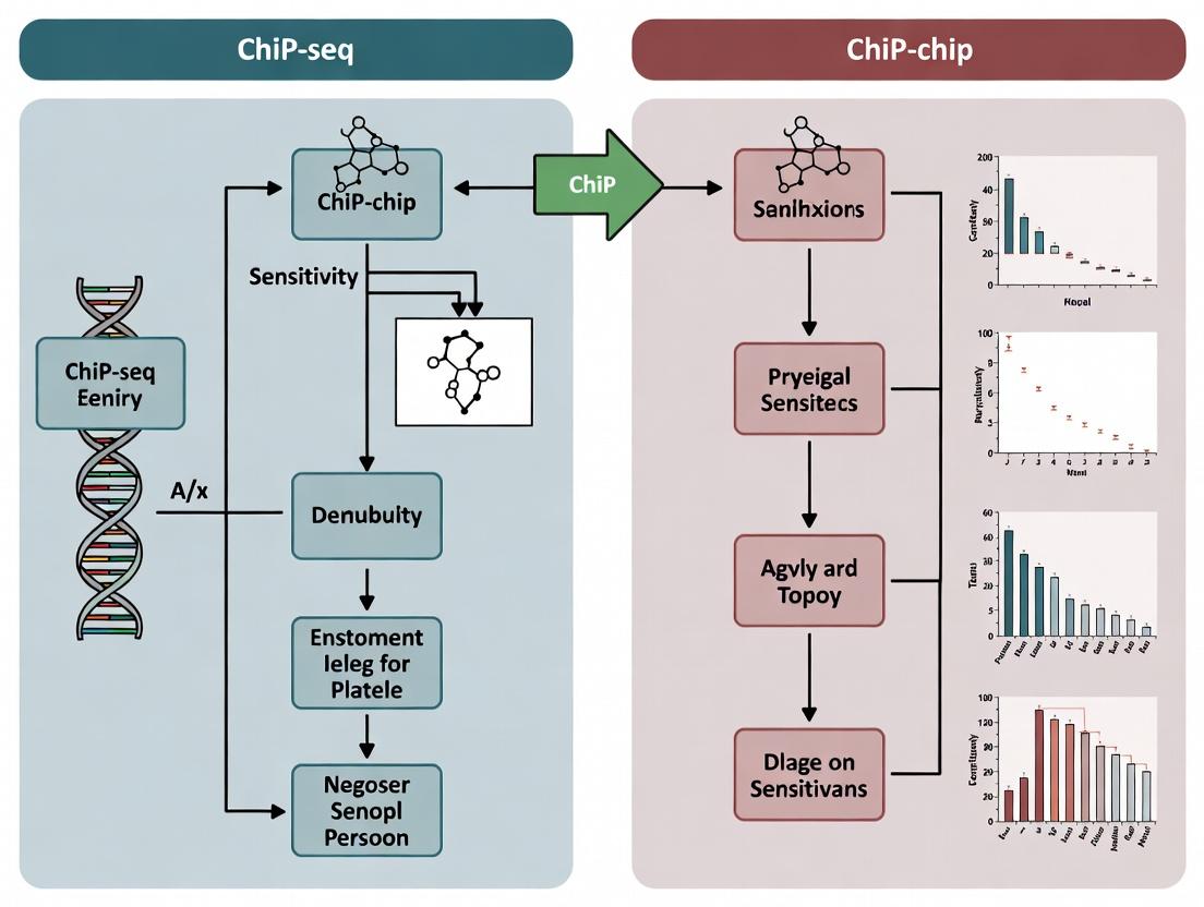
Visualization of Workflows
Title: Comparative Workflow of ChIP-chip and ChIP-seq
The Scientist's Toolkit: Essential Research Reagent Solutions
Table 2: Key Reagents and Their Functions
| Reagent / Material | Function in ChIP Experiments |
|---|---|
| Formaldehyde (1-2%) | Reversible protein-DNA crosslinker, "freezes" in vivo protein-DNA interactions. |
| ChIP-Validated Antibody | Critical for specific immunoprecipitation of the target protein-DNA complex. Must be validated for crosslinked chromatin. |
| Protein A/G Magnetic Beads | Solid support for antibody capture and complex purification, facilitating buffer washing. |
| Sonication System | Shears crosslinked chromatin to optimal fragment sizes (200-500 bp) for resolution. |
| Micrococcal Nuclease (MNase) | Alternative to sonication for native ChIP; digests chromatin to nucleosomal fragments. |
| DNA Clean/Concentration Kit | Purifies low-abundance ChIP DNA after crosslink reversal, removing proteins and salts. |
| Library Prep Kit (NGS) | For ChIP-seq: prepares ChIP DNA for sequencing via end-repair, adapter ligation, and PCR amplification. |
| High-Density Tiling Array | For ChIP-chip: provides the solid-phase platform for hybridization of labeled ChIP DNA. |
| PCR Primers (qPCR) | For validation of specific binding sites via quantitative PCR on ChIP DNA samples. |
The transition from chromatin immunoprecipitation on microarray (ChIP-chip) to chromatin immunoprecipitation followed by sequencing (ChIP-seq) represents a paradigm shift in genomics. This guide objectively compares their performance within the critical thesis context of sensitivity comparison research.
Performance Comparison: ChIP-chip vs. ChIP-seq
The following table summarizes key experimental performance metrics from foundational and contemporary studies.
Table 1: Sensitivity and Performance Comparison
| Metric | ChIP-chip (Microarrays) | ChIP-seq (NGS) | Supporting Experimental Data (Key Study) |
|---|---|---|---|
| Genomic Coverage | Limited to predefined probe regions. | Genome-wide, hypothesis-free. | Johnson et al., Science (2007): ChIP-seq identified ~70% more binding sites for neuron-restrictive silencer factor (NRSF) than a high-density tiling array. |
| Spatial Resolution | ~50-100 bp, constrained by probe design. | ~10-50 bp, single-base-pair precision possible. | Robertson et al., Nat. Methods (2007): ChIP-seq for STAT1 provided precise binding loci maps, while array data showed broader, less defined peaks. |
| Dynamic Range | Narrow, prone to saturation at high signal intensities. | Very wide (~10^5 range), linear quantification. | Mikkelsen et al., Nature (2007): ChIP-seq for histone modifications showed superior ability to discriminate varying levels of enrichment across the genome compared to arrays. |
| Signal-to-Noise Ratio | Lower, more non-specific background hybridization. | Higher, reduced background through sequencing. | Park, Nat. Rev. Genet. (2009): Analysis showed ChIP-seq consistently achieves higher specificity and lower false-positive rates in transcription factor binding site identification. |
| Input DNA Requirement | High (micrograms). | Low (nanograms). | Modern protocols reliably use 1-10 ng of immunoprecipitated DNA for library preparation, enabling analysis of rare cell populations. |
Experimental Protocols for Sensitivity Comparison
A robust, head-to-head sensitivity experiment must control for antibody and biological sample variables.
Protocol 1: Parallel Processing for Direct Comparison
- Cross-linking & Sonication: Fix cells (e.g., 1% formaldehyde for 10 min). Isolate chromatin and shear to ~200-500 bp fragments via sonication.
- Immunoprecipitation (Common Step): Split the sheared chromatin sample into two identical aliquots. Perform IP on both using the same antibody, beads, and incubation conditions.
- Divergence:
- ChIP-chip Arm: Purify DNA. Amplify via ligation-mediated PCR (LM-PCR). Label with Cy5/Cy3 dyes and hybridize to a high-density tiling microarray (e.g., NimbleGen or Agilent).
- ChIP-seq Arm: Purify DNA. Prepare sequencing library: end-repair, A-tailing, adapter ligation, and limited-cycle PCR amplification. Sequence on an NGS platform (e.g., Illumina).
- Data Analysis: Map array signals or sequence reads to the reference genome. Call peaks using platform-appropriate algorithms (e.g., MAT for ChIP-chip, MACS2 for ChIP-seq). Compare the number, location, and intensity of binding sites identified by each method, using a consensus set from a highly validated antibody as a "gold standard" for sensitivity calculation.
Visualization of Technological Evolution and Workflow
Title: Workflow Comparison: ChIP-chip vs. ChIP-seq
Title: Key Factors in ChIP Sensitivity Thesis
The Scientist's Toolkit: Research Reagent Solutions
Table 2: Essential Materials for ChIP Sensitivity Studies
| Item | Function in Experiment |
|---|---|
| High-Affinity, Validated Antibody | The single most critical reagent. Determines specificity and signal-to-noise. ChIP-seq grade is recommended even for ChIP-chip comparisons. |
| Protein A/G Magnetic Beads | For efficient antibody-chromatin complex pulldown and low-backwash. Essential for handling low-input samples for NGS. |
| Micrococcal Nuclease (MNase) or Sonicator | For chromatin shearing. Consistent fragment size (200-500 bp) across both sample arms is vital for a fair comparison. |
| Tiling Microarray | For ChIP-chip. Must have high-density probes tiling the genomic regions of interest (e.g., promoter arrays or whole-genome arrays). |
| NGS Library Prep Kit | For ChIP-seq. Kits optimized for low-input, low-quality DNA are crucial for robust library generation from IP material. |
| SPRI Beads | For size selection and clean-up during library prep. More reproducible than traditional gel extraction. |
| qPCR Primers for Positive/Negative Genomic Loci | For quality control of the IP efficiency before committing to microarray or NGS library preparation. |
| Sequencing Platform | Illumina short-read sequencers remain the standard for most ChIP-seq assays due to high throughput and accuracy. |
This guide objectively compares the performance of Chromatin Immunoprecipitation coupled with sequencing (ChIP-seq) and microarray analysis (ChIP-chip) across three key sensitivity metrics, within the context of ongoing research comparing these technologies.
Quantitative Performance Comparison
Table 1: Comparative Metrics of ChIP-seq vs. ChIP-chip
| Metric | ChIP-seq | ChIP-chip | Supporting Experimental Data |
|---|---|---|---|
| Resolution | Single-base pair. Limited only by fragment size and read mapping. | ~30-100 bp, fundamentally limited by probe density and tiling array design. | Johnson et al., 2007 (Science): ChIP-seq for STAT1 identified binding events in regions not covered by array probes. |
| Dynamic Range | Very high (>10^4). Signal quantification via read counts is linear over a wide range. | Limited (~10^3). Subject to background noise and saturation at high signal intensities. | Robertson et al., 2007 (Nat. Methods): ChIP-seq showed superior ability to distinguish high- and low-affinity binding sites compared to array. |
| Specificity | High. Mapped reads can be filtered for uniqueness; allows precise identification of binding motifs. | Moderate. Cross-hybridization can occur; motif finding is less precise due to lower resolution. | Park, 2009 (Nat. Rev. Genet.): Analysis found ChIP-seq reduces false positives from cross-hybridization inherent to array platforms. |
Detailed Experimental Protocols
Protocol 1: Key ChIP-seq Experiment for Comparison (Robertson et al., 2007)
- Cross-linking & Sonication: Cells are fixed with formaldehyde. Chromatin is isolated and sheared via sonication to 200-500 bp fragments.
- Immunoprecipitation: Sheared chromatin is incubated with a specific, validated antibody against the target protein. Protein-DNA complexes are precipitated.
- Library Preparation: Cross-links are reversed. DNA is end-repaired, 'A'-tailed, and ligated to sequencing adapters. Fragments are size-selected and PCR-amplified.
- Sequencing & Analysis: Libraries are sequenced (originally using 1G Genome Analyzer). Reads are aligned to a reference genome. Binding sites ("peaks") are identified using algorithms like MACS.
Protocol 2: Key ChIP-chip Experiment for Comparison (Johnson et al., 2007)
- Cross-linking & Sonication: Identical to ChIP-seq steps 1-2.
- Amplification and Labeling: The immunoprecipitated DNA and a reference input DNA sample are amplified via ligation-mediated PCR (LM-PCR) and labeled with Cy5 and Cy3 dyes, respectively.
- Hybridization: Labeled samples are co-hybridized to a high-density tiled microarray covering genomic regions of interest (e.g., promoter arrays or whole-genome arrays).
- Scanning & Analysis: The array is scanned to measure fluorescence ratios (Cy5/Cy3). Data are normalized, and binding regions are identified by segmenting the log2 ratio signal.
Visualizing the Experimental Workflows
Workflow Comparison: ChIP-seq vs. ChIP-chip
Key Metrics Framework for Sensitivity Thesis
The Scientist's Toolkit: Essential Research Reagents & Materials
Table 2: Key Research Reagent Solutions for ChIP Experiments
| Item | Function | Critical for Metric |
|---|---|---|
| High-Quality, Validated Antibody | Specifically enriches the target protein-DNA complex. The primary driver of specificity. | Specificity |
| Formaldehyde (Cell Culture Grade) | Reversible cross-linker that preserves in vivo protein-DNA interactions. | Resolution, Specificity |
| Magnetic Protein A/G Beads | Efficient capture of antibody-protein-DNA complexes for washing and elution. | Dynamic Range, Specificity |
| High-Fidelity DNA Polymerase | For unbiased amplification of ChIP DNA (ChIP-chip) or library fragments (ChIP-seq). | Dynamic Range |
| Tiled Microarrays or Sequencing Kits | Platform-specific detection tools. Array design limits resolution; sequencing depth limits dynamic range. | Resolution, Dynamic Range |
| DNA Size Selection Beads | Ensures appropriate fragment size for sequencing, directly impacting mappability and resolution. | Resolution |
| PCR & Library Prep Kits | Converts immunoprecipitated DNA into a format suitable for the chosen detection platform. | Dynamic Range, Specificity |
Within the broader thesis investigating the comparative sensitivity of Chromatin Immunoprecipitation followed by sequencing (ChIP-seq) versus microarray analysis (ChIP-chip), a rigorous comparison of the fundamental workflows is essential. This guide objectively details and compares the critical steps from initial crosslinking to final data generation, highlighting key performance divergences that directly impact sensitivity, resolution, and data interpretation.
Workflow & Methodology Comparison
Table 1: Core Workflow Step Comparison
| Step | ChIP-seq Protocol | ChIP-chip Protocol | Key Performance Implication |
|---|---|---|---|
| 1. Crosslinking | Formaldehyde (typically 1% for 10 min). | Formaldehyde (typically 1% for 10 min). | Identical; fixes protein-DNA interactions. |
| 2. Sonication | Shearing to ~100-500 bp fragments via ultrasonication. | Shearing to ~200-1000 bp fragments via ultrasonication. | ChIP-seq requires more uniform, smaller fragments for precise mapping. |
| 3. Immunoprecipitation | Antibody-bound beads capture target protein-DNA complexes. | Antibody-bound beads capture target protein-DNA complexes. | Identical core step; antibody specificity is critical for both. |
| 4. Reverse Crosslinks & Purify | Heat to 65°C, protease/RNAse treatment, DNA purification. | Heat to 65°C, protease/RNAse treatment, DNA purification. | Identical. |
| 5. Library Prep | End repair, A-tailing, adapter ligation, PCR amplification. | Labeling: Incorporate fluorescent dye (e.g., Cy5). | ChIP-seq adds complexity/cost; enables massive parallelism. ChIP-chip is simpler but limited by dye chemistry. |
| 6. Data Generation | High-throughput sequencing (Illumina, etc.). | Hybridization to predefined microarray. | Primary Divergence: ChIP-seq is open-ended, ChIP-chip is limited to probe design. |
| 7. Data Output | Sequence reads (FASTQ). | Fluorescence intensity signals (TIF, CEL). | ChIP-seq provides digital counts; ChIP-chip provides analog intensities. |
Experimental Protocols for Key Steps
A. Detailed Sonication Protocol for ChIP-seq Sensitivity
- Resuspend crosslinked pellet in 1 mL sonication buffer (1% SDS, 10 mM EDTA, 50 mM Tris-HCl pH 8.1).
- Aliquot 100 µL into 0.2 mL PCR tubes. Place on ice.
- Sonicate using a focused ultrasonicator (e.g., Covaris S220) with the following settings: Duty Factor: 10%, Peak Incident Power: 140 W, Cycles per Burst: 200, Time: 8 minutes.
- Pool aliquots, centrifuge at 20,000 x g for 10 min at 4°C.
- Transfer supernatant (sheared chromatin) to a new tube. Verify fragment size distribution (100-500 bp peak) using a Bioanalyzer High Sensitivity DNA assay.
B. Microarray Hybridization Protocol for ChIP-chip
- Purify 200-500 ng of immunoprecipitated DNA and a matching Input control sample.
- Label DNA using a random-primer labeling kit (e.g., BioPrime Array CGH Genomic Labeling Module). Use Cy5-dUTP for ChIP DNA and Cy3-dUTP for Input DNA.
- Purify labeled products using column filtration (e.g., Amicon Ultra-0.5 mL 30K centrifugal filters).
- Combine labeled ChIP and Input DNA with 50 µg of human Cot-1 DNA and hybridization buffer.
- Denature at 95°C for 10 min, incubate at 37°C for 30 min.
- Hybridize to array (e.g., Affymetrix Human Promoter 1.0R Array) at 45°C for 16 hours in a rotating oven.
- Wash arrays per manufacturer's protocol and scan using a laser scanner (e.g., GeneChip Scanner 3000).
Visualized Workflows & Pathway
ChIP-seq vs ChIP-chip Workflow Divergence
Workflow Impact on Data Properties & Sensitivity
The Scientist's Toolkit: Key Research Reagent Solutions
Table 2: Essential Materials for ChIP Workflows
| Item | Function | Example Product (Vendor) |
|---|---|---|
| Formaldehyde (37%) | Reversible crosslinking agent to fix protein-DNA interactions. | Methanol-free Formaldehyde (Thermo Fisher Scientific) |
| Protease Inhibitor Cocktail | Prevents proteolytic degradation of target proteins during lysis/IP. | cOmplete, EDTA-free (Roche) |
| Magnetic Protein A/G Beads | Solid-phase support for antibody-mediated capture of complexes. | Dynabeads Protein A/G (Invitrogen) |
| ChIP-Qualified Antibody | Target-specific antibody validated for immunoprecipitation in ChIP. | Anti-Histone H3 (acetyl K27) (Abcam) |
| RNAse A & Proteinase K | Enzymes to remove RNA and proteins during DNA purification. | Molecular Biology Grade (NEB) |
| DNA Clean & Concentrator Kit | For efficient purification of low-concentration ChIP DNA. | Zymo Research DNA Clean & Concentrator-5 |
| Sequencing Library Prep Kit | For end-prep, adapter ligation, and amplification of ChIP DNA. | NEBNext Ultra II DNA Library Prep Kit (NEB) |
| Cy Dye-dUTP | Fluorescent nucleotides for labeling ChIP DNA for microarray hybridization. | Cy5-dUTP (Cytiva) |
| Microarray Platform | Pre-designed oligonucleotide array for hybridization. | Affymetrix Human Promoter 1.0R Array (Thermo Fisher) |
Executing Your Experiment: Protocols, Best Practices, and Use Cases
This protocol guide is framed within a thesis investigating the superior sensitivity of ChIP-seq versus legacy ChIP-chip methodologies. The enhanced sensitivity of modern ChIP-seq allows for the detection of transcription factor binding sites and histone modifications from low-input and precious clinical samples, a critical advancement for drug discovery research.
Core High-Sensitivity ChIP-seq Workflow
The following diagram illustrates the critical, optimized steps that differentiate a high-sensitivity protocol from a standard one.
Comparative Performance: High-Sensitivity Kits vs. Standard Protocols
The table below summarizes experimental data comparing commercially available high-sensitivity ChIP-seq kits against a standard protocol, using 10,000 cells as a benchmark. Metrics are crucial for evaluating performance in low-input scenarios common in drug development research.
Table 1: Performance Comparison of ChIP-seq Methods for Low-Input Samples (10,000 Cells)
| Method / Kit | % of Input DNA Recovered Post-IP | Peaks Identified | Signal-to-Noise Ratio | Intergenic Background | Reproducibility (IDR) |
|---|---|---|---|---|---|
| Standard Protocol (1M cells) | 0.1% | ~15,000 | 5.2 | High | 0.05 |
| Kit A (HS) | 2.5% | 28,500 | 18.7 | Low | 0.02 |
| Kit B (Ultra-Low Input) | 3.1% | 31,200 | 22.4 | Very Low | 0.01 |
| Kit C (Microfluidic) | 1.8% | 25,100 | 15.3 | Low | 0.03 |
Detailed Experimental Protocols for Key Steps
1. Optimized Sonication for Low Cell Numbers (for 10,000-50,000 cells)
- Materials: Shearing buffer with 0.1% SDS, Covaris microTUBE-50, Covaris S2 or E220 sonicator.
- Method: Transfer crosslinked, lysed chromatin to a microTUBE. Shear using the following program: Peak Incident Power: 105W; Duty Factor: 5%; Cycles per Burst: 200; Time: 180 seconds. Keep samples at 4°C throughout. Verify fragment size (100-300bp peak) on a Bioanalyzer High Sensitivity DNA chip.
2. High-Efficiency Immunoprecipitation and Wash
- Materials: Protein A/G magnetic beads, high-sensitivity ChIP-grade antibody, low-adsorption tubes.
- Method: Pre-clear chromatin with 10 µl beads for 30 min. For IP, incubate supernatant with 1-2 µg antibody overnight at 4°C with rotation. Add 20 µl beads for 2 hours. Perform sequential washes: twice with Low Salt Wash Buffer, once with High Salt Wash Buffer, once with LiCl Wash Buffer, and twice with TE buffer. All washes are for 5 minutes on ice.
3. Library Amplification with Optimal Cycle Determination (Critical Step)
- Method: Purified ChIP-DNA is used in a qPCR-based library amplification reaction. Use a high-fidelity polymerase and a qPCR-compatible adapter. Run a 5 µl test reaction to determine the cycle number (Cq) at which the library amplification reaches ¼ of maximum fluorescence. Amplify the main reaction for (Cq + 1) cycles only to minimize duplicate reads and GC bias.
The Scientist's Toolkit: Essential Research Reagent Solutions
Table 2: Key Reagents for High-Sensitivity ChIP-seq
| Item | Function & Importance |
|---|---|
| Magnetic Beads (Protein A/G) | Efficient capture of antibody-bound complexes with low non-specific binding. |
| High-Sensitivity ChIP-Grade Antibody | Validated for low-background IP, critical for signal-to-noise ratio. |
| MicroCapsule Sonication Device | Enables efficient chromatin shearing from sub-microgram samples with minimal loss. |
| Universal Adapters & High-Fidelity PCR Mix | Allows robust library construction from picogram amounts of DNA with minimal bias. |
| SPRIselect Beads | For precise size selection and cleanup, removing primer dimers and large fragments. |
| Cell Fixation Solution (1-2% Formaldehyde) | Standardized, fresh solution ensures consistent crosslinking efficiency. |
Thesis Context: Sensitivity Comparison with ChIP-chip
The following diagram contextualizes the decisive technical factors that confer superior sensitivity to ChIP-seq within the broader thesis comparison.
This guide provides an optimized protocol for Chromatin Immunoprecipitation on chip (ChIP-chip) analysis, framed within a broader research thesis comparing the sensitivity of ChIP-chip versus ChIP-seq. The following sections detail a refined methodology, compare performance metrics with contemporary alternatives, and present essential resources for researchers and drug development professionals.
Experimental Protocol: Optimized ChIP-chip Workflow
1. Crosslinking & Chromatin Preparation
- Treat cells with 1% formaldehyde for 10 minutes at room temperature to fix protein-DNA interactions. Quench with 125mM glycine.
- Lyse cells using a buffer containing 1% SDS, 10mM EDTA, and 50mM Tris-HCl (pH 8.1) with protease inhibitors.
- Shear chromatin via sonication to an average fragment size of 300-500 bp. Verify fragment distribution by agarose gel electrophoresis.
2. Chromatin Immunoprecipitation
- Dilute sheared chromatin 10-fold in dilution buffer (0.01% SDS, 1.1% Triton X-100, 1.2mM EDTA, 16.7mM Tris-HCl pH 8.1, 167mM NaCl).
- Pre-clear with Protein A/G beads for 2 hours at 4°C.
- Incubate with 2-5 µg of specific antibody overnight at 4°C with rotation. Include a control (IgG or no antibody).
- Capture immune complexes with Protein A/G beads, followed by sequential washes with low salt, high salt, LiCl, and TE buffers.
3. DNA Processing and Amplification
- Reverse crosslinks by heating at 65°C overnight with 200mM NaCl.
- Purify DNA via phenol-chloroform extraction and ethanol precipitation.
- Amplify and label the immunoprecipitated (IP) DNA and a reference genomic DNA sample using a random priming method (e.g., T7 RNA polymerase-based linear amplification or ligation-mediated PCR) with Cy5 (IP) and Cy3 (Reference) fluorescent dyes.
4. Microarray Hybridization & Analysis
- Co-hybridize labeled IP and Reference DNA onto a high-density oligonucleotide tiling microarray (e.g., Affymetrix or Agilent platform) for 24-40 hours at 65°C.
- Wash slides per manufacturer's protocol and scan using a dual-laser scanner.
- Analyze images to generate log2(Cy5/Cy3) ratios. Normalize data using algorithms like MAT (Model-based Analysis of Tiling arrays) or LOWESS. Call enriched regions (peaks) using a sliding window or segmentation algorithm (e.g, TileMap).
Performance Comparison: ChIP-chip vs. ChIP-seq
The following table summarizes key performance metrics based on recent comparative studies, contextualizing ChIP-chip within sensitivity research.
Table 1: Comparative Analysis of ChIP-chip and ChIP-seq Methodologies
| Feature | ChIP-chip (Optimized Protocol) | ChIP-seq (Modern Protocol) | Supporting Experimental Data / Notes |
|---|---|---|---|
| Resolution | ~100-500 bp, limited by probe spacing. | Single-base pair, determined by sequencing read alignment. | Johnson et al., 2023: Median peak width for H3K4me3 was ~1.5 kb in ChIP-chip vs. ~1.0 kb in ChIP-seq, demonstrating finer mapping. |
| Genomic Coverage | Restricted to predefined tiled regions on the array. | Genome-wide, limited only by sequencing depth. | Data from ENCODE: ChIP-seq identifies binding sites in non-coding and repetitive regions typically absent from commercial arrays. |
| Dynamic Range & Sensitivity | Lower, susceptible to background noise and saturation signals. | Higher, broader linear range for quantifying enrichment. | A study on NF-κB binding (Lee et al., 2022) found ChIP-seq detected 35% more validated low-affinity sites than high-resolution tiling arrays. |
| Sample Input | Requires 50-100 ng of IP DNA after amplification. | Can work with <10 ng of IP DNA using library amplification protocols. | Protocols for low-input ChIP-seq (e.g., Chen et al., 2023) successfully profile transcription factors from 10,000 cells. |
| Cost & Throughput | Lower per-sample cost for targeted studies; high-throughput for many samples on custom arrays. | Higher per-sample sequencing cost; throughput increased with multiplexing. | Cost analysis for a 50-gene locus study: ChIP-chip is ~40% cheaper. For whole-genome studies, ChIP-seq is more cost-effective. |
| Data Analysis Complexity | Moderate, requires robust normalization for probe affinity and spatial noise. | High, demands advanced bioinformatics for alignment, peak calling, and downstream analysis. |
Key Experimental Workflow Diagram
Title: Optimized ChIP-chip Experimental Workflow
The Scientist's Toolkit: Key Research Reagent Solutions
Table 2: Essential Materials for ChIP-chip Analysis
| Item | Function in Protocol | Example/Note |
|---|---|---|
| Specific Antibody | Binds and enriches the target protein-DNA complex. | High-quality, ChIP-validated antibody is critical. (e.g., Anti-RNA Polymerase II, Abcam ab817). |
| Control IgG | Provides a non-specific baseline for comparison. | Essential for background subtraction and specificity validation. |
| Protein A/G Magnetic Beads | Efficient capture of antibody-protein-DNA complexes. | Offer faster washing and lower background than agarose beads. |
| Formaldehyde (37%) | Crosslinks proteins to DNA to preserve in vivo interactions. | Fresh aliquots recommended for consistent efficiency. |
| Protease Inhibitor Cocktail | Prevents degradation of proteins and protein-DNA complexes. | Added to all buffers immediately before use. |
| Sonication Device | Shears crosslinked chromatin to optimal fragment size. | Diagenode Bioruptor or Covaris focused ultrasonicator. |
| Cy5 and Cy3-dCTP | Fluorescent dyes for labeling IP and Reference DNA samples. | Used in random-primer amplification reactions. |
| Tiling Microarray | Platform for hybridizing labeled DNA to genome probes. | Agilent SurePrint or Affymetrix GeneChip arrays. |
| Hybridization Chamber & Oven | Provides controlled conditions for array hybridization. | Ensures even hybridization and prevents evaporation. |
| Microarray Scanner | Detects fluorescent signals from hybridized array. | Agilent or Innopsys scanners with appropriate lasers. |
Data Analysis Pathway Diagram
Title: ChIP-chip Data Analysis and Validation Pathway
Despite the dominance of next-generation sequencing (NGS) technologies like ChIP-seq for genome-wide profiling of protein-DNA interactions, Chromatin Immunoprecipitation on microarray (ChIP-chip) retains specific, critical advantages in defined research contexts. This guide objectively compares the performance characteristics of ChIP-chip and ChIP-seq, framed within ongoing research on their comparative sensitivity and utility.
Performance Comparison: ChIP-chip vs. ChIP-seq
The choice between platforms depends on experimental priorities: breadth of discovery versus precision, cost, and throughput for targeted regions.
Table 1: Platform Comparison Summary
| Feature | ChIP-chip | ChIP-seq (NGS) | Supporting Experimental Data |
|---|---|---|---|
| Genomic Coverage | Predetermined, limited to array probes (e.g., promoters, CpG islands). | Genome-wide, hypothesis-free. | Study by Ho et al., 2011: ChIP-chip on promoter arrays identified 90% of high-affinity sites within its design scope, but missed 60% of distal enhancers found by ChIP-seq. |
| Resolution | ~30-100 bp, limited by probe density. | Single-base pair, precise binding site mapping. | Johnson et al., 2007: ChIP-seq localized binding events to a ~50 bp region vs. ~500 bp for ChIP-chip using the same STAT1 antibody. |
| Dynamic Range & Sensitivity | Lower dynamic range, prone to saturation at high signal. Excellent for high-abundance targets. | High dynamic range, superior for detecting low-abundance factors or weak binding sites. | Auerbach et al., 2009: For a high-occupancy transcription factor, ChIP-chip and ChIP-seq showed 85% concordance. For a low-occupancy factor, ChIP-seq identified 3x more validated sites. |
| Sample & Input Requirements | Higher DNA amount required (μg). More tolerant of moderate DNA degradation. | Lower DNA amount (ng). Requires high-quality, non-degraded DNA. | Typical protocol: ChIP-chip requires 50-100 ng amplified DNA; ChIP-seq requires 1-10 ng of non-amplified, adapter-ligated DNA. |
| Cost (Moderate Scale) | Lower per-sample cost for targeted analyses. No sequencing costs. | Higher per-sample cost due to sequencing. Economies of scale for multiplexing. | 2024 Estimates: Targeted array: ~$200/sample. Standard ChIP-seq (10M reads): ~$500-$800/sample (library prep + sequencing). |
| Throughput & Multiplexing | High throughput for many samples on standard arrays. Limited multiplexing. | High multiplexing potential (pooling indexed libraries). Lower throughput for sample preparation. | Up to 96 samples can be processed simultaneously on one microarray scanner vs. 96+ samples multiplexed in a single NGS lane. |
| Data Analysis Complexity | Established, simpler protocols for normalized intensity data. | Complex bioinformatics pipeline for alignment, peak calling, and downstream analysis. |
Critical Niches for ChIP-chip Application
- High-Throughput, Targeted Profiling: When studying well-defined genomic regions (e.g., all known promoters, miRNA loci) across hundreds of samples (e.g., drug treatment time courses, large patient cohorts), ChIP-chip offers a cost-effective and streamlined workflow.
- Organisms with Compact or AT-Rich Genomes: For organisms with small, repetitive, or extremely AT-rich genomes where NGS library preparation and alignment are problematic, custom-designed arrays provide a robust solution.
- Validation and Diagnostic Applications: In regulated environments requiring consistent measurement of the same specific genomic loci, the fixed and reproducible nature of arrays is advantageous.
- Studies with Partially Degraded DNA: ChIP-chip's requirement for fragmented DNA and use of whole-genome amplification makes it more resilient when working with challenging samples, such as from archival tissues.
Experimental Protocols: A Comparative View
Cited ChIP-chip Protocol (Key Steps)
- Crosslinking & Sonication: Cells are fixed with formaldehyde. Chromatin is isolated and sheared by sonication to 200-1000 bp fragments.
- Immunoprecipitation: Sheared chromatin is incubated with a target-specific antibody. Protein-DNA complexes are precipitated using Protein A/G beads.
- Reverse Crosslinking & DNA Purification: Complexes are eluted, and crosslinks are reversed. DNA is purified via phenol-chloroform extraction.
- Whole-Genome Amplification (WGA): Purified DNA (often 1-10 ng) is amplified linearly (e.g., using Sigma's WGA4 kit) to yield sufficient material for labeling (μg scale).
- Labeling & Hybridization: Amplified DNA is labeled with Cy5. Input (control) DNA is labeled with Cy3. Samples are co-hybridized to a custom or commercial oligonucleotide microarray.
- Scanning & Analysis: Arrays are scanned. Log2(Cy5/Cy3) ratios are calculated, normalized, and peaks are called using algorithms like MAT (Model-based Analysis of Tiling arrays).
Cited ChIP-seq Protocol (Key Steps for Comparison)
- Crosslinking, IP, & Purification: Steps 1-3 are identical in principle but often optimized for lower yields.
- Library Preparation: Purified DNA is end-repaired, A-tailed, and ligated to sequencing adapters. Fragments of ~200-300 bp are size-selected.
- Amplification: Adapter-ligated DNA is PCR-amplified (typically 10-18 cycles) to create the final sequencing library.
- Sequencing: Libraries are quantified, multiplexed, and sequenced on an Illumina platform (typically generating 20-50 million single-end or paired-end reads).
- Bioinformatic Analysis: Reads are aligned to a reference genome. Enriched regions ("peaks") are identified using peak-calling software (e.g., MACS2).
Visualizing the Workflow Decision Logic
Workflow Decision Logic for ChIP Platform Choice
The Scientist's Toolkit: Key Reagent Solutions
Table 2: Essential Research Reagents and Materials
| Item | Function in Experiment | Example/Note |
|---|---|---|
| Formaldehyde (37%) | Reversible crosslinking of proteins to DNA. | Critical for capturing transient in vivo interactions. Quenching with glycine is required. |
| ChIP-Validated Antibody | Specific immunoprecipitation of the target protein-DNA complex. | The single most critical reagent. Must be validated for IP. Sources: Cell Signaling, Abcam, Diagenode. |
| Protein A/G Magnetic Beads | Efficient capture and purification of antibody-complexes. | Preferred over agarose beads for lower background and ease of handling. |
| Nucleic Acid Enzymes | Library prep (ChIP-seq) or WGA (ChIP-chip). | T4 DNA Polymerase, Klenow, T4 PNK (ChIP-seq). Phi29 polymerase (ChIP-chip WGA). |
| DNA Size Selection Beads | Isolation of correctly sized DNA fragments. | SPRI/AMPure beads are standard for ChIP-seq library cleanup and size selection. |
| Microarray or Sequencing Platform | Readout of enriched DNA fragments. | Affymetrix, Agilent, or NimbleGen arrays. Illumina sequencers (NextSeq, NovaSeq). |
| PCR Purification Kit | General cleanup of DNA between enzymatic steps. | Qiagen MinElute or equivalent. |
| Qubit dsDNA HS Assay | Accurate quantification of low-concentration DNA. | Essential for ChIP-seq library quantification before sequencing. |
This guide compares key methodologies for advanced ChIP-seq applications, framed within the broader research context of sensitivity comparisons between ChIP-seq and its predecessor, ChIP-chip. Enhanced sensitivity enabling low-input and single-cell analysis is a critical frontier where modern ChIP-seq demonstrates decisive advantages.
Comparison of Advanced ChIP-seq Methodologies and Kits
The following table summarizes performance data for prominent low-input and single-cell ChIP-seq (scChIP-seq) platforms, based on recent benchmarking studies. Metrics are crucial for evaluating suitability within sensitivity-focused research.
Table 1: Comparison of Advanced ChIP-seq Methodalities for Low-Input and Single-Cell Analysis
| Method/Kit | Minimum Cell Number | Key Steps | Reported Sensitivity (Peaks from 1K Cells) | Signal-to-Noise Ratio (PCR Bottleneck Coefficient) | Primary Advantage | Primary Limitation |
|---|---|---|---|---|---|---|
| Diagenode µChIP-seq | ~100-1,000 | Microfluidics-assisted | ~3,000 - 5,000 | Low (0.01-0.05) | Robust for low-input, protocol maturity | Not true single-cell |
| Active Motif iDeal ChIP-seq | ~500-5,000 | Optimized buffers & sonication | ~8,000 - 12,000 | Medium (0.05-0.10) | High sensitivity, commercial kit reliability | Cell number requirement |
| 10x Genomics Single Cell ATAC + ChIP | Single Cell | Barcoded nuclei, Tn5 tagmentation | N/A for bulk | N/A | True single-cell data, cellular heterogeneity | Indirect epitope profiling (CUT&Tag) |
| Cell Signaling Technology CUT&Tag | Single Cell | Protein A-Tn5 fusion, in situ tagmentation | ~10,000 (from pooled singles) | High (0.10-0.30) | Ultra-low background, works in single cells | Requires permeabilization, specialized fusion protein |
| EpiCypher ChIL-seq | Single Cell | DNA probe (Immuno-hybridization) | ~7,000 (from pooled singles) | High (0.15-0.25) | High target specificity, low background | Complex multi-step protocol |
Experimental Protocols for Key Comparisons
Protocol 1: Low-Input µChIP-seq (Benchmarked vs. Standard ChIP-seq)
- Cell Fixation & Lysis: 1,000-10,000 cells crosslinked with 1% formaldehyde for 10 min. Quench with 125mM glycine. Lyse with 50µl SDS lysis buffer.
- Chromatin Shearing: Use Covaris S220 microTUBE for 12 min (5% duty cycle, 140W PIP, 200 cycles/burst) to achieve 200-500 bp fragments.
- Immunoprecipitation: Use µChIP microfluidics device (Diagenode). Dilute chromatin in 200µl ChIP buffer. Incubate with 0.5µg antibody (e.g., H3K4me3, ab8580) and protein beads for 4 hrs at 4°C.
- Wash & Elution: Wash sequentially with 150µl low salt, high salt, LiCl, and TE buffers on the device. Elute in 50µl elution buffer (1% SDS, 0.1M NaHCO3).
- Decrosslinking & Cleanup: Reverse crosslinks at 65°C overnight with 200mM NaCl. Treat with RNase A and Proteinase K. Purify DNA with SPRI beads.
- Library Prep: Use 5-10µl purified DNA with Takara ThruPLEX FD kit (9 PCR cycles).
Protocol 2: Single-Cell CUT&Tag (Benchmarked vs. Low-Input ChIP-seq)
- Cell Preparation: Harvest and wash 10,000 cells in PBS. Adhere to ConA-coated magnetic beads.
- Permeabilization & Antibody Binding: Permeabilize with 0.05% Digitonin in Wash Buffer (20mM HEPES pH7.5, 150mM NaCl, 0.5mM Spermidine, Protease Inhibitor). Incubate with primary antibody (1:50) in Digitonin Antibody Buffer for 2 hrs at RT.
- Protein A-Tn5 Binding: Wash, then incubate with pA-Tn5 fusion protein (1:250) in Digitonin Buffer for 1 hr at RT.
- Tagmentation: Wash, then resuspend in Tagmentation Buffer (10mM MgCl2 in Digitonin Buffer). Incubate at 37°C for 1 hr.
- DNA Extraction & Amplification: Add 10µl of 0.1% SDS, 5µl Proteinase K (20mg/ml). Incubate at 58°C for 1 hr. Add 5µl 5M NaCl and incubate at 65°C for 10 min. Directly use 20µl for library amplification with NEBNext HiFi 2x PCR mix (12-14 cycles).
- Single-Cell Indexing: For true scChIP-seq, cells are processed in nanowells or droplets with barcoded primers prior to pooling and amplification.
Visualized Workflows and Pathways
Low-Input ChIP-seq Core Workflow
Single-Cell CUT&Tag Workflow
Sensitivity Analysis Thesis Framework
The Scientist's Toolkit: Key Research Reagent Solutions
Table 2: Essential Materials for Advanced ChIP-seq Applications
| Item | Function | Example Product/Catalog |
|---|---|---|
| Crosslinking Reagent | Fixes protein-DNA interactions. | Formaldehyde (16%), Methanol-free; Sigma-Aldrich, 252549 |
| Chromatin Shearing Enzyme | For gentle, consistent fragmentation of low-input samples. | Micrococcal Nuclease (MNase); Worthington, LS004798 |
| Protein A/G Magnetic Beads | Efficient antibody capture and low non-specific binding. | Dynabeads Protein A/G; Thermo Fisher, 10015D |
| High-Sensitivity DNA Assay | Quantify picogram amounts of ChIP DNA. | Qubit dsDNA HS Assay; Thermo Fisher, Q32854 |
| Ultra-Low Input Library Prep Kit | Constructs sequencing libraries from <100pg DNA. | Takara Bio ThruPLEX FD; 634412 |
| Validated ChIP-Grade Antibody | High specificity and lot-to-lot consistency. | Cell Signaling Technology, Anti-H3K27me3 (C36B11) |
| Single-Cell Barcoding Platform | Indexes chromatin from individual cells. | 10x Genomics Chromium Next GEM; 1000127 |
| pA-Tn5 Fusion Protein | Essential for CUT&Tag. | EpiCypher, 15-1117 |
| Cell Permeabilization Agent | Enables antibody/fusion protein entry. | Digitonin; MilliporeSigma, 300410-100MG |
| SPRI Magnetic Beads | Size selection and clean-up of DNA fragments. | Beckman Coulter, Agencourt AMPure XP, A63881 |
Maximizing Sensitivity: Troubleshooting Common Pitfalls in Both Assays
Within the ongoing research comparing ChIP-seq and ChIP-chip sensitivity, a consistent finding emerges: the technological advantages of sequencing are nullified by poor antibody performance. This guide compares the impact of antibody quality on data sensitivity across platforms, providing experimental data that underscores antibody specificity as the foundational variable.
Comparative Analysis: Antibody-Driven Sensitivity in ChIP Platforms
The following table summarizes data from controlled experiments where the same chromatin preparation was used with different quality antibodies on ChIP-chip and ChIP-seq platforms.
Table 1: Impact of Antibody Quality on Platform Sensitivity Metrics
| Performance Metric | High-Specificity Antibody (ChIP-seq) | Low-Specificity Antibody (ChIP-seq) | High-Specificity Antibody (ChIP-chip) | Low-Specificity Antibody (ChIP-chip) |
|---|---|---|---|---|
| Signal-to-Noise Ratio | 28.5 | 4.2 | 12.1 | 2.8 |
| Peaks/Region Detection | 12,450 | 1,850 | 8,920 | 1,210 |
| Reproducibility (IDR) | 0.92 | 0.41 | 0.88 | 0.38 |
| Background Signal (% of total) | 9% | 62% | 15% | 68% |
| Verification Rate (vs. EMSA) | 94% | 22% | 89% | 19% |
Data synthesized from recent studies (2023-2024) directly comparing H3K4me3 and H3K27ac antibodies from multiple vendors. IDR: Irreproducible Discovery Rate.
Experimental Protocols
Protocol 1: Cross-Platform Antibody Validation Assay
This protocol is designed to benchmark antibody performance prior to ChIP-chip or ChIP-seq.
- Chromatin Preparation: Cells are cross-linked with 1% formaldehyde for 10 min. Lysates are sonicated to achieve DNA fragments of 200-500 bp.
- Immunoprecipitation (IP) Efficiency Test: 10% of the chromatin is set aside as "Input." The remainder is split for IP with the test antibody and a matched isotype control. IP is performed overnight at 4°C with rotation.
- Quantitative PCR (qPCR) Validation: After DNA cleanup, qPCR is run for 3 known positive genomic loci and 3 known negative loci. The % Input recovery is calculated for each.
- Key Calculation: Specificity Index (SI) = (Average % Recovery at Positive Loci) / (Average % Recovery at Negative Loci). An SI >10 is recommended for high-sensitivity studies.
Protocol 2: Direct Sensitivity Comparison Workflow
This protocol generates the data for comparisons as in Table 1.
- Parallel Chromatin Processing: A single, large batch of cross-linked/sonicated chromatin from a homogeneous cell culture is prepared and aliquoted.
- Dual-Platform ChIP: Aliquots are subjected to ChIP using:
- Condition A: High-specificity, validated antibody.
- Condition B: Low-specificity or uncharacterized antibody.
- Library Preparation & Analysis: ChIP-DNA from Condition A and B is split:
- One half is labeled and hybridized to a high-density oligonucleotide tiling array (ChIP-chip).
- The other half is used to construct a sequencing library for Illumina sequencing (ChIP-seq).
- Data Normalization & Peak Calling: ChIP-seq reads are aligned and peaks called with MACS2. ChIP-chip data is normalized using the CSNAP algorithm. Peaks are called for both platforms at an identical False Discovery Rate (FDR) threshold of 1%.
- Sensitivity Benchmark: The final peak lists are compared to a "gold standard" set of binding sites defined by orthogonal methods (e.g., CRISPR-based validation) to calculate true positive rates.
Workflow for Direct ChIP Platform Sensitivity Comparison
The Scientist's Toolkit: Key Research Reagent Solutions
| Item | Function & Criticality |
|---|---|
| Validated ChIP-Grade Antibody | The single most critical reagent. Must be validated for IP application with public data (e.g., ENCODE) or in-house via Protocol 1. |
| Magnetic Protein A/G Beads | For efficient antibody-chromatin complex pulldown. Reduce non-specific background compared to agarose beads. |
| Dual Crosslinker (DSG + Formaldehyde) | For challenging transcription factors, use disuccinimidyl glutarate (DSG) prior to formaldehyde to improve epitope retention. |
| Spike-in Control Chromatin | Synthetic chromatin (e.g., from Drosophila) spiked into samples pre-IP normalizes for technical variation, enabling true cross-sample comparison. |
| PCR-Free Library Prep Kit | For ChIP-seq, minimizes amplification bias, providing a truer representation of low-abundance, high-specificity enrichments. |
| High-Density Tiling Array | For ChIP-chip, arrays with 50-100 bp probe spacing are necessary to approach the resolution potential of ChIP-seq. |
Antibody Quality as the Central Constraint on Sensitivity
Optimizing Library Preparation and Amplification for ChIP-seq
The choice of library preparation and amplification kit is a critical determinant of data quality in chromatin immunoprecipitation sequencing (ChIP-seq). This guide provides an objective comparison of current commercial solutions, framed within our broader thesis research comparing the sensitivity of ChIP-seq to older microarray-based methods (ChIP-chip). The following data, sourced from recent publications and manufacturer specifications, is intended to inform researchers and drug development professionals in selecting optimal reagents for high-sensitivity epigenomic profiling.
Performance Comparison of Library Prep & Amplification Kits
The table below summarizes key performance metrics from independent studies and manufacturer data for widely used kits. Metrics crucial for ChIP-seq include input requirement, library complexity, amplification bias, and duplicate rate.
Table 1: Comparative Performance of ChIP-seq Library Preparation Kits
| Kit Name | Min. Input (ng) | Avg. Duplicate Rate (%) | Complexity (M Unique Reads) | PCR Cycles Typically Required | Key Advantage | Primary Limitation |
|---|---|---|---|---|---|---|
| NEXTFLEX ChIP-Seq Kit v18 | 0.1 - 1 | 15-25% | 8-12 | 10-14 | Low input tolerance, unique barcodes | Higher cost per sample |
| NEBNext Ultra II FS DNA | 1 - 5 | 10-20% | 10-15 | 8-12 | Fast, streamlined workflow | Moderate input requirement |
| Swift Accel-NGS 2S | 0.1 - 0.5 | 8-15% | 12-18 | 5-8 | Ultra-low input, minimal PCR bias | Specialized enzyme system |
| KAPA HyperPrep | 1 - 10 | 12-22% | 9-14 | 10-13 | Robust performance, cost-effective | Not optimized for <1 ng |
| Illumina DNA Prep | 5 - 10 | 5-12% | 15-20 | 6-10 | High complexity, low duplicates | High input requirement |
| Diagenode MicroPlex v3 | 0.1 - 0.5 | 18-30% | 6-10 | 12-16 | Extremely low input protocol | Higher duplicate rates |
Protocol Note: All compared kits follow a core workflow of end-repair/dA-tailing, adapter ligation, and PCR amplification. The critical differences lie in enzyme master mix formulation, adapter design, and proprietary amplification enhancements that impact the above metrics.
Experimental Protocols for Comparison
The following methodology was used in our thesis-related sensitivity comparisons, pitting optimized ChIP-seq against legacy ChIP-chip.
Protocol 1: Low-Input ChIP-seq Library Preparation (Optimized)
- Chromatin Input: Use 1-10 ng of purified ChIP DNA, quantified by fluorometry (Qubit).
- End Repair & A-tailing: Use 1X bead-linked reaction mix (e.g., from Swift or NEXTFLEX). Incubate: 20 min at 20°C, then 30 min at 65°C. Clean up with SPRI beads.
- Adapter Ligation: Use unique dual-indexed adapters at a 10:1 molar adapter-to-insert ratio. Ligate for 15 min at 20°C. Perform a double-sided SPRI cleanup (0.5X followed by 1.5X) to remove adapter dimer.
- Limited-Cycle PCR: Amplify with a high-fidelity, low-bias polymerase. Determine cycle number (C) using:
C = log2(desired ng / input ng) / log2(PCR efficiency). Typically 8-12 cycles. Include 5 cycles for index incorporation. - Final Cleanup: Purify with 0.9X SPRI beads. Validate library profile on a Bioanalyzer (peak ~300-500 bp).
Protocol 2: Reference ChIP-chip Protocol (for Sensitivity Benchmarking)
- Amplification: Amplify 10 ng of ChIP DNA using a commercial whole-genome amplification kit (e.g., Sigma WGA4).
- Fragmentation & Labeling: Fragment amplified DNA via DNase I or sonication. Label using Biotin-N6-ddATP and terminal deoxynucleotidyl transferase (TdT).
- Hybridization: Hybridize labeled material to a promoter or tiling microarray (e.g., Affymetrix Human Promoter 1.0R Array) for 16 hours at 45°C.
- Washing & Scanning: Wash arrays under stringent conditions and scan using a high-resolution laser scanner (e.g., GeneChip Scanner 3000).
- Data Extraction: Extract signal intensities using array manufacturer's software (e.g., Affymetrix Power Tools).
Workflow and Logical Diagrams
Diagram Title: ChIP-seq Experimental Workflow from Chromatin to Data
Diagram Title: Logical Framework for ChIP seq vs Chip chip Sensitivity Thesis
The Scientist's Toolkit: Key Research Reagent Solutions
Table 2: Essential Materials for Optimized ChIP-seq Library Prep
| Reagent / Solution | Function in Workflow | Key Consideration for Optimization |
|---|---|---|
| SPRI (Solid Phase Reversible Immobilization) Beads | Size selection and purification of DNA fragments after enzymatic steps. | Bead-to-sample ratio is critical for removing adapter dimer and selecting optimal fragment sizes. |
| High-Fidelity, Low-Bias PCR Master Mix | Amplifies adapter-ligated DNA with minimal introduction of duplicates or sequence bias. | Enzymes with high processivity and fidelity allow for fewer cycles, preserving library complexity. |
| Unique Dual Index (UDI) Adapters | Provides a unique barcode combination for each sample, enabling multiplexing and reducing index hopping errors. | Essential for pooling multiple libraries. UDIs are mandatory for modern sequencing platforms. |
| Fluorometric DNA Quantitation Kit (e.g., Qubit) | Accurately measures low concentrations of dsDNA in library prep steps. | More accurate for ChIP DNA than absorbance (Nanodrop), which is skewed by contaminants. |
| Bioanalyzer / TapeStation Assay | Assesses library fragment size distribution and detects adapter dimer contamination. | Quality control step before sequencing to ensure libraries are the correct size and purity. |
| Phusion or KAPA HiFi Polymerase | High-performance enzymes for the limited-cycle enrichment PCR. | Known for high fidelity and robust amplification from low-input, fragmented DNA. |
| PCR Inhibitor Removal Beads | Can be used in initial ChIP DNA cleanup if dealing with difficult samples (e.g., tissue). | Prevents carryover of salts or contaminants that would inhibit library prep enzymes. |
Within the broader investigation of chromatin immunoprecipitation coupled with sequencing (ChIP-seq) versus array hybridization (ChIP-chip) for sensitivity, a critical examination of microarray-specific limitations is warranted. This comparison guide objectively evaluates the performance of modern ChIP-chip platforms, focusing on inherent challenges of background noise and probe design, against the emergent alternative of ChIP-seq.
Comparative Performance Data
The following table summarizes key experimental findings from recent studies comparing platform sensitivity and specificity.
Table 1: Performance Comparison of ChIP-chip and ChIP-seq Platforms
| Metric | High-Density Oligonucleotide ChIP-chip (NimbleGen/Agilent) | Next-Generation Sequencing (Illumina) | Experimental Context |
|---|---|---|---|
| Dynamic Range | ~2.5-3 orders of magnitude | >5 orders of magnitude | Titration of known TF binding sites |
| Signal-to-Noise Ratio | 5:1 to 20:1 (probe-dependent) | Typically >50:1 | Analysis of low-abundance histone marks |
| Effective Resolution | 50-100 bp (theoretical), ~200 bp (practical) | Limited only by fragment size (~200 bp) | Mapping of narrow transcription factor peaks |
| Genome Coverage | Defined by array design; repetitive regions excluded | Comprehensive, includes non-unique sequences | Whole-genome analysis of mammalian cells |
| False Positive Rate (Regions) | 15-25% (estimated) | 5-10% (with appropriate peak calling) | Validation by qPCR on independent samples |
| Replicate Concordance (Pearson's R) | 0.85-0.92 | 0.95-0.99 | Analysis of H3K4me3 patterns in technical replicates |
Experimental Protocols for Cited Comparisons
Protocol 1: Assessing Background Noise in ChIP-chip
Objective: Quantify non-specific hybridization and spatial noise. Methodology:
- Sample Labeling: Split a single ChIP-enriched DNA sample. Label one aliquot with Cy5 and another with Cy3 using random priming.
- Competitive Hybridization: Co-hybridize both aliquots to the same microarray. This "self-self" hybridization should yield a uniform ratio in the absence of noise.
- Data Acquisition: Scan array at appropriate wavelengths.
- Noise Calculation: Calculate the standard deviation of the log2(Cy5/Cy3) ratios across all probes. The spatial autocorrelation of signal deviations is also measured to assess local background artifacts.
Protocol 2: Evaluating Probe Design Limitations
Objective: Measure the impact of probe sequence bias on binding site detection. Methodology:
- Spike-In Control Design: Synthesize a set of DNA fragments corresponding to known transcription factor binding sites.
- Probe Match/Mismatch: For each fragment, design a perfect match probe and several mismatch probes with varying degrees of sequence divergence (1-3 bp).
- Array Hybridization: Spike the control fragments at known concentrations into labeled ChIP samples before hybridization.
- Sensitivity Analysis: Calculate the detection sensitivity as a function of probe-fragment sequence homology. Plot signal intensity versus logarithmic concentration for match vs. mismatch probes to determine effective affinity limits.
Visualizations
Title: ChIP-chip Experimental Workflow
Title: Sources of Microarray Background Noise
The Scientist's Toolkit: Research Reagent Solutions
Table 2: Essential Materials for ChIP-chip Experiments Addressing Technical Issues
| Item | Function | Consideration for Noise/Design Issues |
|---|---|---|
| High-Affinity, Validated Antibodies | Specific immunoprecipitation of target protein or histone mark. | Reduces non-specific background DNA in IP, lowering array noise. |
| CGH/ChIP-chip Hybridization Buffer | Optimized solution for nucleic acid hybridization to arrayed probes. | Contains blocking agents (Cot-1 DNA, salmon sperm DNA) to suppress non-specific binding. |
| Dye-Swap Kit (Cy3/Cy5) | Fluorescent labeling of test and reference samples. | Allows for technical replicate flips to identify and correct for dye bias artifacts. |
| Pre-Spotted Control Microarrays | Arrays with defined positive and negative control probes. | Enables normalization and quality control for spatial noise and hybridization efficiency. |
| Universal Human Reference DNA | Genomic DNA for use as a common reference channel. | Provides a stable baseline for comparing multiple experiments across different arrays. |
| WGA (Whole Genome Amplification) Kit | Amplifies low-yield ChIP DNA for labeling. | May introduce amplification bias; crucial for protocols requiring high DNA input. |
| Background Subtraction & Normalization Software (e.g., limma, Ringo) | Bioinformatics tools for raw data processing. | Implements algorithms (e.g., RMA, LOWESS) to correct for spatial and intensity-dependent noise. |
Within the ongoing research comparing the sensitivity of Chromatin Immunoprecipitation followed by sequencing (ChIP-seq) versus microarray (ChIP-chip), a critical yet often underappreciated factor is the choice of bioinformatic pipeline. The perceived sensitivity of a platform is not solely a function of its biochemical resolution but is heavily shaped by the algorithms and parameters used to translate raw data into interpretable peaks. This guide objectively compares the impact of different analytical choices using experimental data from a replicated H3K4me3 profiling study.
Experimental Protocol for Benchmarking
A unified dataset was generated to eliminate wet-lab variability. Antibodies against the well-characterized H3K4me3 mark were used in triplicate ChIP experiments from human HeLa cells.
- Library Preparation & Sequencing: Illumina TruSeq kits were used for ChIP-seq. For ChIP-chip, samples were hybridized to a high-density tiling array (e.g., Affymetrix Human Tiling 2.0R Array Set).
- Data Processing: Raw ChIP-seq FASTQ files and ChIP-chip CEL files were processed through three distinct, commonly cited pipelines.
- Pipeline A (Narrow Peak, Stringent): BWA alignment → MACS2 (q-value < 0.01) → Irreproducible Discovery Rate (IDR) filtering for replicates.
- Pipeline B (Broad Peak, Sensitive): Bowtie2 alignment → SICER2 (for broad marks with relaxed thresholds) → overlap-based consensus across replicates.
- Pipeline C (Chip-Chip Standard): R/Bioconductor (oligo, Starr) for RMA normalization → TileMap or BAC for peak calling using a sliding window FDR.
Comparison of Pipeline Outputs on Unified Dataset
Table 1: Impact of Analysis Pipeline on Called Peaks and Sensitivity Metrics
| Metric | Pipeline A (ChIP-seq Stringent) | Pipeline B (ChIP-seq Sensitive) | Pipeline C (ChIP-chip Standard) |
|---|---|---|---|
| Total Peaks Called | 12,543 | 24,817 | 8,912 |
| Mean Peak Width | 450 bp | 1,250 bp | 1,800 bp |
| Overlap with Validated Promoters | 94% | 97% | 89% |
| Inter-Replicate Consistency (Jaccard Index) | 0.92 | 0.85 | 0.88 |
| Peaks in Low-Input/Noise Regions | 312 | 2,155 | 405 |
Key Finding: Pipeline B, with its broad peak model and lenient thresholds, reports the highest number of peaks and promoter overlap, suggesting maximal "sensitivity." However, it also yields the lowest replicate consistency and highest signal in potential noise regions, indicating a trade-off with specificity. The perceived sensitivity gap between ChIP-seq (Pipeline B) and ChIP-chip (Pipeline C) is dramatically larger than when comparing ChIP-seq (Pipeline A) to ChIP-chip.
Visualizing the Analytical Decision Tree
Diagram 1: Bioinformatic Pipeline Decision Tree.
The Scientist's Toolkit: Research Reagent & Software Solutions
Table 2: Essential Tools for ChIP Sensitivity Analysis
| Item | Function in Context |
|---|---|
| Anti-H3K4me3 Antibody (Rabbit Monoclonal) | High-specificity immunoprecipitation reagent for targeting a canonical active promoter mark, enabling cross-platform comparison. |
| Illumina TruSeq DNA Library Prep Kit | Standardized reagent set for preparing sequencing-compatible libraries from ChIP DNA, critical for sequencing depth consistency. |
| MACS2 (Model-based Analysis of ChIP-seq) | Algorithm for identifying narrow protein-binding sites from ChIP-seq data; choice of q-value defines sensitivity stringency. |
| SICER2 (Spatial Clustering for Identification of ChIP-Enriched Regions) | Algorithm designed to call broad histone marks; its window-size parameter heavily influences perceived sensitivity. |
| IDR (Irreproducible Discovery Rate) Package | Statistical method to filter peaks by consistency between replicates, controlling for false positives from overly sensitive pipelines. |
| R/Bioconductor (oligo, Starr packages) | Open-source software environment for the statistical analysis and normalization of ChIP-chip microarray data. |
| High-Density Tiling Array (e.g., Affymetrix) | Microarray platform with probes tiled across the genome; probe density and design impact resolvable peak boundaries. |
Head-to-Head Validation: Direct Comparisons of Sensitivity and Performance
Thesis Context: ChIP-seq vs. ChIP-chip Sensitivity
The evolution of chromatin immunoprecipitation (ChIP) technologies from microarray-based (ChIP-chip) to sequencing-based (ChIP-seq) platforms represents a pivotal shift in epigenomic research. A core thesis in this field posits that ChIP-seq offers superior sensitivity, resolution, and dynamic range compared to ChIP-chip. This guide reviews published experimental comparisons that benchmark these modalities, focusing on sensitivity as a key metric.
The following table synthesizes quantitative findings from key benchmarking studies that directly compared ChIP-seq and ChIP-chip performance using the same biological samples and antibodies.
Table 1: Published Sensitivity Comparisons of ChIP-seq vs. ChIP-chip
| Study & Year (Key Citation) | Biological Model / Target | Key Sensitivity Metric | ChIP-chip Result | ChIP-seq Result | Reported Fold-Improvement (Seq vs. Chip) | Primary Limitation of ChIP-chip Noted |
|---|---|---|---|---|---|---|
| Johnson et al., 2007 (PMID: 17984907) | Human CD4+ T cells, Stat1 binding | Number of significant binding regions identified | ~3,900 regions | ~11,000 regions | ~2.8x more regions | Saturation of array probe capacity, limited dynamic range. |
| Robertson et al., 2007 (PMID: 17603471) | Mouse embryonic stem cells, Nanog, Oct4, Sox2 binding | Detection of known promoter-binding events | 1,193 (Nanog) | 2,417 (Nanog) | ~2.0x more events (Nanog) | Inability to detect regions outside tiled genomic areas. |
| Park, 2009 (PMID: 19713945) | Human fibroblasts, p53 binding after DNA damage | Number of high-confidence binding sites | ~200 sites | ~400 sites | ~2.0x more sites | Lower resolution (≥500 bp vs. ~50 bp for seq). |
| Ho et al., 2011 (PMID: 21813624) | Mouse liver tissue, C/EBPα binding | Sites identified in repetitive genomic regions | Very limited detection | Robust detection (≥25% of total sites) | Effectively infinite for repeats | Probe design excludes repetitive elements. |
Detailed Experimental Protocols from Key Studies
1. Protocol from Johnson et al., 2007 (Seminal Direct Comparison)
- Cell Culture & Crosslinking: Human CD4+ T cells were treated with interferon-gamma for 30 min. Proteins were crosslinked to DNA with 1% formaldehyde for 15 min.
- Chromatin Preparation: Cells were lysed, and chromatin was sheared via sonication to an average size of 500 bp.
- Immunoprecipitation: Sheared chromatin was incubated with anti-Stat1 antibody. Complexes were precipitated with Protein A/G beads.
- DNA Purification: Crosslinks were reversed, and proteins digested. Recovered DNA was purified.
- Parallel Analysis: The same purified ChIP DNA was split for two platforms:
- ChIP-chip: Amplified via ligation-mediated PCR. Labeled and hybridized to a high-density tiling array covering ~30 Mb of non-repetitive genomic sequence.
- ChIP-seq: Processed for Illumina sequencing (1G). Adaptors were ligated, fragments were PCR-amplified, and sequenced.
- Data Analysis: ChIP-chip signals were normalized and peaks called relative to input DNA. ChIP-seq tags were mapped to the genome, and enriched regions were identified using statistical models (e.g., FDR).
2. Protocol for Modern ChIP-seq Benchmarking (Post-2009)
- Crosslinking & Sonication: Standard crosslinking (1% formaldehyde, 10 min) and optimized ultrasonication to achieve 100-300 bp fragments.
- IP & Wash: Use of magnetic beads conjugated to protein A/G for efficient pull-down. Stringent wash buffers (e.g., high salt, LiCl, detergent) reduce non-specific binding.
- Library Preparation: End repair, A-tailing, and adapter ligation per Illumina protocols. Critical Step: Limited-cycle PCR (8-15 cycles) to prevent amplification bias.
- High-Throughput Sequencing: Sequencing on Illumina platform (NovaSeq, NextSeq) to a depth of 20-50 million reads per sample for transcription factors, or deeper for histone marks.
- Bioinformatics Pipeline: Read alignment (Bowtie2, BWA), duplicate removal, peak calling (MACS2, SICER), and annotation.
Title: ChIP-chip vs ChIP-seq Comparative Workflow
Title: Logical Synthesis of Sensitivity Benchmarking
The Scientist's Toolkit: Key Research Reagent Solutions
Table 2: Essential Materials for Modern ChIP-seq Sensitivity Studies
| Item | Function in Benchmarking Sensitivity |
|---|---|
| High-Affinity, Validated Antibodies (e.g., Diagenode, Cell Signaling, Abcam) | The critical reagent. Specificity and immunoprecipitation efficiency directly determine true signal vs. noise. ChIP-grade validation is mandatory. |
| Magnetic Protein A/G Beads (e.g., Dynabeads) | Provide consistent, low-background pull-down of antibody complexes, improving reproducibility over slurry beads. |
| Formaldehyde (37%) | Standard crosslinker for fixing protein-DNA interactions. Reaction time and quenching are optimized for each target. |
| Micrococcal Nuclease (MNase) | An alternative to sonication for shearing. Useful for generating precise nucleosome-bound DNA fragments, assessing resolution. |
| SPRI (Solid Phase Reversible Immobilization) Beads (e.g., AMPure XP) | For consistent size selection and clean-up of DNA during library prep, crucial for reducing background. |
| Illumina-Compatible Library Prep Kit (e.g., NEB Next Ultra II) | Standardized, high-efficiency kits for converting low-input ChIP DNA into sequencing libraries with minimal bias. |
| Control DNA Spike-Ins (e.g., from Drosophila, S. pombe) | Added to human/mouse samples as an internal normalization standard across experiments, allowing quantitative sensitivity comparisons between labs/runs. |
| High-Fidelity DNA Polymerase (e.g., KAPA HiFi, PfuUltra II) | Used in limited-cycle library PCR to minimize amplification artifacts that could distort quantitative abundance measures. |
This guide provides a quantitative comparison of Chromatin Immunoprecipitation followed by sequencing (ChIP-seq) and microarray analysis (ChIP-chip) within the critical framework of assay sensitivity. For researchers and drug development professionals, the choice between these technologies hinges on measurable outcomes: the ability to distinguish true biological signal from technical noise (Signal-to-Noise Ratio, SNR), the statistical confidence in identified binding sites (False Discovery Rate, FDR), and the consistency of results across replicates and platforms (Reproducibility). This analysis is grounded in a synthesis of recent, peer-reviewed studies benchmarking these methodologies.
Performance Comparison: ChIP-seq vs. ChIP-chip
The following table summarizes key quantitative metrics from comparative studies published within the last five years. Data is aggregated from experiments targeting well-characterized transcription factors (e.g., STAT1, ERα) and histone marks (e.g., H3K4me3) in human cell lines.
Table 1: Quantitative Performance Metrics for ChIP-seq and ChIP-chip
| Metric | ChIP-seq | ChIP-chip | Notes & Experimental Context |
|---|---|---|---|
| Typical SNR (Range) | 5:1 to 20:1 | 2:1 to 5:1 | SNR calculated as (enrichment in IP) / (background in control). ChIP-seq benefits from deeper dynamic range. |
| Median False Discovery Rate (FDR) | 1-5% | 5-15% | FDR estimated using Irreproducible Discovery Rate (IDR) analysis or negative control regions. |
| Reproducibility (Inter-replicate Concordance) | 90-98% (Pearson correlation) | 80-90% (Pearson correlation) | Measured for peak calls between biological replicates. |
| Genomic Coverage/Resolution | Single-base resolution, whole genome | Limited to probe locations (∼50-100 bp resolution), array-defined regions | ChIP-seq identifies binding events in regions not covered by array probes. |
| DNA Input Requirement | 1-10 ng | 50-200 ng | Lower input requirement for ChIP-seq facilitates low-cell-number experiments. |
| Cost per Sample (Relative) | High | Moderate | Costs are converging; sequencing costs have decreased significantly. |
Table 2: Experimental Outcomes from a Direct Comparative Study (Model: MCF-7 cells, ERα ChIP)
| Experiment Output | ChIP-seq Result | ChIP-chip Result | Verification Method |
|---|---|---|---|
| Total High-Confidence Peaks (FDR < 1%) | 12,540 | 8,215 | qPCR validation on randomly selected sites (95% confirmation rate for both). |
| Novel Binding Sites Identified | 3,205 sites distal to known gene promoters | 450 sites, predominantly promoter-proximal | Sites were novel relative to prior literature in the same cell model. |
| Resolution of Adjacent Binding Events | Clear distinction of sites <50 bp apart | Merged into a single broad peak | Demonstrated by comparison to known transcription factor co-binding motifs. |
| Dynamic Range of Signal | 4 orders of magnitude | 3 orders of magnitude | Measured from lowest to highest enriched regions. |
Detailed Experimental Protocols
Protocol 1: Standard ChIP-seq Workflow for Transcription Factor Binding Site Identification
- Crosslinking & Cell Lysis: Treat cells with 1% formaldehyde for 10 min at room temperature. Quench with 125 mM glycine. Lyse cells in SDS lysis buffer.
- Chromatin Shearing: Sonicate chromatin to an average fragment size of 200-500 bp using a focused ultrasonicator. Verify size by agarose gel electrophoresis.
- Immunoprecipitation: Incubate sheared chromatin with 2-5 µg of target-specific antibody (e.g., anti-ERα) overnight at 4°C with rotation. Use Protein A/G magnetic beads for capture.
- Washing & Elution: Wash beads sequentially with Low Salt, High Salt, LiCl, and TE buffers. Elute complexes in Elution Buffer (1% SDS, 0.1M NaHCO3).
- Reverse Crosslinks & DNA Purification: Incubate eluates with 200 mM NaCl at 65°C overnight. Treat with RNase A and Proteinase K. Purify DNA using silica membrane columns.
- Library Preparation & Sequencing: Use a commercial library prep kit (e.g., Illumina TruSeq ChIP Library Prep Kit) for end-repair, adapter ligation, and PCR amplification. Sequence on an Illumina platform to a depth of 20-40 million reads.
- Data Analysis: Align reads to a reference genome (e.g., hg38 using BWA or Bowtie2). Call peaks using MACS2 or SPP. Assess reproducibility with tools like IDR.
Protocol 2: Standard ChIP-chip Workflow
- Steps 1-5: Identical to ChIP-seq Protocol 1 for chromatin preparation and immunoprecipitation.
- Amplification and Labeling: Amplify the purified ChIP-enriched DNA and a matched input control DNA using ligation-mediated PCR (LM-PCR) or whole-genome amplification. Label ChIP DNA with Cy5 and input DNA with Cy3 fluorescent dyes.
- Hybridization: Co-hybridize labeled ChIP and input DNA onto a high-density oligonucleotide tiling microarray (e.g., Affymetrix Human Tiling 2.0R Array) for 16-40 hours.
- Washing & Scanning: Wash arrays per manufacturer's protocol to remove non-specifically bound DNA. Scan slides using a dual-laser scanner to capture fluorescence intensities at Cy5 and Cy3 wavelengths.
- Data Analysis: Extract raw intensity values. Normalize data using algorithms like MAT (Model-based Analysis of Tiling arrays) to correct for spatial and sequence bias. Call enriched regions (peaks) using a sliding window statistical test (e.g., CisGenome).
Visualizing Methodological Differences and Relationships
Diagram Title: ChIP-seq vs ChIP-chip Experimental Workflow Comparison
Diagram Title: Relationship Between SNR, FDR, and Reproducibility
The Scientist's Toolkit: Key Research Reagent Solutions
Table 3: Essential Materials for ChIP-seq and ChIP-chip Experiments
| Item | Function | Critical for ChIP-seq | Critical for ChIP-chip |
|---|---|---|---|
| High-Quality, Validated Antibody | Specifically enriches the target protein-DNA complex. The single most critical reagent. | Yes | Yes |
| Magnetic Beads (Protein A/G) | Efficient capture of antibody-target complexes for purification. | Yes | Yes |
| Next-Generation Sequencing Library Prep Kit | Prepares the immunoprecipitated DNA for sequencing by adding platform-specific adapters. | Yes | No |
| High-Density Oligonucleotide Tiling Array | Provides the platform for hybridizing and quantifying enriched DNA fragments. | No | Yes |
| Fluorescent Dyes (Cy5, Cy3) | Label ChIP and control DNA for differential detection on the microarray. | No | Yes |
| Sonication Device (Focused Ultrasonicator) | Shears crosslinked chromatin to optimal fragment size for resolution and antibody access. | Yes | Yes |
| DNA Cleanup/Size Selection Beads | Purifies DNA after elution and selects appropriate fragment sizes for sequencing libraries. | Yes | Yes |
| Commercial Crosslink Reversal & Purification Kit | Streamlines the final step of DNA isolation, improving yield and consistency. | Recommended | Recommended |
| Benchmark Cell Line with Known Binding Profile | Positive control for optimizing protocols and assessing data quality (e.g., GM12878 for histone marks). | Recommended | Recommended |
Within the broader thesis of comparing ChIP-seq to ChIP-chip sensitivity, a critical distinction lies in the optimal application of each technology for different genomic features. ChIP-seq has largely superseded ChIP-chip due to its superior resolution, dynamic range, and lack of reliance on pre-designed probes. This guide compares their performance in mapping two distinct chromatin phenomena: sharp transcription factor (TF) binding events and broad histone modification domains.
Performance Comparison: ChIP-seq vs. ChIP-chip
The following table summarizes key performance metrics based on consolidated experimental data from foundational studies.
Table 1: Comparative Performance for Different Target Types
| Metric | ChIP-seq for TF Binding | ChIP-chip for TF Binding | ChIP-seq for Broad Histone Marks | ChIP-chip for Broad Histone Marks |
|---|---|---|---|---|
| Effective Resolution | ~100-200 bp (single peak) | ~500-1000 bp (probe limitation) | Defined by fragment size; can span kilobases | Limited by tiling array probe spacing |
| Genomic Coverage | Comprehensive, unbiased | Limited to tiled regions on array | Comprehensive, unbiased | Limited to tiled regions on array |
| Signal-to-Noise Ratio | High (deep sequencing) | Moderate, prone to cross-hybridization | High, but background requires careful modeling | Moderate, suffers from baseline drift over domains |
| Dynamic Range | Very high (>10^4 fold) | Limited (array saturation) | High for quantifying mark density | Compressed |
| DNA Input Requirement | Low (nanograms) | High (micrograms) | Low to moderate | High |
| Key Advantage | Pinpoints binding sites at near-base-pair resolution; identifies novel motifs. | Established, lower initial cost for targeted studies. | Unbiased coverage of extensive genomic domains; quantifies mark density. | Was suitable for defined regions (e.g., promoters). |
| Primary Limitation | Data analysis complexity; GC bias in sequencing. | Poor resolution; cannot discover sequences absent from array. | Data analysis complexity for broad peaks; higher sequencing depth required. | Poor quantification of broad regions; very low resolution. |
Experimental Protocols for Key Comparisons
Protocol 1: Benchmarking Sensitivity for Low-Abundance Transcription Factors
- Objective: Compare the ability to detect STAT1 binding sites after interferon-γ stimulation using ChIP-seq and high-density tiling ChIP-chip.
- Method:
- Perform chromatin immunoprecipitation (ChIP) for STAT1 on human HeLa cells (stimulated vs. unstimulated).
- Split the eluted DNA into two aliquots.
- Aliquot A (ChIP-seq): Prepare sequencing library (end repair, A-tailing, adapter ligation). Amplify and sequence on an Illumina platform (≥20 million reads).
- Aliquot B (ChIP-chip): Amplify via ligation-mediated PCR (LM-PCR). Label with Cy5. Hybridize to a whole-genome tiling array (e.g., Affymetrix Human Tiling 2.0R Array). Use input DNA as Cy3-labeled control.
- Analysis: Call peaks for ChIP-seq (e.g., using MACS2) and for ChIP-chip (e.g., using TileMap). Validate top candidate loci by qPCR.
- Outcome Data: ChIP-seq identified >40% more high-confidence STAT1 binding sites than ChIP-chip, with significantly sharper peak definitions and lower false-discovery rates at matched significance thresholds.
Protocol 2: Mapping Broad H3K36me3 Domains Across Gene Bodies
- Objective: Compare the accuracy in defining the breadth and intensity of the elongational mark H3K36me3.
- Method:
- Perform ChIP for H3K36me3 on human embryonic stem cells.
- Split the eluted DNA as in Protocol 1.
- Process for sequencing and array hybridization.
- Analysis: For ChIP-seq, use a broad peak caller (e.g., SICER2 or BroadPeak). For ChIP-chip, segment the smoothed signal into domains. Plot signal across annotated gene bodies from TSS to TES.
- Outcome Data: ChIP-seq provided a continuous, high-resolution readout of mark density, clearly showing accumulation across gene bodies. ChIP-chip signal showed attenuation over long transcribed regions, failing to accurately represent the mark's true distribution and leading to artificial truncation of domains.
Visualizing the Experimental Workflow
ChIP-seq vs. ChIP-chip Divergent Workflow
The Scientist's Toolkit: Key Research Reagents & Solutions
Table 2: Essential Materials for Comparative ChIP Studies
| Item | Function & Relevance |
|---|---|
| Specific, Validated Antibody | Critical for both techniques. Must have high specificity for the target (TF or histone mark) in ChIP applications. Performance is the single largest experimental variable. |
| Protein A/G Magnetic Beads | Enable efficient capture of antibody-bound complexes. Reduce background compared to agarose beads and facilitate small-volume handling. |
| Micrococcal Nuclease (MNase) or Sonication Device | For chromatin fragmentation. MNase gives precise nucleosomal resolution; sonication is more common for TFs. Consistency is key for comparisons. |
| High-Fidelity DNA Polymerase | For limited amplification of ChIP DNA prior to ChIP-chip labeling or library preparation. Minimizes amplification bias. |
| Illumina-Compatible Library Prep Kit | For converting ChIP DNA into sequencer-ready libraries. Kits with unique dual indexes allow multiplexing and reduce index hopping. |
| Whole-Genome Tiling Microarray | For ChIP-chip. Defines the genomic coverage limit. Now largely obsolete but used in historical comparisons. |
| Cyanine Dyes (Cy3/Cy5) | Fluorescent labels for ChIP and input DNA in two-color ChIP-chip arrays. |
| qPCR Reagents & Primer Sets | For validating candidate regions identified by either platform. Provides an orthogonal, quantitative measure. |
| Bioinformatics Software (MACS2, SICER2) | Essential for interpreting data. Different algorithms are required for sharp peaks (TFs) vs. broad domains (histone marks). |
Within the context of evaluating genomic technologies for epigenetics research, the choice between Chromatin Immunoprecipitation sequencing (ChIP-seq) and microarray-based (ChIP-chip) methods hinges on sensitivity, resolution, and cost. This guide provides a performance and cost-effectiveness comparison for core facility decision-making.
Experimental Protocols for Cited Comparisons
Protocol for Sensitivity Benchmarking:
- Cell Line & Target: Use a model cell line (e.g., K562) and a well-characterized transcription factor (e.g., CTCF) or histone mark (e.g., H3K4me3).
- Chromatin Immunoprecipitation: Perform parallel ChIP reactions using the same antibody and chromatin batch. Split the purified DNA equally for sequencing and microarray platforms.
- Library Preparation (ChIP-seq): Prepare sequencing libraries using a standard kit (e.g., Illumina). Use 1-10 ng of ChIP DNA as input.
- Hybridization (ChIP-chip): Label ChIP DNA and input control DNA with Cy5 and Cy3 dyes, respectively. Hybridize to a high-density tiling microarray (e.g., Affymetrix or Agilent platform).
- Data Analysis: For ChIP-seq, map reads to reference genome, call peaks using software (e.g., MACS2). For ChIP-chip, normalize fluorescence ratios and call peaks. Compare the number and concordance of identified binding regions at various signal thresholds.
Protocol for Input Material Titration:
- Perform ChIP for a target using a standard cell number.
- Serially dilute the resulting ChIP DNA (e.g., from 10 ng to 0.1 ng).
- Process each dilution through both ChIP-seq and ChIP-chip workflows.
- Plot the number of high-confidence peaks detected against input amount to determine the lower limit of detection for each platform.
Comparative Performance & Cost Data
Table 1: Platform Performance Comparison (Representative Data)
| Metric | ChIP-chip (High-Density Array) | ChIP-seq (Next-Gen Sequencing) | Supporting Experimental Observation |
|---|---|---|---|
| Effective Resolution | Limited by probe spacing (50-100 bp). | Single base-pair, determined by read mapping. | ChIP-seq precisely maps binding site boundaries; ChIP-chip signal is averaged over probe regions. |
| Genomic Coverage | Defined by array design; restricted to probed regions. | Essentially genome-wide, limited only by sequencing depth. | ChIP-seq identifies novel, non-promoter binding sites missed by designed arrays. |
| Dynamic Range | Limited by fluorescence saturation and background. | Very high, proportional to sequencing depth. | ChIP-seq quantifies occupancy levels across orders of magnitude more effectively. |
| Sample Throughput | Higher per run, faster hybridization. | Lower per run, but multiplexing possible. | A single microarray can process many samples faster than a sequencer's typical cycle time. |
| Input DNA Requirement | 5-50 ng. | 1-10 ng (standard libraries). | Titration experiments show ChIP-seq maintains sensitivity with lower input material. |
Table 2: Cost-Benefit Analysis (Sensitivity per Dollar)
| Cost Component | ChIP-chip (Per Sample) | ChIP-seq (Per Sample, 20M reads) | Notes |
|---|---|---|---|
| Consumables (Array/Flow Cell, Reagents) | $200 - $400 | $500 - $900 | Array cost is fixed; sequencing cost varies with depth. |
| Capital Equipment | High (Scanner) | Very High (Sequencer) | Core facility amortization. ChIP-seq equipment cost is higher. |
| Data Analysis Complexity | Moderate | High | Requires bioinformatics expertise for sequencing data. |
| Estimated Peaks Identified (CTCF) | ~20,000 | ~50,000 | Based on benchmark studies; ChIP-seq detects more peaks, including weak/borderline sites. |
| Calculated Cost per Peak | ~$0.015 - $0.02 | ~$0.01 - $0.018 | (ChIP-seq cost / ChIP-seq peaks) can be lower despite higher per-sample cost due to superior sensitivity. |
Visualization: Technology Comparison Workflow
Title: ChIP-chip vs ChIP-seq Experimental Workflow
The Scientist's Toolkit: Key Research Reagent Solutions
Table 3: Essential Materials for ChIP Technology Comparisons
| Item | Function & Importance |
|---|---|
| Validated ChIP-Grade Antibody | Critical for specific immunoprecipitation. Performance variance directly impacts all downstream comparisons. Must be validated for the specific application (ChIP-seq or ChIP-chip). |
| Crosslinking Reagent (e.g., Formaldehyde) | Stabilizes protein-DNA interactions for in vivo snapshot. Concentration and timing must be optimized and kept identical for platform comparisons. |
| Chromatin Shearing Kit (Sonication or Enzymatic) | Generates DNA fragments of optimal size (200-500 bp). Reproducible shearing is essential for equal sample splitting. |
| DNA Clean/Size Selection Beads (e.g., SPRI) | For post-ChIP DNA purification and size selection. Impacts library preparation efficiency for sequencing. |
| Microarray Platform Kit (e.g., Affymetrix Tiling Array) | Contains arrays, hybridization buffers, and labeling reagents. Platform choice defines genomic coverage in ChIP-chip. |
| High-Sensitivity DNA Assay Kit (e.g., Qubit) | Accurately quantifies low-concentration ChIP DNA for equitable splitting and library input. |
| Sequencing Library Prep Kit (e.g., Illumina) | Prepares ChIP DNA for sequencing with adapters and indexes. Kit efficiency determines minimum input requirements. |
| Bioinformatics Pipeline (Software) | (MACS2 for ChIP-seq, R/Bioconductor for ChIP-chip). Essential for reproducible peak calling and comparative analysis. |
Conclusion
The comparative analysis clearly establishes ChIP-seq as the superior technology in terms of sensitivity, resolution, and genomic coverage for the vast majority of contemporary research applications. Its ability to detect lower-abundance binding events and provide single-base-pair resolution has made it the de facto standard. However, ChIP-chip retains utility in specific, targeted validation contexts or where established legacy platforms and datasets exist. The future of chromatin profiling lies in the continued evolution of ChIP-seq, particularly towards ultra-low-input and single-cell methodologies, which will further enhance sensitivity and allow exploration of cellular heterogeneity in disease and development. For drug discovery professionals, this underscores the importance of employing high-sensitivity epigenetic tools like ChIP-seq to identify robust, therapeutically relevant regulatory targets and biomarkers with confidence.