ADAR Enzymes and A-to-I RNA Editing: Mechanisms, Methods, and Therapeutic Applications in Modern Biomedicine
This comprehensive review explores the intricate mechanisms of adenosine-to-inosine (A-to-I) RNA editing catalyzed by ADAR enzymes, a crucial post-transcriptional regulatory process.
ADAR Enzymes and A-to-I RNA Editing: Mechanisms, Methods, and Therapeutic Applications in Modern Biomedicine
Abstract
This comprehensive review explores the intricate mechanisms of adenosine-to-inosine (A-to-I) RNA editing catalyzed by ADAR enzymes, a crucial post-transcriptional regulatory process. Targeted at researchers, scientists, and drug development professionals, the article provides a foundational understanding of ADAR biology and its physiological roles, details cutting-edge methodologies for detecting and manipulating editing events, addresses common experimental challenges and optimization strategies, and critically evaluates validation techniques and compares ADAR-based tools to other gene-editing platforms. The synthesis of these core intents offers a practical and current resource for leveraging RNA editing in basic research and therapeutic development.
Decoding ADAR Biology: The Fundamental Mechanisms and Physiological Impact of A-to-I RNA Editing
Abstract This technical guide delves into the core enzymology of adenosine deamination to inosine (A-to-I), the foundational reaction catalyzed by Adenosine Deaminases Acting on RNA (ADARs). Framed within contemporary research on A-to-I RNA editing, this document details the biochemical transformation, its quantitative dynamics, the resultant consequences for RNA sequence and structure, and its profound implications in physiology and disease. Methodologies for detection and quantification, alongside essential research tools, are provided to empower ongoing scientific and therapeutic exploration.
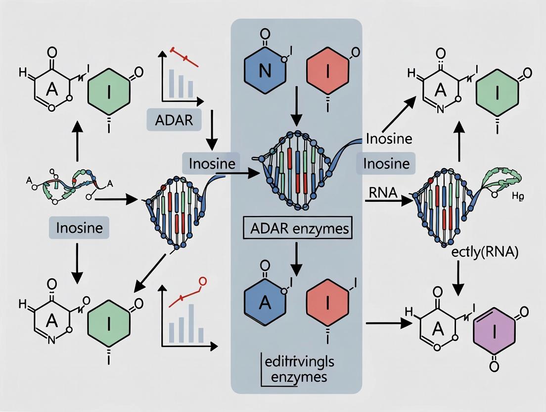
The Biochemical Transformation: Adenosine to Inosine
The core reaction is a hydrolytic deamination occurring at the C6 position of the adenosine nucleoside within an RNA molecule. ADAR enzymes facilitate the substitution of the exocyclic amine group (-NH2) with a carbonyl oxygen (=O), converting adenosine (A) to inosine (I). Crucially, inosine is biochemically recognized as guanosine (G) by most cellular machinery—ribosomes, splicing factors, and reverse transcriptases.
Table 1: Key Biophysical Properties of Adenosine vs. Inosine
| Property | Adenosine (A) | Inosine (I) | Consequence |
|---|---|---|---|
| Base Pairing | Normally pairs with Uridine (U) | Pairs with Cytidine (C) | An A-to-I edit effectively creates an A->G mutation in the RNA sequence. |
| Chemical Formula | C10H13N5O4 | C10H12N4O5 | Loss of NH3 (deamination). |
| Molecular Weight | 267.24 g/mol | 268.23 g/mol | Minimal mass change. |
| Recognition by Polymerases | Template for Thymidine (T) in cDNA | Template for Cytidine (C) in cDNA | Fundamental for PCR-based detection methods (e.g., RNA-seq mismatches). |
Enzymatic Catalysts: The ADAR Family
ADARs are a conserved family of RNA-binding proteins. In humans, three active enzymes exist: ADAR1 (p150 and p110 isoforms), ADAR2, and ADAR3 (catalytically inactive). ADAR1 p150 is interferon-inducible and primarily cytoplasmic, while ADAR1 p110 and ADAR2 are nuclear. Their core structure comprises double-stranded RNA binding domains (dsRBDs) and a C-terminal deaminase domain.
Diagram: ADAR Enzyme Domain Architecture and RNA Interaction
The Inosine Consequence: Molecular and Cellular Outcomes
The A-to-I edit has cascading effects depending on its location:
- Coding Regions: Can alter the amino acid sequence of the protein (recoding), potentially changing function, localization, or stability. Example: Glutamate (Q) to Arginine (R) change in the GluA2 subunit of AMPA receptors.
- Non-Coding Regions: Alters RNA secondary structure, impacting microRNA binding sites, splicing patterns (e.g., in NARF), and RNA stability. This is the most frequent type of editing.
- Immune Recognition: Inosine in double-stranded RNA reduces its immunogenicity by disrupting perfect duplex structure, dampening the MDA5-mediated interferon response—a key function of ADAR1.
Table 2: Functional Consequences of A-to-I Editing by Genomic Location
| Genomic Context | Primary Consequence | Example Gene | Potential Biological Impact |
|---|---|---|---|
| Synonymous (Coding) | None (silent) | Various | Neutral; can be used as an editing "footprint." |
| Non-Synonymous (Coding) | Amino Acid Substitution | GRIA2 (GluA2) | Alters calcium permeability of ion channel. |
| Intronic / Splicing | Alters Splice Site | NARF, AZIN1 | Generates alternative protein isoforms. |
| 3' UTR / miRNA Seed | Alters miRNA Binding | Numerous targets | Modulates mRNA stability and translation. |
| Long dsRNA | Destabilizes Duplex | Alu elements | Prevents aberrant innate immune activation. |
Experimental Protocols for Detection and Quantification
RNA Isolation and Reverse Transcription Protocol
- Reagent: TRIzol or equivalent phenol-guanidine isothiocyanate.
- Key Step: Treat purified total RNA with DNase I (RNase-free) to eliminate genomic DNA contamination.
- Reverse Transcription: Use random hexamers or gene-specific primers. Critical: Use a reverse transcriptase with high processivity but standard fidelity (e.g., SuperScript IV). Do not use mutants with reduced RNA template specificity, as they may misincorporate opposite inosine.
Gold-Standard Validation: Sanger Sequencing of Cloned PCR Products
- PCR Amplify the region of interest from cDNA using high-fidelity DNA polymerase.
- Clone the PCR product into a plasmid vector (e.g., using TA or blunt-end cloning kits).
- Transform competent E. coli and pick at least 20-30 individual colonies.
- Prepare plasmid DNA from each colony and perform Sanger sequencing.
- Analysis: Manually inspect chromatograms of individual clones for A-to-G changes (indicative of A-to-I editing). The editing level is calculated as (number of clones with G) / (total clones sequenced) * 100%.
High-Throughput Quantification: RNA Sequencing Analysis
- Library Prep: Use strand-specific, ribosomal RNA-depleted RNA-seq protocols. Avoid 3'-end focused methods (e.g., some single-cell protocols) which lose internal editing information.
- Alignment: Map sequencing reads to the reference genome using splice-aware aligners (STAR, HISAT2) without hard-clipping soft-clipped portions, as these may contain mismatches.
- Variant Calling: Use specialized tools (e.g., REDItools2, JACUSA2, GIREMI) that are designed to distinguish true RNA editing events from DNA polymorphisms and sequencing errors. Parameters must be stringent.
- Validation: A subset of high-confidence calls should be validated by the cloning method in Section 4.2.
The Scientist's Toolkit: Research Reagent Solutions
Table 3: Essential Reagents for ADAR and A-to-I Editing Research
| Item / Reagent | Function / Purpose | Example / Notes |
|---|---|---|
| Recombinant ADAR Proteins | In vitro deamination assays, structural studies, substrate profiling. | Human ADAR1 (p110) or ADAR2 catalytic domains. |
| ADAR-Specific Antibodies | Immunoprecipitation (RIP), Western blotting, immunofluorescence. | Validate isoform-specific knockdown/knockout. |
| Chemical Inhibitors | Probe ADAR function in cells. | 8-Azaadenosine (non-specific); newer compounds are in development. |
| Synthetic dsRNA Oligos | In vitro activity assays, defining sequence/structure preferences. | Fluorescently labeled substrates allow real-time kinetic measurement. |
| ADAR Knockout Cell Lines | Study phenotypic consequences, identify bona fide editing sites. | Available via CRISPR-Cas9 (e.g., from ATCC or academic sources). |
| Inosine-Specific Chemical Labeling | Enrichment and sequencing of inosine-containing RNA. | icSHAPE or CLEAR methodologies. |
| Validated Positive Control RNA | Assay standardization. | Synthetic RNA with known editing site (e.g., from GluA2 pre-mRNA). |
| High-Fidelity & RT Enzymes | Accurate cDNA synthesis and amplification for detection. | Critical to avoid misinterpreting polymerase errors as editing. |
Diagram: A-to-I Editing Detection and Analysis Workflow
The deamination of adenosine to inosine represents a precise and reversible mechanism for expanding the informational content of the RNA transcriptome. Its consequences—from fine-tuning protein function to maintaining cellular self-tolerance—are vast. As research progresses, the quantitative measurement of editing dynamics and the development of tools to modulate ADAR activity are becoming critical, not only for understanding neurodevelopment, immunity, and cancer but also for pioneering RNA-targeted therapeutics that harness or correct this fundamental RNA modification.
Adenosine-to-inosine (A-to-I) RNA editing, catalyzed by the Adenosine Deaminase Acting on RNA (ADAR) enzyme family, is a critical post-transcriptional modification in metazoans. Inosine is interpreted as guanosine by cellular machineries, leading to recoding events that expand the transcriptome and proteome diversity. This whitepaper details the structural and functional characteristics of ADAR isoforms, framing this knowledge within the broader thesis that precise modulation of ADAR activity holds therapeutic potential for treating neurological disorders, cancers, and autoimmune conditions linked to editing dysregulation.
Structures and Isoforms of the ADAR Family
Three ADAR genes (ADAR1, ADAR2, ADAR3) encode functionally distinct enzymes, with ADAR1 producing two major isoforms via alternative transcription/translation initiation.
Comparative Structural and Functional Analysis
Table 1: Human ADAR Isoforms: Key Characteristics
| Isoform | Gene | Length (aa) | Nuclear Localization | Cytoplasmic Localization | Primary Function | Essential for Life |
|---|---|---|---|---|---|---|
| ADAR1 p150 | ADAR1 | 1,226 | Yes (weak) | Yes (dominant) | Innate immune suppression; global editing of dsRNA | Yes (embryonic lethality if KO) |
| ADAR1 p110 | ADAR1 | 931 | Yes (dominant) | Yes (weak) | Editing of specific sites; housekeeping | No (viable but deficient) |
| ADAR2 | ADAR2 | 801 | Yes | Minimal | Site-specific editing (e.g., GluA2 Q/R site) | Yes (lethal seizures if KO) |
| ADAR3 | ADAR3 | ~735 | Yes | No | Putative negative regulator; brain-specific | No (KO viable) |
Table 2: Key Functional Domains in ADAR Proteins
| Domain | Structure/Features | Function in ADAR1 p150/p110 | Function in ADAR2 | Function in ADAR3 |
|---|---|---|---|---|
| Z-DNA/RNA binding domains (Zα, Zβ) | Zα: Canonical Z-nucleic acid binding. Zβ: Less conserved. | p150: Contains both Zα and Zβ. p110: Lacks Zα. | Absent | Absent |
| Double-stranded RNA Binding Domains (dsRBDs) | 2-3 dsRBDs; ~65-70 aa each; bind dsRNA non-specifically. | Three dsRBDs (I, II, III). Mediate substrate recognition and binding. | Two dsRBDs. Critical for substrate specificity and affinity. | Two dsRBDs; one may be non-functional. |
| Deaminase Domain | Catalytic core; zinc-coordinating motif (HxE/C...C). | Catalyzes A-to-I hydrolysis. Requires dsRNA for activity. | Catalyzes A-to-I hydrolysis. More efficient on certain substrates than ADAR1. | Catalytically inactive (mutations in zinc-coordinating residues). |
| Nuclear Localization Signal(s) (NLS) | Basic amino acid clusters. | Present in both isoforms. | Strong NLS. | Contains NLS. |
| Nuclear Export Signal (NES) | Leucine-rich motif. | Present in p150, conferring cytoplasmic shuttling. | Not clearly defined. | Absent? |
| ADAR3-specific R-domain | Arginine-rich, basic domain. | Absent | Absent | Unique domain; proposed to sequester substrates. |
Key Experimental Protocols in ADAR Research
Protocol: Measuring A-to-I Editing Efficiency via Deep Sequencing
Objective: Quantify editing levels at specific sites or transcriptome-wide. Methodology:
- RNA Isolation & DNase Treatment: Extract total RNA from cells/tissue. Treat with rigorous DNase I to remove genomic DNA.
- Reverse Transcription: Use random hexamers or gene-specific primers with a high-fidelity reverse transcriptase.
- PCR Amplification: Amplify target region(s) using primers with Illumina adaptor overhangs. Keep PCR cycles low to prevent duplicates.
- Library Preparation & Sequencing: Index PCR, purify amplicons, and pool for high-throughput sequencing (Illumina MiSeq/NovaSeq).
- Bioinformatic Analysis:
- Align reads to the reference genome (STAR, HISAT2).
- Use variant callers (GATK) or specialized tools (REDItools, JACUSA2) to identify A-to-G mismatches.
- Filter for known SNPs and alignability. Editing level = (G reads) / (G + A reads) * 100% at a specific genomic coordinate.
Protocol: In Vitro Deamination Assay
Objective: Assess catalytic activity of purified recombinant ADAR protein. Methodology:
- Substrate Preparation: Synthesize a short dsRNA oligo (e.g., 30-50 bp) containing a known editable adenosine (e.g., from GluR-B R/G site).
- Protein Purification: Express and purify N-terminally tagged (e.g., GST, His6) ADAR protein or catalytic domain from E. coli or insect cells.
- Reaction Setup: Incubate dsRNA substrate (radiolabeled or fluorescent) with purified ADAR in reaction buffer (25 mM Tris-HCl pH 7.5, 100 mM KCl, 5% glycerol, 1 mM DTT, 0.1 mg/mL BSA) at 30°C for 30-60 min.
- Analysis:
- TLC Method: Stop reaction with ethanol, digest RNA to nucleosides with nuclease P1. Spot on cellulose TLC plate. Resolve in solvent (e.g., saturated (NH4)2SO4 / isopropanol). Quantify inosine vs. adenosine spots.
- CE Method: Use fluorescently labeled RNA. Treat reaction with RNase T1, which cleaves after guanosine and inosine. Analyze fragments by capillary electrophoresis. The appearance of a new cleavage fragment indicates editing.
Visualizations: ADAR Pathways and Experimental Workflows
The Scientist's Toolkit: Key Research Reagents & Materials
Table 3: Essential Reagents for ADAR Structure-Function and Editing Analysis
| Reagent/Material | Supplier Examples | Function/Application |
|---|---|---|
| Recombinant Human ADAR Proteins (His/GST-tagged) | Sino Biological, Origene, in-house purification | In vitro deamination assays, binding studies (EMSA), structural studies (crystallography, Cryo-EM). |
| ADAR-Specific Antibodies | Santa Cruz (sc-73408), Proteintech, Cell Signaling | Western blot, immunofluorescence, immunoprecipitation to assess expression, localization, and interactions. |
| Site-Specific Editing Reporter Plasmids (e.g., GluA2 Q/R site) | Addgene (#111172, pSELECT-GFPzeo-GluR-B R/G) | Functional validation of ADAR activity in cultured cells; high-throughput screening for activators/inhibitors. |
| Inosine-Specific Chemical Labeling Reagents (e.g., acrylonitrile) | Sigma-Aldrich | Chemical conversion of inosine to cytidine for sequencing-based detection (ICE-seq). |
| Selective ADAR Inhibitors (e.g., 8-azaadenosine, CRON) | Tocris, Sigma, research compounds | Probe ADAR function in disease models; potential therapeutic leads. |
| dsRNA Substrates (e.g., GluR-B R/G site RNA) | IDT, Dharmacon | Defined substrates for in vitro kinetic assays and enzyme characterization. |
| ADAR Knockout Cell Lines (e.g., HEK293 ADAR1-/-) | Commercial or CRISPR-generated | Isogenic controls to define ADAR-specific editing events and phenotypic consequences. |
| Specialized NGS Analysis Software (REDItools, JACUSA2) | Open source | Accurate identification and quantification of A-to-I editing sites from RNA-seq data. |
Adenosine-to-inosine (A-to-I) RNA editing, catalyzed by Adenosine Deaminases Acting on RNA (ADARs), is a crucial post-transcriptional modification. Its role in diversifying the transcriptome, regulating innate immunity, and its implications in neurological disorders and oncology positions it as a critical focus for therapeutic intervention. A central, unresolved question in the field is the precise mechanism of substrate recognition. This whitepaper delves into the core determinants: the structural context provided by double-stranded RNA (dsRNA) and the sequence motifs surrounding the editing site. Understanding this interplay is fundamental to the broader thesis of predicting editing outcomes, deciphering its regulatory logic, and developing targeted therapies that modulate ADAR activity.
The dsRNA Structural Context: Length, Stability, and Imperfections
ADARs bind dsRNA via multiple dsRNA-binding domains (dsRBDs). Substrate affinity and editing efficiency are not uniform but are heavily influenced by the dsRNA's architectural features.
Table 1: Impact of dsRNA Structural Features on ADAR1 Editing Efficiency
| Structural Feature | Experimental Range/Type | Observed Impact on Editing Efficiency (Relative) | Key Experimental Insight |
|---|---|---|---|
| Duplex Length | Short (<30 bp) | Low to Moderate | Minimal processivity; highly site-dependent. |
| Optimal (∼50-150 bp) | High | Allows for stable ADAR binding and sliding; peak efficiency. | |
| Very Long (>500 bp) | High but Selective | Efficient binding but editing often restricted to specific regions near imperfections. | |
| Duplex Perfection | Perfectly Base-Paired | Very Low | Inhibits deamination; ADARs bind but do not efficiently edit. |
| Mismatches/Bulges (especially A-C mismatches) | High | Disrupt helical geometry, flipping target adenosine into the deaminase active site. Essential for site-selectivity. | |
| Duplex Location | Intronic/3’ UTR dsRNA | High | Common genomic context for bona fide editing sites (e.g., GluA2 Q/R site). |
| Intergenic/Repetitive Elements | Variable | Often hyper-edited; role in immune response (prevent MDA5 activation). |
Experimental Protocol: Electrophoretic Mobility Shift Assay (EMSA) for dsRNA-ADAR Binding Affinity
- Purpose: To quantitatively measure the binding affinity (Kd) between a purified ADAR protein (or its dsRBDs) and a defined dsRNA substrate.
- Materials: Purified recombinant ADAR protein, (^{32})P- or fluorescently end-labeled dsRNA probes, non-specific competitor RNA (e.g., yeast tRNA), binding buffer (HEPES/KCl/MgCl2/DTT/glycerol), polyacrylamide gel, electrophoresis apparatus.
- Procedure:
- Probe Preparation: Generate complementary RNA strands, anneal, and purify the dsRNA. Label one strand at the 5' or 3' end.
- Binding Reactions: Set up a series of reactions with a constant, low concentration of labeled dsRNA probe and increasing concentrations of ADAR protein (e.g., 0 nM to 1 µM). Include a large excess of non-specific tRNA to suppress non-specific binding. Incubate at 30°C for 30 min.
- Non-Denaturing Gel Electrophoresis: Load reactions onto a pre-run, chilled non-denaturing polyacrylamide gel (e.g., 6%). Run at constant voltage in a low-ionic-strength buffer (0.5x TBE) at 4°C to preserve complexes.
- Detection & Analysis: Visualize using phosphorimaging or fluorescence. Quantify the fraction of bound vs. free RNA for each protein concentration. Fit the data to a hyperbolic binding equation to determine the dissociation constant (Kd).
Editing Site Sequence Motifs: The Neighborhood Code
While dsRNA structure directs ADAR binding, local sequence motifs (typically -2 to +2 relative to the target adenosine at position 0) govern site selection and efficiency.
Table 2: Sequence Preferences for Human ADAR1-p110 and ADAR2
| Position Relative to Editing Site (A=0) | ADAR1-p110 Preference (5'→3' on opposite strand) | ADAR2 Preference (5'→3' on opposite strand) | Interpretation |
|---|---|---|---|
| -2 (5' neighbor) | U ≈ A > C > G | U > C > A > G | A 5' Uridine (or adenosine for ADAR1) is strongly favored. |
| -1 (immediate 5') | G > A > U > C | A > G > U > C | A 5' guanosine is optimal for ADAR1; adenine for ADAR2. |
| 0 (Editing Site) | A (deaminated) | A (deaminated) | The substrate nucleotide. |
| +1 (immediate 3') | G >> U > A > C | G >> U > A > C | A 3' guanosine is critically important for both enzymes. |
| +2 (3' neighbor) | C ≈ U > A > G | A > C ≈ U > G | Variable preference; ADAR2 shows a clearer preference for A. |
Integrated Model: Structure and Motif Interplay
Recognition is a two-tiered process: 1) initial docking and sliding on dsRNA via dsRBDs, and 2) local interrogation of the sequence motif and structural distortion to flip the target adenosine into the catalytic zinc-containing deaminase domain.
Diagram 1: ADAR Substrate Recognition Pathway
Title: Two-Step ADAR Recognition Process
Experimental Protocol: In Vitro Editing Assay with Mutational Analysis
- Purpose: To functionally validate the contribution of a specific dsRNA structural feature or sequence motif to editing efficiency.
- Materials: Purified ADAR enzyme, synthetic dsRNA substrates (wild-type and mutants), reaction buffer, ATP (optional), EDTA, phenol-chloroform, ethanol.
- Procedure:
- Substrate Design: Synthesize RNA oligonucleotides to form a short (e.g., 30-50 bp) dsRNA containing the target site. Create mutant substrates: (a) disrupting the duplex (e.g., perfect vs. bulged), (b) altering the -1 and +1 nucleotides.
- Editing Reaction: Incubate a fixed concentration of each dsRNA substrate with ADAR enzyme in appropriate buffer. Use time points or enzyme titration to stay in the linear range of the reaction. Quench with EDTA.
- Product Analysis: Extract RNA. Use reverse transcription (RT) followed by either:
- Restriction Fragment Length Polymorphism (RFLP): If editing creates/destroys a restriction site.
- Sanger Sequencing or Pyrosequencing: For quantitative measurement of editing percentage.
- Next-Generation Sequencing (NGS): For high-throughput analysis of multiple substrates.
- Quantification: Compare editing efficiency (%) of mutant substrates to the wild-type control.
The Scientist's Toolkit: Key Research Reagents
Table 3: Essential Reagents for dsRNA/ADAR Substrate Recognition Studies
| Reagent / Material | Function / Application | Key Consideration |
|---|---|---|
| Recombinant Human ADAR1/ADAR2 (full-length or catalytic domain) | In vitro binding and editing assays. Source from commercial vendors or express in insect/bacterial systems. | Requires proper folding and zinc coordination for deaminase activity. |
| Synthetic, Site-Specifically Modified RNA Oligonucleotides | Creating defined dsRNA substrates with mismatches, bulges, and sequence motif variants. | Chemical synthesis allows for precise control; HPLC purification is essential. |
| Fluorescent or Isotopic RNA Labeling Kits (e.g., Cy5, (^{32})P) | Tagging dsRNA substrates for EMSA, in vitro editing, or cellular tracking. | Choice depends on detection method (gel imaging, fluorescence polarization). |
| Anti-ADAR Antibodies (specific for ADAR1 or ADAR2) | Immunoprecipitation (RIP/CLIP) to identify endogenous RNA targets, or Western blot analysis. | Validation for specific applications (IP, IF) is critical. |
| Dual-Luciferase Reporter Vectors with Engineered dsRNA Structures | Validating substrate rules in a cellular context. | Enables high-throughput screening of sequence/structural determinants. |
| Inosine-Specific Chemical Sequencing Reagents (e.g., CEU) | Genome-wide mapping of in vivo editing sites. | Key for differentiating A-to-I editing from other modifications or SNPs. |
Adenosine-to-Inosine (A-to-I) RNA editing, catalyzed by the Adenosine Deaminase Acting on RNA (ADAR) enzyme family, is a critical post-transcriptional mechanism that diversifies the transcriptome. Within the context of a broader thesis on A-to-I editing and ADAR research, this whitepaper explores the dual and seemingly disparate cellular functions of this system: its non-immunogenic role in preventing aberrant activation of the innate immune sensor MDA5 (Melanoma Differentiation-Associated protein 5) and its role in fine-tuning neurotransmission through the recoding of neurotransmitter receptor and ion channel transcripts. These functions underscore ADAR's pivotal position at the intersection of innate immunity and neurobiology, with significant implications for autoimmune disease, viral infection response, and neurological disorders.
Core Mechanism: A-to-I Editing and ADAR Enzymes
A-to-I editing is mediated by three ADAR proteins: ADAR1 (with interferon-inducible p150 and constitutive p110 isoforms), ADAR2, and the catalytically inactive ADAR3. ADAR1 and ADAR2 deaminate adenosine to inosine within double-stranded RNA (dsRNA) structures, which is recognized as guanosine by cellular machinery. This process occurs both in non-coding regions (e.g., Alu repeats in 3'UTRs) and coding sequences, where it can alter amino acid sequences.
Preventing Innate Immune Activation: Silencing Endogenous dsRNA
Endogenous dsRNA, often formed by inverted repeats or transposable elements like Alu sequences, can be mistaken for viral RNA by cytosolic pattern recognition receptors, primarily MDA5. Unedited endogenous dsRNA activates MDA5, leading to a type I interferon (IFN) response and autoinflammation. ADAR1, by editing these dsRNAs, disrupts their perfect complementarity, preventing MDA5 recognition and activation.
Quantitative Data on ADAR1 and Immune Signaling
Table 1: Key Quantitative Findings in ADAR1-MDA5 Pathway
| Observation / Parameter | Experimental System | Quantitative Outcome | Reference (Example) |
|---|---|---|---|
| ADAR1 loss-of-function | Mouse embryonic fibroblasts (MEFs) | >1000-fold increase in Ifnb1 mRNA levels | Liddicoat et al., 2015 |
| MDA5 dependency | Adar1 −/− MEFs + MDA5 knockout | Complete rescue of embryonic lethality; IFNβ reduced to baseline | Mannion et al., 2014 |
| Editing sites in immune genes | Human PBRNA-seq | >150 A-to-I sites in Alu elements near interferon-stimulated genes (ISGs) | Chung et al., 2018 |
| Adar1 p150 requirement | Adar1 (p150-only) mice | Viable; no interferon signature | Ward et al., 2011 |
| Correlation of editing & ISG expression | Aicardi-Goutières Syndrome (AGS) patients | Inverse correlation (r ≈ -0.7) between editing index in Alu repeats and ISG expression | Rice et al., 2012 |
Experimental Protocol: Measuring MDA5 Activation and Interferon Response
Protocol: Assessing IFNβ Activation via qPCR and Luciferase Reporter Assay
Objective: To quantify the innate immune response upon ADAR1 knockdown or knockout.
Materials:
- Cells: HEK293T, A549, or primary fibroblasts.
- Transfection Reagents: Lipofectamine RNAiMAX (for siRNA) or Fugene/PEI (for plasmids).
- siRNAs: Targeting ADAR1 and non-targeting control.
- Plasmids: IFNβ promoter-driven firefly luciferase reporter, Renilla luciferase control (pRL-TK).
- qPCR Reagents: SYBR Green master mix, primers for IFNB1, ISG15 (response), and GAPDH (control).
- Luciferase Assay Kit: Dual-Luciferase Reporter Assay System.
Procedure:
- Cell Seeding: Seed cells in 24-well plates for 24 hours (70-80% confluency).
- Knockdown: Transfect with 50 nM ADAR1-specific or control siRNA using RNAiMAX per manufacturer's protocol.
- Reporter Assay (Parallel): Co-transfect cells with 100 ng IFNβ-firefly luciferase reporter and 10 ng Renilla control plasmid 24 hours post-siRNA transfection.
- Incubation: Harvest cells 48 hours post-siRNA transfection.
- qPCR:
- Isolate total RNA (TRIzol).
- Perform reverse transcription (1 µg RNA).
- Run qPCR in triplicate: 95°C for 10 min; 40 cycles of 95°C for 15 sec, 60°C for 1 min.
- Calculate ΔΔCt for target genes normalized to GAPDH and relative to control.
- Luciferase Measurement:
- Lyse cells in Passive Lysis Buffer.
- Measure firefly and Renilla luciferase luminescence sequentially.
- Normalize firefly luciferase activity to Renilla.
- Validation: Confirm ADAR1 knockdown by western blot (anti-ADAR1 antibody).
Pathway Diagram: ADAR1 Prevents MDA5-Mediated Interferon Activation
Diagram 1: ADAR1 editing prevents MDA5 sensing of endogenous dsRNA.
Modulating Neurotransmission: Recoding Synaptic Proteins
In the nervous system, ADAR2-mediated editing recodes key neurotransmitter receptors, altering their functional properties. The most characterized edit is in the glutamate receptor subunit GluA2 (Q/R site), which controls calcium permeability. Disruption of this editing is linked to neurological pathologies.
Quantitative Data on Editing in Neurotransmission
Table 2: Key Quantitative Findings in Neuronal RNA Editing
| Transcript / Site | Editing Level (Tissue) | Functional Consequence | Disease Link |
|---|---|---|---|
| GRIA2 (GluA2) Q/R site (R/G) | ~100% in adult brain (human cortex) | Reduces Ca²⁺ permeability of AMPA receptors; alters single-channel conductance | Amyotrophic lateral sclerosis (ALS), epilepsy |
| GRIA2 R/G site | ~50-80% (human cortex) | Accelerates recovery from desensitization | |
| GRIK2 (GluK2) Q/R site | ~80-90% (human brain) | Reduces Ca²⁺ permeability of kainate receptors | Epilepsy, schizophrenia |
| HTR2C (5 sites: A-E) | Varies (15-70% per site, human prefrontal cortex) | Alters G-protein coupling efficacy; generates 24 protein isoforms | Depression, suicide, neuropsychiatric disorders |
| CYFIP2 (K/E site) | ~70% (mouse hippocampus) | Alters actin dynamics, dendritic spine morphology | Alzheimer's disease pathology |
Experimental Protocol: Assessing Editing Efficiency via Deep Sequencing
Protocol: High-Throughput Validation of Editing Sites (PCR-Amplicon Seq)
Objective: Precisely quantify A-to-I editing levels at specific genomic loci from tissue or cell RNA.
Materials:
- RNA: High-quality total RNA (RIN > 8).
- Reverse Transcription: SuperScript IV, random hexamers/oligo-dT.
- PCR: High-fidelity DNA polymerase (e.g., Q5 Hot Start).
- Primers: Specific primers flanking editing site(s) with Illumina overhang adapters.
- Library Prep Kit: Illumina Nextera XT or equivalent.
- Sequencing Platform: Illumina MiSeq (for amplicons).
Procedure:
- cDNA Synthesis: Synthesize cDNA from 1 µg DNase-treated RNA.
- First-PCR (Amplification):
- Use locus-specific primers with overhangs.
- PCR conditions: 98°C 30s; 25 cycles of (98°C 10s, 65°C 30s, 72°C 30s); 72°C 2 min.
- Purify amplicons (SPRI beads).
- Indexing PCR (Add Indices):
- Use Illumina dual index primers (Nextera XT Index Kit).
- 8 cycles of PCR.
- Purify final library.
- Quality Control: Assess library size (Bioanalyzer/TapeStation); quantify (qPCR).
- Sequencing: Pool and sequence on MiSeq (2x250 bp), aiming for >10,000 reads/sample.
- Analysis:
- Align reads to reference (BWA/Bowtie2).
- Use tools like REDItools or GATK to identify mismatches (A->G changes in cDNA).
- Calculate editing efficiency as (G reads) / (G + A reads) × 100% at the locus.
Pathway Diagram: ADAR2 Editing Modulates Synaptic Signaling
Diagram 2: ADAR2-mediated recoding modulates synaptic function.
The Scientist's Toolkit: Key Research Reagents
Table 3: Essential Reagents for ADAR/RNA Editing Research
| Reagent / Material | Function / Application | Example (Vendor) |
|---|---|---|
| Anti-ADAR1 Antibody | Detects ADAR1 p150/p110 isoforms in WB, IP, IF. | Rabbit mAb, DSHB (15.8.6) or Santa Cruz (sc-73408). |
| Anti-ADAR2 Antibody | Specific detection of ADAR2 protein. | Rabbit pAb, Sigma (HPA038524). |
| ADAR1 siRNA/sgRNA | Knockdown/knockout for functional loss-of-function studies. | ON-TARGETplus Human ADAR1 siRNA (Dharmacon). |
| MDA5 (IFIH1) siRNA | Validate MDA5-dependency of interferon response. | Silencer Select siRNA (Thermo). |
| IFNβ Reporter Plasmid | Luciferase-based reporter for interferon pathway activation. | pGL4-IFNβ-luc (Addgene plasmid #102597). |
| Editing-Site Specific Primers | Amplify loci for Sanger or deep-seq quantification of editing. | Custom-designed (IDT). |
| Inosine-Specific Cleavage Kit | Detect inosine sites in RNA (RTL-P method). | ICE Kit (NEB). |
| Recombinant ADAR Protein | In vitro editing assays, substrate specificity studies. | His-tagged human ADAR1 (p110) (Origene). |
| 8-Azaadenosine | Small molecule inhibitor of ADAR deaminase activity. | Tocris Bioscience. |
| High-Fidelity RNA-Seq Library Kit | For transcriptome-wide editing analysis (reduce false positives). | KAPA RNA HyperPrep (Roche). |
Within the broader study of A-to-I RNA editing mechanisms and ADAR enzyme research, a fundamental challenge is the accurate identification and characterization of editing sites across the genome. This process is complicated by the starkly different genomic contexts of hyper-edited, repetitive Alu elements and sparsely edited, non-repetitive regions. Understanding this landscape is crucial for elucidating the biological functions of editing in innate immunity, neural development, and its dysregulation in diseases like cancer and autoimmune disorders.
Genomic Contexts of A-to-I Editing
1AluElements
Alu elements are short (~300 bp), primate-specific SINEs that constitute ~11% of the human genome. They are frequently found in introns and untranslated regions (UTRs). Their high density of inverted repeats forms long, double-stranded RNA (dsRNA) structures, making them prime substrates for the constitutively expressed ADAR1 p110 isoform and the inducible ADAR1 p150 isoform. Editing in Alu elements is often hyper-editing, with dozens to hundreds of adenosine deaminations within a single dsRNA duplex.
Non-Repetitive Regions
Editing sites in non-repetitive, coding or structured non-coding regions are less frequent but often functionally consequential (e.g., recoding events). These sites are typically targeted by ADAR2 and require specific, localized dsRNA structures. The editing level at such sites is usually tightly regulated.
Table 1: Key Characteristics of Editing Sites in Different Genomic Contexts
| Feature | Alu Elements | Non-Repetitive Regions |
|---|---|---|
| Genomic Abundance | Millions of potential loci | Thousands of loci |
| Typical Location | Introns, 3' UTRs | Exons, 5' UTRs, miRNAs, lincRNAs |
| dsRNA Structure | Long, perfect/imperfect IRs from neighboring Alus | Short, imperfect stem-loops |
| Primary ADAR Enzyme | ADAR1 (p110 & p150) | ADAR2 (ADARB1) |
| Editing Density | Hyper-editing (clustered sites) | Isolated, specific sites |
| Typical Function | Immune tolerance, RNA stability, miRNA regulation | Proteome diversification, splicing regulation |
Core Experimental Protocols for Identification & Cataloging
RNA Sequencing & Bioinformatics Pipeline
This protocol outlines the standard workflow for genome-wide editing site discovery.
Materials:
- Total RNA (RIN > 8) from relevant tissue/cell line.
- Poly(A) selection or rRNA depletion kit.
- Strand-specific RNA-seq library prep kit.
- High-throughput sequencer (Illumina NovaSeq, etc.).
- High-performance computing cluster.
Procedure:
- Library Preparation & Sequencing: Perform stranded, paired-end RNA-seq (≥ 100 bp reads, depth ≥ 50M reads per sample). Include technical replicates.
- Read Alignment: Map reads to the human reference genome (GRCh38) using a splice-aware aligner (e.g., STAR) in two-pass mode. Critical: For Alu region mapping, use parameters that permit soft-clipping and do not filter multi-mapping reads excessively (
--outFilterMultimapNmax 100). - Duplicate Marking: Use Picard Tools to mark PCR duplicates.
- Variant Calling: Use a specialized RNA-editing caller (e.g., REDItools2, JACUSA2, or JACUSA2call-2) to identify A-to-G (T-to-C on the opposite strand) mismatches against the genome.
- Stringent Filtering:
- Remove known SNPs (dbSNP, 1000 Genomes).
- Remove variants in simple repeats/low-complexity regions (RepeatMasker).
- Apply a minimum read coverage filter (e.g., ≥ 10 reads).
- Apply a minimum editing level filter (e.g., ≥ 1% for non-repetitive, ≥ 0.1% for Alu clusters).
- For Non-Repetitive Sites: Require sites to be in annotated non-repetitive regions and often validate with Sanger sequencing or targeted amplicon-seq.
- For Alu Sites: Aggregate editing events within inverted Alu pairs. Use tools like REDItools2 or SAILOR to analyze clustered hyper-editing.
- Validation: Perform independent validation via amplicon sequencing of cDNA (with control genomic DNA) for high-priority sites.
Diagram: RNA-seq Editing Detection Workflow
Protocol forAlu-Specific Editing Analysis (CLIP-ADAR & Hyper-Editing Detection)
This protocol combines biochemical purification of ADAR-RNA complexes with hyper-editing detection.
Materials:
- Crosslinker (UV 254 nm).
- ADAR1-specific antibody (validated for CLIP).
- Protein G/A magnetic beads.
- RNase I (for partial digestion).
- Phosphatase, polynucleotide kinase, and ligation reagents.
- Reverse transcription primers with unique molecular identifiers (UMIs).
- High-sensitivity DNA kit (Bioanalyzer/TapeStation).
Procedure:
- UV Crosslinking: Irradiate cells (e.g., HEK293T, primary astrocytes) with 254 nm UV light to crosslink ADAR proteins to bound RNA.
- Cell Lysis & Partial RNase Digestion: Lyse cells and treat lysate with RNase I to fragment bound RNA, leaving ~50-100 nt protein-protected footprints.
- Immunoprecipitation: Incubate with anti-ADAR1 antibody coupled to magnetic beads. Include an isotype control IgG sample.
- RNA Adapter Ligation: On-bead, dephosphorylate, then ligate a pre-adenylated 3' RNA adapter to the RNA.
- Radiolabeling & Purification: Label the 5' end with P³², run on SDS-PAGE, transfer to nitrocellulose, and excise the ADAR-RNA complex band. Extract RNA.
- Reverse Transcription & Library Prep: Reverse transcribe with a primer containing a UMI and a 5' adapter sequence. Circularize PCR-amplify.
- Bioinformatic Analysis:
- Map reads, identify crosslink sites (mutations/truncations).
- Overlap CLIP clusters with Alu annotations from RepeatMasker.
- Integrate with RNA-seq derived editing sites to distinguish binding from active editing.
Diagram: ADAR1 CLIP-seq for Alu Binding Sites
The Scientist's Toolkit: Research Reagent Solutions
Table 2: Essential Reagents for Editing Site Research
| Reagent / Material | Function / Role | Example Product/Catalog |
|---|---|---|
| RNase Inhibitor | Prevents RNA degradation during extraction and library prep. | Murine RNase Inhibitor (M0314L) |
| Poly(A) Selection Beads | Enriches for polyadenylated mRNA, reducing ribosomal RNA background. | NEBNext Poly(A) mRNA Magnetic Isolation Module (E7490) |
| Strand-Specific Library Prep Kit | Maintains strand orientation of RNA, crucial for mapping to repetitive elements. | Illumina Stranded mRNA Prep |
| ADAR1-p150 Specific Antibody | For immunoprecipitation in CLIP experiments to distinguish from p110 isoform. | Sigma-Aldrich HPA021358 (validated for CLIP) |
| UV Crosslinker (254 nm) | Covalently links ADAR enzymes to bound RNA in vivo. | Spectrolinker XL-1000 |
| Protein G Magnetic Beads | Capture antibody-protein-RNA complexes during CLIP. | Dynabeads Protein G (10004D) |
| Pre-Adenylated 3' Adapter | For ligation to RNA 3' ends without ATP (prevents circularization). | TruSeq Small RNA 3' Adapter |
| UMI Adapters/Primers | Unique Molecular Identifiers to eliminate PCR duplicates and bias. | NEBNext Multiplex Small RNA Library Prep Set (E7300) |
| Editing-Specific Software | Bioinformatics tools for accurate calling and cataloging. | REDItools2, JACUSA2, SAILOR |
Integrated Analysis & Catalogs
Modern catalogs (e.g., REDIportal, RADAR) distinguish between Alu and non-repetitive sites. Key quantitative findings from recent studies are summarized below.
Table 3: Quantitative Summary of Human Editing Landscapes (Recent Data)
| Metric | Alu-Associated Sites | Non-Repetitive Sites | Notes |
|---|---|---|---|
| Estimated Total Sites | >4.5 million | ~10,000 - 15,000 | Non-repetitive sites are evolutionarily more conserved. |
| Editing Level Range | 0.1% - 50% (typically low) | 1% - 100% (often ~20-80%) | Alu editing is frequent but low-level; non-repetitive can be stoichiometric. |
| Tissue Specificity | Widespread, high in brain | Highly tissue-specific (CNS enriched) | ADAR2 expression drives brain-specific recoding. |
| Disease Association | Autoimmunity (AGS), cancer | Neurological disorders, epilepsy, cancer | Alu editing loss triggers MDA5-mediated interferon response. |
| Validation Rate | Lower (due to mapping ambiguity) | High (>90% with amplicon-seq) | Alu sites require specialized mapping algorithms. |
Diagram: Integrated Pathway of ADAR Function & Editing Consequences
From Detection to Design: Methodologies and Therapeutic Applications of RNA Editing
Within the broader research on A-to-I RNA editing, catalyzed by ADAR (Adenosine Deaminase Acting on RNA) enzymes, precise detection and quantification of editing events are paramount. This technical guide details core methodologies for identifying and measuring these post-transcriptional modifications, which are crucial in neuroscience, immunology, and cancer research. The integration of high-throughput RNA sequencing (RNA-seq) with targeted validation techniques like the ICE method and Sanger sequencing forms the cornerstone of rigorous A-to-I editing analysis.
RNA-seq Analysis for A-to-I Editing Detection
RNA-seq provides a genome-wide survey of editing sites but requires specific analytical pipelines to distinguish true A-to-I events from single-nucleotide polymorphisms (SNPs) and technical artifacts.
Core Computational Workflow
A standard bioinformatic pipeline involves the following stages:
- Alignment: Map cleaned reads to the reference genome using splice-aware aligners (e.g., STAR, HISAT2). It is critical to not perform standard SNP-aware realignment, as this would mask true editing sites.
- Variant Calling: Identify mismatches relative to the reference genome using tools like GATK HaplotypeCaller or specialized editors like REDItools or JACUSA2.
- A-to-I Filtering: Apply stringent filters:
- Remove known SNPs (using dbsNP, 1000 Genomes).
- Select only A-to-G (on the transcript) or T-to-C (on the genome) mismatches in the context of double-stranded RNA.
- Require a minimum read depth (e.g., ≥10x) and editing level (e.g., ≥1%).
- Filter sites present in intronic Alu repetitive regions (common for ADAR1) or non-Alu regions (common for ADAR2).
- Quantification: Calculate the editing level/rate as: (Number of G reads / Total reads covering the site) * 100%.
Quantitative Data from Recent Studies
Table 1: Representative A-to-I Editing Metrics from Recent RNA-seq Studies (2023-2024)
| Tissue/Cell Type | Total Sites Detected | Hyper-edited Sites (>50%) | Average Editing Level (Non-Alu) | Key ADAR Enzyme | Reference |
|---|---|---|---|---|---|
| Human Cerebral Cortex | >2 million | ~3,500 | 15-75% (site-dependent) | ADAR2 (predominant) | Reichold et al., 2023 |
| Glioblastoma Stem Cells | ~1.2 million | ~1,800 | 5-60% | ADAR1 (upregulated) | He et al., 2024 |
| Mouse Spleen (Immune Activated) | ~850,000 | ~950 | 10-40% | ADAR1 | Pestal et al., 2024 |
| HEK293T (ADAR1-KO) | <50,000 | <10 | <1% | - | Validation control |
Detailed Protocol: RNA-seq Library Prep for Editing Analysis
Aim: Prepare stranded, total RNA libraries to preserve strand information, critical for distinguishing A-to-G edits from T-to-C transcriptional variants.
Reagents:
- RiboCop rRNA Depletion Kit (for human/mouse) or Poly(A) Selection Kit.
- NEBNext Ultra II Directional RNA Library Prep Kit.
- RNase inhibitor.
- Agencourt AMPure XP beads.
Method:
- RNA Quality Control: Verify RIN > 8.5 (Agilent Bioanalyzer).
- rRNA Depletion: Remove ribosomal RNA using 1μg of total RNA with the RiboCop kit.
- Fragmentation and First-Strand Synthesis: Fragment enriched mRNA and synthesize cDNA using random hexamers.
- Second-Strand Synthesis: Use dUTP incorporation to preserve strand orientation.
- End Repair, A-tailing, and Adapter Ligation: Prepare ends for indexed adapter ligation.
- Size Selection and PCR Enrichment: Select fragments ~300-500 bp using AMPure beads. Perform 10-12 cycles of PCR.
- QC and Sequencing: Validate library size (Bioanalyzer) and quantify (qPCR). Sequence on Illumina NovaSeq platform, aiming for >50 million 150bp paired-end reads per sample.
The ICE Method for Site-Specific Validation
The Inverse CE-PCR (ICE) method is a robust, PCR-based technique for validating and quantifying specific editing sites without cloning.
ICE Method Workflow
Diagram Title: ICE Method Workflow for A-to-I RNA Editing Quantification
Detailed Protocol: ICE Assay
Aim: Quantify editing level at a specific site in GRIA2 (Q/R site).
Reagents:
- High-specificity DNA polymerase (e.g., Phusion Hot Start).
- Restriction enzyme BtgZI (recognizes GCGATGC, where * is edited base? Note: Requires primer design to create site for unedited allele).
- Reverse transcriptase.
- Agarose gel equipment.
Method:
- Primer Design: Design a reverse primer for cDNA synthesis ~100bp downstream of the editing site. Design PCR primers that amplify a 200-300bp fragment. Critically, modify the forward primer to introduce a single mismatch that creates a BtgZI restriction site only if the genomic base is an unedited A.
- cDNA Synthesis: Synthesize cDNA from 500ng DNase-treated total RNA.
- PCR: Amplify the target from cDNA using high-fidelity polymerase. Use 25-30 cycles.
- Restriction Digest: Purify the PCR product. Digest 200ng of product with 10U BtgZI at 60°C for 3 hours.
- Quantification: Run digested products on a 3% agarose gel. Measure band intensities using ImageJ. The editing percentage is calculated as: [Intensity of Uncut Band / (Intensity of Cut Band + Intensity of Uncut Band)] * 100%. Validate with known mixed templates.
Sanger Sequencing for Validation and Cloning Assessment
Sanger sequencing provides definitive validation of editing sites and is essential for analyzing editing patterns in cloned PCR products.
Detailed Protocol: Direct and Clonal Sanger Sequencing
Aim: Validate an RNA-seq-identified site and assess editing heterogeneity across transcripts.
Reagents:
- TOPO-TA Cloning Kit.
- BigDye Terminator v3.1 Cycle Sequencing Kit.
- Competent E. coli.
- Capillary sequencer.
Method: Part A: Direct Sequencing from PCR Products:
- Amplify the region of interest from cDNA (as in ICE, but without engineered restriction site).
- Purify the PCR product.
- Perform Sanger sequencing reaction with a nested primer.
- Analyze the chromatogram. A mixed A/G peak at the edited position indicates partial editing. The relative peak heights (A vs G) provide a semi-quantitative estimate.
Part B: Clonal Sequencing:
- Clone the purified PCR product (from Step A.1) into a TA vector. Transform competent cells.
- Pick 20-30 individual bacterial colonies for colony PCR.
- Sequence each colony PCR product.
- Count the number of clones containing 'G' (edited) versus 'A' (unedited) at the site. The editing level = (Number of 'G' clones / Total clones sequenced) * 100%. This provides a quantitative, digital readout.
Integrated Analysis Pathway
The relationship between these techniques is synergistic, not sequential. RNA-seq discovers sites, while ICE and Sanger validate and precisely quantify them in specific biological or experimental contexts.
Diagram Title: Integrated Workflow for A-to-I Editing Analysis in ADAR Research
The Scientist's Toolkit: Essential Reagents & Materials
Table 2: Key Research Reagent Solutions for A-to-I RNA Editing Studies
| Reagent/Material | Supplier Examples | Function in Protocol | Critical Consideration for Editing Research |
|---|---|---|---|
| RiboCop rRNA Depletion Kit | Lexogen, Illumina | Removes ribosomal RNA prior to RNA-seq library prep. Preserves non-polyadenylated transcripts. | Preferred over poly(A) selection for capturing editing in introns and non-coding RNAs. |
| NEBNext Ultra II Directional RNA Library Prep Kit | New England Biolabs | Stranded RNA-seq library construction. | Maintains strand information to correctly assign A-to-G vs T-to-C changes. |
| Phusion Hot Start DNA Polymerase | Thermo Fisher | High-fidelity PCR for ICE assay and amplicon generation. | Minimizes PCR-induced mutations that could be mistaken for editing events. |
| BtgZI Restriction Enzyme | NEB | Key enzyme for ICE assay. Cuts specifically at the sequence context of the unedited allele. | Success depends on perfect primer design to introduce enzyme site only for the 'A' allele. |
| TOPO-TA Cloning Kit | Thermo Fisher | Cloning of PCR amplicons for clonal Sanger sequencing. | Allows for assessment of editing heterogeneity across individual transcript molecules. |
| BigDye Terminator v3.1 Kit | Thermo Fisher | Cycle sequencing for Sanger methodology. | Provides clean chromatograms for base calling at suspected editing sites. |
| ADAR1-p110 Specific Antibody | Santa Cruz, Cell Signaling | Immunoprecipitation or validation of ADAR expression in study models. | Essential for correlating editing changes with ADAR protein levels in thesis research. |
| Synthetic RNA Controls | IDT, Sigma | Spike-in controls with known editing levels. | Validates the accuracy and sensitivity of both RNA-seq and ICE quantification pipelines. |
This technical guide details the engineering of guide RNAs (gRNAs) for Site-Directed RNA Editing (SDRE), a programmable technology leveraging endogenous Adenosine Deaminase Acting on RNA (ADAR) enzymes for the precise conversion of adenosine (A) to inosine (I) at specific transcriptomic loci. Framed within the broader thesis of exploiting the A-to-I editing mechanism for therapeutic and research applications, this document provides a comprehensive resource for researchers aiming to design, optimize, and implement gRNA-based editing systems.
A-to-I RNA editing, catalyzed by ADAR enzymes, is a conserved post-transcriptional mechanism that diversifies the transcriptome. Inosine is interpreted as guanosine (G) by cellular machinery, effectively resulting in an A-to-G change. The thesis central to this field posits that harnessing this endogenous, RNA-based system for programmable correction of disease-causing mutations or modulation of gene function offers distinct advantages over DNA-editing approaches, including reduced off-target genomic risk and transient, tunable effects. Natural ADAR substrates are long, double-stranded RNAs (dsRNAs). SDRE redirects this activity to specific single adenosines on endogenous messenger RNAs (mRNAs) using engineered gRNAs that form a defined, editable dsRNA structure with the target site.
Core Principles of gRNA Engineering for ADAR Recruitment
Effective gRNA design must satisfy two primary constraints: target specificity and efficient ADAR recruitment/editing. The gRNA is typically an antisense oligonucleotide complementary to the target RNA region, with strategic mismatches to position the target adenosine within an optimal editing context.
Key Structural and Sequence Determinants
- Editing Window: The target adenosine is positioned opposite a strategic mismatch (often a cytidine or another adenosine) in the gRNA, typically at positions -2 to 0 relative to the 5' end of the gRNA's complementary region. This creates a local structural mimic of ADAR's preferred substrate.
- gRNA Length & Complementarity: The complementary region usually spans 15-35 nucleotides. Longer lengths increase affinity but may promote promiscuous editing and reduce cellular delivery efficiency.
- 3' and 5' Handles: Non-complementary flanking sequences, often derived from natural ADAR substrates (e.g., portions of the GluR2 R/G site), can enhance ADAR binding and editing efficiency.
- Chemical Modifications: To improve nuclease resistance, pharmacokinetics, and cellular uptake, gRNAs are chemically modified (e.g., 2'-O-methyl, phosphorothioate backbone, locked nucleic acids).
Table 1: Comparison of ADAR Isoforms for SDRE Applications
| Parameter | ADAR1 (p150) | ADAR1 (p110) | ADAR2 | Engineered Deaminase (e.g., dADAR) |
|---|---|---|---|---|
| Primary Location | Cytoplasm & Nucleus | Nucleus | Nucleus | Cytoplasm (by design) |
| Endogenous Role | Innate immunity, editing of Alu elements | Unknown | Neurotransmission, coding site editing | N/A |
| Preferred Substrate | Long dsRNA | Long dsRNA | Short, structured dsRNA | Engineered for short gRNA recognition |
| Editing Efficiency (Model System)* | Variable, can be high with optimal gRNA | Moderate | High for specific motifs | Very High (engineered for specificity) |
| Immunogenicity Risk | Higher (cytosolic dsRNA sensor) | Lower | Low | Low (engineered domain) |
| Common Use in SDRE | Yes, for cytoplasmic targets | Less common | Yes, for nuclear targets | Increasingly common for high efficiency |
*Efficiency is highly context-dependent.
Table 2: Impact of gRNA Design Parameters on Editing Outcomes
| Design Parameter | Typical Range | Effect on Efficiency | Effect on Specificity |
|---|---|---|---|
| Complementarity Length | 15-35 nt | Increases up to a point, then plateaus or decreases | Longer lengths may increase off-target binding |
| Optimal Editing Window | Position -2 to 0* | Critical for activity; position 0 often highest | Defines primary on-target site |
| 5' Handle Sequence | e.g., 5'-GGACU-3' | Can increase efficiency 2-10 fold | Minimal impact |
| 3' Handle Sequence | Variable | Stabilizes complex; moderate impact (1.5-3 fold) | Minimal impact |
| Chemical Modifications | PS backbone, 2'-O-Me | Increases stability & delivery efficiency (>10-fold in vivo) | Can slightly reduce off-targets by limiting gRNA half-life |
| Mismatch at Target A | C (best), A, U, G | C >> A > U > G in efficiency ranking | Critical for defining the target base |
*Position 0 is the nucleotide opposite the target A. Position -1 is 5' adjacent to pos 0 on the gRNA.
Experimental Protocols
Protocol: In Vitro Validation of gRNA Editing Efficiency
Objective: To test and quantify the editing efficiency of candidate gRNAs using purified ADAR enzyme. Materials:
- Purified recombinant ADAR protein (e.g., human ADAR2 catalytic domain)
- Synthetic target RNA oligonucleotide (containing the target adenosine in context)
- Synthetic candidate gRNAs (with varying designs)
- Reaction buffer (100 mM HEPES pH 7.0, 100 mM KCl, 5 mM MgCl2, 1 mM DTT, 0.1 mg/mL BSA)
- RNase inhibitor
- Phenol:chloroform:isoamyl alcohol, ethanol for cleanup
- RT-PCR and sequencing reagents (or restriction enzyme assay if editing creates a site)
Methodology:
- Annealing: Combine 1 pmol of target RNA with 5 pmol of gRNA in annealing buffer (10 mM Tris pH 7.5, 50 mM NaCl). Heat to 85°C for 2 min, then cool slowly to room temperature.
- Editing Reaction: Assemble a 20 µL reaction containing 1X reaction buffer, 10 U RNase inhibitor, 0.5 pmol annealed duplex, and 100-500 nM ADAR protein. Incubate at 30°C for 1-2 hours.
- Reaction Termination: Add 200 µL of Phenol:Chloroform:IAA, vortex, and centrifuge. Recover the aqueous phase.
- RNA Precipitation: Add 2.5 volumes of ethanol and 1/10 volume of 3M NaOAc (pH 5.2). Precipitate at -20°C for 1 hour. Pellet, wash with 70% ethanol, and resuspend in nuclease-free water.
- Analysis:
- Sanger Sequencing: Reverse transcribe the RNA, PCR amplify the region, and sequence. Quantify editing efficiency by chromatogram peak height ratio (G vs A) at the target site using software like EditR or TIDE.
- High-Throughput Sequencing (Recommended): Perform RT-PCR with barcoded primers, sequence on an Illumina platform, and analyze with pipelines like REDItools or SAILOR to obtain precise editing percentages and detect bystander editing.
Protocol: Cellular Delivery and Assessment of SDRE
Objective: To evaluate gRNA performance in a relevant mammalian cell line. Materials:
- HEK293T cells (or other relevant cell line)
- Lipofectamine 3000 or similar transfection reagent
- Plasmid expressing ADAR enzyme (e.g., hyperactive ADAR2(E488Q)) or purified protein for delivery
- Synthetic, chemically modified gRNAs
- Total RNA extraction kit (e.g., TRIzol)
- cDNA synthesis kit
- qPCR reagents, sequencing primers.
Methodology:
- Cell Seeding: Seed HEK293T cells in a 24-well plate to reach 70-80% confluence at transfection.
- Transfection Complex Formation:
- For plasmid-based ADAR delivery: Co-transfect 250 ng ADAR expression plasmid and 25 pmol of synthetic gRNA per well using Lipofectamine 3000 per manufacturer's protocol.
- For protein-based delivery: Pre-complex gRNA with recombinant ADAR protein (e.g., as a fusion with cell-penetrating peptides) and add directly to cell medium.
- Incubation: Incubate cells for 48-72 hours to allow editing to occur.
- Harvest and Analysis:
- Extract total RNA using TRIzol, treat with DNase I.
- Reverse transcribe 500 ng of RNA using a gene-specific primer or random hexamers.
- Analysis: Amplify the target region by PCR and perform Sanger or Next-Generation Sequencing as described in Protocol 4.1. Normalize editing efficiency to control conditions (e.g., gRNA only, ADAR only).
Visualizations
Title: gRNA Engineering and Testing Workflow
Title: gRNA-Target mRNA Hybrid Structure
Title: Mechanism of ADAR Recruitment by gRNA
The Scientist's Toolkit: Research Reagent Solutions
Table 3: Essential Reagents for gRNA Engineering and SDRE Research
| Reagent / Material | Function / Purpose | Example Vendor / Product |
|---|---|---|
| Synthetic gRNAs (Chemically Modified) | Programmable oligonucleotide to bind target mRNA and recruit ADAR. Modifications (PS, 2'-O-Me) enhance stability. | Integrated DNA Tech (IDT), Horizon Discovery |
| Recombinant ADAR Protein (Catalytic Domain) | For in vitro screening and biochemical characterization of gRNA efficiency. Purified enzyme ensures controlled conditions. | Creative BioMart, Abcam, in-house purification |
| Hyperactive ADAR Expression Plasmids | For cellular overexpression to boost editing rates. Common mutants: ADAR2(E488Q), ADAR2(T375G). | Addgene (plasmids #113865, #138898) |
| ADAR Knockout Cell Lines | Isogenic background to study editing by exogenous ADAR without confounding endogenous activity. | Horizon Discovery, generated via CRISPR-Cas9 |
| Next-Generation Sequencing Kit | For unbiased, quantitative assessment of on-target and off-target editing efficiency from cellular RNA. | Illumina (TruSeq), NEB (NEBNext) |
| Editing Analysis Software | Bioinformatics pipeline to quantify A-to-G changes from sequencing data. | REDItools, SAILOR, EditR (web tool) |
| Lipid Nanoparticle (LNP) Formulation Kits | For in vivo delivery of gRNA/ADAR components. Encapsulation protects nucleic acids and facilitates cell uptake. | Precision NanoSystems, Avanti Polar Lipids |
| Fluorescent Reporter Assay Plasmids | Rapid, medium-throughput screening of gRNA activity. Contains a disrupted fluorescent protein restored by editing. | Addgene (e.g., plasmid #138897 for GFP rescue) |
G-to-A point mutations represent a significant class of pathogenic variants, accounting for approximately 20-25% of all known human disease-causing single nucleotide substitutions. In disorders like Rett Syndrome (often caused by MECP2 G>A mutations) and Hurler Syndrome (IDUA G>A mutations), these mutations lead to loss-of-function of critical proteins. The broader thesis in A-to-I (adenosine-to-inosine) RNA editing and ADAR (Adenosine Deaminase Acting on RNA) enzyme research posits that the endogenous cellular machinery for deaminating adenosine (A) to inosine (I) in RNA can be repurposed. Since inosine is read as guanosine (G) by the translational machinery, this process can therapeutically reverse the phenocopy of a G-to-A mutation at the RNA level, restoring functional protein expression without altering the genome.
Core Mechanism: Exploiting Endogenous ADAR Enzymes
The human ADAR family (ADAR1, ADAR2, ADAR3) catalyzes the hydrolytic deamination of adenosine to inosine in double-stranded RNA (dsRNA) substrates. Therapeutic strategies engineer complementary antisense oligonucleotides (AONs) that bind to the mutant mRNA transcript, forming a dsRNA structure with a mismatched A:C pair at the target site. This recruits endogenous ADARs, primarily ADAR1, to catalyze the corrective A-to-I edit.
Diagram 1: Core Mechanism of ADAR-Mediated Therapeutic RNA Editing
Quantitative Landscape of Key Preclinical Studies
Table 1: Summary of Key Preclinical Studies in G-to-A Mutation Correction
| Target Disease (Gene) | Mutation | Editing Platform | Editing Efficiency (Reported Range) | Functional Protein Restoration | Key Model System | Year (Key Ref) |
|---|---|---|---|---|---|---|
| Rett Syndrome (MECP2) | Multiple G>A | CRISPR-Cas13d-ADAR fusions | 20-40% in vitro | Yes (MeCP2 detected) | Human iPSC-derived neurons | 2023 |
| Hurler Syndrome (IDUA) | c.1205G>A (p.Trp402Ter) | Antisense Oligo (AON) recruiting endogenous ADAR | Up to 30% in vitro | Yes (α-L-iduronidase activity) | Patient fibroblasts | 2022 |
| Dravet Syndrome (SCN1A) | c.5305G>A | Engineered tRNA-ADAR guide (LEAPER) | ~35% in vitro | Yes (NaV1.1 function) | Patient iPSC neurons | 2023 |
| Alpha-1 Antitrypsin Def. (SERPINA1) | c.1096G>A | AON with modified bases (e.g., LNA) | 40-60% | Yes (AAT secretion) | HepG2 cell line | 2021 |
Detailed Experimental Protocol: AON-Mediated Editing in Patient Fibroblasts
This protocol details a standard methodology for assessing AON-mediated correction of a G-to-A mutation in patient-derived fibroblasts, as applicable to Hurler Syndrome.
Title: In Vitro Assessment of AON-Medicated RNA Editing for G-to-A Mutations.
Objective: To deliver a chemically modified antisense oligonucleotide (AON) designed to correct a specific G-to-A point mutation in patient fibroblast mRNA and quantify editing efficiency and functional rescue.
Materials: See "The Scientist's Toolkit" below.
Procedure:
- Cell Culture: Maintain Hurler syndrome patient-derived fibroblasts (e.g., homozygous for IDUA c.1205G>A) in DMEM + 10% FBS at 37°C, 5% CO2.
- AON Design & Preparation: Design a 20-30 nt AON fully complementary to the target mRNA region but with a guanosine (G) opposite the target adenosine (A). Incorporate 2'-O-methyl and/or LNA modifications and a 5'-terminal phosphorothioate linkage for stability. Resuspend AON in nuclease-free water to 100 µM stock.
- Transfection: Seed fibroblasts in a 24-well plate at 70% confluency. For each well, complex 5 µL of Lipofectamine 3000 reagent with 50 pmol of AON in 100 µL Opti-MEM. Incubate for 20 min, then add dropwise to cells in 400 µL fresh medium. Include a nonsense AON control.
- RNA Harvest & Analysis (48h post-transfection): a. Extract total RNA using a column-based kit, including DNase I treatment. b. Perform reverse transcription using a gene-specific primer or random hexamers. c. PCR Amplification: Amplify the target region (~200-300 bp) using high-fidelity polymerase. d. Editing Quantification: Clone PCR products into a sequencing vector and transform bacteria. Sequence 50-100 individual colonies via Sanger sequencing. Calculate editing efficiency as (number of clones with corrected G / total clones sequenced) * 100%. OR Use deep sequencing (NGS) for higher accuracy.
- Functional Assay (72-96h post-transfection): a. Enzyme Activity: Harvest cell lysates. Measure α-L-iduronidase activity using a fluorogenic substrate (4-methylumbelliferyl α-L-iduronide). Express activity as nmol substrate cleaved/hour/mg protein, normalized to untreated patient and wild-type control cells. b. Protein Detection: Perform western blotting for IDUA protein using a specific antibody (note: may require enrichment due to low endogenous levels).
Workflow Diagram
The Scientist's Toolkit: Key Reagent Solutions
Table 2: Essential Research Reagents for ADAR-Mediated RNA Editing Studies
| Reagent / Material | Function / Role | Example Product/Type |
|---|---|---|
| Chemically Modified AONs | Binds target mRNA, forms dsRNA substrate, contains corrective base. Essential for stability and recruiting ADAR. | 2'-O-methyl, LNA, or PNA-modified bases with phosphorothioate backbone. |
| CRISPR-Cas13-ADAR Fusion Constructs | Cas13d guides to transcript, fused ADAR domain performs editing. Offers programmable targeting. | pCMV-based plasmids expressing PspCas13d-ADAR2dd (E488Q). |
| Patient-Derived Cells | Biologically relevant model containing the endogenous genetic context and mutation. | Hurler Syndrome fibroblasts (IDUA G>A), Rett Syndrome iPSC-derived neurons (MECP2 G>A). |
| Lipofectamine 3000 / RNAiMAX | Lipid nanoparticles for efficient delivery of AONs or plasmids into mammalian cells. | Thermo Fisher Lipofectamine 3000 reagent. |
| High-Fidelity Polymerase | For accurate amplification of the target cDNA region prior to sequencing analysis. | Q5 Hot-Start (NEB) or KAPA HiFi. |
| Sanger Sequencing Service / NGS Kit | Gold-standard for quantifying editing efficiency at the RNA level. | Illumina MiSeq for amplicon-seq; standard Sanger services. |
| Fluorogenic Enzyme Substrate | To measure functional rescue of the corrected enzyme (e.g., IDUA). | 4-Methylumbelliferyl α-L-iduronide (for IDUA assay). |
| Anti-ADAR1 Antibody | To monitor endogenous ADAR protein levels, which can impact editing efficiency. | Rabbit monoclonal anti-ADAR1 (e.g., Abcam #ab126745). |
Signaling Pathways and Cellular Context
The success of ADAR-mediated editing is influenced by the cellular environment. Key pathways regulating ADAR expression and localization include the interferon (IFN) response and cellular stress pathways.
Diagram 3: Key Pathways Influencing Therapeutic ADAR Editing
Current Challenges and Future Directions
Key challenges include: Delivery Efficiency (systemic in vivo delivery to target tissues), Off-Target Editing (A's in similar dsRNA contexts), Variable Endogenous ADAR Levels, and Immune Recognition (especially of bacterial Cas proteins or long dsRNA). Future research is focused on engineering hyperactive and more specific ADAR variants, developing novel AON chemistries, optimizing viral (AAV) and non-viral delivery vehicles, and employing machine learning for guide RNA design to maximize on-target and minimize off-target effects. The integration of RNA editing into the therapeutic pipeline for monogenic disorders represents a promising avenue of the broader A-to-I editing thesis.
This technical guide explores advanced therapeutic strategies centered on A-to-I RNA editing, mediated by ADAR enzymes, for precision oncology. Within the broader thesis of leveraging endogenous RNA-editing machinery, this document details methodologies for manipulating immune checkpoint transcripts and oncogenic sequences to reprogram the tumor microenvironment and induce anti-tumor immunity.
Core Mechanisms: ADAR Enzymes in Oncological Context
Adenosine Deaminases Acting on RNA (ADARs) catalyze the hydrolytic deamination of adenosine (A) to inosine (I) in double-stranded RNA (dsRNA). Inosine is read as guanosine (G) by translation and splicing machinery, enabling precise recoding of genetic information. This mechanism presents a unique tool for correcting gain-of-function mutations and modulating immunogenic signaling.
Key ADAR Family Members
| ADAR Enzyme | Primary Localization | Key Function in Cancer | Editing Specificity |
|---|---|---|---|
| ADAR1 (p110 & p150) | Nucleus & Cytoplasm | Immune evasion; edits dsRNA to prevent MDA5 sensing. | Promiscuous editing of long dsRNA regions. |
| ADAR2 | Nucleus | Neuronal function; potential tumor suppressor in glioma. | Highly specific for defined substrates (e.g., GluA2 Q/R site). |
| ADAR3 | Nucleus (Brain) | Catalytically inactive; potential dominant-negative regulator. | Binds dsRNA but lacks deaminase activity. |
Table 1: Quantitative Overview of ADAR Expression in Select Cancers (TCGA Data Summary)
| Cancer Type | ADAR1 Expression (FPKM, Mean) | Correlation with Survival (Hazard Ratio) | Frequently Edited Transcripts |
|---|---|---|---|
| Hepatocellular Carcinoma | 18.7 | HR: 1.92 (p<0.01) | AZIN1, COPA, GRIA2 |
| Breast Invasive Carcinoma | 15.3 | HR: 1.41 (p=0.03) | FLNB, PNP, GLI1 |
| Glioblastoma | 12.1 | HR: 0.87 (p=0.21) | CDC14B, CYFIP2, IGFBP7 |
| Lung Adenocarcinoma | 20.5 | HR: 1.65 (p<0.01) | NEIL1, PODXL, WWOX |
Modulating Immune Checkpoint Transcripts via RNA Editing
Therapeutic A-to-I editing can be directed to transcripts encoding immune checkpoint proteins to dampen their inhibitory signals and revitalize T-cell function.
Target Checkpoints and Editing Strategies
| Immune Checkpoint | Editing Strategy | Intended Outcome | Experimental System (Cited) |
|---|---|---|---|
| PD-1 (PDCD1) | Introduce premature termination codon (PTC) in mRNA via A-to-I edit. | Deplete PD-1 protein on T-cells, enhancing cytotoxic activity. | Primary human CAR-T cells in vitro & mouse tumor model. |
| PD-L1 (CD274) | Edit splice acceptor site to induce exon skipping and frame shift. | Reduce PD-L1 surface expression on tumor cells. | Human melanoma cell line (A375) & co-culture assay. |
| CTLA-4 | Recode specific amino acid (e.g., Y139) in cytoplasmic domain. | Disrupt protein interaction required for inhibitory signaling. | Murine T-cell hybridoma & in vivo CT26 model. |
Experimental Protocol: Directed Editing of PD-1 mRNA in Primary T-Cells
Objective: To specifically edit a target adenosine in human PD-1 mRNA, generating a stop codon. Materials:
- Primary human CD3+ T-cells isolated from healthy donor PBMCs.
- Nucleofection kit (e.g., Lonza P3 Primary Cell 96-well Nucleofector Kit).
- Chemically synthesized guide RNA (gRNA, 70-100 nt) complementary to PD-1 transcript around target A, forming a dsRNA structure with the target site.
- Recombinant catalytic domain of ADAR2 (ADAR2d) or engineered hyperactive variant (ADAR2dd_E488Q).
- Flow cytometry antibodies: anti-human CD3, CD8, PD-1.
- RNA extraction & RT-PCR reagents; Sanger sequencing primers for target region.
Methodology:
- Design & Synthesis: Design a ~90 nt guide RNA with a 5'-biotin tag and a central 20-30 nt region of perfect complementarity to the PD-1 mRNA sequence surrounding the target A (e.g., within exon 3). Include a 3' hairpin for stability.
- Complex Formation: Incubate guide RNA (200 nM) with recombinant ADAR2d enzyme (100 nM) in editing buffer (20 mM HEPES, 150 mM KCl, 0.5 mM DTT, pH 7.4) for 15 min at 30°C to form ribonucleoprotein (RNP) complexes.
- T-Cell Activation & Transfection: Activate isolated T-cells with anti-CD3/CD28 beads for 48 hours. Harvest 1x10^6 cells, resuspend in nucleofection solution with 5 pmol of RNP complex. Perform nucleofection using program EH-115.
- Analysis:
- At 48h post-nucleofection: Stain cells for surface CD3, CD8, and PD-1. Analyze PD-1 protein mean fluorescence intensity (MFI) by flow cytometry.
- At 24h post-nucleofection: Extract total RNA, perform RT-PCR on the PD-1 target region, and analyze editing efficiency by Sanger sequencing trace decomposition (using software like EditR or ICE) or deep sequencing.
Diagram Title: Workflow for Directed RNA Editing of PD-1 in T-Cells
Editing Cancer-Specific Transcripts: Oncogenes and Tumor Suppressors
Beyond immune modulation, A-to-I editing can directly correct pathogenic point mutations in cancer driver genes.
Promising Targets for Recoding
| Target Gene | Cancer Type | Common Mutation | Editing Aim | Technical Challenge |
|---|---|---|---|---|
| KRAS | Pancreatic, CRC | G12D (GGT>GAT) | Recode A to G (GAT>GGT) to restore wild-type Glycine. | High specificity required to avoid editing wild-type transcript. |
| PI3KCA | Breast, Glioma | H1047R (CAT>CGT) | Recode A to I in CGT, read as CGG (Arg), subtly altering function. | Efficient delivery to tumor tissue in vivo. |
| CTNNB1 (β-catenin) | HCC | S45F (TCT>TTT) | Recode central A in TTT to I, read as TCT (Ser). | Target site accessibility in mRNA structure. |
Experimental Protocol: In Vitro Editing of KRAS G12D in Cell Lines
Objective: To correct the KRAS G12D mutation in human pancreatic cancer cell lines using guide RNA-directed ADAR editing. Materials:
- KRAS G12D mutant cell line (e.g., MIA PaCa-2).
- Lipofectamine 3000 transfection reagent.
- Plasmid expressing engineered ADAR1 variant (e.g., ADAR1dd) fused to MS2 coat protein.
- Plasmid expressing guide RNA containing MS2 binding loops, targeting KRAS G12D codon.
- Western blot antibodies: anti-KRAS, anti-p-ERK, anti-β-actin.
- Cell Titer-Glo viability assay kit.
Methodology:
- Construct Design: Clone the hyperactive ADAR1dd (E1008Q) domain fused to MS2 coat protein into a mammalian expression vector (pCMV). Clone the guide RNA sequence, containing two MS2 aptamers, into a U6-promoter driven vector.
- Cell Transfection: Plate MIA PaCa-2 cells at 70% confluence in a 6-well plate. Co-transfect with 1.5 µg of ADAR1dd-MS2 plasmid and 0.5 µg of gRNA plasmid using Lipofectamine 3000 per manufacturer's protocol.
- Validation:
- 72h post-transfection: Harvest RNA for RT-PCR and deep sequencing of the KRAS exon 2 region. Calculate percentage editing from sequencing reads.
- 96h post-transfection: Lyse cells for western blot to assess KRAS protein levels and downstream p-ERK signaling.
- 120h post-transfection: Perform cell viability assay (Cell Titer-Glo) and colony formation assay over 10 days to assess functional impact of editing.
- Control: Include cells transfected with a non-targeting gRNA and the ADAR1dd-MS2 construct.
Diagram Title: KRAS G12D Correction via MS2-ADAR System
Research Toolkit: Key Reagents & Materials
| Reagent/Material | Function/Application | Example Product/Catalog # (Representative) |
|---|---|---|
| Recombinant ADAR Proteins | Catalytic engine for in vitro or delivered editing. | His-tagged ADAR2d (ActiveMotif, #81127); ADAR1 p150 (Novus, #NBP2-59015). |
| Chemically Modified Guide RNA | Enhances stability and specificity for target mRNA. | 2'-O-methyl, phosphorothioate backbone gRNA (synthesized by IDT or Trilink). |
| ADAR Overexpression Plasmids | For transient or stable expression of editing enzymes in cells. | pCMV-ADAR1-FLAG (Addgene #146585); pUMVC-ADAR2dd. |
| Editing Detection Kits | Quantify A-to-I editing efficiency from genomic DNA or cDNA. | EditR Sanger Sequencing Analysis Tool (IDT); ICE Analysis for CRISPR edits (Synthego). |
| dsRNA Sensor Cell Lines | Report innate immune activation via MDA5 upon dsRNA presence. | HEK293T ISG54-Luciferase reporter cell line. |
| Next-Gen Sequencing Library Prep Kits | Targeted RNA sequencing to assess off-target editing. | NEBNext Ultra II Directional RNA Library Prep Kit. |
Pathway Visualization: ADAR Editing in Tumor-Immune Axis
Diagram Title: ADAR Action on Immune & Oncogenic Pathways
Challenges and Future Directions
While promising, clinical translation requires overcoming hurdles: Delivery efficiency of editing machinery to target tissues, minimizing off-target editing, and managing potential immunogenicity of exogenous ADAR proteins or guide RNAs. Future work will focus on developing more specific ADAR variants, optimizing lipid nanoparticle (LNP) delivery for in vivo applications, and combinatorial approaches with existing immunotherapies.
The therapeutic promise of A-to-I RNA editing, mediated by Adenosine Deaminases Acting on RNA (ADAR) enzymes, hinges on the precise, efficient, and safe delivery of editing machinery to target cells in vivo. This guide details the core delivery modalities, each offering distinct advantages and limitations for ADAR-based therapeutics. The choice of strategy directly impacts tropism, immunogenicity, payload capacity, durability of effect, and the fundamental feasibility of transient versus permanent editing approaches.
Viral Vector Delivery Systems
Viral vectors are engineered viruses stripped of pathogenicity, leveraging natural viral transduction mechanisms for high-efficiency gene delivery.
Adeno-Associated Virus (AAV)
AAVs are small, non-enveloped, single-stranded DNA viruses favored for their low immunogenicity, long-term transgene expression in non-dividing cells, and extensive library of tissue-specific serotypes (e.g., AAV9 for CNS and muscle, AAV-LK03 for liver).
- Application in ADAR Editing: Typically used to deliver:
- Engineered ADAR Enzyme: A mutant ADAR2 (e.g., E488Q) with deaminase domain fused to an RNA-binding domain (e.g., λN22) to impart guide RNA (gRNA) specificity.
- Guide RNA (gRNA): Expressed from a U6 or Pol II promoter, containing the target-complementary sequence and the binding motif for the engineered ADAR (e.g., BoxB stem-loops for λN22).
- Key Limitation: Limited cargo capacity (~4.7 kb), restricting the size of the ADAR fusion construct and promoter choices.
Lentivirus (LV)
Lentiviruses are enveloped, single-stranded RNA retroviruses capable of integrating into the host genome, leading to stable, long-term expression in both dividing and non-dividing cells.
- Application in ADAR Editing: Ideal for ex vivo applications (e.g., editing hematopoietic stem cells) or for delivering larger, more complex payloads. Can package both ADAR enzyme and gRNA expression cassettes within its ~8 kb capacity.
- Key Limitation: Risk of insertional mutagenesis and stronger pre-existing immune responses in humans compared to AAV.
Table 1: Quantitative Comparison of Viral Vectors for ADAR Delivery
| Feature | Adeno-Associated Virus (AAV) | Lentivirus (LV) |
|---|---|---|
| Payload Capacity | ~4.7 kb | ~8 kb |
| Genomic Integration | Non-integrating (episomal) | Integrating |
| Expression Kinetics | Slow onset (weeks), long-term (years) | Rapid onset (days), permanent |
| Tropism Range | Broad, serotype-dependent | Broad (pseudotyping) |
| Immunogenicity | Relatively low; pre-existing Abs common | Moderate; stronger cellular immunity |
| Typical ADAR Use Case | In vivo delivery of split systems | Ex vivo cell engineering or large construct delivery |
| Primary Safety Concern | Capsid immunity, hepatotoxicity at high doses | Insertional mutagenesis |
Protocol 1: Production & Purification of Recombinant AAV for ADAR Delivery
- Method: Transient transfection of HEK293T cells.
- Steps:
- Co-transfection: Plate HEK293T cells at 70% confluency. Co-transfect with three plasmids:
- pAAV-Rep2/Cap9: Provides AAV2 replication (Rep) and serotype 9 capsid (Cap) proteins.
- pHelper: Provides adenoviral helper functions (E4, E2a, VA RNA).
- pAAV-ITR-ADAR2dd-λN22-U6-gRNA: ITR-flanked vector expressing the ADAR deaminase domain (E488Q)-λN22 fusion and the U6-driven gRNA.
- Harvest: 72 hrs post-transfection, collect cells and medium. Lyse cells via freeze-thaw cycles and benzonase treatment.
- Purification: Purify viral particles via iodixanol gradient ultracentrifugation.
- Concentration & Buffer Exchange: Concentrate using Amicon centrifugal filters (100 kDa MWCO) and exchange into PBS + 5% sorbitol.
- Titration: Quantify viral genome titer (vg/mL) via ddPCR using ITR-specific primers.
- Co-transfection: Plate HEK293T cells at 70% confluency. Co-transfect with three plasmids:
Lipid Nanoparticles (LNPs)
LNPs are non-viral, biodegradable delivery vehicles consisting of ionizable lipids, phospholipids, cholesterol, and PEG-lipids. They are the leading platform for systemic mRNA/sgRNA delivery.
- Application in ADAR Editing: Optimally suited for transient delivery of in vitro transcribed (IVT) mRNA encoding ADAR fusion proteins, co-packaged with chemically modified gRNA.
- Key Advantage: Rapid, high-level protein expression without genomic integration, ideal for transient editing to limit off-target effects. Enables repeat dosing.
Table 2: Typical LNP Formulation Components for ADAR mRNA/gRNA Delivery
| Component | Example Molecule | Function in Formulation |
|---|---|---|
| Ionizable Lipid | DLin-MC3-DMA, SM-102 | Enters negatively charged endosomes, becomes protonated, destabilizes endosomal membrane to release payload. |
| Phospholipid | DSPC | Stabilizes LNP bilayer structure; enhances fusion with cellular membranes. |
| Cholesterol | Animal-derived/Phyto | Modulates membrane fluidity and stability; promotes cellular uptake. |
| PEG-lipid | DMG-PEG2000, ALC-0159 | Shields LNP surface, prevents aggregation, controls particle size, and influences pharmacokinetics. |
Protocol 2: Formulation of LNPs for ADAR mRNA/gRNA via Microfluidic Mixing
- Method: Rapid mixing of aqueous and organic phases using a microfluidic device (e.g., NanoAssemblr).
- Steps:
- Prepare Lipid Stock: Dissolve ionizable lipid, DSPC, cholesterol, and PEG-lipid in ethanol at a molar ratio (e.g., 50:10:38.5:1.5) to a total lipid concentration of ~12.5 mM.
- Prepare Aqueous Phase: Dilute ADAR fusion protein mRNA and chemically modified gRNA in 50 mM citrate buffer (pH 4.0) at an N/P ratio of ~6:1 (amine groups on ionizable lipid to phosphate groups on RNA).
- Mixing: Using a microfluidic chip, rapidly mix the aqueous and organic phases at a 3:1 volumetric flow rate ratio (aqueous:organic).
- Buffer Exchange & Dialysis: Collect LNPs in a Tris buffer (pH 7.4). Dialyze against PBS (pH 7.4) for 4+ hrs to remove ethanol and establish neutral pH.
- Characterization: Measure particle size and PDI via DLS, encapsulation efficiency via RiboGreen assay, and zeta potential.
CRISPR-based Fusion Protein Delivery
This strategy leverages CRISPR-Cas systems not for DNA cleavage, but as a programmable RNA-binding platform. A catalytically dead Cas protein (e.g., dCas13) is fused to the ADAR deaminase domain.
- Mechanism: The dCas13-gRNA complex binds specifically to target RNA transcripts via base pairing. The tethered ADAR domain then catalyzes A-to-I editing on the nearby adenosine(s).
- Advantage: Highly specific RNA targeting via Watson-Crick base pairing of the gRNA, decoupling binding (gRNA) from editing (ADAR).
- Delivery: The fusion protein can be delivered as mRNA (via LNPs) or encoded in a viral vector (AAV, LV), with the gRNA expressed separately.
Diagram Title: CRISPR-dCas13-ADAR RNA Editing Mechanism Workflow
The Scientist's Toolkit: Key Research Reagent Solutions
Table 3: Essential Reagents for ADAR Delivery Research
| Reagent Category | Specific Example | Function & Application |
|---|---|---|
| ADAR Expression Plasmids | pCMV-ADAR2dd(E488Q)-λN22 | Mammalian expression vector for the catalytic core of ADAR2 fused to an RNA-binding domain for gRNA recruitment. |
| gRNA Scaffold Cloning Vector | pU6-BoxB-gRNA-scaffold | Vector with U6 promoter and sequence to clone target-specific guide sequences upstream of BoxB stem-loops for λN binding. |
| AAV Packaging System | pAAV2/9 Rep-Cap, pAdDeltaF6 | Serotype-specific capsid and helper plasmids for high-titer AAV9 production via triple transfection. |
| Ionizable Lipid | SM-102 (MedChemExpress, Cat# HY-135156) | Critical component of LNP formulations for efficient mRNA encapsulation and endosomal escape. |
| In Vitro Transcription Kit | MEGAscript T7 Kit (Thermo) | For high-yield synthesis of capped and tailed ADAR mRNA and chemically modified gRNAs. |
| RNA Quantification Assay | Quant-iT RiboGreen RNA Assay (Thermo) | Specifically measures encapsulated vs. free RNA to determine LNP encapsulation efficiency. |
| Next-Gen Sequencing Kit | Illumina TruSeq RNA UD Indexes | For deep sequencing (e.g., CEL-Seq2, DART-seq) to quantify editing efficiency and transcriptome-wide off-target effects. |
| Cell Line for In Vitro Testing | HEK293T ADAR1/2 DKO (Horizon Discovery) | ADAR1 and ADAR2 double-knockout cell line to eliminate background endogenous editing activity. |
Conclusion The selection of a delivery strategy for A-to-I RNA editing therapeutics is a critical determinant of success, requiring a balanced assessment of payload requirements, target tissue, desired editing durability, and immunogenic profile. Viral vectors (AAV, LV) offer potent, durable delivery, while LNPs provide a flexible, transient platform ideal for iterative dosing. CRISPR-fusion systems marry programmable targeting with the natural ADAR mechanism. Integrating these delivery platforms with ongoing research into ADAR enzyme engineering and guide design is paramount for realizing the full clinical potential of precise RNA editing.
Overcoming Experimental Hurdles: Optimization and Troubleshooting in RNA Editing Workflows
The therapeutic promise of A-to-I RNA editing, mediated by Adenosine Deaminases Acting on RNA (ADAR), hinges on achieving exquisite specificity. Unlike DNA editing, RNA editing offers a transient, potentially safer therapeutic window, but off-target editing can lead to aberrant protein function and unforeseen cellular consequences. This whitepaper, framed within our broader thesis on ADAR enzyme mechanism and application, details the dual-pronged strategy for mitigating off-target effects: computational and experimental optimization of guide RNA (gRNA) design and the engineering of novel ADAR variants with enhanced fidelity.
Part 1: gRNA Design Principles for On-Target Specificity
The guide RNA is the primary determinant of target site selection. Its sequence must form a near-perfect duplex with the target RNA, except for the mismatched adenosine to be edited. Off-targets arise when the gRNA hybridizes to similar, but incorrect, RNA sequences.
Key Design Parameters:
- Seed Region Optimization: The 5' end of the gRNA (positions opposite the target A and upstream) is critical. Mismatches here drastically reduce editing efficiency but can paradoxically increase specificity by preventing promiscuous binding.
- Binding Energy Calculations: The minimum free energy (ΔG) of the gRNA:target duplex and, more importantly, the ΔΔG between the on-target and potential off-target duplexes predict specificity.
- Genome-Wide Specificity Scoring: Algorithms must scan the transcriptome for sites with high sequence similarity, especially in the seed region.
Quantitative Data on gRNA Design Impact:
Table 1: Impact of gRNA Design Features on Specificity
| Design Feature | Parameter | Effect on On-Target Efficiency | Effect on Off-Target Events | Recommended Threshold/Strategy |
|---|---|---|---|---|
| Seed Length | Nucleotides 5' to edit site | High correlation | Strong negative correlation | 8-12 nt; longer seeds increase specificity |
| Thermodynamic Specificity (ΔΔG) | kcal/mol difference (On-target vs. Off-target ΔG) | Minimal direct effect | Strong negative correlation | ΔΔG > 2-3 kcal/mol for high specificity |
| Off-Target Mismatch Position | Location in seed vs. 3' region | Severe reduction if in seed | Major determinant of escape | Avoid gRNAs with off-targets bearing seed mismatches |
| gRNA Length | Total spacer length | Optimal ~20-30 nt | Increases with longer length | Use minimal length for full duplex formation |
Experimental Protocol: In Vitro Specificity Profiling via HIGH-TIME
Method: High-Throughput Identification of Misses Edited by ADAR (HIGH-TIME) involves sequencing the gRNA and its associated target RNA in cis after in vitro editing reactions.
Procedure:
- Library Construction: Clone a pool of potential target sequences (including the intended target and predicted off-targets) into a plasmid vector, each linked to a unique barcode and the gRNA sequence.
- In Vitro Transcription: Generate the pool of RNA targets with their cognate gRNAs.
- ADAR Editing Reaction: Incubate the RNA pool with recombinant ADAR enzyme (wild-type or engineered).
- Reverse Transcription & PCR: Convert RNA to cDNA and amplify regions of interest.
- High-Throughput Sequencing: Sequence the target region and the gRNA barcode.
- Data Analysis: Quantify editing rates at each adenosine for every gRNA-target pair. Correlate gRNA sequence features with off-target profiles.
Part 2: Engineering High-Fidelity ADAR Variants
Wild-type ADAR deaminases, particularly the catalytic domain of ADAR2 (ADAR2dd), exhibit inherent promiscuity when recruited by a gRNA. Protein engineering aims to restrict this activity to the intended site.
Engineering Strategies:
- Mutagenesis of Catalytic Residues: Weakening the catalytic efficiency (e.g., E488Q in ADAR2dd) can reduce off-target editing but also impacts on-target efficiency, requiring careful tuning.
- Allosteric Control: Introducing mutations that make the enzyme's activity dependent on the presence of a specific small molecule or the perfect geometry of the gRNA:target duplex.
- Domain Fusion & Steric Blocking: Fusing inert protein domains (e.g., inactive Cas proteins) to ADAR to sterically hinder access to off-target sites that lack the correct local RNA context.
Quantitative Data on Engineered ADAR Variants:
Table 2: Performance Metrics of Engineered ADAR Variants
| Variant Name | Base Enzyme | Key Mutation/Feature | On-Target Efficiency (Model Site) | Off-Target Reduction (vs. WT) | Primary Mechanism |
|---|---|---|---|---|---|
| ADAR2dd(E488Q) | ADAR2 catalytic domain | E488Q (reduced catalysis) | ~40-60% of WT | ~2-5 fold | Reduced catalytic rate |
| Fidelity-Enhanced ADAR (FE-ADAR) | ADAR2dd | R/G-rich peptide insertion | ~70-90% of WT | ~10-50 fold | Enhanced duplex specificity |
| dCas13-ADAR Fusion | ADAR2dd fused to dCas13 | CRISPR-Cas13 targeting | Context-dependent | >100 fold (vs. free ADAR) | Steric blocking & precise recruitment |
| Small-Molecule Dependent ADAR | ADAR2dd | Allosteric switch domain | Inducible (0 to >80%) | High in "off" state | Chemical control of activity |
Experimental Protocol: Directed Evolution for Fidelity
Method: Yeast surface display or bacterial selection systems to evolve ADAR variants that selectively edit only perfectly matched duplexes.
Procedure (Yeast Display):
- Library Creation: Generate a mutant library of the ADAR deaminase domain on a yeast display vector.
- Dual Target Display: Engineer yeast to co-display two RNA targets on their surface: one perfectly complementary to the gRNA (On-Target) and one with a seed mismatch (Decoy).
- Editing Reaction: Incubate yeast with a gRNA and a fluorescently labeled antibody that recognizes inosine (anti-I antibody).
- FACS Sorting: Use Fluorescence-Activated Cell Sorting (FACS) to select yeast populations that are fluorescent for the On-Target channel but not for the Decoy channel.
- Recovery & Iteration: Recover plasmid DNA from sorted yeast, transform into E. coli for amplification, and repeat the display/sort process for 3-5 rounds.
- Characterization: Isolate individual clones and quantify their editing specificity in vitro and in mammalian cells.
The Scientist's Toolkit: Research Reagent Solutions
Table 3: Essential Reagents for Specificity Research
| Reagent / Material | Function & Rationale |
|---|---|
| Recombinant ADAR2dd (WT & Mutants) | Purified enzyme for in vitro kinetics, specificity profiling (HIGH-TIME), and structural studies. |
| Chemically Modified gRNAs (2'-O-Methyl, PS backbone) | Enhance nuclease resistance and cellular delivery for more accurate in vivo specificity assessment. |
| anti-Inosine Antibody (e.g., J-1 Clone) | Critical for detecting A-to-I editing events via immunofluorescence, IP, or ELISA in directed evolution and cellular assays. |
| Next-Generation Sequencing Kits (for RNA-seq, Ribo-seq) | For unbiased, transcriptome-wide identification of off-target editing sites (e.g., using ICE-seq or REST-seq). |
| Dual-Luciferase Reporter Plasmids | Contain an editable site in the firefly luciferase ORF and a constant Renilla luciferase for normalization. Rapid quantification of editing efficiency and specificity in cells. |
| In Vitro Transcription Kit (T7 Polymerase) | For generating high-yield, pure gRNAs and target RNA transcripts for biochemical assays. |
Visualizations
Title: Dual-Pronged Strategy for Improving Editing Specificity
Title: Mechanism of Off-Target Editing and Engineered Solution
Title: HIGH-TIME Protocol for In Vitro Specificity Profiling
Within the expanding field of A-to-I RNA editing, primarily mediated by Adenosine Deaminases Acting on RNA (ADAR) enzymes, achieving high on-target editing efficiency remains a paramount challenge. Low efficiency directly impedes research into RNA biology and the therapeutic development of RNA editing for genetic disorders. This technical guide dissects three core, interdependent pillars governing editing efficiency: the delivery of editing machinery, the expression and engineering of the ADAR enzyme, and the design of the guide RNA (gRNA) scaffold. Optimization across these domains is essential for translating the promise of RNA editing into robust experimental tools and viable therapeutics.
Core Challenge: The Efficiency Triad
The final observed editing efficiency (E_obs) at a target adenosine is a multiplicative function of three critical components:
Eobs ∝ Cdelivery × Eenz × KgRNA
Where:
- C_delivery: Effective intracellular concentration of functional editing components.
- E_enz: Catalytic efficiency and specificity of the ADAR enzyme (endogenous or engineered).
- K_gRNA: Binding affinity and structural compatibility of the gRNA scaffold for the target RNA and ADAR complex.
Failure to optimize any single factor can collapse the entire system. The following sections provide a detailed analysis and optimization strategies for each.
Pillar I: Optimizing Delivery (C_delivery)
Effective delivery is the foundational step. The choice of vector dictates payload size, duration of expression, immunogenicity, and tropism.
Table 1: Comparison of Delivery Modalities for RNA Editing
| Modality | Payload | Editing Duration | Key Advantages | Key Limitations | Primary Use Case |
|---|---|---|---|---|---|
| Transient Transfection | Plasmid or mRNA + synthetic gRNA | Transient (days) | Simple, rapid screening; low immunogenicity. | Low efficiency in vivo; cytotoxic; non-dividing cells resistant. | In vitro prototyping. |
| Lentiviral (LV) Vector | Integrative (gRNA + ADAR) | Stable (weeks-months) | Stable expression in dividing cells; high titer. | Insertional mutagenesis risk; size constraints (~8kb). | In vitro cell line engineering. |
| Adeno-Associated Viral (AAV) Vector | Non-integrative (gRNA + ADAR) | Long-term (months) in vivo | Low immunogenicity; excellent in vivo tropism; clinical track record. | Very strict cargo limit (~4.7kb); pre-existing immunity. | In vivo therapeutic applications. |
| Virus-Like Particle (VLP) | Pre-assembled ribonucleoprotein (ADAR + gRNA) | Single pulse (hours-days) | Minimal off-target DNA/RNA effects; no viral DNA. | Complex production; transient effect. | Therapeutic applications requiring minimal persistence. |
Experimental Protocol: Side-by-Side Delivery Efficiency Assessment In Vitro
- Construct Design: Clone an optimized ADAR2 variant (e.g., ADAR2dd E488Q) and a GFP-tracer into: a) a standard transfection plasmid, b) a lentiviral transfer plasmid, and c) a dual-AAV system (split-intein or trans-splicing if ADAR exceeds cargo limit).
- gRNA Preparation: Synthesize a chemically modified gRNA targeting a canonical site (e.g., within GFAP or GRIA2 3' UTR reporters).
- Delivery:
- Transfection: Use a lipid-based reagent (e.g., Lipofectamine 3000) to co-deliver plasmid and synthetic gRNA in HEK293T cells.
- Lentivirus: Produce 3rd generation LV particles, titer, and transduce HEK293T cells at an MOI of 5.
- AAV: Produce and titer AAV9 particles, transduce HEK293T or relevant primary cells at a genomic titer of 1e5 vg/cell.
- Efficiency Quantification: At 72h post-delivery, harvest RNA, perform RT-PCR on the target locus, and quantify editing efficiency via Sanger sequencing trace decomposition (e.g., using EditR or ICE analysis) or high-throughput sequencing.
Pillar II: Optimizing ADAR Expression & Engineering (E_enz)
Maximizing the catalytic potential of the editor is crucial. This involves promoter selection, protein engineering, and subcellular localization.
Table 2: Strategies to Enhance ADAR Enzyme Performance
| Strategy | Approach | Rationale & Impact on E_enz | Considerations |
|---|---|---|---|
| Promoter Optimization | Use strong, ubiquitous (EF1α, Cbh) or cell-type-specific promoters. | Increases steady-state ADAR mRNA levels, boosting C_delivery at the protein synthesis level. | Strong promoters may cause cytotoxicity; specific promoters restrict editing to desired tissues. |
| Catalytic Domain Engineering | Mutations like E488Q (p.ADAR2dd) to reduce innate deamination activity; directed evolution for enhanced activity. | Reduces promiscuous off-target editing (E488Q) or increases on-target rate (evolved variants). | Can alter substrate specificity. Requires careful off-target profiling. |
| Fusion Proteins | Fuse ADAR catalytic domain to RNA-binding proteins (e.g., λN, BoxB, MS2) or localization signals. | Recruits editor specifically to gRNA scaffold, drastically improving E_enz at the intended site. | Increases payload size, challenging for AAV delivery. Potential immunogenicity. |
| Subcellular Localization | Co-expression or fusion with nuclear localization signals (NLS). | Concentrates ADAR in the nucleus where transcription occurs, increasing encounter rate with nascent target RNA. | May reduce editing of cytoplasmic transcripts. |
Experimental Protocol: Evaluating Engineered ADAR Variants
- Construct Generation: Create expression constructs for: a) wild-type ADAR2dd, b) ADAR2dd E488Q, and c) an evolved variant (e.g., TadA-ADAR fusions or directed evolution-derived ADAR).
- Stable Cell Line Generation: Use lentiviral delivery to create isogenic HEK293 cell lines expressing each variant under an identical promoter (e.g., EF1α).
- gRNA Transfection: Deliver a panel of 5-10 synthetic gRNAs targeting diverse genomic loci into each stable cell line.
- Dual Assessment: 48h post-transfection:
- On-target Efficiency: Extract RNA, perform amplicon-seq for targeted loci, and calculate editing percentages.
- Global Off-target Analysis: Perform poly-A selected RNA-seq. Use computational pipelines (e.g., RESCUE-SC or CURE-SC) to identify A-to-I editing sites not present in control cells. The ratio of on-target to off-target edits defines the variant's therapeutic index.
Pillar III: Optimizing gRNA Scaffolds (K_gRNA)
The gRNA dictates specificity and facilitates the ADAR-substrate interaction. Its length, sequence, and chemical modifications are critical levers.
Table 3: gRNA Scaffold Design Parameters
| Parameter | Optimization Goal | Effect on K_gRNA & Efficiency | Notes |
|---|---|---|---|
| Target Flanking Sequence | 20-30 nt 5' and 3' to the target A. | Longer flanks increase specificity and affinity but may reduce accessibility. | Must avoid internal secondary structure or extensive complementarity to non-target RNAs. |
| Scaffold Architecture | Use of specific loops (e.g., C. elegans ADR vs. human ADAR2). | Different scaffolds recruit endogenous ADARs with varying efficiencies. Engineered scaffolds (e.g., CRISP RNA) show improved binding. | Must match the delivery method (e.g., encoded vs. synthetic). |
| Chemical Modifications | 2'-O-methyl (M), phosphorothioate (PS) bonds, 5' & 3' inverted dT. | Increases nuclease resistance, prolongs half-life, and enhances cellular uptake for synthetic gRNAs. | Can be cost-prohibitive. Some modifications may interfere with ADAR binding. |
| Mismatch Engineering | Strategic introduction of a mismatch 5' to the target A (e.g., -1 position). | Can bias ADAR enzyme to edit the desired adenosine within a double-stranded region. | Requires empirical testing for each target site. |
Experimental Protocol: High-Throughput gRNA Scaffold Screening
- Library Design: Synthesize a DNA oligonucleotide library encoding 100-1000 gRNA variants. Variables include: flank length (15-35nt), loop type (3-4 different architectures), and the presence of strategic mismatches.
- Cloning & Delivery: Clone the library into a lentiviral vector downstream of a U6 promoter. Co-package with a lentivirus expressing a fixed ADAR editor (e.g., ADAR2dd E488Q).
- Infection & Selection: Infect a reporter cell line (e.g., expressing a BFP-to-GFP editing sensor) at low MOI to ensure single integrations. FACS-sort the top 10% GFP+ cells after 96h.
- Analysis: Recover integrated gRNA sequences from pre-sort and post-sort populations via PCR and NGS. Enrichment scores for each gRNA variant are calculated from the log2 fold-change in abundance, identifying optimal scaffold features.
The Scientist's Toolkit: Research Reagent Solutions
Table 4: Essential Reagents for RNA Editing Research
| Reagent / Material | Function & Role in Optimization | Example Product/Note |
|---|---|---|
| ADAR Expression Plasmids | Source of editor enzyme. Critical for testing variants (E_enz). | pCMV-ADAR2dd, pMAX-ADAR(E488Q). Available from Addgene. |
| Chemically Modified gRNA | Synthetic guide for transient assays. Key for optimizing K_gRNA and stability. | Synthego or Trilink offer base-modified (2'-O-Methyl, PS) gRNAs. |
| Lentiviral Packaging System | For creating stable cell lines or in vivo delivery screening (C_delivery). | 2nd/3rd Gen packaging plasmids (psPAX2, pMD2.G). |
| AAV Serotype Kit | For testing tissue tropism and in vivo delivery efficiency (C_delivery). | AAV1, AAV9, AAV-PHP.eB capsids from academic cores or Virovek. |
| Reporter Cell Lines | Rapid, quantitative assessment of editing efficiency (E_obs). | HEK293T stably expressing BFP-to-GFP or luciferase reporters with target sequences. |
| NGS-Based Off-Target Kit | Comprehensive assessment of editing fidelity (E_enz safety). | Arbor Biosciences myBaits Expert RNA editing panel or custom amplicon-seq services. |
| RNA Isolation Kit (with DNase) | Pure RNA substrate for downstream editing analysis. | Zymo Research Quick-RNA Miniprep Kit. |
| EditR / ICE Analysis Software | Accessible tools for quantifying editing efficiency from Sanger or NGS data. | EditR (web tool), Inference of CRISPR Edits (ICE) (Synthego). |
Visualizing the Workflow and Relationships
Diagram 1: The RNA Editing Efficiency Optimization Triad (89 chars)
Diagram 2: Core Experimental Optimization Workflow (83 chars)
Thesis Context: This whitepaper is framed within a broader thesis on A-to-I RNA editing and ADAR enzymes, focusing on the critical challenge of mitigating interferon-driven immune responses triggered by both endogenous ADAR1-p150 dysregulation and exogenous nucleic acid delivery vehicles—a key hurdle in gene therapy and RNA-based therapeutics.
Adenosine-to-inosine (A-to-I) RNA editing, catalyzed by ADAR enzymes, is a crucial post-transcriptional modification. While ADAR1-p150 plays an essential role in self/non-self discrimination by editing endogenous double-stranded RNA (dsRNA) to prevent aberrant MDA5-mediated interferon (IFN) activation, its dysregulation can contribute to autoimmunity and cancer. Concurrently, viral and synthetic delivery vehicles (e.g., LNPs, AAVs) used in gene therapies can themselves activate innate immune sensors. This guide details strategies to mitigate IFN activation from both fronts.
Core Mechanisms of IFN Activation and ADAR1-p150-Mediated Suppression
The innate immune system detects aberrant cytoplasmic dsRNA via pattern recognition receptors (PRRs) like RIG-I and MDA5. Unedited or exogenous dsRNA activates these sensors, triggering a signaling cascade that culminates in type I IFN production. ADAR1-p150, induced by IFN itself, localizes to the cytoplasm and edits dsRNA, changing its structure and preventing sustained MDA5 activation.
Table 1: Key Sensors, Their Ligands, and Downstream Output
| Sensor Protein | Primary Ligand | Location | Key Adaptor | Output (IFN-β Induction) | Reference |
|---|---|---|---|---|---|
| MDA5 (IFIH1) | Long dsRNA (>1kb) | Cytoplasm | MAVS | High (IFNB1 mRNA ↑ 50-100x) | Rehwinkel et al., 2020 |
| RIG-I (DDX58) | Short dsRNA with 5'PPP | Cytoplasm | MAVS | Very High (IFNB1 mRNA ↑ 100-500x) | Chow et al., 2018 |
| PKR (EIF2AK2) | dsRNA (>30 bp) | Cytoplasm | eIF2α | Translational shutdown (p-eIF2α ↑ 10x) | Garcia et al., 2006 |
| ADAR1-p150 | A-I mismatch in dsRNA | Cytoplasm/Nucleus | N/A | IFN Suppression (Editing can reduce IFN output by 80-90%) | Pestal et al., 2015 |
Diagram 1: IFN Activation Pathway & ADAR1-p150 Inhibition
Experimental Protocols for Assessing IFN Activation and Editing
Protocol: Quantifying IFN-β Response to Delivery Vehicles
Objective: Measure IFN-β induction post-transfection with lipid nanoparticles (LNPs) or viral vectors.
- Cell Seeding: Seed HEK-293T or primary dendritic cells in 24-well plates (2×10^5 cells/well).
- Treatment: Treat cells with:
- Experimental: LNP-formulated mRNA (0.1-1 µg/mL) or AAV vectors (1e4 vg/cell).
- Positive Control: High-molecular-weight poly(I:C) transfection (1 µg/mL).
- Negative Control: PBS or empty LNP.
- Incubate for 6, 12, 24h.
- RNA Extraction & qRT-PCR:
- Lyse cells with TRIzol. Isolate total RNA.
- Synthesize cDNA using a high-capacity kit.
- Perform qPCR with TaqMan probes for IFNB1 (Hs01077958_s1). Use GAPDH as housekeeper.
- Calculate fold change via 2^(-ΔΔCt) method.
- Protein Analysis (ELISA): Collect supernatant. Use Human IFN-β ELISA Kit (standard range 15-1000 pg/mL). Measure absorbance at 450nm.
Protocol: Assessing ADAR1-p150 Editing Activity and Its Impact
Objective: Determine if ADAR1-p150 overexpression or knockdown alters IFN response to dsRNA.
- ADAR1 Modulation:
- Overexpression: Transfect with pCMV-ADAR1-p150-FLAG plasmid (500 ng/well) using a cationic polymer.
- Knockdown: Transfect with siRNA targeting ADAR1 exon 7 (unique to p150) at 20 nM using RNAiMAX.
- dsRNA Challenge: 48h post-modulation, transfect cells with in vitro transcribed dsRNA (1 µg/mL).
- Editing Analysis:
- Extract RNA from treated cells.
- Perform RT-PCR on a known editing site (e.g., GRIA2 Q/R site).
- Purify PCR product and subject to Sanger sequencing. Calculate editing efficiency from chromatogram (peak height of G / (G+A)).
- Correlate Editing % with IFN-β mRNA levels from parallel qPCR assays.
Table 2: Expected Outcomes from ADAR1-p150 Modulation Experiment
| Condition | dsRNA Challenge | Editing % at GRIA2 site | IFN-β mRNA Fold Change vs. Control | Interpretation |
|---|---|---|---|---|
| siRNA Control | Yes | 85% | 1.0 (Baseline) | Normal suppression |
| ADAR1-p150 KD | Yes | <20% | 35.2 ± 5.1 | Loss of editing → High IFN |
| ADAR1-p150 OE | Yes | >95% | 0.3 ± 0.1 | Enhanced suppression |
| No dsRNA | No | N/A | 0.8 ± 0.2 | Baseline signaling |
Strategic Mitigation: Targeting ADAR1 and Engineering Vehicles
Enhancing Endogenous ADAR1-p150 Activity
Small Molecule Agonists: Compounds like 8-azaadenosine can increase A-to-I editing. Protocol: Treat cells with 10 µM compound for 24h prior to dsRNA challenge. Assess editing and IFN output. RNA-Inducing Strategies: Use modified oligonucleotides that form localized, editable dsRNA structures with endogenous transcripts, acting as "decoys."
Engineering Delivery Vehicles for Stealth
Key Principles:
- Purification: Remove dsRNA contaminants from in vitro transcribed mRNA via HPLC or cellulose-based purification. This can reduce IFN induction by >99%.
- Nucleotide Modification: Incorporate N1-methylpseudouridine (m1Ψ) into mRNA. Reduces PKR and RIG-I activation.
- Sequence Design: Avoid GU-rich sequences and optimize codons to minimize secondary structure.
- Co-Delivery of Suppressors: Formulate vehicles with engineered "suppressor RNAs" that recruit ADAR1.
Table 3: Vehicle Engineering Strategies and Efficacy
| Strategy | Method | Target Immune Sensor | Typical Reduction in IFN-β Response |
|---|---|---|---|
| mRNA Purification | HPLC or FPLC | MDA5/RIG-I | 95-99% |
| Nucleotide Modification | m1Ψ substitution | RIG-I/PKR | 70-90% |
| UTR Optimization | Use from human β-globin | General | 50-80% |
| Suppressor RNA Co-formulation | Clustered A-to-I edits in short dsRNA | MDA5 sequestering | 60-85% (in model systems) |
Diagram 2: Co-delivery Strategy for IFN Mitigation
The Scientist's Toolkit: Key Research Reagent Solutions
Table 4: Essential Reagents for Investigating IFN/ADAR1 Axis
| Reagent/Catalog # | Supplier | Function in Experiments |
|---|---|---|
| Human IFN-β ELISA Kit (41410-1) | PBL Assay Science | Quantifies secreted IFN-β protein from cell supernatants. |
| pCMV-ADAR1-p150-FLAG (plasmid #146735) | Addgene | Overexpression of tagged human ADAR1-p150 isoform. |
| ADAR1 (p150) siRNA (sc-44985) | Santa Cruz Biotechnology | Targets exon 7 for specific p150 isoform knockdown. |
| Poly(I:C) HMW (tlrl-pic) | InvivoGen | High molecular weight synthetic dsRNA; potent MDA5 agonist for positive controls. |
| TruSeq RiboProfile Kit (20040523) | Illumina | For ribosome profiling to assess PKR activation via translational shutdown. |
| Lipofectamine MessengerMAX (LMRNA001) | Thermo Fisher | Optimized for mRNA transfection; useful for delivery studies with minimal innate activation. |
| m1Ψ-5'-TP (N-1081) | TriLink BioTechnologies | Modified nucleotide for synthesizing low-immunogenicity mRNA. |
| Anti-phospho-IRF3 (Ser396) (4947S) | Cell Signaling Technology | Detects activation of the IFN pathway via IRF3 phosphorylation (Western). |
| ADAR1 Monoclonal Antibody (8.9) [MA5-47658] | Invitrogen | Specifically recognizes ADAR1-p150 isoform in IF/WB. |
| RNase III (EN0201) | Thermo Fisher | Enzymatically digests dsRNA contaminants; used to validate dsRNA-mediated effects. |
Within the broader thesis on the ADAR-mediated A-to-I RNA editing mechanism, the accurate genome-wide identification of editing sites is paramount. This process is critical for understanding editing's role in cellular homeostasis, neuronal function, and its dysregulation in diseases like cancer and autoimmune disorders. The primary challenge lies in distinguishing true biological editing from the myriad artifacts introduced during sequencing and alignment. This technical guide outlines the core computational pipeline components and best practices for robust A-to-I calling.
Core Pipeline Architecture and Quantitative Challenges
A standard pipeline involves sequencing read alignment, duplicate marking, base quality recalibration, variant calling, and stringent filtering. The major sources of artifacts and their estimated contributions to false positives are summarized below.
Table 1: Major Sources of A-to-I Calling Artifacts and Estimated Frequencies
| Artifact Source | Description | Estimated False Positive Rate* | Primary Mitigation Strategy |
|---|---|---|---|
| Alignment Errors | Mis-mapping of reads from genomically similar regions (e.g., pseudogenes, paralogs). | 20-60% of initial calls | Use spliced-aware aligners (STAR, HISAT2) & genome masking. |
| RNA-seq Library Prep | Reverse transcription errors, PCR amplification biases/errors. | 5-15% | Use duplex sequencing protocols; deduplicate reads. |
| Reference Genome Bias | Calls at positions where the reference genome contains a rare allele or error. | 10-30% | Use population variant databases (gnomAD) for filtering. |
| SNP Contamination | Misinterpretation of common genomic SNPs (A/G polymorphisms). | 15-40% | Strict subtraction of SNPs from dbSNP, gnomAD. |
| Sequence Context Bias | Systemic errors in regions of high/low GC content or specific motifs. | 5-10% | Context-aware filtering; machine learning classifiers. |
| *Rates are highly dependent on tissue type, sequencing depth, and initial data quality. |
Detailed Experimental Protocols for Validation
Protocol 1: In vitro ADAR Overexpression & Validation
This protocol validates candidate sites by recapitulating editing via exogenous ADAR expression.
- Transfection: Transfect HEK293T (ADAR-low) cells with a plasmid expressing wild-type ADAR1p110 or ADAR2, and a control GFP plasmid.
- RNA Extraction: 48 hours post-transfection, extract total RNA using TRIzol reagent, followed by DNase I treatment.
- RT-PCR & Sequencing: Design primers flanking candidate editing sites. Perform reverse transcription using high-fidelity enzymes (Superscript IV). Amplify target regions by PCR and subject the products to Sanger sequencing or deep amplicon sequencing.
- Analysis: Compare chromatogram peaks or read counts between ADAR-overexpressing and control samples. A true site will show a significant increase in A-to-G (T-to-C in cDNA) signal.
Protocol 2: Ribonuclease Protection Assay (RPA) Coupled with Sanger Sequencing
This biochemical protocol validates editing independently of alignment artifacts.
- Probe Preparation: Generate a radiolabeled or fluorescent antisense RNA probe complementary to the RNA sequence spanning the candidate site.
- Hybridization: Hybridize the probe to total cellular RNA (10-20 µg) overnight.
- RNase Digestion: Treat with a mixture of RNase A and RNase T1, which cleave single-stranded RNA but not double-stranded RNA-RNA hybrids.
- Protected Fragment Analysis: Purify the protected RNA fragment and run on a denaturing polyacrylamide gel. Extract the band and perform reverse transcription followed by PCR and Sanger sequencing to confirm the exact base change.
Signaling Pathways and Workflow Visualizations
Diagram Title: Core Computational Pipeline for A-to-I RNA Editing Detection
Diagram Title: ADAR Enzyme Mechanism and Immune Signaling Pathway
The Scientist's Toolkit: Research Reagent Solutions
Table 2: Essential Reagents and Tools for A-to-I Editing Research
| Item | Function & Application | Example/Detail |
|---|---|---|
| ADAR Expression Plasmids | For gain-of-function validation experiments. Mammalian expression vectors for ADAR1 (p110, p150) and ADAR2. | pcDNA3.1-ADAR1-FLAG, pCMV-ADAR2. |
| ADAR Knockout Cell Lines | For loss-of-function studies to establish editing dependency. | CRISPR-generated ADAR1-/-, ADAR2-/- HEK293 or neuronal lines. |
| Duplex Sequencing Kits | Library prep that tags original RNA molecules to eliminate PCR errors. Critical for artifact reduction. | IDT Duplex Seq, Twist NGS Methylation Kit (adapted). |
| High-Fidelity Reverse Transcriptase | Minimizes introduction of base errors during cDNA synthesis. | Superscript IV, PrimeScript. |
| Specific RNase Inhibitors | Protect RNA during extraction, especially important for studying labile editing patterns. | Recombinant RNase Inhibitor (e.g., from Promega). |
| Commercial Editing Databases | Reference sets for benchmarking and filtering. | REDIportal (human), DARNED (multiple species). |
| Motif Analysis Tools | Identify ADAR binding preferences (typically 5' neighbor preference for adenosine). | WebLogo, HOMER suite for motif discovery. |
| Machine Learning Classifiers | Differentiate true sites from artifacts using multiple sequence/read features. | Random Forest or SVM models as in REDITools2 or DeepRed. |
This whitepaper situates recent advancements in engineered ADAR (Adenosine Deaminase Acting on RNA) constructs within the broader thesis of harnessing the A-to-I RNA editing mechanism for therapeutic and research applications. We detail the critical challenge of optimizing both the catalytic hyperactivity of the deaminase domain and the precision of its RNA targeting, providing an in-depth technical guide to the design, validation, and application of hyperactive and fused ADAR systems.
Adenosine-to-inosine (A-to-I) RNA editing, catalyzed by ADAR enzymes, is a fundamental post-transcriptional modification. Inosine is interpreted as guanosine by cellular machinery, enabling precise, single-base recoding of RNA transcripts. The therapeutic potential for correcting disease-causing point mutations is immense. However, wild-type ADARs have limitations: ADAR1 and ADAR2 possess inherent deaminase activity but lack specificity for predetermined genomic targets, and their catalytic efficiency may be suboptimal for therapeutic efficacy. This creates a dual-axis optimization problem: enhancing catalytic activity and engineering targeting specificity. This document explores the convergent solutions: hyperactive deaminase domain mutants and fusion constructs that tether these domains to programmable RNA-binding systems.
Engineering Hyperactive Deaminase Domains
The catalytic core of ADAR2 (dADAR2) serves as the primary scaffold for activity enhancement. Key mutations have been identified that disrupt auto-inhibitory interactions or improve substrate binding.
Key Mutational Sites and Rationale
- E488Q: Permanently neutralizes a critical glutamate involved in proton transfer, effectively mimicking the transition state and increasing reaction rate.
- T375C: Alters a residue in the double-stranded RNA binding motif (dsRBM), reducing non-specific RNA binding affinity and potentially increasing "on-rate" for the intended target site.
- Combination Mutants: The construct E488Q/T375C ("hyperADAR") exhibits synergistic activity enhancement, often yielding >10-fold increase in editing efficiency at certain targets compared to wild-type dADAR2.
Quantitative Comparison of Hyperactive Mutants
Table 1: Comparative Performance of Hyperactive ADAR2 Deaminase Domain Mutants in a Standard Reporter Assay (e.g., HEK293T cells, 48h post-transfection).
| Construct | Key Mutation(s) | Relative Editing Efficiency (%) | Putative Mechanism | Reported Off-target Increase |
|---|---|---|---|---|
| dADAR2(WT) | None | 100 (Baseline) | Baseline catalysis | Baseline |
| dADAR2(E488Q) | E488Q | ~300-500 | Transition-state mimic | Moderate (1.5-2x) |
| dADAR2(T375C) | T375C | ~150-200 | Reduced non-specific binding | Slightly Reduced |
| hyperADAR | E488Q/T375C | >1000 | Synergistic effect | Variable (2-5x, context-dependent) |
| ADAR2(5M)* | G396R, S486A, E488Q, T375A, K350I | ~800-1200 | Multi-parameter optimization | Higher |
*Example of a higher-order combinatorial mutant.
Protocol: In Vitro Screening of Hyperactive Mutants Using a Fluorescent Reporter
Objective: Quantify the editing efficiency and specificity of ADAR variants. Materials:
- Plasmids: pCMV-ADARvariant (test construct), pCMV-GFP-DsRedReporter (contains a stop codon in GFP that can be edited to restore fluorescence).
- Cells: HEK293T cells.
- Reagents: Lipofectamine 3000, flow cytometry buffer (PBS + 2% FBS). Methodology:
- Seed HEK293T cells in a 24-well plate.
- Co-transfect cells with a fixed amount of reporter plasmid (e.g., 200 ng) and varying amounts of ADAR variant plasmid (e.g., 10, 50, 100 ng) using Lipofectamine 3000. Include wild-type dADAR2 and empty vector controls.
- Incubate for 48-72 hours.
- Harvest cells, resuspend in flow cytometry buffer.
- Analyze using a flow cytometer. Gate for live cells, then measure the percentage of cells that are GFP+DsRed+ (successfully edited and transfected) and GFP-DsRed+ (transfected but not edited).
- Editing Efficiency (%) = (GFP+DsRed+ cells) / (Total DsRed+ cells) * 100.
- For off-target assessment, perform RNA-seq on sorted GFP+ cells and analyze transcriptome-wide A-to-G changes.
Diagram 1: Workflow for screening ADAR variants with a reporter.
Fused Constructs: Integrating Targeting Moieties
To direct hyperactive deaminases, they are fused to RNA-targeting proteins. The primary systems are CRISPR/Cas-based (dCas13) and RNA-binding protein (RBP)-based (e.g., λN, BoxB).
Architecture of Fusion Constructs
- N-terminal vs. C-terminal Fusion: The positioning of the deaminase domain relative to the targeting moiety can impact activity and steric access. Common architectures include:
- Targeting Protein - Linker - hyperADAR (e.g., dCas13-ADAR)
- hyperADAR - Linker - Targeting Protein (e.g., ADAR-RBP)
- Linker Design: Flexible glycine-serine (GS) linkers (e.g., (GGGGS)n) of sufficient length (e.g., 15-25 aa) are critical to allow proper folding and spatial coordination between domains.
Comparison of Major Targeting Platforms
Table 2: Characteristics of Major Targeting Platforms for ADAR Fusions.
| Platform | Targeting Component | Guide Molecule | Advantages | Disadvantages |
|---|---|---|---|---|
| dCas13-ADAR | Catalytically dead Cas13 (e.g., dPspCas13b) | CRISPR RNA (crRNA) | Highly programmable; multiplexable; robust expression. | Large size (~4 kb); potential immunogenicity; requires PAM/PFS sequence. |
| RBP-ADAR | λN peptide, MS2 coat protein, etc. | Engineered RNA hairpin (e.g., BoxB, MS2 stem-loop) | Small size; minimal immunogenic profile. | Requires engineering of target RNA with hairpins; lower multiplexing capacity. |
Protocol: Validation of a dCas13-ADAR Fusion Construct
Objective: Assess on-target editing and transcriptome-wide specificity of a dCas13b-hyperADAR fusion. Materials:
- Plasmids: pCMV-dCas13b-GS15-hyperADAR, pU6-crRNA_Expression (targeting gene of interest).
- Reagents: RNA extraction kit, reverse transcription kit, high-fidelity PCR mix, Sanger sequencing reagents, NGS library prep kit. Methodology:
- Co-transfect HEK293T cells with the dCas13b-ADAR plasmid and a crRNA plasmid targeting a specific adenosite in an endogenous gene (e.g., PPIB).
- After 72 hours, extract total RNA and synthesize cDNA.
- On-target Analysis: PCR amplify the target region from cDNA. Quantify editing efficiency via Sanger sequencing trace decomposition (using software like EditR or ICE) or deep amplicon sequencing.
- Global Off-target Analysis: Prepare RNA-seq libraries from total RNA. Use pipelines like RED-ML or SAILOR to identify significant A-to-G changes across the transcriptome, comparing to a non-targeting crRNA control.
Diagram 2: Architecture of a dCas13-ADAR fusion targeting system.
The Scientist's Toolkit: Key Reagent Solutions
Table 3: Essential Research Reagents for ADAR Construct Engineering and Validation.
| Reagent / Material | Supplier Examples | Function / Application |
|---|---|---|
| pCMV-dADAR2(E488Q/T375C) Plasmid | Addgene (#), in-house cloning | Source of the hyperactive deaminase domain for fusion construction. |
| Lenti-dCas13b-ADAR Expression System | Addgene (#), custom lentiviral service | Stable cell line generation for long-term editing studies. |
| Fluorescent STOP-to-GO Reporter Kit | Takara Bio, custom design | Rapid, flow-cytometry-based quantification of editing efficiency. |
| SNAP-ADAR2 Protein (Recombinant) | New England Biolabs, ProteoGenix | In vitro editing assays and biochemical characterization. |
| RNA-seq Library Prep Kit w/ Duplex Sequencing | Illumina, Twist Bioscience | High-fidelity sequencing for accurate detection of off-target edits. |
| Anti-Inosine Antibody (D6D8Z) | Cell Signaling Technology | Immunoprecipitation of edited transcripts (RIP-seq). |
| CRISPR crRNA Cloning Kit | Integrated DNA Technologies | Rapid generation of targeting guide expression constructs. |
Validating Editomes: Comparative Analysis of ADAR Tools and Orthogonal Validation Techniques
Within the rigorous study of A-to-I RNA editing, catalyzed by ADAR enzymes, validation of editing events and their functional consequences is paramount. This guide details the core gold-standard methodologies for confirming editing sites (Inosine Chemical Erasure, ICE), verifying protein recoding (mass spectrometry), and establishing biological impact (functional assays).
Inosine Chemical Erasure (ICE) Analysis
ICE is a biochemical technique that specifically detects inosines in RNA by exploiting the enzyme's ability to deaminate adenosine to inosine. It provides single-nucleotide resolution and quantitative data on editing levels.
Experimental Protocol:
- RNA Isolation & Treatment: Total RNA is isolated and split into two aliquots.
- Reverse Transcription (RT): Aliquot 1 undergoes standard RT-PCR. Aliquot 2 is first treated with E. coli AlkB and α-ketoglutarate, which chemically modifies inosine to a base (N1-methylinosine) that is read as guanosine during RT.
- PCR & Sequencing: Both aliquots are PCR-amplified. The products are Sanger sequenced or analyzed by next-generation sequencing (NGS).
- Data Analysis: Sequencing traces from the treated sample show G peaks at sites of A-to-I editing, while the untreated sample shows A peaks. Editing efficiency is calculated from the relative peak heights (Sanger) or read counts (NGS).
Quantitative Data from ICE-NGS Studies:
Table 1: Typical ICE-NGS Validation Outcomes for Candidate ADAR Targets
| Target Transcript | Genomic Coordinate (A) | Untreated Sample (% 'A' calls) | AlkB-Treated Sample (% 'G' calls) | Validated Editing Efficiency (%) |
|---|---|---|---|---|
| GRIA2 (GluA2) | Chr4: 157,868,101 (Q/R site) | 32% | 95% | 68% |
| AZIN1 | Chr8: 103,642,228 (S/G site) | 55% | 92% | 45% |
| BLCAP | Chr20: 36,209,102 (Y/C site) | 78% | 97% | 22% |
| FLNB | Chr3: 58,097,455 (K/R site) | 90% | 96% | 10% |
Diagram 1: ICE Analysis Experimental Workflow
Mass Spectrometry for Protein Recoding Validation
Confirming that an RNA editing event leads to an amino acid substitution requires direct analysis of the proteome. Mass spectrometry (MS) is the definitive method.
Experimental Protocol:
- Sample Preparation: Cells or tissue are lysed, and proteins are digested with trypsin.
- Peptide Separation: Peptides are fractionated by liquid chromatography (LC).
- Mass Spectrometry Analysis: Peptides are ionized and analyzed by tandem MS (MS/MS). High-resolution instruments (e.g., Orbitrap) are used to distinguish between wild-type and edited peptide sequences, which differ by mass (e.g., K (128.095) vs R (156.101) for +28 Da).
- Data Processing: MS/MS spectra are searched against a custom database containing both wild-type and edited protein sequences. Detection of the edited peptide, with appropriate fragment ion evidence, confirms recoding.
Quantitative Data from Proteomic Studies:
Table 2: Mass Spectrometry Detection of A-to-I Mediated Protein Recoding
| Recoded Protein (Site) | Edited Sequence (Peptide) | Mass Shift (Da) | Detection Method | Typical Frequency in Tissue |
|---|---|---|---|---|
| GluA2 (Q607R) | ...YKSERLA..... | +28.006 (K->R) | PRM / Targeted MS | >99% in brain cortex |
| AZIN1 (S367G) | ...NVDPSGTEDHVR... | -30.011 (S->G) | Shotgun LC-MS/MS | ~40% in hepatocellular carcinoma |
| COG3 (R/K sites) | ...KLEKLAER... | +/- 28.006 | DIA (SWATH-MS) | Variable, up to 30% |
| NCAN (P/L site) | ...APGEPLP... | -15.011 (P->L) | Immunoaffinity-MS | Detected in glioblastoma |
Diagram 2: MS Workflow for Protein Recoding Validation
Functional Assays for Biological Impact
Validation requires linking molecular events to phenotype. Functional assays test the biological consequence of a specific A-to-I edit.
Experimental Protocol (Example: Electrophysiology for GluA2 Q/R editing):
- Model System: Prepare primary neuronal cultures or heterologous cells (e.g., HEK293T).
- Genetic Manipulation: Transfect with constructs: a) wild-type GluA2 (Q), b) genomically recoded GluA2 (R), c) ADAR knockout/rescue, or d) editing-deficient mutants.
- Functional Readout:
- Electrophysiology: Perform whole-cell voltage-clamp recordings. Apply kainate or AMPA to measure Ca²⁺ permeability. Edited (R) GluA2-containing AMPARs are impermeable to Ca²⁺.
- Cell-based Assays: For edits in signaling proteins (e.g., AZIN1), measure downstream pathways via Western blot (p-ERK, p-AKT) or reporter assays.
- Phenotypic Assays: Assess cell growth, viability, or migration in edited vs. non-edited contexts.
The Scientist's Toolkit: Key Research Reagents
Table 3: Essential Reagents for ADAR Editing Validation Studies
| Reagent / Material | Function in Validation | Example / Vendor |
|---|---|---|
| Recombinant AlkB (E. coli) | Key enzyme for ICE analysis; chemically modifies inosine. | NEB, Sigma-Aldrich |
| α-Ketoglutarate | Essential co-substrate for AlkB demethylase activity in ICE. | Thermo Fisher |
| ADAR1/2 Knockout Cell Lines | Isogenic controls to define editing baseline and specificity. | ATCC, Horizon Discovery |
| Site-Directed Mutagenesis Kits | To generate editing-deficient or mimic mutant constructs. | Agilent QuikChange, NEB Q5 |
| High-Fidelity Polymerase | For error-free amplification of RNA/cDNA prior to ICE-seq. | NEB Q5, Takara PrimeSTAR |
| Trypsin, MS-Grade | For proteomic sample preparation prior to LC-MS/MS. | Promega, Thermo Fisher |
| Anti-Inosine Antibody | For immunoprecipitation of edited RNAs or immunofluorescence. | Synaptic Systems, Merck |
| Ion Channel Agonists (e.g., Kainate) | For functional electrophysiology assays on edited receptors. | Hello Bio, Tocris |
| Stable Isotope Labeled (SILAC) Amino Acids | For quantitative MS to compare edited vs. unedited proteomes. | Cambridge Isotopes |
Diagram 3: Linking RNA Editing to Functional Phenotype
The adenosine-to-inosine (A-to-I) RNA editing, catalyzed by Adenosine Deaminases Acting on RNA (ADAR) enzymes, is a fundamental post-transcriptional modification in eukaryotes. Inosine is read as guanosine by cellular machinery, effectively altering the genetic information at the RNA level without changing the genomic DNA. This endogenous mechanism has inspired the development of programmable RNA editing as a therapeutic strategy. This whitepaper compares three major precision genome and transcriptome engineering platforms that leverage or contrast with this principle: ADAR-based Site-Directed RNA Editors (SDREs), DNA-Editing CRISPR-Cas systems, and RNA Base Editors like ABE and RESCUE. The core thesis is that while DNA editors offer permanent correction, ADAR-based and other RNA editors provide transient, reversible modulation, reducing off-target genomic risks and aligning with the natural biology of A-to-I editing.
ADAR-based Site-Directed RNA Editors (SDRE)
SDREs repurpose endogenous ADAR enzymes, primarily ADAR1 or ADAR2, for precise A-to-I conversion. An engineered guide RNA (gRNA), typically an antisense oligonucleotide, binds to a target mRNA transcript and forms a double-stranded RNA (dsRNA) region. This recruits endogenous ADAR, which deaminates a specific adenosine within the duplex. Key platforms include RESTORE (using engineered ADAR2) and approaches using catalytically impaired ADAR1 (dADAR1) fused to RNA-binding domains.
DNA-Editing CRISPR-Cas Platforms (e.g., CRISPR-Cas9, Base Editors)
CRISPR-Cas9 induces double-strand DNA breaks, repaired by non-homologous end joining (NHEJ) or homology-directed repair (HDR). DNA Base Editors (e.g., Adenine Base Editors, ABEs) fuse a catalytically impaired Cas nuclease to a deaminase enzyme (e.g., TadA) to directly convert C-to-T or A-to-G in DNA without double-strand breaks, offering higher efficiency than HDR.
RNA Base Editors (e.g., ABE, RESCUE)
These are engineered proteins that directly edit RNA. RNA Adenosine Base Editors (ABE, based on the RESCUE system) use a catalytically dead Cas13 (dCas13) fused to the adenosine deaminase domain of ADAR2 to achieve A-to-I editing on specific transcripts. The RESCUE platform further evolved the deaminase to also enable C-to-U editing, expanding the toolbox.
Diagram 1: Core Editing Mechanisms Comparison
Title: Central Dogma and Platform Intervention Points
Quantitative Comparison of Platform Characteristics
Table 1: Platform Performance & Characteristics Summary
| Parameter | ADAR-based SDRE | CRISPR-Cas9 DNA Editing | DNA ABE | RNA ABE/RESCUE |
|---|---|---|---|---|
| Primary Target | RNA (A-to-I) | DNA (DSB) | DNA (A-to-G) | RNA (A-to-I, C-to-U) |
| Permanence | Transient (hrs-days) | Permanent | Permanent | Transient (hrs-days) |
| Delivery | gRNA alone (if ADAR present) or gRNA + engineered ADAR | Cas9 + gRNA RNP or vector | BE Protein + gRNA RNP or vector | dCas13-Editor + gRNA |
| Typical Editing Efficiency (in cells) | 20-60%* | 10-80% (HDR <5%) | 10-50% | 20-70%* |
| Primary Repair/Outcome | A-to-I read as A-to-G | NHEJ (indels) or HDR (precise) | A-to-G conversion without DSB | A-to-I or C-to-U conversion |
| Key Risk | Off-target RNA editing, immune activation | Off-target DNA cleavage, p53 activation, large deletions | Off-target DNA/RNA editing, bystander edits | Off-target RNA editing |
| Therapeutic Reversibility | High | Very Low | Very Low | High |
| Key Applications | Correcting G-to-A mutations, transient protein modulation, gain-of-function | Gene knockout, large insertions/deletions, ex vivo cell therapy | Correcting point mutations (A>T, A>G) | Transient correction, disease modeling, metabolic regulation |
*Efficiency highly dependent on gRNA design and endogenous ADAR levels.
Table 2: Specificity & Editing Window Comparison
| Platform | Editing Window | Primary Specificity Determinant | Known Specificity Concerns |
|---|---|---|---|
| ADAR-SDRE | 1-2 nucleotides opposite guide bulge | gRNA complementarity & structure (e.g., mismatches to create "A bulge") | Widespread off-targets due to endogenous ADAR activity on endogenous dsRNA; guide-dependent off-targets. |
| CRISPR-Cas9 | Cut site ~3-4 bp upstream of PAM | gRNA 20-nt spacer sequence & PAM | Off-target cleavage at sites with 1-5 mismatches; chromatin state effects. |
| DNA ABE (7.10) | ~5-nt window within protospacer (A3-A9) | gRNA spacer & deaminase targeting | Bystander edits (other As in window); RNA off-targets from deaminase activity. |
| RNA RESCUE | ~5-nt window within spacer | dCas13 gRNA spacer | RNA off-targets due to promiscuous dCas13 binding; bystander edits. |
Detailed Experimental Protocols
Protocol: Assessing ADAR-SDRE Editing Efficiency & Specificity (RT-PCR & Sequencing)
Objective: Quantify on-target A-to-I editing and identify off-target sites in transfected cells. Materials: See "Scientist's Toolkit" (Table 3). Procedure:
- Cell Transfection: Plate HEK293T cells in a 24-well plate. Transfect with 250 ng of ADAR-guide plasmid (e.g., pCMV-ADAR1(E1008Q)-guide scaffold) and 50 ng of target reporter plasmid using a suitable transfection reagent (e.g., Lipofectamine 3000).
- RNA Extraction (24-48h post-transfection): Lyse cells with TRIzol. Perform chloroform phase separation, precipitate RNA with isopropanol, wash with 75% ethanol, and resuspend in RNase-free water.
- DNase Treatment: Treat total RNA with DNase I to remove residual plasmid DNA.
- Reverse Transcription: Use 500 ng of DNase-treated RNA with a gene-specific reverse primer or random hexamers and a reverse transcriptase (e.g., SuperScript IV) to generate cDNA.
- PCR Amplification: Amplify the target region using high-fidelity DNA polymerase (e.g., Q5). Use primers flanking the edited site.
- Sequencing Analysis:
- Sanger Sequencing: Purify PCR product and submit for Sanger sequencing. Quantify editing efficiency by analyzing chromatogram peak heights (G/A ratio) at the target site using software like EditR or TIDE.
- High-Throughput Sequencing (NGS): Purify PCR amplicons, construct sequencing libraries (e.g., with Illumina adapters), and sequence on a MiSeq. Analyze data with pipelines like REDItools or SAILOR to calculate editing percentages and search for off-target editing across the transcriptome.
Protocol: DNA Off-Target Assessment for CRISPR-Cas Systems (GUIDE-seq)
Objective: Genome-wide identification of Cas9 off-target cleavage sites. Procedure:
- Oligonucleotide Transfection: Co-deliver Cas9 RNP (complex of purified Cas9 protein and target gRNA) and a blunt-ended, double-stranded GUIDE-seq oligonucleotide tag into cells.
- Genomic DNA Extraction: Harvest cells 48-72h post-transfection. Extract genomic DNA.
- Tag Integration & Library Prep: The oligonucleotide tag integrates into Cas9-induced double-strand breaks. Shear genomic DNA, prepare sequencing libraries. Use a primer specific to the GUIDE-seq tag during PCR enrichment to only amplify fragments containing the integrated tag.
- Sequencing & Analysis: Perform paired-end sequencing. Map reads to the reference genome. Sites of tag integration reveal off-target cleavage loci. Analyze with the GUIDE-seq software suite.
Diagram 2: Key Experimental Workflow for Editing Analysis
Title: Post-Editing Molecular Analysis Workflow
The Scientist's Toolkit: Key Research Reagent Solutions
Table 3: Essential Reagents for Platform Research
| Reagent / Solution | Function & Description | Example Vendor/Cat # (Illustrative) |
|---|---|---|
| Engineered ADAR Plasmid | Expresses a mutant ADAR (e.g., ADAR1(E1008Q)) with altered specificity/activity for SDRE. | Addgene (#xxxxxx) |
| gRNA Expression Vector | Plasmid with U6 promoter to express guide RNA for ADAR recruitment or CRISPR system. | Addgene (#xxxxxx) |
| Reporter Plasmid | Contains a target site (e.g., with a premature stop codon) and a fluorescent protein (e.g., mCherry) to visually quantify editing rescue. | Custom synthesis |
| Recombinant ADAR Protein | Purified ADAR enzyme for in vitro editing assays or protein-based delivery. | Sigma-Aldrich (#xxxxxx) |
| Cas9 Nuclease (WT) | Wild-type S. pyogenes Cas9 protein for generating DNA double-strand breaks in RNP format. | IDT, Thermo Fisher |
| Adenine Base Editor (ABE8e) Protein | High-activity purified ABE protein for DNA base editing as an RNP. | Aldevron |
| Lipofectamine 3000 | Lipid-based transfection reagent for plasmid and RNP delivery into mammalian cells. | Thermo Fisher (#L3000015) |
| TRIzol Reagent | Monophasic solution of phenol and guanidine isothiocyanate for simultaneous RNA/DNA/protein extraction. | Thermo Fisher (#15596026) |
| DNase I (RNase-free) | Enzyme to degrade contaminating DNA in RNA preparations prior to RT-PCR. | NEB (#M0303) |
| SuperScript IV RT | Reverse transcriptase with high thermal stability and fidelity for cDNA synthesis. | Thermo Fisher (#18090050) |
| Q5 High-Fidelity DNA Polymerase | PCR enzyme for high-fidelity amplification of target loci prior to sequencing. | NEB (#M0491) |
| Next-Generation Sequencing Kit | Library preparation kit for amplicon sequencing of edited targets (e.g., Illumina MiSeq). | Illumina (#xxxxxx) |
| EditR Software | Web-based tool for quantifying base editing efficiency from Sanger sequencing chromatograms. | https://baseeditr.com/ |
| GUIDE-seq Oligo | Double-stranded, end-protected oligonucleotide tag for genome-wide off-target detection. | Integrated DNA Technologies |
ADAR-based SDREs represent a direct application of fundamental A-to-I RNA editing research, offering a inherently transient and potentially safer alternative to permanent DNA alteration. CRISPR-Cas DNA editors remain unparalleled for permanent knockout or precise DNA correction in ex vivo settings. RNA base editors like RESCUE blend the programmability of CRISPR with the reversibility of RNA editing. The choice of platform hinges on the application: permanent cure of genetic disease vs. transient modulation of protein function or treatment of acute conditions. Future research is focused on improving the efficiency and specificity of all platforms, engineering novel ADAR variants with reduced off-target activity, and developing effective in vivo delivery systems—the major hurdle for all therapeutic genome/transcriptome editing technologies.
Within the A-to-I RNA editing field, primarily catalyzed by ADAR enzymes, the specificity of editing tools is paramount for therapeutic and research applications. Off-target edits can confound experimental results and pose significant safety risks in drug development. This whitepaper provides an in-depth technical evaluation of two primary strategies for identifying off-target events: genome-wide and transcriptome-wide profiling. We place this analysis in the context of developing precise ADAR-based therapeutics and understanding endogenous editing mechanisms.
Adenosine-to-inosine (A-to-I) RNA editing, mediated by ADAR (Adenosine Deaminase Acting on RNA) enzymes, is a crucial post-transcriptional modification. Therapeutically, engineered ADARs or guide RNAs are being developed to correct disease-causing mutations. Evaluating the specificity of these interventions—distinguishing on-target edits from off-target adenosine deamination—is a critical challenge. Off-target profiling methods broadly fall into two categories: those assessing DNA-level changes (genome-wide) and those assessing RNA-level changes (transcriptome-wide).
Genome-wide Off-Target Profiling Methods
These methods aim to identify unwanted DNA mutations, particularly critical for DNA-editing technologies but also relevant for RNA-editing tools that may exhibit collateral DNA deamination (e.g., potential ADAR activity on DNA or DNA:RNA hybrids).
Key Methodologies & Protocols
1. Genome-wide, Unbiased Identification of DSBs Enabled by Sequencing (GUIDE-seq)
- Objective: Detect double-strand breaks (DSBs) resulting from nuclease activity, adapted to test for DNA cleavage induced by RNA-editing constructs.
- Protocol Summary:
- Co-deliver the ADAR construct (e.g., ADAR-guide RNA complex) and a blunt-ended, double-stranded oligonucleotide (dsODN) tag into cells.
- The dsODN tag integrates into DSB sites via non-homologous end joining (NHEJ).
- Harvest genomic DNA, shear, and perform enrichment PCR using a tag-specific primer.
- Sequence the enriched libraries and map integration sites to the genome to identify off-target cleavage loci.
- Critical Control: Include a positive control with a known DNA nuclease (e.g., Cas9) to validate the assay's sensitivity.
2. Circularization for In vitro Reporting of Cleavage Effects by Sequencing (CIRCLE-seq)
- Objective: An in vitro, highly sensitive method to profile nuclease off-target sites on purified genomic DNA.
- Protocol Summary:
- Isolate genomic DNA from relevant cell lines and shear it.
- Repair ends and ligate adapters to create circulizable fragments. Ligate the fragments into circles.
- Incubate the circularized genomic DNA with the ADAR editing system or a control nuclease.
- Any DSB will linearize a circle. Use exonuclease to degrade remaining linear DNA (uncleaved circles are resistant).
- Fragment the linearized DNA, add sequencing adapters, and sequence. Map breakpoints to the reference genome.
3. Digenome-seq
- Objective: In vitro whole-genome sequencing to identify off-target cleavage sites.
- Protocol Summary:
- Isolate high-molecular-weight genomic DNA and treat it in vitro with a high concentration of the ADAR editing complex.
- Perform whole-genome sequencing (WGS) on both treated and untreated DNA samples.
- Bioinformatically identify sites with increased read discontinuities (cleavage sites) in the treated sample compared to the control.
Table 1: Comparison of Genome-wide Off-Target Profiling Methods
| Method | Sensitivity | Throughput | Key Advantage | Key Limitation | Primary Application Context |
|---|---|---|---|---|---|
| GUIDE-seq | High (can detect rare events) | Moderate | Performed in living cells; captures cellular repair context. | Requires dsODN delivery; may miss off-targets in low-division cells. | Validating DNA off-target effects of engineered editors. |
| CIRCLE-seq | Very High | High | In vitro, ultra-sensitive; no cellular context limitations. | High false-positive rate; does not reflect cellular chromatin state. | Comprehensive, unbiased initial screen for DNA cleavage potential. |
| Digenome-seq | High | High | Uses standard WGS; identifies precise cleavage coordinates. | Expensive (WGS); requires high editor concentration in vitro. | Definitive identification of cleavage sites on genomic DNA. |
Transcriptome-wide Off-Target Profiling Methods
These methods directly measure unwanted RNA editing events across the transcriptome, which is the most relevant for ADAR-based technologies.
Key Methodologies & Protocols
1. RNA Sequencing (RNA-seq) with Variant Calling
- Objective: Identify A-to-G (and T-to-C in cDNA) mismatches across the entire transcriptome.
- Protocol Summary:
- Treat cells with the ADAR editing system and an appropriate control (e.g., catalytically dead ADAR).
- Extract total RNA, perform poly-A selection or ribosomal RNA depletion, and prepare stranded RNA-seq libraries.
- Sequence to high depth (≥100M reads per sample).
- Bioinformatic Pipeline: Align reads to the reference genome/transcriptome using a splice-aware aligner (e.g., STAR). Use variant callers (e.g., GATK) tuned for RNA-seq to identify A-to-G changes. Filter against common SNPs (e.g., dbSNP) and control sample variants.
- Statistical Analysis: Compare editing rates (edited reads/total reads) between treatment and control groups.
2. SITE-Seq (Selective Enrichment and Identification of Tagged Endogenous ADAR Substrates)
- Objective: Enrich for edited transcripts to enhance detection sensitivity.
- Protocol Summary:
- Express the ADAR editor fused to a tag (e.g., biotin ligase or an immunoprecipitable tag) along with a specific guide RNA.
- Crosslink cells to freeze protein-RNA interactions.
- Lyse cells and perform immunoprecipitation of the tagged ADAR protein and its bound RNA.
- Reverse crosslink, isolate RNA, and prepare sequencing libraries.
- Sequencing and analysis focus on A-to-G changes within the enriched RNA pool, revealing direct off-target binding and editing sites.
3. APOBEC-Mediated Deamination Sequencing for RNA (amdRNA-seq)
- Objective: Leverage the hyperactive A-to-I editing activity of APOBEC enzymes to detect dsRNA structures bound by ADAR, even with low catalytic activity.
- Protocol Summary:
- Fuse a hyperactive, promiscuous deaminase (e.g., APOBEC1) to the ADAR protein's dsRNA binding domain (dsRBD).
- Express this fusion construct in cells. It will bind endogenous ADAR substrates but edit many adenosines within them.
- Perform RNA-seq. The "hypermutation" signature (clusters of A-to-G changes) marks potential off-target binding sites for the wild-type or engineered ADAR, even if they are poorly edited by the original enzyme.
Table 2: Comparison of Transcriptome-wide Off-Target Profiling Methods
| Method | Sensitivity | Throughput | Key Advantage | Key Limitation | Primary Application Context |
|---|---|---|---|---|---|
| Standard RNA-seq | Moderate (limited by depth) | High | Direct measurement of editing outcomes; captures natural transcriptome. | May miss low-abundance or lowly-edited transcripts; high background. | Standard first-pass assessment of RNA off-targets. |
| SITE-Seq | High (due to enrichment) | Moderate | Identifies RNAs directly bound by the editor; reduces background. | Requires protein tagging; may miss transient interactions. | Mapping direct RNA binding sites of engineered ADAR complexes. |
| amdRNA-seq | Very High for binding | Moderate | Amplifies signal from editor binding sites; reveals latent off-targets. | Does not measure actual editing by the therapeutic editor; overestimates risk. | Identifying all potential RNA binding sites for an ADAR construct. |
Experimental Workflow for Comprehensive Specificity Evaluation
Diagram 1: Comprehensive off-target profiling workflow for ADAR editors.
The Scientist's Toolkit: Research Reagent Solutions
Table 3: Essential Reagents for Off-Target Profiling in A-to-I Editing Research
| Item | Function & Application | Example/Notes |
|---|---|---|
| Recombinant ADAR Proteins | In vitro biochemical assays (CIRCLE-seq, Digenome-seq) to test DNA/RNA editing activity without cellular delivery variables. | Purified, catalytically active ADAR1 or ADAR2 domains; often fused to dCas13 for targeting. |
| Chemically Modified Guide RNAs | Enhance stability and specificity of guide RNAs for in vivo and in vitro off-target assays. | 2'-O-methyl, phosphorothioate, or locked nucleic acid (LNA) modifications at terminal nucleotides. |
| High-Fidelity Reverse Transcriptase | Critical for accurate cDNA synthesis in RNA-seq protocols to prevent misincorporation errors mistaken for editing events. | Enzymes like SuperScript IV or Maxima H Minus with low RNase H activity and high fidelity. |
| Duplex-Specific Nuclease (DSN) | Normalizes cDNA libraries for RNA-seq by degrading abundant double-stranded cDNA (from highly expressed genes), improving coverage of rare transcripts. | Essential for detecting off-targets in low-abundance RNAs. |
| Biotinylated dsODN Tag | The essential tag for integration in GUIDE-seq experiments to mark DNA double-strand break sites. | A defined, blunt-ended 34-bp duplex with phosphorothioate modifications for stability. |
| Crosslinking Reagents | For SITE-seq and related methods to covalently freeze protein-RNA interactions prior to immunoprecipitation. | Formaldehyde or UV crosslinkers (e.g., 254 nm). |
| Strand-Specific RNA-seq Kits | Preserve the directionality of RNA during library prep, allowing unambiguous assignment of A-to-G vs. T-to-C changes. | Kits employing dUTP second-strand marking or adaptor ligation methods. |
| Positive Control Editors | Essential controls to validate the sensitivity of any off-target assay. | For DNA screens: a well-characterized Cas9+gRNA pair. For RNA screens: a promiscuous ADAR mutant (e.g., E488Q). |
| High-Depth Sequencing Services | Ultimate requirement for all genome-wide and transcriptome-wide methods. | Platforms like Illumina NovaSeq for the depth (>100M reads) needed for robust variant calling in RNA. |
A rigorous evaluation of ADAR editor specificity demands a multi-faceted approach. Genome-wide methods (e.g., CIRCLE-seq, GUIDE-seq) are essential to rule out catastrophic DNA deamination, a critical safety checkpoint for therapeutic development. Transcriptome-wide methods (e.g., deep RNA-seq, SITE-seq) directly measure the intended and unintended RNA editing outcomes. Integrating data from both approaches, guided by the workflow above, provides the most comprehensive off-target profile. As the field advances towards clinical applications, standardized implementation of these profiling methods will be crucial for benchmarking the specificity and safety of next-generation A-to-I RNA editing therapeutics.
This whitepaper provides a technical comparison of two distinct genomic intervention platforms: Adenosine Deaminases Acting on RNA (ADAR)-mediated A-to-I editing and CRISPR-Cas9-mediated DNA editing. Framed within ongoing research on the A-to-I RNA editing mechanism, we evaluate these technologies based on their fundamental mechanisms, applications, and therapeutic profiles. ADAR editing offers transient, reversible modulation of RNA, while CRISPR-Cas9 enables permanent DNA alteration. The choice between these platforms hinges on the specific biological question or therapeutic objective, balancing permanence against temporal control and safety.
Core Mechanisms & Comparative Biology
ADAR-Mediated A-to-I RNA Editing
ADAR enzymes catalyze the hydrolytic deamination of adenosine (A) to inosine (I) in double-stranded RNA (dsRNA) substrates. Inosine is interpreted by the cellular machinery as guanosine (G), effectively resulting in an A-to-G change. This process is inherently reversible and transient, as it targets the RNA transcript pool without altering the genomic DNA.
Key ADAR Family Members:
- ADAR1: Ubiquitously expressed, with p150 (interferon-inducible) and p110 isoforms. Essential for preventing aberrant innate immune activation by endogenous dsRNA.
- ADAR2: Primarily expressed in the brain, critical for editing transcripts like the glutamate receptor subunit GluA2 (Q/R site), affecting calcium permeability.
- ADAR3: Predominantly brain-specific, considered catalytically inactive but may regulate editing by binding substrates.
CRISPR-Cas9-Mediated DNA Editing
The CRISPR-Cas9 system creates targeted double-strand breaks (DSBs) in genomic DNA. The cell repairs these breaks via either error-prone Non-Homologous End Joining (NHEJ), leading to insertions/deletions (indels) and gene disruption, or Homology-Directed Repair (HDR), which can incorporate a donor template for precise gene correction or insertion. This results in permanent, heritable genetic changes.
Quantitative Comparison of Key Parameters
Table 1: Core Technology Comparison
| Parameter | ADAR-Mediated RNA Editing | CRISPR-Cas9 DNA Editing |
|---|---|---|
| Molecular Target | RNA (primarily mRNA) | DNA (genomic locus) |
| Editing Outcome | A-to-I (read as A-to-G) | DSB repair via NHEJ (indels) or HDR (precise edit) |
| Permanence | Transient (hours to days) | Permanent & heritable |
| Reversibility | High (depends on transcript turnover) | Very low to none |
| Primary Risk Profile | Off-target RNA editing, immune activation (if dsRNA sensed), overexpression effects | Off-target DNA cleavage, chromosomal rearrangements, p53 activation, on-target genotoxicity |
| Delivery Format | Engineered ADAR enzyme + guide RNA (often as RNA or protein); can be all-in-one constructs. | Cas9 nuclease + sgRNA (DNA, RNA, or RNP). |
| Therapeutic Paradigm | Protein replacement, gain-of-function, transient modulation (e.g., channelopathy). | Gene knockout, gene correction, gene insertion. |
| Typical Editing Efficiency (in vitro) | 20-80% (highly dependent on guide and local context) | 40-90% for NHEJ; 1-30% for HDR (cell-type dependent) |
Table 2: Therapeutic & Safety Metrics
| Metric | ADAR-Mediated RNA Editing | CRISPR-Cas9 DNA Editing |
|---|---|---|
| Potential for Germline Alteration | None (does not affect genome) | High risk if administered in vivo |
| Risk of Chromosomal Aberrations | None | Present (translocations, large deletions) |
| Immunogenicity Concern | Moderate (bacterial/engineered protein, long dsRNA) | High (bacterial Cas9, anti-Cas9 antibodies common) |
| Context for Ideal Use | Diseases requiring temporal, tunable, or reversible protein modulation (e.g., pain, inflammation, acute injury). | Diseases requiring permanent gene disruption or correction in proliferating cell populations (e.g., hemoglobinopathies, certain immunotherapies). |
Experimental Protocols
Protocol for In Vitro ADAR Editing Efficiency Validation (RT-PCR & Sequencing)
Objective: Quantify A-to-I editing efficiency at a specific site in transfected cells. Materials: Cultured cells, ADAR/guide expression vector or RNP, RNA extraction kit, DNase I, reverse transcription kit, PCR master mix, Sanger sequencing or NGS reagents. Procedure:
- Transfection: Deliver ADAR enzyme (e.g., hyperactive ADAR2dd) and guide RNA (chemically modified) constructs into target cells via lipid nanoparticles or electroporation.
- Harvest: Incubate for 24-72 hrs. Collect cells and isolate total RNA using a column-based kit.
- DNase Treatment: Treat RNA with DNase I to remove residual plasmid DNA.
- Reverse Transcription: Convert RNA to cDNA using a gene-specific primer or random hexamers.
- PCR Amplification: Amplify the target region surrounding the edit site using high-fidelity polymerase.
- Analysis:
- Sanger Sequencing: Purify PCR product and sequence. Analyze chromatogram for dual peaks (A and G) at the target site. Editing efficiency ≈ (G peak height) / (A + G peak heights) * 100.
- Next-Generation Sequencing (NGS): Barcode and pool amplicons. Perform deep sequencing (≥10,000x coverage). Align reads and calculate percentage of reads with "G" vs. "A" at the target position.
Protocol for Assessing CRISPR-Cas9 On- & Off-Target Activity (NGS-Based)
Objective: Identify and quantify indels at the predicted on-target site and potential off-target sites. Materials: Genomic DNA extraction kit, PCR primers for on-target/off-target loci, high-fidelity polymerase, NGS library prep kit, bioinformatics pipeline (e.g., CRISPResso2). Procedure:
- Editing & Harvest: Transfect cells with Cas9 + sgRNA (as plasmid, RNA, or RNP). Incubate for 48-72 hrs to allow repair. Harvest cells and extract genomic DNA.
- Amplicon Library Preparation:
- Design primers to amplify ~250-300bp regions surrounding the on-target site and predicted top off-target sites (from tools like ChopChop or Cas-OFFinder).
- Perform PCR with primers containing NGS adapter overhangs.
- Index the amplicons with a second round of limited-cycle PCR.
- Purify and quantify the final library.
- Sequencing & Analysis: Pool libraries and sequence on an Illumina MiSeq or similar platform (≥100,000 reads per site). Use CRISPResso2 to align reads to a reference sequence, quantify the percentage of reads with insertions or deletions, and characterize the spectrum of indel mutations.
Visualizations
Title: ADAR RNA Editing Mechanism Pathway
Title: CRISPR-Cas9 DNA Editing and Repair Outcomes
The Scientist's Toolkit: Research Reagent Solutions
Table 3: Essential Reagents for ADAR and CRISPR Research
| Reagent Category | Specific Item (Example) | Function & Application |
|---|---|---|
| ADAR Editing System | Hyperactive ADAR2 (E488Q) mutant (ADAR2dd) | Engineered deaminase domain with high activity on dsRNA, used as the core effector for directed RNA editing. |
| Chemically Modified Guide RNA (e.g., 2'-O-methyl, phosphorothioate) | Enhances stability, reduces immunogenicity, and improves binding affinity to the target mRNA for efficient editing. | |
| All-in-one ADAR/guide Expression Plasmid (e.g., pSLQ-ADAR) | Enables co-expression of the ADAR enzyme and a guide RNA from a single vector for simplified delivery. | |
| CRISPR-Cas9 System | High-Fidelity SpCas9 (e.g., SpCas9-HF1, eSpCas9) | Cas9 variants with reduced off-target DNA cleavage while maintaining robust on-target activity. |
| Synthetic sgRNA (chemically modified) | Ready-to-use, high-purity guide RNA for rapid RNP complex formation; modifications improve stability. | |
| HDR Donor Template (ssODN or dsDNA) | Single-stranded oligodeoxynucleotide or double-stranded DNA donor containing the desired homology arms and edit for precise repair. | |
| Delivery & Analysis | Lipid Nanoparticle (LNP) Formulation (e.g., for RNA/protein) | Efficient delivery of editing components (mRNA, guide RNA, RNP) into cells in vitro and in vivo. |
| Nucleofection/Electroporation Kit (Cell-type specific) | Physical delivery method for hard-to-transfect cells (e.g., primary T cells, neurons). | |
| T7 Endonuclease I (T7E1) or Surveyor Nuclease | Quick, inexpensive assay to detect CRISPR-induced indels by cleaving heteroduplex DNA. | |
| Deep Sequencing Kit (Amplicon-based) | Gold-standard for quantifying on-target editing efficiency and profiling off-target sites genome-wide. | |
| Controls & Validation | Sanger Sequencing Primers | For initial validation of editing at DNA (CRISPR) or cDNA (ADAR) level. |
| Anti-ADAR1/ADAR2 Antibodies | For Western blot validation of ADAR protein expression levels in experimental systems. | |
| In vitro-Transcribed Control RNA (with target site) | Substrate for testing ADAR enzyme activity and specificity in a cell-free system. |
Adenosine-to-inosine (A-to-I) RNA editing, catalyzed by ADAR (Adenosine Deaminase Acting on RNA) enzymes, represents a critical post-transcriptional regulatory mechanism. Its dysregulation is implicated in neurological disorders, cancers, and autoimmune diseases. The therapeutic reprogramming of this endogenous system—Site-Directed RNA Editing (SDRE)—has emerged as a promising frontier for precise genetic correction without permanent genomic alteration. This whitepaper provides an in-depth technical comparison of three leading SDRE platform technologies: RESTORE, LEAPER, and CRISPR/Cas13-mediated editing (CRISPREAD). We evaluate these systems within the context of advancing the fundamental understanding of ADAR enzyme mechanisms and their translational application.
Comparative Analysis of Core SDRE Systems
The following table summarizes the core architectural and performance characteristics of each platform based on the most current published data.
Table 1: Head-to-Head Comparison of SDRE Platforms
| Feature | RESTORE | LEAPER (v2.0) | CRISPREAD (dCas13-ADAR) |
|---|---|---|---|
| Core Editing Machinery | Engineered, hyperactive ADAR2 (ADAR2dd(E488Q)) tethered to MS2 coat protein. | Endogenous, wild-type ADAR enzymes recruited by circular ADAR-recruiting RNAs (arRNAs). | Catalytically dead Cas13 (dCas13) fused to ADAR deaminase domain (e.g., ADAR2dd). |
| Targeting Component | MS2-binding RNA aptamer embedded in a guide RNA (gRNA). | Chemically synthesized, engineered circular RNA (circ-arRNA). | CRISPR RNA (crRNA) with a spacer sequence complementary to the target RNA. |
| Delivery Format | Typically plasmid or mRNA for editor + separate gRNA expression. | Synthetic, chemically modified circRNA delivered via LNP or nanoparticles. | Plasmid or mRNA for dCas13-ADAR fusion + separate crRNA expression. |
| Primary Editing Window | Primarily around the aptamer, ~20-30 nucleotides. | Editing occurs at a specific adenosine opposite a mismatch in the arRNA duplex. | Editing window defined by crRNA spacer, typically 10-30 nt from 3' end. |
| Key Advantages | High on-target specificity; versatile gRNA design. | Utilizes endogenous ADAR; reduced immunogenicity; long-lasting editing in vitro. | Programmable via crRNA; potential for multiplexing. |
| Key Limitations | Requires exogenous editor expression; potential immunogenicity. | Requires endogenous ADAR expression; efficiency can be cell-type dependent. | Larger cargo size; potential for collateral RNAse activity from Cas13. |
| Reported Efficiency (in cell culture) | Up to ~80% on reporter constructs; ~40-50% on endogenous targets (e.g., SERPINA1). | Up to ~60% on reporter; ~30-40% on endogenous targets (e.g., ACTB). | Up to ~70% on reporter; variable (10-50%) on endogenous targets. |
| Primary References | Merkle et al., Nat Biotechnol. 2019 | Qu et al., Nat Biotechnol. 2019; Yi et al., Nat Biotechnol. 2022 | Cox et al., Science 2017; Katrekar et al., Nat Methods 2019 |
Experimental Protocol: Benchmarking SDRE Efficiency at an Endogenous Locus
A standardized protocol is essential for comparative assessment. Below is a methodology for evaluating the editing efficiency of RESTORE, LEAPER, and CRISPREAD systems at a defined endogenous site, such as the KRAS G12D mutation (c.35G>A, creating an AG>AA mismatch context).
Protocol: Transfection, RNA Harvest, and Deep Sequencing Analysis
- Cell Culture & Plating: Seed HEK293T cells (or a relevant disease model line like AsPC-1 for KRAS) in 24-well plates at 1.5 x 10^5 cells/well 24 hours prior to transfection.
- RNP/RNA/Plasmid Preparation:
- RESTORE: Co-transfect 250 ng of pCMV-ADAR2dd(E488Q)-MS2 plasmid and 100 ng of gRNA-MS2 expression plasmid per well using a lipofectamine-based reagent.
- LEAPER: Transfer 50-100 nM of synthetic, HPLC-purified circ-arRNA (designed with 75-nt antisense arm complementary to target, containing a C mismatch 5' to the target A) using a lipid nanoparticle (LNP) formulation optimized for RNA delivery.
- CRISPREAD: Co-transfect 250 ng of pCMV-dPspCas13b-ADAR2dd fusion plasmid and 100 ng of U6-crRNA expression plasmid per well.
- Include controls: Mock transfection, and an "ADAR-only" control (editor without guide).
- RNA Extraction: 48-72 hours post-transfection, lyse cells directly in the well using TRIzol reagent. Isolate total RNA following the manufacturer's protocol, including a DNase I treatment step.
- RT-PCR & Amplicon Sequencing: Design PCR primers flanking the target site (~150-200 bp amplicon). Perform reverse transcription (RT) using a gene-specific primer or random hexamers. Amplify the cDNA locus by PCR using high-fidelity polymerase. Index the samples and pool for next-generation sequencing (Illumina MiSeq, 2x300 bp).
- Data Analysis: Demultiplex reads. Use a custom bioinformatics pipeline (e.g., based on GATK or A-To-I RNA Editor (SAILOR)) to align reads to the reference genome and quantify the percentage of reads with A-to-G (inosine-read-as-G) changes at the target adenosine. Filter for a minimum base quality score (Q>30) and read depth (>1000x).
Pathway & Workflow Visualizations
SDRE System Comparison Workflow
ADAR-Mediated A-to-I Editing Pathway
The Scientist's Toolkit: Essential Research Reagents & Materials
Table 2: Key Reagent Solutions for SDRE Research
| Reagent / Material | Function / Description | Example Vendor/Catalog |
|---|---|---|
| Hyperactive ADAR2dd Plasmid | Core editor component for RESTORE & CRISPREAD systems. Contains mutations (E488Q) for enhanced activity and abolished dsRNA binding. | Addgene (# restores) |
| Synthetic circ-arRNA (LEAPER) | Chemically synthesized, purified circular RNA for recruiting endogenous ADAR. Requires specialized synthesis services. | Trilink BioTechnologies (Custom Synthesis) |
| Lipid Nanoparticles (LNPs) | Critical delivery vehicle for in vitro and in vivo delivery of RNA-based editors (especially LEAPER). | Precision NanoSystems (NanoAssemblr) |
| High-Fidelity Polymerase | For accurate amplification of target loci prior to sequencing to avoid PCR-introduced errors. | NEB (Q5), Takara (PrimeSTAR GXL) |
| DNase I, RNase-free | Essential for removing genomic DNA contamination from RNA preps before RT-PCR and sequencing. | Thermo Fisher Scientific |
| TRIzol/ TRI Reagent | Standard for simultaneous extraction of RNA, DNA, and protein from precious cell samples. | Invitrogen |
| Next-Gen Sequencing Kit | For preparing deep sequencing libraries to quantify editing efficiency and off-target events. | Illumina (Nextera XT), Swift Biosciences (Accel-NGS 2S) |
| SAILOR / RESCUE-SEQ Bioinformatics Pipeline | Specialized software for identifying and quantifying A-to-I editing events from RNA-seq data. | GitHub Repositories |
The RESTORE, LEAPER, and CRISPREAD platforms each offer distinct advantages and trade-offs in specificity, efficiency, delivery, and reliance on endogenous machinery. The choice of system is highly context-dependent, dictated by the target cell type's ADAR expression, delivery constraints, and the required durability of editing. Future benchmarking must extend beyond reporter assays to challenging endogenous loci in disease-relevant primary cells and in vivo models. As our understanding of ADAR enzyme structure and kinetics deepens, further engineering of both the deaminase and the targeting components will be crucial to realizing the full therapeutic potential of SDRE. This comparative analysis provides a foundational framework for researchers navigating this rapidly evolving field.
Conclusion
The field of A-to-I RNA editing, powered by ADAR enzymes, has evolved from fundamental mechanistic discovery to a platform with significant therapeutic promise. A robust foundational understanding of ADAR biology is essential for designing effective applications, while advanced methodological toolkits enable precise detection and manipulation of the editome. Success hinges on troubleshooting key challenges related to specificity, efficiency, and immune activation. Rigorous validation and comparative analysis position ADAR-mediated editing as a uniquely reversible and transient alternative to permanent genomic changes, offering distinct advantages for treating dynamic conditions. Future directions will focus on developing next-generation editors with enhanced precision, expanding in vivo delivery efficacy, and advancing the pipeline of RNA-editing therapies into clinical trials for genetic diseases, cancer, and beyond. For researchers and drug developers, mastering this integrated framework is key to harnessing the full potential of RNA editing.