A Comprehensive Guide to Validating CRISPR/Cas9 Editing Efficiency: From Foundational Principles to Advanced Genomic Analysis
This article provides a comprehensive guide for researchers, scientists, and drug development professionals on validating CRISPR/Cas9 editing efficiency in genomic DNA.
A Comprehensive Guide to Validating CRISPR/Cas9 Editing Efficiency: From Foundational Principles to Advanced Genomic Analysis
Abstract
This article provides a comprehensive guide for researchers, scientists, and drug development professionals on validating CRISPR/Cas9 editing efficiency in genomic DNA. It covers the foundational principles of CRISPR mechanics and DNA repair pathways, explores methodological approaches for introducing edits and designing templates, details strategies for troubleshooting and optimizing efficiency while minimizing off-target effects, and outlines rigorous validation and comparative analysis techniques. By synthesizing current methodologies and emerging trends, this guide aims to equip scientists with the knowledge to design robust, reproducible, and clinically relevant CRISPR validation workflows.
Understanding the CRISPR/Cas9 Engine: Mechanisms and Key Factors Governing Editing Success
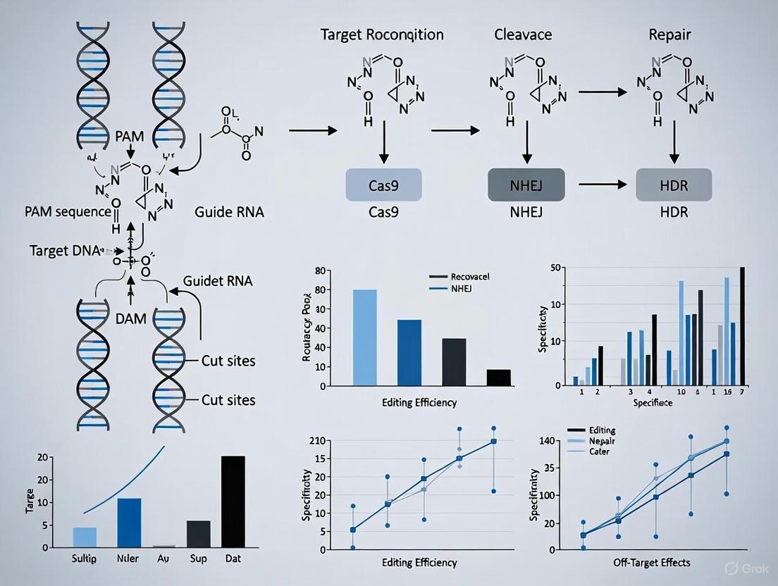
The CRISPR/Cas9 system has revolutionized genomic DNA research by providing an unprecedented ability to perform targeted genome editing. This technology's core consists of three essential components that function as an integrated molecular machine: the guide RNA (gRNA), which provides sequence specificity; the Cas nuclease, an enzyme that acts as the molecular scissor to cut DNA; and the Protospacer Adjacent Motif (PAM), a short DNA sequence that is critical for target recognition [1] [2]. The precise interplay between these components enables researchers to induce double-strand breaks at predetermined locations in the genome, facilitating gene knockouts, insertions, and corrections. As CRISPR technology advances into clinical applications, with the recent FDA approval of the first CRISPR/Cas9-based gene therapy, understanding the function, optimization, and limitations of each component becomes increasingly crucial for validating editing efficiency and ensuring experimental success [3] [4].
Component Deep Dive: Structure, Function, and Experimental Considerations
Guide RNA (gRNA): The Targeting System
The guide RNA serves as the targeting mechanism of the CRISPR/Cas9 system, directing the Cas nuclease to a specific genomic locus through Watson-Crick base pairing. Structurally, the gRNA is a chimeric single guide RNA (sgRNA) that combines two natural RNA elements: the CRISPR RNA (crRNA), which contains a 20-nucleotide spacer sequence complementary to the target DNA, and the trans-activating CRISPR RNA (tracrRNA), which serves as a binding scaffold for the Cas9 protein [2] [4]. This synthetic fusion simplifies the system to a two-component setup while maintaining full functionality.
The targeting specificity of the gRNA is determined by the 20-nucleotide spacer sequence, which must be carefully designed to minimize off-target effects while maintaining high on-target efficiency. Critical considerations for gRNA design include:
- Seed Sequence: The 10-12 nucleotides proximal to the PAM are crucial for target recognition and binding stability [5].
- Specificity: The spacer sequence should be unique within the genome to avoid off-target editing at similar sequences.
- GC Content: An optimal GC content (40-60%) improves gRNA stability and binding efficiency.
- Secondary Structure: Potential hairpin formations in the gRNA can interfere with Cas9 binding and should be minimized [2].
In experimental practice, gRNAs are typically encoded in plasmid vectors under the control of RNA polymerase III promoters such as U6, ensuring high expression levels in mammalian cells [2].
Cas Nuclease: The Molecular Scissor
The Cas nuclease is the effector protein that executes the DNA cleavage function. The most widely used variant, derived from Streptococcus pyogenes (SpCas9), contains two catalytic domains: the HNH domain, which cleaves the DNA strand complementary to the gRNA, and the RuvC domain, which cleaves the non-complementary strand [4]. This results in a blunt-ended double-strand break approximately 3-4 nucleotides upstream of the PAM sequence [1].
Cas9 undergoes a conformational activation process upon gRNA binding, transitioning from an inactive to an active state capable of DNA interrogation. The mechanism involves:
- PAM Scanning: Cas9 first searches for compatible PAM sequences through three-dimensional diffusion along the DNA [1].
- DNA Melting: Upon PAM recognition, Cas9 unwinds the adjacent DNA duplex.
- Target Verification: The gRNA interrogates the unwound DNA for complementarity.
- Conformational Activation: Full complementarity triggers nuclease activity [4].
The discovery and engineering of novel Cas variants with diverse properties have significantly expanded the CRISPR toolkit. Table 1 compares commonly used Cas nucleases and their characteristics, highlighting the importance of selecting the appropriate nuclease for specific experimental requirements.
Table 1: Comparison of Commonly Used Cas Nuclease Variants
| Nuclease | Origin | Size (aa) | PAM Sequence | Editing Efficiency | Key Applications |
|---|---|---|---|---|---|
| SpCas9 | Streptococcus pyogenes | 1,368 | NGG | High | Standard gene editing, knockout generation |
| SaCas9 | Staphylococcus aureus | 1,053 | NNGRRT | Moderate | In vivo applications with AAV delivery |
| NmeCas9 | Neisseria meningitidis | 1,082 | NNNNGATT | Moderate | Applications requiring high specificity |
| CjCas9 | Campylobacter jejuni | 984 | NNNNRYAC | Moderate | Compact nuclease for viral delivery |
| Cas12a (Cpf1) | Lachnospiraceae bacterium | 1,300 | TTTV | High | Multiplexed editing, staggered cuts |
| hfCas12Max | Engineered | ~1,300 | TN and/or TNN | High | Reduced off-target effects |
| OpenCRISPR-1 | AI-designed | N/A | NGG | Comparable/Improved vs. SpCas9 | High-fidelity editing [6] |
PAM Sequence: The Recognition Signal
The Protospacer Adjacent Motif (PAM) is a short, specific DNA sequence (typically 2-6 base pairs) located immediately downstream of the target DNA region. This motif is not part of the gRNA recognition sequence but is essential for Cas nuclease activation [1] [7]. For the commonly used SpCas9, the PAM sequence is 5'-NGG-3', where "N" can be any nucleotide base [1] [7].
The PAM serves two critical biological functions:
- Self vs. Non-Self Discrimination: In bacterial immune systems, the absence of PAM sequences in the host's CRISPR array prevents autoimmunity, ensuring that Cas9 only targets foreign DNA [1].
- Cas9 Activation: PAM binding triggers conformational changes in Cas9 that facilitate DNA unwinding and enable gRNA-DNA hybridization [7].
The PAM requirement represents a fundamental constraint in CRISPR experimental design, as it determines the potential target sites within a genome. Table 2 provides a comprehensive overview of PAM sequences for various Cas nucleases, highlighting the expanding targeting range through nuclease engineering.
Table 2: PAM Sequences and Recognition Patterns for Cas Nuclease Variants
| CRISPR Nucleases | Organism Isolated From | PAM Sequence (5' to 3') |
|---|---|---|
| SpCas9 | Streptococcus pyogenes | NGG |
| hfCas12Max | Engineered from Cas12i | TN and/or TNN |
| SaCas9 | Staphylococcus aureus | NNGRRT or NNGRRN |
| NmeCas9 | Neisseria meningitidis | NNNNGATT |
| CjCas9 | Campylobacter jejuni | NNNNRYAC |
| StCas9 | Streptococcus thermophilus | NNAGAAW |
| LbCpf1 (Cas12a) | Lachnospiraceae bacterium | TTTV |
| AsCpf1 (Cas12a) | Acidaminococcus sp. | TTTV |
| AacCas12b | Alicyclobacillus acidiphilus | TTN |
| BhCas12b v4 | Bacillus hisashii | ATTN, TTTN and GTTN |
| Cas14 | Uncultivated archaea | T-rich PAM sequences, eg. TTTA for dsDNA cleavage |
| Cas3 | in silico analysis of various prokaryotic genomes | No PAM sequence requirement |
Recent advances have significantly expanded PAM flexibility through protein engineering approaches such as:
- Directed Evolution: Creating Cas9 mutants with altered PAM specificities (e.g., xCas9, SpCas9-NG) [1].
- Structure-Guided Engineering: Modifying PAM-interacting domains to recognize alternative sequences.
- AI-Assisted Design: Using large language models to generate novel editors with tailored PAM preferences, as demonstrated by OpenCRISPR-1 [6].
Experimental Validation: Assessing CRISPR Component Performance
Protocol for Validating Editing Efficiency in Genomic DNA
To establish a robust framework for evaluating CRISPR/Cas9 component performance, we outline a standardized protocol adapted from successful genome editing studies in eukaryotic systems [8]:
1. Target Selection and gRNA Design:
- Identify target sites within the gene of interest containing appropriate PAM sequences.
- Design multiple gRNAs (typically 3-5) targeting different regions to account for variability in efficiency.
- Evaluate potential off-target sites using bioinformatic tools (e.g., Cas-OFFinder) and select gRNAs with minimal predicted off-target activity.
2. Plasmid Construction:
- Clone selected gRNA sequences into a CRISPR expression vector containing the U6 promoter.
- Select an appropriate Cas9 expression vector (e.g., with CMV promoter for mammalian cells).
- For transfection efficiency monitoring, include a fluorescent marker (e.g., GFP) or antibiotic resistance gene.
3. Cell Transfection and Delivery:
- Culture appropriate cell lines (HEK293T is commonly used for initial validation).
- Transfect using preferred method (lipofection, electroporation) with 1:3 mass ratio of Cas9:gRNA plasmids.
- Include untransfected and vector-only controls.
4. Editing Efficiency Analysis (72-96 hours post-transfection):
- Harvest genomic DNA using standard extraction protocols.
- Amplify target region by PCR (amplicon size: 400-800 bp).
- Quantify editing efficiency using T7 Endonuclease I assay or tracking of indels by decomposition (TIDE) analysis.
- For precise quantification, perform next-generation sequencing of amplicons.
5. Off-Target Assessment:
- Analyze top 5-10 predicted off-target sites by PCR and sequencing.
- Alternatively, use genome-wide methods like GUIDE-seq for comprehensive off-target profiling.
This protocol enables systematic comparison of different gRNA designs, Cas nuclease variants, and their combinations, providing quantitative data on editing efficiency and specificity.
Workflow Visualization: Experimental Process for CRISPR Validation
The following diagram illustrates the key stages in the experimental validation of CRISPR/Cas9 components:
Quantitative Comparison of Editing Efficiency Across Systems
Recent advances in CRISPR technology have yielded numerous engineered systems with varied performance characteristics. Table 3 summarizes quantitative efficiency data for different CRISPR systems, providing a reference for selecting appropriate components based on experimental needs.
Table 3: Editing Efficiency Metrics for CRISPR Systems in Genomic DNA Research
| CRISPR System | Average On-Target Efficiency | Relative Off-Target Rate | Optimal Cell Types | Key Advantages |
|---|---|---|---|---|
| SpCas9 (WT) | 40-60% | Baseline | Mammalian cells, plants | Reliable, well-characterized |
| High-Fidelity SpCas9 | 30-50% | 2-5x lower than WT | All cell types | Reduced off-target effects |
| SaCas9 | 20-40% | Comparable to SpCas9 | Neuronal cells, in vivo applications | Compact size for AAV delivery |
| Cas12a (Cpf1) | 25-45% | 2-4x lower than SpCas9 | Mammalian cells, plants | Staggered cuts, simpler gRNA |
| Base Editors | 15-50% (varies by type) | 10-100x lower than nuclease | Non-dividing cells | No double-strand breaks, precise base changes |
| Prime Editors | 10-30% | Extremely low | All cell types | Versatile, all possible base changes |
| CAST Systems (I-F) | Up to 100% in prokaryotes | Minimal (no DSBs) | Prokaryotes, early development in eukaryotes | Large DNA insertions without DSBs |
| AI-Designed Editors (OpenCRISPR-1) | Comparable/Improved vs. SpCas9 | Improved specificity | Human cells | Novel sequences, optimized properties [6] |
Advanced Applications and Integration with Emerging Technologies
Specialized CRISPR Systems for Advanced Genome Engineering
Beyond standard gene editing, specialized CRISPR systems have been developed to address specific research needs:
CRISPR-Associated Transposase (CAST) Systems: CAST systems represent a breakthrough for large DNA insertions without creating double-strand breaks. The type I-F CAST system has demonstrated nearly 100% insertion efficiency in E. coli with donor sequences up to 15.4 kb, while type V-K variants can accommodate inserts up to 30 kb [9]. Although current editing efficiency in mammalian cells remains low (approximately 1-3%), ongoing engineering efforts show promise for future applications [9].
Base and Prime Editing: These precision editing systems enable specific nucleotide changes without double-strand breaks. Base editors directly convert one DNA base to another (C→T, A→G) using catalytically impaired Cas proteins fused to deaminase enzymes, achieving efficiencies of 15-50% with dramatically reduced off-target effects [4]. Prime editors offer even greater versatility, supporting all 12 possible base-to-base conversions with minimal indels, though at lower efficiencies (10-30%) [4].
Anti-CRISPR Systems for Enhanced Control: Recent developments in anti-CRISPR proteins provide temporal control over Cas9 activity, addressing safety concerns related to prolonged nuclease expression. The LFN-Acr/PA system, derived from anthrax toxin components, enables rapid shutdown of Cas9 activity within minutes of application, reducing off-target effects and improving editing specificity by up to 40% [3].
Mechanism Visualization: CRISPR-Cas9 Molecular Machinery
The molecular mechanism of CRISPR/Cas9 can be visualized as follows:
Integration with Artificial Intelligence and Machine Learning
The application of artificial intelligence and machine learning represents the cutting edge of CRISPR technology development. Large language models (LMs) trained on biological diversity at scale have demonstrated remarkable success in designing novel gene editors. Recently, researchers curated a dataset of over 1 million CRISPR operons through systematic mining of 26 terabases of assembled genomes and metagenomes, using this information to fine-tune protein language models [6]. This approach generated 4.8 times the number of protein clusters across CRISPR-Cas families found in nature, with AI-designed editors like OpenCRISPR-1 showing comparable or improved activity and specificity relative to SpCas9, despite being 400 mutations away in sequence [6].
Machine learning tools are also being deployed to predict gRNA efficiency and off-target effects with increasing accuracy. As more sequence features are identified and incorporated into deep learning tools, predictions are expected to better align with experimental results, streamlining the CRISPR design process [10]. These computational approaches leverage large CRISPR databases to identify patterns and relationships that would be difficult to discern through manual analysis alone.
The Scientist's Toolkit: Essential Research Reagent Solutions
Successful validation of CRISPR/Cas9 editing efficiency requires access to specialized reagents and tools. The following table catalogs essential research solutions for CRISPR experimentation:
Table 4: Essential Research Reagents and Tools for CRISPR/Cas9 Experiments
| Reagent/Tool | Function | Examples/Specifications |
|---|---|---|
| Cas9 Expression Vectors | Delivery of Cas nuclease | CMV or CAG promoters, nuclear localization signals, codon optimization for target species |
| gRNA Cloning Vectors | gRNA expression | U6 or H1 promoters, BsaI/BbsI restriction sites for golden gate assembly |
| Bioinformatic Tools | gRNA design and off-target prediction | CATS [5], Cas-OFFinder, CHOPCHOP, CRISPOR |
| Validation Enzymes | Editing efficiency quantification | T7 Endonuclease I, Surveyor Nuclease |
| Delivery Reagents | Introduction of CRISPR components | Lipofectamine CRISPRMAX, electroporation systems (Neon, Amaxa) |
| Cell Lines | Editing validation | HEK293T (high transfection efficiency), target cell lines relevant to research |
| Control Plasmids | Experimental standardization | Non-targeting gRNA, GFP reporter constructs |
| Sequencing Primers | Amplification of target loci | Custom-designed for each target site, flanking edited region |
| Anti-CRISPR Proteins | Control of editing duration | LFN-Acr/PA system for rapid Cas9 inhibition [3] |
| HDR Donor Templates | Precise gene editing | Single-stranded or double-stranded DNA templates with homology arms |
The CATS (Comparing Cas9 Activities by Target Superimposition) bioinformatic tool deserves special mention, as it automates the detection of overlapping PAM sequences across different Cas9 nucleases and identifies allele-specific targets, particularly those arising from pathogenic mutations [5]. This tool significantly reduces the time and effort required for CRISPR/Cas9 experimental design by enabling direct comparison of nucleases with different PAM requirements in the same genomic context.
The core components of the CRISPR/Cas9 system—gRNA, Cas nuclease, and PAM sequence—function as an integrated molecular machine that can be strategically optimized for specific research applications. Validation of editing efficiency in genomic DNA research requires careful consideration of each component's properties and their interactions. The expanding repertoire of Cas nucleases with diverse PAM specificities, coupled with advances in gRNA design and delivery methods, provides researchers with unprecedented flexibility in genome engineering applications.
As CRISPR technology continues to evolve, integration with artificial intelligence and machine learning promises to further enhance the precision and efficiency of genome editing. The development of specialized systems like base editors, prime editors, and CAST systems addresses limitations of early CRISPR platforms, while anti-CRISPR proteins provide enhanced temporal control. By strategically selecting and validating the appropriate combination of CRISPR components for each experimental context, researchers can maximize editing efficiency while minimizing off-target effects, advancing both basic research and therapeutic applications.
The CRISPR/Cas9 system has revolutionized genetic research by enabling precise, targeted modifications to the genome. This powerful gene-editing tool functions by introducing a site-specific double-strand break (DSB) in the DNA, after which the cell's own endogenous repair mechanisms are activated to resolve the break [11] [12]. The two primary competing pathways for repairing these CRISPR-induced breaks are Non-Homologous End Joining (NHEJ) and Homology-Directed Repair (HDR). The fundamental difference between these pathways lies in their mechanisms and fidelity: NHEJ is an error-prone process that directly ligates broken ends, while HDR uses a homologous template for precise repair [13] [14]. Understanding the dynamics between NHEJ and HDR is crucial for researchers aiming to optimize CRISPR/Cas9 editing efficiency, particularly for applications requiring precise genetic modifications such as gene therapy, disease modeling, and functional genomics.
The competition between these pathways presents a significant challenge in genome engineering. In most eukaryotic cells, both repair pathways remain active; however, NHEJ generally dominates as it functions throughout the cell cycle and does not require a homologous template [14]. This inherent competition often results in low HDR efficiency, especially in non-dividing cells, making precise genome editing a considerable hurdle [13]. Consequently, developing strategies to enhance HDR efficiency or inhibit NHEJ has become a major focus in the field, requiring researchers to employ sophisticated validation methods to accurately quantify editing outcomes.
Molecular Mechanisms of NHEJ and HDR
The Non-Homologous End Joining (NHEJ) Pathway
Overview and Biological Function Non-Homologous End Joining represents the dominant and most active DSB repair pathway in mammalian cells, functioning throughout all phases of the cell cycle but particularly predominant during G1 [11] [15]. As an error-prone repair mechanism, NHEJ directly ligates broken DNA ends without requiring a homologous template, often resulting in small insertions or deletions (indels) at the repair site [12]. This characteristic makes NHEJ particularly suitable for gene knockout studies where the goal is to disrupt gene function by introducing frameshift mutations [12].
Key Molecular Steps and Protein Components The NHEJ pathway initiates when the Ku heterodimer complex (composed of Ku70 and Ku80 subunits) recognizes and binds to the broken DNA ends [13]. This complex then recruits and activates DNA-dependent protein kinase catalytic subunit (DNA-PKcs), forming a stable complex that protects the DNA ends from resection [11]. Subsequently, the Artemis:DNA-PKcs complex is activated and processes the DNA ends by removing overhangs, creating ligatable blunt ends [13]. In cases where nucleotide loss has occurred, DNA polymerases μ and λ fill in the gaps by adding missing nucleotides [11] [13]. The final ligation step is catalyzed by the DNA Ligase IV complex in conjunction with XRCC4 and XLF [11] [13]. This multi-step process, while efficient, often results in mutagenic outcomes due to the potential for nucleotide loss or addition during end processing.
Beyond the canonical NHEJ pathway, alternative end-joining mechanisms exist, including microhomology-mediated end joining (MMEJ) which operates during S and G2 phases and relies on short homologous sequences (5-25 base pairs) for repair [11]. The MMEJ pathway involves different protein components including PARP1 for end recognition, MRN complex and CtIP for end resection, and Ligase I/III for the final ligation step [11].
Table 1: Key Protein Components of the NHEJ Pathway
| Protein Component | Function in NHEJ Pathway |
|---|---|
| Ku70/Ku80 heterodimer | Initial recognition and binding to DSB ends |
| DNA-PKcs | Recruitment and activation of processing enzymes |
| Artemis | Endonuclease activity for processing DNA overhangs |
| Polymerase μ and λ | Fill missing nucleotides at DSB ends |
| DNA Ligase IV | Catalyzes final ligation of broken ends |
| XRCC4/XLF | Stabilizes and enhances Ligase IV activity |
| 53BP1 | Promotes NHEJ pathway choice, particularly in G1 phase |
| PARP1 | Involved in alternative NHEJ pathways (MMEJ) |
The Homology-Directed Repair (HDR) Pathway
Overview and Biological Function Homology-Directed Repair represents a precise, error-free repair mechanism that utilizes homologous DNA sequences as templates for accurate DSB restoration [12]. Unlike NHEJ, HDR is restricted to the late S and G2 phases of the cell cycle when sister chromatids are available as natural templates [14]. This cell cycle dependency significantly impacts HDR efficiency in various cell types, particularly post-mitotic cells where HDR rates are substantially lower [13]. Researchers leverage HDR for precise genetic modifications including gene knock-ins, point mutations, or insertion of tagged gene versions by providing exogenous donor templates with homology arms [12].
Key Molecular Steps and Protein Components HDR initiation begins with extensive 5' to 3' end resection of the DNA break, a process coordinated by the MRN complex (MRE11-RAD50-NBS1) in conjunction with CtIP [11]. This resection creates 3' single-stranded DNA overhangs that are essential for subsequent steps. The single-stranded DNA is rapidly bound and protected by Replication Protein A (RPA). BRCA2 then facilitates the replacement of RPA with RAD51, forming a nucleoprotein filament that catalyzes the central homologous pairing and strand invasion reaction [11]. This RAD51 nucleoprotein filament invades the homologous donor sequence, forming a D-loop structure that serves as a primer for DNA synthesis using the donor template. After DNA synthesis, the resulting intermediate structures are resolved through various subpathways including double-strand break repair (DSBR), synthesis-dependent strand annealing (SDSA), or break-induced replication (BIR), with SDSA being the predominant pathway mitigating crossover events in mitotic cells.
The choice between NHEJ and HDR is regulated by several key factors. 53BP1 promotes NHEJ by limiting DNA end resection, while BRCA1 antagonizes 53BP1 to favor HDR [11]. Additional regulators include TIRR, which masks the H4K20me2 binding surface targeted by 53BP1, and SCAI, which disrupts 53BP1-RIF1 interaction to promote HDR [11]. The POH1 deubiquitinating enzyme also contributes to HDR promotion by removing ubiquitin modifications that recruit 53BP1 [11].
Table 2: Key Protein Components of the HDR Pathway
| Protein Component | Function in HDR Pathway |
|---|---|
| MRN Complex | Initial DSB recognition and 5' to 3' end resection |
| CtIP | Cooperates with MRN complex in end resection |
| BRCA1 | Promotes HDR pathway choice; antagonizes 53BP1 |
| RPA | Binds and protects single-stranded DNA after resection |
| BRCA2 | Loads RAD51 onto single-stranded DNA |
| RAD51 | Forms nucleoprotein filament; catalyzes strand invasion |
| RAD52 | Facilitates strand annealing in alternative HDR pathways |
| TIRR | Promotes HDR by inhibiting 53BP1 recruitment |
Quantitative Comparison of NHEJ and HDR Efficiency
Systematic quantification of HDR and NHEJ outcomes reveals complex relationships between these competing pathways that are highly dependent on experimental conditions. Using a novel droplet digital PCR (ddPCR) assay capable of simultaneously detecting HDR and NHEJ events at endogenous loci, researchers have demonstrated that the relative efficiency of these pathways varies significantly based on gene locus, nuclease platform, and cell type [15]. Contrary to the widespread assumption that NHEJ generally occurs more frequently than HDR, studies have found that more HDR than NHEJ can be induced under multiple conditions, with HDR/NHEJ ratios showing remarkable context dependency [15].
Kinetic studies in human pluripotent stem cells (hPSCs) have revealed distinct temporal patterns for these repair pathways. HDR events plateau approximately 24 hours after Cas9 introduction, while NHEJ repair continues until 48 hours post-transfection [16]. Cut but unrepaired alleles reach their maximum level within 12-24 hours after DSB induction before gradually declining as repair processes complete [16]. These kinetic differences have important implications for experimental design, particularly regarding the optimal timing for analysis after CRISPR editing.
Cell type significantly influences pathway efficiency, with notable differences observed between naïve and primed pluripotent stem cells. Research demonstrates that the rate of HDR is approximately 40% lower in naïve hPSCs compared to conventional primed hPSCs, correlating with a higher proportion of naïve cells in the G1 phase of the cell cycle where HDR is less active [16]. This finding contradicts earlier assumptions that naïve hPSCs might be superior for gene editing and highlights the importance of considering cell cycle distribution when planning HDR-based experiments.
Table 3: Quantitative Comparison of NHEJ and HDR Efficiencies Across Experimental Conditions
| Experimental Condition | Impact on NHEJ | Impact on HDR | Key Findings |
|---|---|---|---|
| Cell Type (naïve vs. primed hPSCs) | Similar levels observed | 40% lower in naïve hPSCs | Correlates with increased G1 population in naïve cells [16] |
| Cell Cycle Phase | Active throughout all phases | Restricted to late S and G2 phases | HDR efficiency cell cycle-dependent [14] |
| Repair Kinetics | Continues until 48 hours post-transfection | Plateaus after 24 hours | Unrepaired alleles peak at 12-24h [16] |
| Gene Locus | Variable depending on chromatin context | Variable depending on chromatin context | Locus-specific effects on HDR/NHEJ ratio [15] |
| Nuclease Platform | Varies with nuclease type | Varies with nuclease type | Affects HDR/NHEJ ratios [15] |
| Template Design (TFO-tailed ssODN) | Minimal direct effect | Increase from 18.2% to 38.3% | Enhanced spatial accessibility improves HDR [17] |
Experimental Strategies for Modifying NHEJ/HDR Balance
Molecular and Chemical Interventions
Researchers have developed multiple strategic approaches to enhance HDR efficiency by modulating the balance between NHEJ and HDR pathways. These interventions typically target three key aspects: inhibiting NHEJ components, stimulating HDR factors, or synchronizing the cell cycle to favor HDR [11].
NHEJ Inhibition Strategies Small molecule inhibitors targeting key NHEJ components have shown promise in improving HDR efficiency. Compounds such as SCR7 (targeting DNA Ligase IV) and KU0060648 (inhibiting DNA-PKcs) can effectively suppress the canonical NHEJ pathway, thereby reducing competing error-prone repair and increasing the relative frequency of HDR [11]. Additionally, genetic approaches including knockdown of 53BP1 or other essential NHEJ factors (Ku70/Ku80, DNA-PKcs) through RNA interference can achieve similar effects, though these methods may be less practical for therapeutic applications [11].
HDR Stimulation Approaches Alternatively, enhancing HDR activity directly represents another viable strategy. RS-1, a small molecule activator of RAD51, can stimulate the core homologous pairing activity central to HDR, thereby increasing precise editing efficiency [11]. Timing Cas9 delivery to coincide with S/G2 phase through cell cycle synchronization protocols can also significantly boost HDR rates by ensuring the homologous repair machinery is active and accessible when DSBs occur [14].
CRISPR/Cas9 System Optimization
Cas9 Engineered Variants Substantial efforts have focused on engineering optimized Cas9 variants that favor HDR outcomes. These include chimeric Cas9 proteins fused with HDR-promoting factors, Cas9 nickase systems that generate single-strand breaks instead of DSBs, and modified Cas9 versions that can directly recruit the donor template to the cleavage site [11]. For instance, eCas9 and HiFi Cas9 variants offer improved specificity while maintaining robust on-target activity, with HiFi Cas9 demonstrating particularly favorable kinetics for HDR-based editing in stem cells [16].
Donor Template Design Innovations The design and delivery of donor templates significantly impact HDR efficiency. Recent research demonstrates that modifying single-stranded oligodeoxynucleotides (ssODNs) with triplex-forming oligonucleotide (TFO) tails that form PolyPurine Reverse Hoogsteen (PPRH) hairpins can increase HDR efficiency from 18.2% to 38.3% by improving spatial accessibility of the donor template to the break site [17]. Optimization of homology arm length and strategic incorporation of silent mutations in the donor sequence to prevent re-cleavage of edited sites further enhance HDR outcomes [13].
Validation Methods for CRISPR Editing Efficiency
Accurate validation of CRISPR editing outcomes is essential for evaluating the efficiency of both NHEJ and HDR events. Various methods with differing sensitivities, advantages, and limitations have been developed for this purpose.
T7 Endonuclease I (T7E1) Assay The T7E1 assay represents a traditional, cost-effective method for detecting NHEJ-derived indels. This approach relies on the T7 endonuclease I enzyme, which cleaves heteroduplex DNA formed when wild-type and mutant PCR amplicons are annealed [18] [19]. However, comparative studies have revealed significant limitations in T7E1 accuracy, particularly for detecting editing efficiencies above 30% or distinguishing between sgRNAs with similar activity levels [19]. When evaluated against next-generation sequencing, T7E1 consistently underestimated editing efficiency and failed to detect poorly performing sgRNAs with less than 10% NHEJ activity [19].
Tracking of Indels by Decomposition (TIDE) TIDE analysis utilizes Sanger sequencing of edited populations followed by computational decomposition of the resulting chromatograms to quantify indel frequencies [19]. While TIDE shows improved correlation with next-generation sequencing results compared to T7E1, it can miscall alleles in edited clones and deviates by more than 10% from sequencing-predicted indel frequencies in approximately 50% of cases [19].
Next-Generation Sequencing (NGS) Approaches Next-generation sequencing, particularly amplicon-based deep sequencing, provides the most comprehensive and quantitative assessment of CRISPR editing outcomes. NGS enables simultaneous detection of both HDR and NHEJ events with single-base resolution, sensitivity for alleles with frequency below 1%, and full characterization of complex editing patterns [15] [20]. Droplet digital PCR (ddPCR) offers an alternative highly sensitive quantification method capable of detecting one HDR or NHEJ event among 1,000 genomic copies, making it suitable for kinetic studies and precise efficiency measurements [15] [16]. High-throughput genotyping services like genoTYPER-NEXT leverage NGS to enable automated analysis of thousands of samples simultaneously, significantly streamlining the validation pipeline for large-scale CRISPR screening projects [20].
Table 4: Comparison of CRISPR Validation Methods
| Validation Method | Detection Principle | Sensitivity | Advantages | Limitations |
|---|---|---|---|---|
| T7E1 Assay | Enzyme cleavage of heteroduplex DNA | Limited, fails below 10% editing | Cost-effective; technically simple | Low dynamic range; underestimates high efficiency; subjective interpretation [19] |
| TIDE Analysis | Decomposition of Sanger sequencing chromatograms | Moderate | More accurate than T7E1; quantitative | Deviates >10% in 50% of clones; miscalls alleles [19] |
| Next-Generation Sequencing | Amplicon sequencing with high coverage | High (<1% allele frequency) | Comprehensive; quantitative; detects all variants | Requires bioinformatics; higher cost [20] [19] |
| Droplet Digital PCR | Probe-based quantification in partitioned samples | Very high (1 in 1,000 copies) | Absolute quantification; high precision; kinetic studies | Requires specific probe design; limited multiplexing [15] [16] |
Successful CRISPR genome editing experiments require careful selection of reagents and resources tailored to specific research goals. The following essential components represent the core toolkit for investigators working with NHEJ and HDR pathways:
Table 5: Essential Research Reagents for CRISPR DNA Repair Studies
| Reagent Category | Specific Examples | Function and Application |
|---|---|---|
| Nuclease Systems | Wild-type Cas9, Cas9 D10A nickase, Cas9 H840A nickase, FokI-dCas9 | Generate DSBs or nicks at target sites; different platforms affect HDR/NHEJ ratios [15] |
| Donor Templates | ssODNs, dsDNA donors with homology arms, TFO-tailed ssODNs | Provide repair template for HDR; design significantly impacts efficiency [17] |
| Pathway Modulators | SCR7 (Ligase IV inhibitor), KU0060648 (DNA-PKcs inhibitor), RS-1 (RAD51 activator) | Enhance HDR efficiency by inhibiting NHEJ or stimulating HDR components [11] |
| Validation Tools | T7E1 enzyme, ddPCR assays, NGS library prep kits | Detect and quantify editing outcomes; vary in sensitivity and throughput [15] [19] |
| Cell Line Models | HEK293T, HeLa, human iPSCs, mouse embryos | Different cell types show variable HDR/NHEJ ratios; selection impacts efficiency [15] [16] |
Visualizing the DNA Repair Pathway Decision Process
The following diagram illustrates the key decision points and molecular interactions that determine whether NHEJ or HDR pathways are activated following a CRISPR/Cas9-induced double-strand break:
CRISPR-Induced DNA Break Repair Pathway Decision Map
Additionally, the following workflow diagram illustrates a modern experimental approach for quantifying and validating CRISPR editing efficiency:
CRISPR Editing Validation Workflow
The competition between NHEJ and HDR pathways represents a fundamental biological constraint that significantly impacts the efficiency and precision of CRISPR/Cas9 genome editing. While NHEJ operates as the dominant, error-prone pathway throughout the cell cycle, HDR provides a precise, template-dependent alternative restricted to specific cell cycle phases. The dynamic balance between these pathways is influenced by multiple factors including cell type, gene locus, nuclease platform, and experimental conditions [15].
Understanding these relationships enables researchers to develop strategic approaches for manipulating the repair pathway choice through small molecule inhibitors, optimized Cas9 variants, cell cycle synchronization, and innovative donor template designs [11] [17]. Concurrently, advances in validation methodologies, particularly ddPCR and NGS-based approaches, provide researchers with increasingly sensitive tools to quantify editing outcomes accurately and optimize experimental conditions [15] [16].
As CRISPR applications continue to expand toward therapeutic implementations, further elucidating the complex regulation of DNA repair pathways will remain essential for achieving predictable and precise genomic modifications. The ongoing development of strategies to enhance HDR efficiency while minimizing off-target effects represents a critical frontier in advancing both basic research and clinical applications of genome editing technologies.
The validation of CRISPR/Cas9 editing efficiency is a cornerstone of modern genomic research and therapeutic development. While the design of guide RNAs and the choice of Cas nuclease are critical, the ultimate editing outcome is not determined solely by these tools. Instead, it is profoundly influenced by the cellular environment and the methods used to deliver the editing machinery [2] [21]. This guide provides an objective comparison of how key biological and technical factors—specifically the cell cycle, delivery methods, and broader cellular context—govern the efficiency and precision of CRISPR/Cas9 genome editing, equipping researchers with the data needed to optimize their experimental strategies.
The Interplay of Cell Cycle and DNA Repair Pathways
At the heart of CRISPR/Cas9 editing is the creation of a DNA double-strand break (DSB), which the cell must repair. The choice of repair pathway is heavily constrained by the cell's position in the cell cycle, directly determining the editing outcome [2] [22].
- Homology-Directed Repair (HDR) is restricted to the S and G2 phases of the cell cycle, as it requires a sister chromatid template. This makes HDR, which enables precise gene knock-ins or corrections, highly inefficient in non-dividing, postmitotic cells [22].
- Non-Homologous End Joining (NHEJ) is active throughout the cell cycle and is the dominant pathway in postmitotic cells. It ligates the broken DNA ends together, often resulting in small insertions or deletions (indels) that disrupt gene function [2] [22].
- Microhomology-Mediated End Joining (MMEJ), another error-prone pathway, is also restricted to certain cell cycle stages (S/G2/M) and is predominant in dividing cells, leading to larger deletions [22].
Recent research reveals that DNA repair is not only cell-cycle-dependent but also varies by cell type. A 2025 study in Nature Communications demonstrated that postmitotic human neurons repair Cas9-induced DNA damage over a much longer timeframe (up to two weeks) compared to genetically identical dividing cells [22]. Furthermore, neurons exhibited a narrower distribution of indel outcomes, heavily favoring small indels typical of classical NHEJ, while dividing cells showed a broader range, including MMEJ-like larger deletions [22]. This fundamental difference in the DNA damage response underscores the critical importance of cellular context.
The diagram below illustrates how cellular context dictates the available DNA repair pathways and, consequently, the resulting editing outcomes.
A Comparative Analysis of CRISPR/Cas9 Delivery Methods
The vehicle used to deliver CRISPR components is a major determinant of editing success, influencing everything from efficiency and specificity to immunogenicity. The three primary cargo types—plasmid DNA, mRNA/gRNA, and pre-assembled Ribonucleoprotein (RNP) complexes—each present distinct advantages and limitations [21] [23].
The following table compares the most common delivery vehicles, their applications, and their documented editing efficiencies.
Table 1: Comparison of CRISPR/Cas9 Delivery Vehicles and Their Performance
| Delivery Method | Cargo Type | Application Context | Reported Editing Efficiency | Key Advantages | Key Limitations |
|---|---|---|---|---|---|
| Electroporation (e.g., CASGEVY) [21] | RNP (preferred), mRNA | Ex vivo (e.g., HSCs, T cells) | Up to 90% indel frequency [21] | High efficiency for hard-to-transfect cells; direct delivery of RNP minimizes off-targets. | Can compromise cell viability; primarily suitable for ex vivo use. |
| Lipid Nanoparticles (LNPs) [21] [24] | mRNA/gRNA, RNP | In vivo (systemic or local) | ~90% protein reduction (e.g., in hATTR trials) [24] | Biodegradable; low immunogenicity; potential for redosing; organ-targeted versions in development. | Requires endosomal escape; liver-tropic by default; editing efficiency depends on cargo. |
| Adeno-Associated Virus (AAV) [2] [23] | DNA (plasmid) | In vivo, ex vivo | Varies widely; can be high with optimized systems [25] | Low immunogenicity; high transduction efficiency; tissue-specific serotypes. | Limited packaging capacity (~4.7 kb); can trigger immune responses; prolonged expression raises off-target risk. |
| Virus-Like Particles (VLPs) [22] [23] | RNP (protein) | Ex vivo, in vivo (preclinical) | Up to 97% transduction in iPSC-derived neurons [22] | Transient delivery; no viral genome integration; high efficiency for sensitive cells like neurons. | Manufacturing challenges; cargo size constraints; stability issues. |
| Adenoviral Vectors (AdVs) [23] | DNA (plasmid) | In vitro, ex vivo | Not quantified in results | Large cargo capacity (~36 kb); does not integrate into genome. | Can cause undesirable immune responses. |
| Lentiviral Vectors (LVs) [2] [23] | DNA (plasmid) | Ex vivo (common) | Not quantified in results | Can infect dividing and non-dividing cells; no cargo size limit. | Integrates into host genome, raising safety concerns for therapeutics. |
Cellular Context: A Determinant of Repair and Outcome
Beyond the cell cycle, the broader cellular context—encompassing cell type, tissue origin, and metabolic state—significantly influences editing outcomes through its effect on the native DNA repair machinery.
- Cell Type-Specific Repair Kinetics: As highlighted earlier, the 2025 study by [22] found that indel accumulation in human neurons continued for up to 16 days post-delivery of Cas9 RNP, a dramatically longer timeline than the 1-2 days typical in dividing cells. This prolonged repair process was also observed in other clinically relevant nondividing cells, such as cardiomyocytes and primary T cells in a resting state [22].
- Differential Repair Factor Expression: The same study demonstrated that neurons upregulate non-canonical DNA repair factors in response to Cas9 exposure compared to dividing cells. This unique repair signature presents both a challenge and an opportunity; by manipulating this response with chemical or genetic perturbations, researchers were able to direct DNA repair toward desired outcomes in neurons and cardiomyocytes [22].
- Implications for Experimental Design: These findings necessitate that editing strategies be tailored to the specific cell model. For example, achieving precise HDR in primary neurons is inherently difficult, making NHEJ-based gene knockout or newer base/prime editing approaches more viable. Furthermore, the extended timeline for full editing manifestation in non-dividing cells means that analysis performed too early may significantly underestimate editing efficiency [22].
Experimental Protocols for Assessing Key Factors
To validate CRISPR/Cas9 efficiency in light of these factors, researchers can employ the following detailed methodologies, drawn from recent literature.
Protocol 1: Assessing Cell-Cycle Dependence of Editing Outcomes
This protocol is adapted from approaches used to compare dividing and non-dividing cells [22].
- Cell Preparation and Synchronization: Utilize a matched cellular model, such as induced pluripotent stem cells (iPSCs) and iPSC-derived postmitotic neurons, or activated vs. resting primary human T cells.
- CRISPR Delivery: Deliver a consistent dose of CRISPR-Cas9 ribonucleoprotein (RNP) complexes. For neurons and resting T cells, use efficient vehicles like virus-like particles (VLPs) or electroporation, respectively. For dividing cells (iPSCs, activated T cells), electroporation or chemical transfection can be used.
- Longitudinal Sampling and Analysis: Harvest genomic DNA at multiple time points post-delivery (e.g., days 1, 2, 4, 7, 14). Amplify the target locus by PCR and analyze the editing outcomes using next-generation sequencing (NGS).
- Data Interpretation: Quantify the percentage of indels over time and classify the types of mutations (small indels vs. large deletions). Compare the kinetics and distribution of outcomes between dividing and non-dividing cell populations.
Protocol 2: Evaluating Delivery Method Efficiency
This protocol provides a framework for comparing different delivery vehicles in a target cell type.
- Vehicle and Cargo Selection: Choose 2-3 relevant delivery methods (e.g., Electroporation of RNP, LNP-mediated mRNA delivery, Lentiviral transduction). Standardize the gRNA sequence and target locus across all conditions.
- Transfection/Transduction: Perform delivery according to optimized protocols for each method, ensuring consistent cell numbers and health status across conditions.
- Efficiency and Specificity Assessment: At 48-72 hours post-delivery (or later for non-dividing cells), harvest cells for analysis.
- Editing Efficiency: Isolate genomic DNA and perform T7 Endonuclease I (T7EI) assay or, preferably, NGS to quantify indel percentage at the on-target site.
- Off-Target Effects: Use in silico prediction tools to identify potential off-target sites, followed by amplicon sequencing of those loci to assess off-target activity.
- Cell Viability: Perform a cell viability assay (e.g., MTT, flow cytometry with viability dye) 24 hours after delivery to assess cytotoxicity.
Essential Research Reagent Solutions
The following table details key reagents and their functions for investigating the factors discussed in this guide.
Table 2: Research Reagent Solutions for CRISPR Workflow Optimization
| Reagent / Tool | Function & Application | Key Considerations |
|---|---|---|
| Cas9 RNP Complex | Pre-assembled complex of Cas9 protein and sgRNA; considered the gold standard for high efficiency and low off-target effects [21] [23]. | Ideal for electroporation and VLP delivery; transient activity; reduced cytotoxicity compared to plasmid DNA. |
| Virus-Like Particles (VLPs) | Engineered delivery vehicle for RNP complexes; enables efficient transduction of hard-to-modify cells like neurons [22]. | Pseudotype (e.g., VSVG, BaEVRless) must be chosen for target cell type; allows transient RNP delivery without viral genome integration. |
| Lipid Nanoparticles (LNPs) | Synthetic nanoparticles for in vivo delivery of mRNA or RNP cargo; target organs like the liver [21] [24]. | Enable systemic administration; potential for redosing; efficiency depends on endosomal escape and cargo release. |
| Selective Organ Targeting (SORT) LNPs | Engineered LNPs with added molecules to alter tropism; can target spleen, lungs, or specific liver cell types [23]. | Emerging technology for expanding in vivo delivery beyond the liver. |
| T7 Endonuclease I Assay | Mismatch-cleavage assay for rapid, cost-effective detection of indel mutations [25]. | Semi-quantitative; less sensitive than NGS but useful for initial screening. |
| Single-Cell DNA Sequencing (e.g., Tapestri platform) | High-resolution analysis of editing outcomes (zygosity, complex structural variations) at the single-cell level [26]. | Reveals clonality and heterogeneity of editing that bulk sequencing masks; critical for safety assessment. |
The journey to a predictable and precise CRISPR/Cas9 editing outcome is multifaceted. This guide has underscored that successful genomic research and therapeutic development depend on a holistic strategy that integrates the cellular context, including cell cycle status and cell-type-specific repair mechanisms, with a rational selection of delivery methods based on cargo, target cells, and application. By adopting the detailed experimental protocols and leveraging the reagent solutions outlined here, researchers can systematically dissect and optimize these key factors, thereby enhancing the reliability, efficiency, and safety of their CRISPR-based genomic validations.
CRISPR-Cas systems have revolutionized genomic DNA research, offering unprecedented precision in genetic engineering. For researchers and drug development professionals, selecting the appropriate Cas enzyme is crucial for experimental success. This guide provides an objective comparison of three widely used nucleases—SpCas9, SaCas9, and Cas12a—focusing on their editing efficiency, specificity, and practical applications. The evaluation of these enzymes is framed within the broader thesis of validating CRISPR/Cas9 editing efficiency, presenting key performance data to inform reagent selection for specific genomic contexts.
Molecular Mechanisms and Key Characteristics
The CRISPR-Cas system functions as a programmable DNA-targeting complex. The Cas nuclease is directed to a specific genomic locus by a guide RNA (gRNA for Cas9; crRNA for Cas12a). Target recognition and cleavage are contingent upon the presence of a short protospacer adjacent motif (PAM) sequence adjacent to the target site, which varies by nuclease [27] [28].
- SpCas9: Derived from Streptococcus pyogenes, it remains the most widely adopted nuclease. It requires a 5'-NGG PAM (where N is any nucleotide) downstream of its target sequence. Upon binding, its two nuclease domains create a double-strand break (DSB) with blunt ends [29].
- SaCas9: Isolated from Staphylococcus aureus, it recognizes a more complex 5'-NNGRRT PAM (where R is A or G). Its key advantage is a significantly smaller size (about 1 kb smaller than SpCas9), facilitating packaging into delivery vectors like adeno-associated viruses (AAVs) for therapeutic applications. It also produces blunt-ended DSBs [28] [29].
- Cas12a (formerly Cpf1): Representative variants include AsCas12a (Acidaminococcus sp.) and LbCas12a (Lachnospiraceae bacterium). This nuclease differs from Cas9 in several ways: it recognizes a T-rich 5'-TTTV PAM (where V is A, C, or G) upstream of the target, requires only a single crRNA without a tracrRNA, and introduces DSBs with staggered ends that create 5' overhangs [27] [28].
CRISPR-Cas Nuclease Workflow: This diagram illustrates the shared workflow of target recognition and cleavage by SpCas9, SaCas9, and Cas12a, highlighting the critical divergence at the PAM recognition and cleavage stages.
Comparative Performance Data
Direct comparisons of Cas nuclease efficiency under controlled conditions are critical for reagent selection. A study comparing nucleases in plants using identical regulatory elements and vector backbones found SaCas9 to be the most efficient at inducing mutations, though the nucleotide content of the target itself also correlated with efficiency [27]. Performance can also be organism-dependent; in rice calli, SpCas9 was the most efficient nuclease at both 27°C and 37°C, whereas AsCas12a showed no detectable activity under the tested conditions [30].
Table 1: Comparative Analysis of Common Cas Nucleases
| Feature | SpCas9 | SaCas9 | Cas12a (As/Lb) |
|---|---|---|---|
| Origin | Streptococcus pyogenes [29] | Staphylococcus aureus [29] | Acidaminococcus sp. / Lachnospiraceae bacterium [27] |
| PAM Sequence | 5'-NGG-3' [30] | 5'-NNGRRT-3' [28] [30] | 5'-TTTV-3' [27] [30] |
| Guide RNA | sgRNA (crRNA + tracrRNA) [29] | sgRNA (crRNA + tracrRNA) [28] | crRNA only [27] |
| Cleavage Type | Blunt ends [29] | Blunt ends [29] | Staggered ends (5' overhangs) [27] |
| Protein Size | ~1368 aa (large) [29] | ~1053 aa (small) [29] | ~1300 aa (large) [27] |
| Reported Editing Efficiency | High (e.g., >95% in HEK293T with optimal gRNAs) [31] | High efficiency; found "comparatively most efficient" in one plant study [27] | Variable; can be highly efficient with optimized crRNAs [32] |
| Specificity & Off-Targets | Moderate; improved by high-fidelity variants [27] [31] | Can exhibit high specificity; engineered variants available [29] | Generally high specificity; can be enhanced with modified crRNAs [32] |
| Key Advantage | Well-characterized, versatile, extensive toolkit [29] | Small size ideal for viral delivery (e.g., AAV) [29] | Simplified guide RNA, staggered ends for HDR [27] |
Experimental Protocols for Validation
Validating the efficiency of CRISPR-Cas editing requires standardized and reliable experimental workflows. Below are detailed protocols for two common assessment methods.
Protocol 1: Transient Assay in Plant Protoplasts for gRNA Screening
This protocol, adapted from a study in maize, is designed to rapidly evaluate the performance of multiple gRNAs or nucleases before undertaking stable transformation [33].
- Target Site Selection & gRNA Design: Use bioinformatics tools (e.g., CRISPRdirect, CRISPOR) to identify potential target sites with minimal off-targets in the genome of interest [28].
- Construct Assembly: Clone selected gRNA or crRNA sequences into appropriate CRISPR expression vectors. Using a unified vector system for different nucleases minimizes experimental variability [28].
- Protoplast Isolation:
- Harvest young leaves or embryonic tissue.
- Digest cell walls using enzyme mixtures (e.g., cellulase and macerozyme) to release protoplasts.
- Purify protoplasts through filtration and washing.
- Protoplast Transformation:
- Incubate purified protoplasts with the CRISPR plasmid DNA constructs.
- Use polyethylene glycol (PEG)-mediated transformation to introduce the DNA.
- Incubate the transformed protoplasts in the dark for 48-72 hours to allow for gene editing to occur.
- DNA Extraction & Analysis:
Protocol 2: Evaluating Editing in Rice Callus at Different Temperatures
This protocol assesses how temperature, a critical environmental factor, influences the activity of different Cas nucleases [30].
- Vector Construction: Assemble vectors encoding the nuclease (e.g., SpCas9, SaCas9, AsCas12a) and their respective guide RNAs using a modular cloning system. The nuclease should be driven by a constitutive plant promoter like OsUbi2 [30].
- Stable Transformation:
- Use biolistic transformation to deliver the constructed vectors into embryogenic rice calli (e.g., cultivar Taipei 309).
- After transformation, transfer calli to selection media containing hygromycin to select for transformed events.
- Temperature Treatment:
- Divide the selected calli into two groups.
- Incubate one group at 27°C (standard plant tissue culture temperature) and the other at 37°C (reported optimal temperature for many bacterial Cas nucleases) for the duration of the experiment.
- Genotyping and Efficiency Calculation:
- After a suitable incubation period, harvest calli and pool them for genomic DNA extraction.
- Amplify the target locus via PCR and subject the amplicons to high-throughput sequencing.
- Analyze sequencing data to calculate the percentage of modified reads, distinguishing between monoallelic and biallelic edits [30].
Nuclease Selection Logic: This diagram outlines the primary delivery methods available for different nucleases and the key application considerations that should guide the selection process.
The Scientist's Toolkit: Essential Reagents and Materials
Successful CRISPR experiment design and execution relies on a core set of reagents and tools.
Table 2: Essential Research Reagents and Materials
| Reagent / Material | Function in Experiment | Application Notes |
|---|---|---|
| CRISPR Expression Vector | Provides a backbone for the expression of the Cas nuclease and guide RNA in cells. | Use standardized, modular systems (e.g., Golden Gate Assembly-compatible) for seamless testing of different nucleases and targets [28]. |
| Endogenous Promoters | Drives the expression of CRISPR components in the host organism. | Using highly expressed, species-specific promoters (e.g., LarPE004 in larch) can significantly boost editing efficiency over standard viral promoters [34]. |
| Modified Guide RNA Scaffolds | Optimizes guide RNA structure to enhance stability and expression. | Replacing the standard 4T-sequence in the gRNA scaffold with a 3TC sequence can significantly increase gRNA transcript levels from U6 promoters, boosting editing efficiency, especially for difficult targets or with limited vector [31]. |
| Chemically Modified crRNA | Enhances stability and performance of Cas12a crRNA. | Using crRNA with a ribosyl-2'-O-methylated uridinylate-rich 3'-overhang improves Cas12a dsDNA digestibility and increases editing efficiency and specificity in zygotes and cell lines [32]. |
| Lipid Nanoparticles (LNPs) | A delivery vehicle for in vivo CRISPR therapy. | LNPs are effective for delivering CRISPR components to the liver and have been used successfully in clinical trials for systemic administration, with the advantage of allowing re-dosing [24]. |
| Adeno-Associated Virus (AAV) | A delivery vehicle for in vivo gene therapy. | The small size of SaCas9 makes it particularly suited for AAV delivery, enabling targeted in vivo editing in tissues like the liver and nervous system [29]. |
Advanced Engineering and Clinical Applications
The continuous engineering of Cas enzymes has expanded their utility and performance, paving the way for clinical applications.
- High-Fidelity Variants: Engineered versions of SpCas9 (e.g., eSpCas9, SpCas9-HF1) and SaCas9 (e.g., SaCas9-HF) have been developed to reduce off-target effects by incorporating mutations that decrease non-specific interactions with DNA, which is critical for therapeutic safety [27] [31] [29].
- AI-Designed Editors: Artificial intelligence and large language models are now being used to design novel CRISPR-Cas proteins with optimal properties. These AI-generated editors, such as OpenCRISPR-1, can exhibit comparable or improved activity and specificity relative to SpCas9 while being highly divergent in sequence [6].
- Therapeutic Breakthroughs: CRISPR-based medicines have transitioned to the clinic. Casgevy, a therapy for sickle cell disease and beta-thalassemia, was the first to receive approval. Furthermore, in vivo treatments are showing remarkable success, such as LNP-delivered Cas9 therapies for hereditary transthyretin amyloidosis (hATTR) and hereditary angioedema (HAE), which achieve deep, sustained reduction of disease-causing proteins in the liver [24].
The choice between SpCas9, SaCas9, and Cas12a is not a matter of identifying a single superior enzyme, but rather of selecting the right tool for a specific experimental or therapeutic goal. SpCas9 remains a versatile and powerful workhorse for many applications, while SaCas9's compact size makes it ideal for viral delivery. Cas12a offers a distinct mechanism with its simple guide RNA and staggered cuts. Critical evaluation of editing efficiency must account for factors such as target sequence, delivery method, and host organism. The ongoing development of high-fidelity and AI-designed nucleases, coupled with promising clinical data, underscores the dynamic nature of this field and the continuous evolution of the CRISPR toolkit for genomic research and medicine.
From Design to Delivery: Practical Strategies for Achieving High-Efficiency Genome Editing
The CRISPR/Cas9 system has revolutionized genomic DNA research by providing an unprecedented ability to perform targeted genome editing. At the heart of this technology lies the guide RNA (gRNA), a short nucleic acid sequence that directs the Cas9 nuclease to specific genomic locations. The design of this gRNA represents perhaps the most critical determinant of experimental success, as it must achieve two often competing objectives: high on-target efficiency to ensure effective editing at the intended locus, and sufficient specificity to minimize off-target effects that could compromise experimental results or therapeutic applications [35] [36].
For researchers and drug development professionals, navigating the complex landscape of gRNA design requires understanding the computational tools, design parameters, and validation strategies that collectively ensure reliable outcomes. This challenge is particularly acute in therapeutic contexts, where off-target mutations could have serious clinical consequences. The fundamental components of the CRISPR-Cas9 system include the Cas9 nuclease and a synthetic single-guide RNA (sgRNA) that combines the functions of crRNA (which provides target specificity through a 20-nucleotide guide sequence) and tracrRNA (which serves as a scaffold for Cas9 binding) [37] [38]. Successful targeting requires both base pairing between the gRNA and target DNA and the presence of a protospacer adjacent motif (PAM) sequence immediately adjacent to the target site—typically 5'-NGG-3' for the most commonly used Streptococcus pyogenes Cas9 (SpCas9) [35] [38].
Key Parameters for Optimal gRNA Design
Predicting and Maximizing On-Target Efficiency
On-target efficiency refers to the capability of a gRNA to direct Cas9-mediated cleavage at the intended genomic location. Multiple studies have identified specific sequence features that correlate with high editing activity, leading to the development of various predictive algorithms. Research has consistently demonstrated that nucleotide composition at specific positions within the gRNA significantly influences efficiency. For instance, guanine at position 20 (adjacent to the PAM) and cytosine at the variable position of the PAM site are associated with higher activity, while poly-N sequences (especially consecutive guanines) tend to reduce efficiency [35].
The following features have been identified as significant modulators of cleavage efficiency:
- Position-specific nucleotides: G or A at position 19; C at positions 16 and 18; G at position 20; avoidance of U at positions 17-20 [35]
- Overall nucleotide usage: Preference for A counts; avoidance of excessive G or U counts [35]
- Motifs: Favorable dinucleotides include AG, CA, AC, and UA; unfavorable dinucleotides include UU and GC [35]
- GC content: Optimal range between 40-60%; efficiency drops significantly with GC content exceeding 80% [35] [39]
- PAM preference: CGG PAM sequences show higher efficiency than TGG [35]
Several scoring systems have been developed to quantitatively predict gRNA efficiency based on these features, with the Rule Set series (Rule Set, Rule Set 2, and Rule Set 3) being among the most widely adopted. Rule Set 3, published in 2022, incorporates the tracrRNA sequence into its model and uses a gradient boosting framework trained on data from 47,000 gRNAs, showing improved predictive accuracy over earlier versions [37]. Other notable efficiency prediction algorithms include CRISPRscan, based on in vivo validation of 1,280 gRNAs in zebrafish, and Lindel, which employs a logistic regression model trained on approximately 1.16 million mutation events to predict insertion and deletion patterns resulting from Cas9 cleavage [37].
Ensuring Specificity and Minimizing Off-Target Effects
Off-target effects occur when Cas9 cleaves at genomic sites with significant similarity to the intended target sequence, potentially leading to unintended mutations and confounding experimental results. The specificity of a gRNA is influenced primarily by the number and position of mismatches between the gRNA and potential off-target sites, with mismatches closer to the PAM-distal region (positions 1-8) being more tolerant than those near the PAM-proximal region (positions 12-20) [37] [35].
Three principal methods have emerged for assessing off-target potential:
- Homology Analysis: This approach identifies sequences across the genome with significant similarity to the gRNA, with particular attention to those fitting the PAM sequence and having fewer than three nucleotide mismatches. Sequences with only one mismatch indicate high off-target potential, while those with two or three mismatches should be limited as much as possible [37].
- MIT Specificity Score: Also known as the Hsu-Zhang score, this method was developed based on data from over 700 gRNA variants with 1-3 mismatches and provides a quantitative assessment of off-target risk [37].
- Cutting Frequency Determination (CFD): Developed by Doench et al. in 2016, CFD uses a scoring matrix based on the activity of 28,000 gRNAs with single variations. CFD scores below 0.05 (or sometimes 0.023) are generally considered to indicate low off-target risk [37].
Recent advances in specificity analysis include GuideScan2, which employs a novel algorithm using the Burrows-Wheeler transform for memory-efficient genome indexing and exhaustive off-target enumeration. This tool has demonstrated a 50× improvement in memory efficiency compared to its predecessor while maintaining high accuracy in identifying potential off-target sites [40].
Comparative Analysis of gRNA Design Tools
Researchers have access to numerous computational tools for gRNA design, each with distinct strengths, scoring methodologies, and applications. The table below provides a comparative analysis of major design platforms:
Table 1: Comparison of Major gRNA Design Tools
| Tool | On-Target Scoring | Off-Target Scoring | Key Features | Applications |
|---|---|---|---|---|
| CRISPick | Rule Set 3 | CFD | Simple interface; updated logic from large-scale experiments; supports SpCas9 and AsCas12a | Knockout, CRISPRi, CRISPRa [37] |
| CHOPCHOP | Rule Set, CRISPRscan | Homology analysis | Visual off-target representations; supports various CRISPR-Cas systems; batch processing | Multiple species and applications [37] [36] |
| CRISPOR | Rule Set 2, CRISPRscan | MIT, CFD | Detailed off-target analysis with position-specific mismatch scoring; restriction enzyme sites for cloning | Experimental cloning considerations [37] |
| GuideScan2 | Not specified | Custom specificity scoring | Memory-efficient genome indexing; handles non-coding regions; allele-specific gRNA design | Genome-wide screens, non-coding targeting [40] |
| GenScript sgRNA Design Tool | Rule Set 3 | CFD | Overall score balancing multiple parameters; transcript coverage; downstream ordering capability | SpCas9 and AsCas12a designs [37] |
Recent benchmarking studies have evaluated the performance of various prediction tools across multiple datasets spanning different cell types and species. Deep learning models such as CRISPRon and DeepHF have demonstrated superior performance compared to conventional machine learning approaches, exhibiting greater accuracy and higher Spearman correlation coefficients across diverse experimental conditions [41]. However, the optimal tool choice often depends on the specific application, as performance can vary across cell types and species [35].
Experimental Validation of Editing Efficiency
Validation Methodologies
Computational predictions of gRNA efficiency and specificity must be confirmed through experimental validation. Several methods have been established for this purpose, each with distinct advantages and limitations:
Table 2: Comparison of gRNA Validation Methods
| Method | Principle | Sensitivity | Advantages | Limitations |
|---|---|---|---|---|
| T7E1 Assay | Detects structural deformities in heteroduplexed DNA | Low to moderate (saturates at ~30% efficiency) | Cost-effective; technically simple; easy interpretation | Underestimates high efficiency; requires heteroduplex formation; subjective interpretation [19] |
| TIDE Analysis | Decomposes sequencing chromatograms to quantify indels | Moderate | Quantitative; provides information on indel types; no special equipment | Can miscall alleles in clones; deviations >10% in 50% of clones [19] |
| IDAA | Uses fluorescent primers to detect length variations | Moderate | Medium-throughput; size resolution | Can miscall alleles; accurately predicted only 25% of indel sizes and frequencies [19] |
| Targeted NGS | High-throughput sequencing of target locus | High (gold standard) | Most accurate; identifies precise indel sequences; broad dynamic range | Higher cost; computational requirements [42] [19] |
A comprehensive study comparing these validation methods revealed significant discrepancies in their estimates of editing efficiency. The T7E1 assay consistently underestimated efficiency, particularly for highly active gRNAs, and failed to distinguish between sgRNAs with substantially different activities. For example, two sgRNAs (M2 and M6) that showed approximately 28% activity by T7E1 demonstrated dramatically different efficiencies by targeted next-generation sequencing (NGS)—40% for M6 versus 92% for M2 [19]. These findings highlight the importance of using sensitive validation methods, with targeted NGS emerging as the gold standard for accurate quantification of editing efficiency.
Recommended Validation Workflow
Based on comparative studies, the following workflow is recommended for validating gRNA editing efficiency:
- Initial screening using the T7E1 assay or TIDE analysis for rapid assessment of multiple gRNA candidates
- Quantitative validation of selected gRNAs through targeted NGS for accurate efficiency measurement and indel characterization
- Off-target assessment using specialized methods such as GUIDE-seq or CIRCLE-seq for therapeutic applications where comprehensive off-target profiling is essential
This workflow balances practical considerations with the need for accurate efficiency quantification, ensuring reliable selection of high-performing gRNAs for downstream applications.
Advanced Strategies for Enhanced Specificity
Addressing the Challenges of Complex Genomes
The challenges of gRNA design are particularly pronounced in organisms with complex genomic architectures. For example, wheat—a hexaploid crop with a large genome size (17.1 Gb) and high repetitive DNA content (>80%)—requires specialized design strategies to ensure specificity across homologous subgenomes [39]. Successful approaches include:
- Comprehensive homology analysis using tools like WheatCRISPR to identify unique target sites with minimal off-target potential across subgenomes
- Leveraging pan-genome databases to design cultivar-specific gRNAs or target conserved regions across varieties
- Structural analysis of gRNA properties including secondary structure, Gibbs free energy, and self-complementarity to optimize functionality [39]
These principles can be extended to other complex genomes, including mammalian systems, where repetitive elements and gene families present similar challenges for specific targeting.
Emerging Approaches: AI-Designed Editors and Specificity Enhancement
Recent advances have introduced novel strategies for enhancing CRISPR specificity. Large language models (LMs) trained on diverse CRISPR-Cas sequences have demonstrated the ability to generate highly functional genome editors with optimal properties. For instance, the AI-designed OpenCRISPR-1 exhibits comparable or improved activity and specificity relative to SpCas9 while being 400 mutations away in sequence space [6]. This approach bypasses evolutionary constraints to create editors with tailored properties.
Additionally, the development of high-specificity gRNA libraries through tools like GuideScan2 has revealed confounding effects in published CRISPR screens, where low-specificity gRNAs produced strong negative fitness effects even for non-essential genes, likely through toxicity from non-specific cuts [40]. Newly designed libraries that prioritize specificity while maintaining efficiency demonstrate reduced off-target effects in essentiality screens, highlighting the importance of specificity-oriented design for reliable genetic screening.
Table 3: Key Research Reagent Solutions for CRISPR Experiments
| Reagent/Resource | Function | Examples/Applications |
|---|---|---|
| TrueGuide Synthetic gRNAs | Pre-designed, validated gRNAs for specific targets | Human AAVS1, HPRT, CDK4; Mouse Rosa26; negative controls [42] |
| GeneArt Genomic Cleavage Detection Kit | Rapid estimation of CRISPR cleavage efficiency | Gel-based detection of indels in pooled cell populations [42] |
| CRISPR Plasmids | Expression vectors for gRNA and/or Cas9 | Available from repositories such as AddGene; enable customizable gRNA expression [38] |
| Cas9 Variants | Engineered nucleases with enhanced properties | High-fidelity variants; altered PAM specificity; orthogonal systems [6] [19] |
| gRNA Design Software | Computational selection of optimal gRNAs | CRISPick, CHOPCHOP, CRISPOR, GuideScan2 [37] [40] |
The following diagram illustrates a comprehensive workflow for gRNA design and validation that integrates the principles and tools discussed in this guide:
Diagram 1: Comprehensive gRNA Design and Validation Workflow
Achieving optimal balance between on-target efficiency and specificity requires a systematic approach that integrates computational design with experimental validation. By leveraging the expanding toolkit of design algorithms, validation methods, and novel CRISPR systems, researchers can significantly enhance the reliability and reproducibility of their genome editing experiments. As the field continues to evolve, the integration of artificial intelligence and deep learning approaches promises to further refine our ability to design highly functional gene editors with minimal off-target effects, accelerating both basic research and therapeutic development.
The transformative potential of CRISPR-Cas9 in genomic DNA research and therapeutic development is undeniable, yet its success is profoundly influenced by the method chosen to deliver its molecular components into target cells. The delivery strategy directly dictates key outcomes, including editing efficiency, specificity, and cell viability [43]. For researchers and drug development professionals, selecting the optimal delivery platform is a critical step in experimental design and therapeutic translation. The three primary cargo formats—plasmid DNA, Cas9 messenger RNA (mRNA) with guide RNA (gRNA), and pre-assembled ribonucleoprotein (RNP) complexes—each present a distinct profile of advantages and limitations [43]. This guide provides an objective, data-driven comparison of these methods, framing the analysis within the broader thesis of validating CRISPR-Cas9 editing efficiency for robust genomic research. By integrating recent experimental data and detailed protocols, we aim to equip scientists with the evidence needed to make an informed choice for their specific application.
Performance Comparison of Delivery Methods
The choice of cargo format influences nearly every aspect of a CRISPR experiment. The table below summarizes a direct comparison of key performance metrics, synthesizing data from multiple recent studies.
Table 1: Performance Comparison of CRISPR/Cas9 Delivery Cargo Formats
| Feature | Plasmid DNA | mRNA + gRNA | Ribonucleoprotein (RNP) |
|---|---|---|---|
| Editing Efficiency | Variable; can be lower due to reliance on transcription/translation [44] | High; faster than plasmids [43] | Consistently high; often the highest reported efficiency [45] [44] |
| Specificity (Off-target Effects) | Higher risk due to prolonged Cas9 expression [44] | Lower than plasmids due to transient activity [43] | Highest specificity; minimal off-targets due to rapid degradation [46] [44] |
| Toxicity & Cell Viability | Can be stressful and cytotoxic; viability often reduced [44] | Generally low toxicity and good biocompatibility [43] | Notably less cytotoxic; maintains high cell viability (>80%) [45] [44] |
| Onset of Editing | Slow (24-72 hours); requires transcription and translation [44] | Faster than plasmids; requires only translation [43] | Most rapid (a few hours); Cas9 is immediately active [44] |
| Risk of Foreign DNA Integration | Yes; random integration of plasmid fragments can occur [47] | No (if using synthetic RNA) | No; non-viral, DNA-free editing [44] [47] |
| Key Supporting Data | 30% unwanted plasmid integration in chicory study [47] | Efficient editing with lipid nanoparticles (LNPs) [43] | 50% KI efficiency in CHO-K1 cells; 58.3-87.5% precise editing with NanoMEDIC [45] [46] |
The following diagram illustrates the core mechanistic differences and cellular fates of these three cargo formats, which underpin their performance characteristics.
Experimental Data and Protocols
Case Study: Evaluating Delivery Methods in Chicory
A rigorous 2023 study on root chicory (Cichorium intybus L.) provides a compelling direct comparison of three delivery methods using the same sgRNA, offering a model for validation in genomic research [47].
- Experimental Objective: To inactivate the germacrene A synthase (CiGAS) genes and create chicory variants free of bitter-tasting compounds.
- Methodology: The research team compared Agrobacterium-mediated stable transformation (T-DNA integration), transient plasmid transfection, and transient RNP delivery into protoplasts.
- Key Findings:
- Editing Efficiency: All three methods successfully induced mutations in the target CiGAS genes.
- Unintended Integration: Crucially, plasmid delivery resulted in a 30% frequency of unwanted plasmid fragment integration into the plant genome, a significant risk for genomic research [47].
- Regulatory Status: The RNP method produced non-transgenic plants, as there is no incorporation of foreign DNA, simplifying regulatory pathways [47].
This case highlights that while all methods can be effective, RNP delivery stands out for applications where minimizing genomic scarring and achieving a non-transgenic status are priorities.
Protocol: RNP Delivery via Cationic Cyclodextrin Polymer
A 2025 study demonstrated highly efficient gene knock-in using a novel cationic hyper-branched cyclodextrin-based polymer (Ppoly) to deliver RNP complexes [45]. The following protocol details the key steps.
Step 1: RNP Complex Formation
- Procedure: Pre-complex purified Cas9 protein with in vitro-transcribed sgRNA at an optimal molar ratio to form the RNP complex. The RNP is then mixed with the cationic cyclodextrin polymer (Ppoly) in a suitable buffer to form stable nanoparticles via electrostatic interactions.
- Quality Control: Characterize the resulting RNP/Ppoly complexes using Dynamic Light Scattering (DLS) and Zeta potential analysis. The study reported a complex size of approximately 130 nm and a positive surface charge, which facilitates cellular uptake. Encapsulation efficiency for the RNP was over 90% [45].
Step 2: Cell Transfection and Selection
- Procedure: Transfer the RNP/Ppoly complexes into target cells (e.g., CHO-K1) using a standard transfection protocol. Subsequently, apply antibiotic selection to enrich for successfully edited cells.
- Critical Parameter: The TILD-CRISPR method was employed, which couples the RNP delivery with a linearized double-stranded DNA donor template featuring long homology arms to enhance Homology-Directed Repair (HDR) efficiency [45].
Step 3: Validation and Clonal Isolation
- Procedure: After selection, perform single-cell cloning to isolate homogeneous edited populations. Validate the knock-in efficiency using junction PCR and DNA sequencing on the isolated clones.
- Performance Outcome: This method achieved a 50% integration efficiency of a GFP gene in CHO-K1 cells, significantly outperforming a commercial reagent (CRISPRMAX, at 14%) while maintaining cell viability above 80% [45].
Protocol: RNP Delivery via NanoMEDIC for Precision Editing
Another advanced RNP delivery system, NanoMEDIC, showcases the application of virus-like particles for high-precision editing [46].
- Procedure: The NanoMEDIC system was used to deliver pre-assembled Cas9/gRNA RNP complexes into human cell lines (293FT and NP-2). The system was designed to excise a specific DNA sequence.
- Performance Outcome: Sequencing of edited loci revealed that NanoMEDIC produced 58.3% to 87.5% of the desired "removal-edited" DNA without any insertions or deletions (indels). In contrast, plasmid transfection resulted in only 8.3% to 29.4% of this precise outcome, with the majority of edits being less precise indels [46].
- Interpretation: The high precision of NanoMEDIC-mediated delivery is attributed to the transient presence of the Cas9 nuclease, which enables rapid DNA repair via the Non-Homologous End Joining (NHEJ) pathway without prolonged nuclease activity that can cause genetic heterogeneity at the cut site [46].
The Scientist's Toolkit: Key Research Reagents
The following table catalogues essential materials and their functions, as utilized in the cited studies, to aid in experimental planning.
Table 2: Key Research Reagent Solutions for CRISPR/Cas9 Delivery Experiments
| Reagent / Material | Function in Experiment | Example Use Case |
|---|---|---|
| Cationic Cyclodextrin Polymer (Ppoly) | Forms stable, low-toxicity nanoparticles with RNP complexes; enhances cellular uptake and endosomal escape [45]. | Efficient knock-in editing in CHO-K1 and other mammalian cell lines [45]. |
| NanoMEDIC System | A virus-like particle system designed for the efficient delivery of pre-assembled Cas9 RNP complexes [46]. | Achieving highly precise, indel-free gene excision in human cell lines [46]. |
| Cell-Penetrating Peptide Nanoparticles (ADGN) | Self-assemble with long RNA (e.g., Cas9 mRNA); cross cell membranes and safeguard RNA prior to cytoplasmic delivery [48]. | In vivo delivery of CRISPR-Cas9 RNA for gene knockout in murine lung tumor models [48]. |
| Pre-complexed RNP (Commercial) | High-purity, pre-validated Cas9 protein and synthetic sgRNA; ensures consistent editing efficiency and reduces experimental preparation time [44]. | Standardized RNP transfection across various primary and immortalized cell types. |
| Linearized dsDNA Donor Template | Serves as the repair template for Homology-Directed Repair (HDR); linearization with long homology arms boosts knock-in efficiency [45]. | Targeted gene integration using the TILD-CRISPR method [45]. |
The collective experimental data strongly supports the use of RNP complexes as the superior cargo format for CRISPR-Cas9 experiments where the goals are high editing efficiency, minimal off-target effects, low cytotoxicity, and the avoidance of foreign DNA integration. Plasmid DNA, while cost-effective, carries inherent risks that can compromise data integrity and experimental safety. mRNA delivery offers a strong balance of efficiency and transient activity.
The field continues to advance rapidly with innovations in delivery vehicles, such as the cationic cyclodextrin polymers [45] and peptide-based nanoparticles [48] highlighted in this guide. Furthermore, the emergence of AI-designed editors like OpenCRISPR-1 [6] promises to expand the toolkit available to researchers. When validating CRISPR-Cas9 editing efficiency for genomic DNA research, the choice of delivery method is not merely a technical detail but a fundamental variable that must be aligned with the core objectives of the study. The evidence presented here makes a compelling case for the adoption of RNP-based delivery to achieve the most reliable and precise genomic edits.
In the context of validating CRISPR/Cas9 editing efficiency for genomic DNA research, achieving high rates of homology-directed repair (HDR) is a pivotal challenge. The structure of the donor repair template (DRT) is a critical factor influencing the success of precise gene editing experiments [49]. Two of the most essential design parameters are the length of the homology arms (HAs) and the strandedness (single-stranded vs. double-stranded) of the donor, including the orientation of single-stranded DNA (ssDNA) donors. This guide objectively compares the performance of different donor template designs based on recent experimental data, providing researchers and drug development professionals with evidence-based recommendations to enhance their experimental outcomes.
Homology Arm Length: A Systematic Comparison
The length of the homology arms flanking the desired edit is a primary design consideration. Research indicates that optimal HA length is highly dependent on the form of the donor template (ssDNA vs. dsDNA) and the intended application.
Table 1: Comparison of Optimal Homology Arm Lengths for Different Donor Templates
| Donor Template Type | Recommended Homology Arm Length | Key Experimental Findings and Context |
|---|---|---|
| Single-Stranded Oligodeoxynucleotide (ssODN) | 30–60 nucleotides [50] | Sufficient for high HDR efficiency; as short as 30 nt can facilitate targeted insertion, though sometimes predominantly via MMEJ pathway [49]. |
| ssODN (Alternative Guidelines) | ~40 nucleotides [51] | Used in a large-scale study across hundreds of genomic loci in Jurkat and HAP1 cells to achieve high HDR frequencies. |
| Linear Double-Stranded DNA (dsDNA) Donor Blocks | 200–300 base pairs [50] | Found to be sufficient for HDR with modified, linear dsDNA donors. |
| Plasmid Donor | 500–1000 base pairs [52] | Typical range for plasmid donors used for inserting larger sequences, such as fluorescent reporters. |
| dsDNA (Animal Studies) | 200–2000+ base pairs [49] | Efficiency increases sharply from 200 bp to 2,000 bp, with moderate gains up to 10,000 bp, as demonstrated in mouse models. |
Experimental Evidence on HA Length
A 2025 study on potato protoplasts provides insightful data challenging conventional wisdom about HA length. Using ribonucleoprotein (RNP) complexes and ssDNA donors, researchers found that HDR efficiency was independent of HA length within the tested range of 30 to 97 nucleotides. Notably, a ssDNA donor with HAs as short as 30 nucleotides led to targeted insertions in up to 24.89% of sequencing reads on average, though this was predominantly via alternative imprecise repair pathways like microhomology-mediated end joining (MMEJ) [49].
For dsDNA donors, evidence from animal models shows a clearer relationship between length and efficiency. A systematic evaluation in mice demonstrated that HDR efficiency increases sharply as HAs extend from 200 bp to 2,000 bp, with more moderate gains observed for HAs longer than 2,000 bp and up to 10,000 bp [49]. Similarly, in human cells, HDR efficiency gradually increased as HAs extended from 50 bp to 900 bp, although sequences as short as 50 bp still enabled 6%–10% HDR efficiency [49].
Donor Template Strandedness and Orientation
The strandedness of the donor template (single-stranded vs. double-stranded) and the orientation of ssDNA donors are equally critical design parameters that significantly impact HDR efficiency.
ssDNA vs. dsDNA Donors
Single-stranded DNA donors, particularly ssODNs, are often preferred for introducing point mutations or short insertions and have been shown to outperform dsDNA donors in various systems [49]. A key advantage is that high HDR efficiency is achievable with ssDNA donors even with short homology arms of 50–100 nucleotides [49].
Strand Orientation in ssDNA Donors
For ssDNA donors, the orientation relative to the sgRNA recognition sequence is a crucial factor. The "target" orientation coincides with the strand recognized by the sgRNA, while the "non-target" orientation corresponds to the opposite strand containing the PAM sequence [49].
Table 2: Comparison of ssDNA Donor Strand Orientation on HDR Efficiency
| Strand Orientation | Definition | Experimental Performance |
|---|---|---|
| Target Strand | Coincides with the strand recognized by the sgRNA. | Outperformed other configurations in potato, achieving 1.12% HDR efficiency; effective at 3 out of 4 tested loci [49]. |
| Non-Target Strand | Corresponds to the opposite strand containing the PAM sequence. | Generally lower efficiency compared to target strand in potato study [49]. |
| Context-Dependent Preference | Optimal orientation may vary by target locus and sequence. | Some studies in animals indicate preference may depend on the target locus and its sequence [49]. |
Evidence from a comprehensive 2021 study in mammalian cells presents a more nuanced picture. When testing HDR efficiency at 254 genomic loci in Jurkat cells and 239 loci in HAP1 cells using ssODN donors with 40-nt homology arms, researchers found that strand preference was cell-line dependent. In Jurkat cells, there was no statistical difference in total editing when using either the target or non-target strand. However, a significant difference in editing efficiency was observed in HAP1 cells, though the specific preference was not detailed in the available excerpt [51].
Advanced Design Strategies and Experimental Protocols
Donor Template Modification and Enhancement
Chemical modifications of donor templates can significantly improve HDR efficiency by enhancing oligo stability. Comparative experiments have shown that donor oligos with proprietary Alt-R HDR modifications provide increased HDR rates compared to unmodified donors or those with only phosphorothioate (PS) linkages [53]. Furthermore, combining modified donor templates with HDR enhancer compounds can have an additive effect. Research demonstrates that using Alt-R HDR modified donors together with Alt-R HDR Enhancer V2 leads to the highest HDR rates, significantly improving knock-in efficiency [53].
Experimental Workflow for HDR Efficiency Assessment
The following diagram illustrates a generalized experimental workflow for assessing HDR efficiency using RNP transfection and NGS quantification, as employed in recent studies:
Strategic Blocking Mutations
A critical design consideration is preventing re-cleavage of successfully edited alleles. CRISPR-Cas9 can re-cut dsDNA after a desired repair outcome if the protospacer and PAM sequence remain unaltered, thereby lowering final HDR efficiency [51]. This can be prevented by strategically incorporating "blocking mutations" — silent mutations that disrupt the PAM sequence or the protospacer target region in the donor template without altering the amino acid sequence of the resulting protein [51] [52].
Table 3: Key Research Reagent Solutions for HDR Experiments
| Reagent/Resource | Function and Application | Examples/Features |
|---|---|---|
| RNP Complexes | Pre-formed complexes of Cas9 protein and sgRNA for precise editing with reduced off-target effects. | Alt-R S.p. Cas9 Nuclease; can include HiFi variants for enhanced specificity [51] [53]. |
| HDR Donor Templates | Single- or double-stranded DNA templates containing desired edits and homology arms. | Alt-R HDR Donor Oligos (ssDNA, up to 200 nt); Alt-R HDR Donor Blocks (dsDNA); proprietary stability modifications [53] [50]. |
| HDR Enhancers | Chemical compounds or proteins that inhibit NHEJ pathway or promote HDR. | Alt-R HDR Enhancer V2 (small molecule); Alt-R HDR Enhancer Protein (inhibits 53BP1) [53]. |
| Design Tools | Online bioinformatics platforms for designing optimal donor templates and gRNAs. | Alt-R CRISPR HDR Design Tool; Horizon Discovery HDR Donor Designer Workflow [54] [55]. |
| Positive Controls | Validated reagents for establishing HDR efficiency benchmarks in specific model systems. | Alt-R CRISPR-Cas9 HPRT Positive Controls for human, mouse, and rat [53]. |
Designing effective donor templates for HDR requires careful consideration of multiple interacting parameters. The optimal homology arm length is strongly influenced by donor template type, with ssODNs performing efficiently with short arms (30-60 nt) while dsDNA donors require longer arms (200-1000+ bp). Strand orientation of ssDNA donors shows a general preference for the target strand, though this can be context-dependent. Incorporating strategic blocking mutations and chemical modifications to donors, combined with HDR-enhancing reagents, can significantly improve precise editing outcomes. By applying these evidence-based design principles and utilizing the available toolkit of reagents and resources, researchers can systematically optimize HDR efficiency for their specific CRISPR/Cas9 applications in genomic research and therapeutic development.
CRISPR-Cas9 has revolutionized genetic research and therapeutic development, enabling precise modifications to genomic sequences with unprecedented accuracy and efficiency. For researchers and drug development professionals, selecting the appropriate Cas variant is paramount to experimental success, particularly within the critical framework of validating editing efficiency in genomic DNA research. The fundamental choice between creating knockouts, knock-ins, or base edits dictates which CRISPR system and validation methodologies will yield the most reliable and interpretable results. This guide provides a comprehensive comparison of Cas variants and their specialized applications, supported by experimental data and detailed protocols for efficiency verification, to inform strategic decision-making in genome editing workflows.
Understanding Core CRISPR-Cas9 Applications
The CRISPR-Cas9 system functions as precise molecular scissors, utilizing a guide RNA (gRNA) to direct the Cas nuclease to a specific DNA sequence where it creates a double-strand break (DSB). The cellular response to this break determines the editing outcome, primarily through two distinct repair pathways [56].
Knockouts via Non-Homologous End Joining (NHEJ)
Knockout strategies aim to disrupt a gene's function by exploiting the error-prone Non-Homologous End Joining (NHEJ) repair pathway. Once CRISPR-Cas9 creates a DSB, NHEJ repairs the damage by directly ligating the broken ends, often resulting in small insertions or deletions (indels). When these indels are not multiples of three base pairs, they cause a frameshift mutation that disrupts the reading frame, leading to a premature STOP codon and a non-functional, truncated protein [56]. This approach is highly efficient and ideal for loss-of-function studies, allowing researchers to infer gene function by observing the phenotypic consequences of its disruption [56] [57].
Knock-ins via Homology-Directed Repair (HDR)
Knockin approaches are designed to insert a specific exogenous DNA sequence, such as a fluorescent reporter or a disease-relevant point mutation, into a predefined genomic locus. This strategy relies on the more precise Homology-Directed Repair (HDR) pathway. For HDR to occur, a donor DNA template containing the desired insertion flanked by homology arms (sequences identical to the regions surrounding the cut site) must be provided to the cell [56]. While powerful for creating precise edits, HDR is inherently less efficient than NHEJ and requires careful optimization [56].
Base Editing
Beyond DSBs, advanced CRISPR systems like base editors enable direct, irreversible conversion of one base pair to another without cleaving the DNA backbone. This is achieved by fusing a catalytically impaired Cas nuclease (dCas9 or nickase Cas9) to a deaminase enzyme. For instance, cytosine base editors (CBEs) convert C•G to T•A base pairs, while adenine base editors (ABEs) convert A•T to G•C [58]. Base editing offers a highly efficient method for installing single-nucleotide changes with minimal indel formation, making it superior to HDR for many point mutation applications.
The following diagram illustrates the core mechanisms behind these three primary editing strategies.
Cas Variants and Their Suitability for Different Applications
While the native Streptococcus pyogenes Cas9 (SpCas9) is a versatile tool, its application is limited by its relatively large size and specific PAM requirement (NGG). To overcome these limitations, a suite of engineered Cas variants has been developed, each with unique properties optimized for specific tasks.
Cas9: The Foundational Nuclease
The wild-type SpCas9 is a robust nuclease suitable for generating knockouts via NHEJ. It can also facilitate knock-ins via HDR, though efficiency is often low. Its primary constraint is the requirement for an NGG PAM sequence adjacent to the target site, which can limit targeting density in the genome.
High-Fidelity Cas9 Variants
A significant challenge with wild-type SpCas9 is the potential for off-target effects, where editing occurs at unintended genomic sites with sequences similar to the target. High-fidelity variants like eSpCas9(1.1) and SpCas9-HF1 were engineered to reduce off-target activity by introducing mutations that weaken non-specific interactions between Cas9 and the DNA backbone, thereby increasing specificity without substantially compromising on-target efficiency [3]. These are the preferred choice for all applications, especially therapeutic development, where precision is critical.
Cas9 Nickases
To further improve specificity, Cas9 can be converted into a nickase (nCas9) by inactivating one of its two nuclease domains (RuvC or HNH). The nCas9 creates a single-strand break, or "nick," in the DNA. Using a pair of nCas9s targeting opposite DNA strands can create a DSB, which significantly increases specificity because it requires two closely spaced, adjacent binding events for a full break to occur. Nickases are also a key component of dual base editing systems.
Dead Cas9 (dCas9) and CRISPR Interference (CRISPRi)
The catalytically "dead" Cas9 (dCas9) has both nuclease domains inactivated. It can no longer cut DNA but can still bind to specific sequences based on the gRNA. dCas9 serves as a programmable DNA-binding platform that can be fused to various effector domains. When fused to transcriptional repressors (CRISPRi), it can silence gene expression without altering the DNA sequence, creating a reversible knockdown—a useful alternative to permanent knockouts, especially for studying essential genes [57].
Table 1: Comparison of Key Cas Variants and Their Primary Applications
| Cas Variant | PAM Requirement | Key Features | Best Suited For | Editing Efficiency Considerations |
|---|---|---|---|---|
| Wild-Type SpCas9 | NGG | Standard nuclease, robust activity | General knockout generation, basic research | High on-target efficiency, but potential for off-target effects [3] |
| High-Fidelity SpCas9 (eSpCas9, SpCas9-HF1) | NGG | Reduced off-target binding | Knockouts and knock-ins where high specificity is required (e.g., therapeutic development) | High specificity with maintained on-target efficiency; ideal for validation workflows [3] |
| Cas9 Nickase (nCas9) | NGG | Creates single-strand breaks; used in pairs | Base editor fusions, dual-nicking for enhanced specificity | Paired nicking reduces off-targets; base editing achieves high efficiency with low indels [58] |
| Dead Cas9 (dCas9) | NGG | DNA binding only, no cleavage | CRISPRi/CRISPRa (transcriptional modulation), epigenetic editing | Highly specific binding; efficient gene repression without DNA modification [57] |
Validating CRISPR Editing Efficiency: Key Methodologies
Rigorous validation is non-negotiable in CRISPR experimentation. The choice of validation method depends on the application (knockout vs. knock-in), required sensitivity, and throughput.
Genomic Cleavage Detection Assays
For a rapid, initial assessment of knockout efficiency in a pooled cell population, enzyme mismatch cleavage assays like the T7 Endonuclease I (T7E1) assay are commonly used. This method involves PCR-amplification of the target region from genomic DNA. The amplicons are denatured and reannealed, creating heteroduplexes if indels are present. The T7E1 enzyme cleaves these mismatched heteroduplexes, and the cleavage products are visualized on a gel. The ratio of cleaved to uncleaved DNA provides an estimate of editing efficiency [59]. While cost-effective and quick, this method does not reveal the specific sequence of the indels.
Sequencing-Based Validation
Sequencing provides the most accurate and detailed view of editing outcomes.
- TIDE (Tracking of Indels by DEcomposition): This method utilizes Sanger sequencing of the PCR-amplified target region from a mixed cell population. The sequencing chromatogram is then analyzed by a software algorithm that decomposes the complex trace to quantify the spectrum and frequency of indel mutations [59]. It is a quick and cost-effective way to assess knockout efficiency without the need for cloning.
- Next-Generation Sequencing (NGS): For the highest sensitivity and comprehensive analysis, NGS of amplicons is the gold standard. It can detect low-frequency indels, precisely identify the exact sequences of edits, and is capable of screening for off-target effects in parallel. It is the preferred method for rigorous validation, especially in clinical applications, despite being more costly and computationally intensive [42] [59].
Table 2: Comparison of Methods for Validating CRISPR Editing Efficiency
| Method | Principle | Information Provided | Throughput | Sensitivity | Best For |
|---|---|---|---|---|---|
| T7E1 Assay | Enzyme cleavage of DNA heteroduplexes | Estimated indel frequency | Medium | Low-Moderate | Rapid, low-cost initial screening of knockouts [59] |
| Sanger Sequencing + TIDE | Chain-termination sequencing & computational decomposition | Identifies and quantifies major indel types | Low-Medium | Moderate | Cost-effective validation of knockout efficiency with indel detail [59] |
| Next-Generation Sequencing (NGS) | Massively parallel sequencing of amplicons | Precise sequence of all edits; quantifies frequency of each variant | High | Very High | Gold standard for knockout/knock-in validation; off-target assessment [42] |
| Western Blot / Flow Cytometry | Protein-level detection | Confirmation of protein loss (KO) or tag expression (KI) | Low | High | Functional confirmation of editing at the protein level [59] |
The following workflow chart outlines a standard process for executing and validating a CRISPR experiment, integrating the key methodologies discussed.
The Scientist's Toolkit: Essential Reagents and Bioinformatics
A successful CRISPR experiment relies on a suite of high-quality reagents and computational tools.
Key Research Reagent Solutions
- Synthetic Single-Guide RNA (sgRNA): High-purity, chemically modified sgRNAs significantly improve editing efficiency and reduce off-target effects compared to plasmid-based expression systems [57].
- Cas9 Protein: Recombinantly produced Cas9 protein, used in the Ribonucleoprotein (RNP) complex delivery format, leads to faster editing, higher efficiency, and reduced off-target activity [57].
- Positive Control gRNAs: Pre-validated gRNAs targeting housekeeping genes (e.g., human HPRT1 or AAVS1) are essential for optimizing transfection and verifying that the entire CRISPR workflow is functioning correctly [42] [59].
- Donor DNA Template: For knock-in experiments, a donor template (single-stranded oligodeoxynucleotide or double-stranded DNA plasmid) with homology arms is required to guide the HDR repair process [56].
Essential Bioinformatics Tools
The design phase is critical for success. Multiple bioinformatics tools are available to optimize gRNA selection.
- Guide Design & Off-Target Prediction: Tools like CHOPCHOP and Cas-OFFinder are indispensable for selecting gRNAs with high on-target efficiency and predicting potential off-target sites across the genome, allowing researchers to choose the most specific guide [60].
- Data Analysis: After NGS, tools like CRISPResso2 analyze the sequencing reads to precisely quantify the spectrum and frequency of editing outcomes, providing a clear and validated measure of editing efficiency [60].
Selecting the optimal Cas variant is a foundational decision that dictates the strategy, execution, and validation of a CRISPR experiment. For robust knockouts, high-fidelity Cas9 variants offer the best balance of efficiency and specificity. Precise knock-ins require careful optimization of HDR conditions and are increasingly aided by high-fidelity nucleases. For point mutations, base editors provide a superior alternative with higher efficiency and cleaner outcomes. Across all applications, a rigorous validation workflow—progressing from initial screening with T7E1 or TIDE to definitive confirmation with NGS and protein-level analysis—is essential for generating reliable, publication-quality data. By aligning the choice of Cas variant with the experimental goal and employing a comprehensive validation strategy, researchers can fully leverage the power of CRISPR technology to advance genomic research and therapeutic development.
Leveraging Computational Tools for gRNA Design and Specificity Prediction
The precision of CRISPR/Cas9 genome editing is fundamentally dependent on the selection of highly efficient and specific guide RNAs (gRNAs). Computational tools for gRNA design have therefore become indispensable in genomic research, enabling researchers to predict on-target efficiency and minimize off-target effects before conducting experiments. These tools leverage sophisticated algorithms to navigate the complex landscape of genomic sequences, balancing multiple factors including sequence composition, specificity, and protospacer adjacent motif (PAM) requirements. Within the broader thesis of validating CRISPR/Cas9 editing efficiency in genomic DNA research, the strategic selection and application of these computational resources directly determines the success, reliability, and interpretability of experimental outcomes. This guide provides an objective comparison of leading gRNA design platforms, supported by recent experimental data, to inform researchers and drug development professionals in selecting the most appropriate tools for their specific applications.
The consequences of inadequate gRNA design are well-documented in scientific literature. gRNAs with low specificity can produce strong negative cell fitness effects even when targeting non-essential genes, likely through toxicity induced by numerous non-specific cuts [40]. Furthermore, in CRISPR inhibition (CRISPRi) screens, genes targeted by gRNAs with lower average specificity are systematically less likely to be identified as hits, potentially obscuring genuine biological discoveries [40]. These confounding effects underscore why computational gRNA design is not merely a preliminary step but a critical component in ensuring the validity of CRISPR-based research.
Comprehensive Comparison of gRNA Design Tools
The landscape of computational tools for gRNA design has evolved significantly, with platforms now offering diverse functionalities ranging from basic gRNA identification to advanced off-target prediction and library design. The following analysis compares the features, strengths, and optimal use cases of leading tools based on recent evaluations.
Table 1: Comparison of Major CRISPR gRNA Design Tools and Platforms
| Tool Name | Primary Functionality | Key Features | Best For | Considerations |
|---|---|---|---|---|
| GuideScan2 [40] | gRNA design & specificity analysis | Memory-efficient genome indexing; off-target enumeration; user-friendly web interface | Genome-wide library design; specificity-critical applications | Command-line version offers maximum flexibility |
| Benchling [61] | Collaborative gRNA design & annotation | Cloud-based interface; team collaboration features; molecular biology suite | Collaborative projects; academic-commercial teams | Subscription-based model |
| IDT Alt-R [61] | gRNA & HDR donor design | Focus on HDR experiments; validated oligos; multiple model organisms | Precision editing with HDR | Commercial platform with associated costs |
| CRISPOR/CHOPCHOP [61] [62] | gRNA design & off-target scoring | Flexible web-based options; advanced off-target analysis; academic development | Academic researchers; flexible design needs | May require validation across multiple tools |
| Synthego [61] | gRNA design & analysis | Broad genome coverage; integration with synthetic gRNAs | High-throughput experiments; industrial research | Platform tied to company's products |
| WGE [61] | gRNA design & visualization | Genome browser integration; regulatory element visualization | Preclinical research; human/mouse models | Focused on human and mouse genomes |
| GuideMaker [61] | Custom gRNA design | Non-standard organisms; uncommon Cas systems | Agricultural biotech; microbial engineering | Specialized for non-model organisms |
Recent experimental validations have demonstrated the tangible impact of tool selection on research outcomes. GuideScan2, for instance, has shown capabilities in designing gRNA libraries with higher predicted specificity while maintaining similar cutting efficiency compared to other libraries [40]. Its novel algorithm enables exhaustive enumeration of potential off-targets, addressing a limitation of earlier tools that often missed suboptimal alignments [40]. Meanwhile, tools like CHOPCHOP and CRISPOR remain popular in academic settings for their accessibility and robust feature sets, including integrated off-target scoring and genomic locus visualization [62].
For researchers working with non-standard organisms or novel Cas systems, GuideMaker offers unique capabilities by allowing gRNA design for custom genomes and less common enzymes [61]. This flexibility is particularly valuable in emerging fields such as agricultural biotechnology and microbial engineering, where pre-validated systems may not be available.
Experimental Data: Performance Benchmarks and Validation
Library Performance in Essentiality Screens
Recent benchmark studies provide quantitative comparisons of gRNA library performance. One comprehensive evaluation compared multiple genome-wide libraries by assessing their performance in essentiality screens across four colorectal cancer cell lines (HCT116, HT-29, RKO, and SW480) [63]. The study employed a benchmark human CRISPR-Cas9 library targeting essential and non-essential genes, with gRNA sequences sourced from six pre-existing libraries (Brunello, Croatan, Gattinara, Gecko V2, Toronto v3, and Yusa v3).
Table 2: Performance Comparison of gRNA Libraries in Essentiality Screens
| Library/Strategy | Average Guides per Gene | Relative Depletion of Essential Genes | Key Findings | Reference |
|---|---|---|---|---|
| Top3-VBC | 3 | Strongest | Performed no worse than best libraries with more guides | [63] |
| Vienna (Top6-VBC) | 6 | Strongest | Outperformed larger libraries in lethality screens | [63] |
| Yusa v3 | 6 | Moderate | Consistently weaker performance in benchmark screens | [63] |
| Croatan | 10 | Strong | Among best performing conventional libraries | [63] |
| Bottom3-VBC | 3 | Weakest | Demonstrated importance of guide selection criteria | [63] |
| MinLib-Cas9 (2-guide) | 2 | Strong (incomplete comparison) | Suggested as potentially best performing | [63] |
Notably, libraries with fewer but better-designed guides (selected using Vienna Bioactivity CRISPR scores) performed as well as or better than larger libraries, demonstrating that library size alone does not determine performance [63]. The top3-VBC guides exhibited the strongest depletion curves for essential genes, while the bottom3-VBC guides showed the weakest depletion, highlighting the critical importance of guide selection criteria over mere quantity [63].
Dual vs. Single Targeting Strategies
The same benchmark study also evaluated dual-targeting strategies, where two sgRNAs target the same gene simultaneously. The results demonstrated that dual-targeting guide pairs produced stronger depletion of essential genes compared to single-targeting approaches [63]. However, researchers observed a potential trade-off: dual-targeting guides also exhibited weaker enrichment of non-essential genes, possibly indicating a fitness cost associated with creating twice the number of double-strand breaks in the genome [63]. This suggests that while dual-targeting can enhance gene knockout efficiency, it may trigger a heightened DNA damage response that could be undesirable in certain screening contexts.
Specificity Analysis Across Platforms
GuideScan2 analysis of published CRISPR knockout screens revealed that a substantial number of gRNAs in commonly used libraries have numerous off-targets and consequently low specificity [40]. This low specificity can confound essentiality screens by producing strong negative fitness effects even for non-essential genes, likely through toxicity from multiple non-specific cuts [40]. The analysis further identified a previously unobserved confounding effect in CRISPRi screens, where genes targeted by low-specificity gRNAs were systematically underrepresented among screen hits, potentially due to reduced inhibition efficiency at the intended primary target when dCas9 is diluted across excessive off-target sites [40].
Experimental Protocols for gRNA Validation
Protocol 1: Essentiality Screen Benchmarking
Objective: Compare the performance of different gRNA libraries in identifying essential genes.
- Library Design: Assemble a benchmark library comprising gRNAs targeting defined sets of essential and non-essential genes. Include gRNAs from multiple public libraries (e.g., Brunello, Gecko V2, Yusa v3) and guides selected by predictive algorithms (e.g., top and bottom VBC-scored guides) [63].
- Cell Line Selection: Utilize multiple relevant cell lines (e.g., HCT116, HT-29, RKO, SW480 for colorectal cancer models) to assess consistency across biological contexts [63].
- Screen Execution: Conduct pooled CRISPR lethality screens with appropriate replication and controls.
- Data Analysis: Analyze gRNA depletion using the Chronos algorithm, which models screen data as a time series to produce a single gene fitness estimate [63]. Generate precision-recall curves to compare library performance in distinguishing essential from non-essential genes.
- Validation: Compare identified hits to previously validated essential genes or resistance hits from drug-gene interaction screens [63].
Protocol 2: gRNA Specificity Assessment
Objective: Evaluate the specificity of gRNAs designed by different tools and their impact on screen outcomes.
- gRNA Selection: Select gRNAs for the same target genes using multiple design platforms (e.g., GuideScan2, CRISPOR, CHOPCHOP) [61] [40].
- Specificity Scoring: Use GuideScan2 to enumerate off-targets and calculate specificity scores for all gRNAs, including those not in its original database [40].
- Experimental Validation: Employ sequencing-based methods like GUIDE-seq or CIRCLE-seq for empirical off-target detection [64] [65].
- Phenotypic Correlation: In essentiality screens, correlate gRNA specificity with fitness effects for non-essential genes (low-specificity gRNAs should not affect fitness) [40].
- Hit Identification Analysis: In CRISPRi/a screens, compare the average specificity of gRNAs targeting hit versus non-hit genes to identify potential specificity-related biases [40].
Protocol 3: Editing Efficiency Validation at Scale
Objective: Assess the efficiency of gRNAs designed by computational tools.
- Library Construction: Generate a targeted library containing top-ranked gRNAs from multiple design tools for a set of genes [63].
- Cell Transduction: Conduct screens in biologically relevant cell lines.
- Outcome Measurement:
- Data Analysis:
Visualization of gRNA Design and Validation Workflow
The following diagram illustrates the critical decision points and processes in a comprehensive gRNA design and validation workflow, integrating computational design with experimental verification:
gRNA Design and Validation Workflow
Table 3: Key Research Reagents and Computational Resources for gRNA Design and Validation
| Resource Category | Specific Examples | Function/Application | Key Features |
|---|---|---|---|
| gRNA Design Tools | GuideScan2, CRISPOR, CHOPCHOP, Benchling | Computational gRNA design and specificity prediction | Off-target scoring; efficiency prediction; library design |
| Analysis Software | ICE (Inference of CRISPR Edits), TIDE, MAGeCK | Analysis of CRISPR editing outcomes and screen data | Indel characterization; statistical analysis of screen hits |
| Validation Assays | GUIDE-seq, CIRCLE-seq, DISCOVER-seq, TEG-seq | Empirical detection of off-target effects | Genome-wide identification of CRISPR off-target activity |
| Cas Nuclease Variants | SpCas9-HF1, eSpCas9, xCas9, HiFi Cas9 | High-specificity genome editing | Engineered for reduced off-target activity while maintaining on-target efficiency |
| Sequencing Platforms | Tapestri (single-cell), Illumina NGS | Comprehensive editing outcome analysis | Single-cell resolution; detection of structural variations |
Computational tools for gRNA design have evolved from simple sequence identifiers to sophisticated platforms that integrate multiple factors influencing CRISPR efficiency and specificity. The experimental data clearly demonstrates that the choice of design tool and library significantly impacts screening outcomes, with properly selected smaller libraries often outperforming larger but less optimized collections. Tools like GuideScan2 excel in specificity-critical applications and library-scale design, while platforms like Benchling offer superior collaboration features for team science. For specialized applications involving non-model organisms or novel editors, GuideMaker and AI-designed systems like OpenCRISPR-1 provide pathways to expand editing capabilities beyond conventional boundaries.
The optimal tool selection depends fundamentally on research objectives, experimental scale, and biological context. Researchers should prioritize tools with validated performance in similar applications, robust off-target prediction algorithms, and appropriate specificity for their experimental models. As CRISPR applications continue to expand into therapeutic development, agricultural biotechnology, and functional genomics, the strategic integration of computational design with experimental validation will remain essential for generating reliable, interpretable, and impactful research outcomes.
Maximizing Efficiency and Minimizing Off-Targets: A Troubleshooting Playbook
Strategies to Enhance HDR Efficiency in Difficult-to-Edit Cells like Primary B Cells
CRISPR-Cas9-based therapeutics have shown promising results in treating hematological diseases, opening new avenues for precise genome manipulation in immune cells [67]. However, achieving efficient Homology-Directed Repair (HDR) remains a significant challenge in difficult-to-edit cells like primary human B cells. These cells often reside in a quiescent state and preferentially utilize the error-prone Non-Homologous End Joining (NHEJ) pathway over HDR for repairing CRISPR-induced Double-Strand Breaks (DSBs) [67] [68]. This biological preference poses a substantial barrier to precise gene knock-ins. This article compares strategic methodologies designed to overcome these limitations, providing a structured analysis of their performance and practical implementation for researchers working in genomic DNA validation and therapeutic drug development.
Comparative Analysis of HDR Enhancement Strategies
The table below summarizes the core strategies for enhancing HDR efficiency, comparing their key features and reported outcomes.
Table 1: Comparison of Strategies to Enhance HDR Efficiency in Primary B Cells
| Strategy Category | Specific Method/Agent | Key Feature/Mechanism | Relative HDR Efficiency Increase (Reported Range) | Key Advantages | Primary Limitations |
|---|---|---|---|---|---|
| HDR Template Design | Long ssDNA donors (30-60 nt arms) [67] | Optimal homology arm length for small insertions | 2-5 fold vs. suboptimal design [67] | High specificity; reduced toxicity | Limited to small inserts (e.g., tags, point mutations) |
| Plasmid/dsDNA donors (200-300 nt arms) [67] | Optimal for large inserts (e.g., fluorescent proteins) | Variable; highly dependent on cell type and delivery [67] | Suitable for large genetic payloads | Lower efficiency compared to ssDNA for small edits | |
| Cell Cycle Synchronization | Nocodazole, Lovastatin [68] | Arrests cells in G2/M phase where HDR is active | Not Quantified | Exploits native biology; no genetic modification needed | Can be cytotoxic; effect is transient |
| NHEJ Pathway Inhibition | Small Molecule Inhibitors (e.g., SCR7) [68] | Suppresses key NHEJ proteins (e.g., DNA Ligase IV) | 2-4 fold in various cell types [68] | Easy to administer; broad applicability | Potential for off-target cellular toxicity |
| RNAi Knockdown of Ku70/80 [68] | Reduces core NHEJ complex formation | 3-5 fold in various cell types [68] | High specificity | Requires co-delivery, adding complexity | |
| Advanced Cas9 Delivery | Cas9 Ribonucleoprotein (RNP) Complexes [69] | Direct delivery of pre-formed Cas9-gRNA complex; short-lived activity | 2-3 fold vs. plasmid delivery [69] | Reduces off-target effects; rapid degradation | Requires optimized delivery (e.g., electroporation) |
| Combined Approaches | RNP Delivery + NHEJ Inhibitor [68] [69] | Synchronized activity and repair pathway manipulation | 5-10 fold vs. baseline plasmid transection [68] | Synergistic effect leading to highest reported gains | Highest complexity in protocol optimization |
Strategic Methodologies and Experimental Protocols
HDR Template Design and Optimization
The design of the donor template is a critical determinant of HDR success. For precise edits, such as introducing specific mutations or short tags (e.g., FLAG, HIS), single-stranded oligodeoxynucleotides (ssODNs) with homology arms of 30–60 nucleotides are recommended [67]. The placement of the edit relative to the Cas9 cut site influences strand preference; the targeting strand is preferred for PAM-proximal edits, while the non-targeting strand shows benefits for PAM-distal edits [67]. For larger insertions, such as fluorescent reporter genes, double-stranded DNA (dsDNA) templates with longer homology arms (200–300 bp) are more effective [67].
Pharmacological and Molecular Inhibition of NHEJ
A direct approach to enhance HDR is to suppress the competing NHEJ pathway. This can be achieved using small-molecule inhibitors targeting key NHEJ components. For instance, SCR7 inhibits DNA Ligase IV, a critical enzyme for the final ligation step in NHEJ [68]. Treatment with SCR7 has been shown to increase HDR efficiency by 2–4 fold in various cell types. Alternatively, RNA interference (RNAi) can be used to knock down core NHEJ factors like the Ku70/Ku80 heterodimer, which can increase HDR by 3–5 fold [68]. These strategies should be used with caution due to potential cytotoxic effects and the fundamental role of NHEJ in maintaining genomic integrity.
Synchronized Expression and Cas9 RNP Delivery
The HDR pathway is primarily active in the S and G2 phases of the cell cycle. Synchronizing cells at these stages using chemicals like nocodazole (G2/M arrest) can create a favorable cellular environment for HDR [68]. Furthermore, the mode of Cas9 delivery significantly impacts editing outcomes. Delivering the Cas9 protein pre-complexed with guide RNA as a Ribonucleoprotein (RNP) complex is superior to plasmid-based methods [69]. RNP delivery leads to rapid genome editing and rapid degradation of the nuclease, reducing off-target effects and providing a shorter window for the HDR template to be used, which can boost HDR efficiency by 2–3 fold compared to plasmid transfection [69].
Experimental Workflow for HDR in Primary B Cells
A robust protocol for achieving knock-ins in primary B cells involves a coordinated sequence of steps from design to validation.
Figure 1: A sequential workflow for achieving precise gene editing via HDR in primary B cells, encompassing target design, strategic enhancement, delivery, and critical validation steps.
Validation of CRISPR Editing Efficiency
Validating editing outcomes is a crucial final step. Following the experimental workflow, efficiency must be confirmed before functional assays.
Genomic Cleavage Detection (GCD) Assay
The GeneArt Genomic Cleavage Detection Kit provides a rapid method to estimate editing efficiency in a pooled cell population soon after editing [42]. This assay uses a mismatch-specific enzyme to cleave heteroduplex DNA formed by re-annealing PCR products from a mixed population of edited and unedited sequences. The cleavage products are visualized on an agarose gel, and the efficiency is calculated based on band intensity [42]. While rapid, this method provides an estimate of total indels rather than precise HDR frequency.
Sequencing-Based Validation
For accurate quantification of HDR efficiency and precise indel characterization, sequencing is required.
- Sanger Sequencing: PCR products from edited cells can be cloned into plasmids, and multiple clones can be sequenced to determine the percentage that contains the HDR edit [42].
- Next-Generation Sequencing (NGS): This is the gold standard for validation. Target-specific amplicons from edited samples are sequenced, providing a highly accurate measure of HDR efficiency, the spectrum of indel mutations, and can be used to detect low-frequency off-target effects [42] [69]. NEB offers kits such as the NEBNext Ultra II DNA Library Prep Kit for this application [69].
Table 2: Key Research Reagent Solutions for HDR Enhancement and Validation
| Research Reagent | Primary Function | Example Product/Source |
|---|---|---|
| TrueGuide Synthetic gRNA [42] | A synthetic, highly pure sgRNA for complexing with Cas9 protein to form RNP complexes. | Thermo Fisher Scientific |
| Cas9 Nuclease, S. pyogenes [69] | The Cas9 endonuclease protein for forming RNP complexes for delivery. | New England Biolabs (NEB #M0386) |
| HDR Donor Templates (ssODN, dsDNA) [67] | Provides the homologous template for the desired precise edit during DNA repair. | Custom synthesized (e.g., IDT) |
| NHEJ Inhibitors (e.g., SCR7) [68] | Small molecule chemical that inhibits the NHEJ pathway to favor HDR. | Available from multiple chemical suppliers (e.g., Sigma-Aldrich) |
| GeneArt Genomic Cleavage Detection Kit [42] | Enzymatic assay kit for rapid estimation of total editing efficiency (indel formation). | Thermo Fisher Scientific |
| NEBNext Ultra II DNA Library Prep Kit [69] | Kit for preparing sequencing libraries from amplicons for NGS-based validation of editing. | New England Biolabs (NEB #E7645) |
| Electroporation System | Instrument for delivering RNP complexes and HDR templates efficiently into hard-to-transfect primary B cells. | Systems from Lonza, Bio-Rad, etc. |
Enhancing HDR in primary B cells requires a multi-faceted approach that combines optimal HDR template design, suppression of the NHEJ pathway, and efficient delivery of editing components via RNP complexes. As shown in the comparative data, combined approaches, such as using RNPs with NHEJ inhibitors, yield the most substantial gains, potentially increasing HDR efficiency by 5–10 fold over baseline methods. The future of B cell engineering will likely involve refining these strategies further and developing novel Cas variants with higher inherent HDR activity. The continued development of these methodologies is crucial for advancing functional genomics, disease modeling, and the creation of next-generation B cell therapies.
Approaches to Suppress NHEJ and Favor HDR for Precise Knock-ins
The efficacy of CRISPR/Cas9-based precise genome editing, essential for creating knock-in models and for therapeutic applications, hinges on the successful execution of Homology-Directed Repair (HDR). However, in most mammalian cells, the error-prone Non-Homologous End Joining (NHEJ) pathway dominates the repair of CRISPR-induced double-strand breaks (DSBs), acting as the primary competitor to HDR [70] [12]. This competition arises because NHEJ is active throughout the cell cycle and is generally a faster process, whereas HDR is restricted to the S and G2 phases when a sister chromatid template is available [70] [71]. Consequently, achieving high efficiencies of precise editing requires deliberate experimental strategies to tilt the intracellular balance away from NHEJ and in favor of HDR. This guide objectively compares the performance of various chemical, molecular, and design-based approaches designed to achieve this goal, providing a framework for researchers to optimize their knock-in strategies within the critical context of validating true editing outcomes.
Understanding the Core DNA Repair Pathways
Before delving into suppression strategies, it is vital to understand the key pathways involved. When the CRISPR-Cas9 system creates a double-strand break, the cell's innate repair machinery is activated. The two primary competing pathways for repair are NHEJ and HDR, each with distinct mechanisms and outcomes.
The following diagram illustrates the core competitive relationship between these two pathways following a CRISPR-induced break.
Diagram 1: Core NHEJ and HDR Pathway Competition. Following a CRISPR/Cas9-induced double-strand break, the cell's repair machinery can proceed down two main competing pathways. The faster, error-prone NHEJ pathway typically dominates, resulting in insertions or deletions (INDELs) ideal for gene knockouts. The slower, precise HDR pathway uses a homologous template to repair the break, enabling precise knock-ins, but is naturally less frequent [70] [12] [71].
A multi-pronged approach is often most effective for enhancing HDR rates. Strategies can be broadly categorized into chemical inhibition of NHEJ components, manipulation of cellular states, and optimization of the experimental reagents and donor template design. The following table summarizes the key approaches, their mechanisms, and reported quantitative efficacies.
Table 1: Comparison of Strategies to Suppress NHEJ and Enhance HDR
| Strategy Category | Specific Method / Reagent | Mechanism of Action | Reported Efficacy (HDR Increase) | Key Considerations / Risks |
|---|---|---|---|---|
| Chemical Inhibition | DNA-PKcs inhibitor (AZD7648) | Inhibits a key kinase in the canonical NHEJ pathway, diverting repair to HDR [72]. | Significant increase, with apparent HDR rates nearing 100% in short-read sequencing [72]. | Causes frequent large-scale genomic alterations (kb-scale deletions, chromosome arm loss, translocations) that evade standard PCR-based detection [72]. |
| Protein-Based Enhancement | RAD52 Supplementation | Promotes single-stranded DNA annealing and integration, a key step in HDR [73]. | ~4-fold increase in single-stranded DNA integration efficiency [73]. | Can be accompanied by a higher rate of template multiplication (concatemer formation) [73]. |
| Alt-R HDR Enhancer Protein (IDT) | Proprietary protein that shifts repair pathway balance toward HDR (specific mechanism not fully detailed) [74]. | Up to 2-fold increase in challenging cells (iPSCs, HSPCs) [74]. | Maintains cell viability and genomic integrity with no reported increase in off-target edits or translocations [74]. | |
| Donor Template Engineering | 5′-Biotin Modification | Enhances recruitment of the donor template to the Cas9 complex, improving single-copy integration [73]. | Up to 8-fold increase in single-copy HDR integration [73]. | Effective with both ssDNA and dsDNA donors [73]. |
| 5′-C3 Spacer Modification | 5′-end modification (5′-propyl) of the donor DNA to enhance HDR-mediated integration [73]. | Up to 20-fold rise in correctly edited mice [73]. | Highly effective across different experimental conditions [73]. | |
| Donor Denaturation (ssDNA) | Using single-stranded DNA templates instead of dsDNA to mimic natural HDR intermediates and reduce concatemerization [73]. | Near 4-fold increase in correctly targeted animals vs. dsDNA [73]. | Reduces unwanted template multiplications (concatemer formation) [73]. | |
| Cell Cycle Manipulation | Cell Synchronization | Synchronizing cells to S/G2 phase, where sister chromatids are available as natural templates for HDR [70] [71]. | Variable; can significantly improve HDR efficiency as it is restricted to these cell cycle phases [70]. | Can be technically challenging and may impact cell health. |
Detailed Experimental Protocols for Key Strategies
Protocol: Using DNA-PKcs Inhibitor AZD7648
This protocol outlines the steps for using AZD7648 in a CRISPR editing experiment in human cell lines, based on the methodology described in the research [72].
Materials:
- Cell culture of the target cell line (e.g., RPE-1, K-562, CD34+ HSPCs).
- CRISPR-Cas9 components (RNP or plasmid).
- HDR donor template (e.g., ssODN).
- AZD7648 inhibitor (reconstituted in DMSO).
- Appropriate cell culture plates and media.
Procedure:
- Prepare Cells and CRISPR Components: Seed the cells and complex the Cas9 protein with the sgRNA to form Ribonucleoprotein (RNP) complexes.
- Co-Delivery: Deliver the RNP complexes and the HDR donor template into the cells using an appropriate method (e.g., nucleofection).
- Inhibitor Treatment: Immediately after delivery, add AZD7648 to the cell culture medium. The specific concentration and duration should be optimized (e.g., treatment for 24-48 hours).
- Post-Treatment Culture: After the inhibitor treatment period, replace the medium with standard culture medium.
- Validation and Analysis: Allow cells to recover for several days before analyzing editing outcomes. Crucially, due to the risk of large-scale deletions, employ long-read sequencing (e.g., Oxford Nanopore, PacBio) or ddPCR-based copy number analysis in addition to short-read NGS to comprehensively profile editing outcomes [72].
Protocol: Using 5'-Modified Single-Stranded Donor Templates
This protocol details the use of chemically modified single-stranded oligodeoxynucleotides (ssODNs) as donor templates, a strategy highlighted for its significant efficacy [73].
Materials:
- Chemically synthesized ssODN with 5′-biotin or 5′-C3 spacer modification.
- ssODN designed with ~30-40 nt homology arms on each side of the Cas9 cut site.
- CRISPR-Cas9 components (RNP recommended).
- Equipment for delivery (e.g., nucleofector).
Procedure:
- Donor Design: Design the ssODN to place the desired edit as close as possible to the Cas9 cut site. Incorporate "blocking" mutations in the protospacer or PAM region to prevent re-cleavage of the successfully edited allele [51].
- Template Preparation: Order the ssODN with the desired 5′ modification (biotin or C3 spacer). For long templates, consider using denatured dsDNA [73].
- Co-Delivery: Co-deliver the Cas9 RNP complex and the modified ssODN donor template into the target cells. RNP delivery is preferred for its rapid activity and reduced off-target effects [51].
- Analysis: Culture cells and analyze editing outcomes using NGS. The use of modified ssODNs is not reported to increase genomic instability, making standard validation methods sufficient.
The workflow for implementing a combined strategy using optimized donor templates and NHEJ inhibition is summarized below.
Diagram 2: Combined HDR Enhancement Workflow. A recommended workflow begins with the design and preparation of optimized donor templates and CRISPR RNP complexes. These are co-delivered into cells, followed by the application of an NHEJ-suppressing agent. The process culminates in comprehensive genomic validation to confirm precise editing and detect potential unintended alterations [51] [74] [72].
The Scientist's Toolkit: Essential Reagent Solutions
Successful implementation of the aforementioned strategies requires a set of core reagents. The following table lists key solutions for researchers designing HDR-enhancement experiments.
Table 2: Research Reagent Solutions for HDR Experiments
| Reagent / Solution | Function in HDR Experiment | Examples / Specifications |
|---|---|---|
| CRISPR Nuclease | Generates a targeted double-strand break to initiate the repair process. | S.p. Cas9 Nuclease; S.p. Cas9 D10A Nickase (for paired nicking); A.s. Cas12a (for targeting AT-rich regions) [51]. |
| HDR Donor Template | Serves as the homologous template for precise repair, carrying the desired mutation or insertion. | Single-stranded ODNs (ssODNs, up to 200 nt); double-stranded DNA (dsDNA) donors with ~800 bp homology arms for large insertions; should include homology arms (30-40 nt for ssODNs) [51] [75]. |
| NHEJ Inhibitors / HDR Enhancers | Shifts the cellular repair balance from error-prone NHEJ to precise HDR. | Small molecule inhibitors (e.g., DNA-PKcs inhibitors like AZD7648); proprietary enhancer proteins (e.g., Alt-R HDR Enhancer Protein) [74] [72]. |
| Delivery Tools | Introduces CRISPR components and donor templates into cells. | Electroporation/nucleofection kits optimized for RNP delivery; chemical transfection reagents; viral vectors (e.g., AAV for donor delivery) [51]. |
| Validation Assays | Confirms the efficiency and precision of the gene edit, detecting both intended and unintended outcomes. | Next-Generation Sequencing (NGS - essential for indel and HDR quantification); T7E1 assay (less accurate, not recommended for quantitative analysis [19]); long-read sequencing (e.g., Nanopore, PacBio for detecting large deletions); ddPCR (for copy number variation) [42] [19] [72]. |
Validating CRISPR Editing Efficiency and Outcomes
Validation is a critical, non-negotiable step in precise genome editing. The choice of validation assay is paramount, as some commonly used methods can be misleading.
- Next-Generation Sequencing (NGS): This is the gold standard for quantifying editing efficiency, including the percentage of HDR and INDELs in a pooled population [42] [19]. It provides a high dynamic range and accurate quantification, unlike the T7E1 assay, which often incorrectly reports sgRNA activities, especially at high editing efficiencies [19].
- Long-Read Sequencing and Advanced Cytogenetic Methods: When using potent NHEJ inhibitors like AZD7648, short-read NGS is insufficient. It can miss kilobase-scale and even megabase-scale deletions and rearrangements [72]. Long-read sequencing (Oxford Nanopore, PacBio) and ddPCR-based copy number quantification are necessary to fully characterize the on-target genomic integrity after editing [72].
In conclusion, suppressing NHEJ to favor HDR is achievable through multiple strategies, each with distinct efficacy and risk profiles. While chemical inhibition of NHEJ can dramatically increase HDR reads, the associated risk of large-scale genomic alterations necessitates robust and comprehensive validation well beyond standard short-read sequencing. Strategies focusing on donor template engineering and the use of novel enhancer proteins that maintain genomic integrity present promising paths forward for achieving high-efficiency precise knock-ins suitable for both basic research and therapeutic development.
Advanced Cas9 Variants and gRNA Modifications to Reduce Off-Target Effects
The CRISPR-Cas9 system has revolutionized genomic research and therapeutic development by enabling precise genome editing. However, a significant challenge that persists is off-target effects, where the Cas9 nuclease cleaves DNA at unintended genomic sites. These effects occur due to the tolerance of mismatches between the guide RNA (gRNA) and DNA target sequence, with the wild-type Streptococcus pyogenes Cas9 (SpCas9) capable of tolerating between three and five base pair mismatches [65]. The clinical implications are substantial, as off-target edits in tumor suppressor genes or oncogenes could drive malignant transformation, raising critical safety concerns for therapeutic applications [76] [65]. This guide provides a comprehensive comparison of advanced Cas9 variants and gRNA modification strategies, presenting experimental data to help researchers select optimal approaches for validating CRISPR/Cas9 editing efficiency while minimizing unintended genomic alterations.
Advanced Cas9 Variants: Mechanisms and Performance Data
Engineering high-fidelity Cas9 variants represents a primary strategy for reducing off-target effects while maintaining on-target efficiency. These variants incorporate specific mutations that decrease mismatch tolerance between the gRNA and DNA target sites.
Table 1: Comparison of Advanced Cas9 Variants for Reduced Off-Target Effects
| Cas9 Variant | Key Mutations/Features | Off-Target Reduction | On-Target Efficiency | Primary Applications |
|---|---|---|---|---|
| HiFi Cas9 | Engineered mutations reducing non-specific DNA binding | Significant reduction compared to wild-type | Maintains high efficiency [76] | Therapeutic applications requiring high precision |
| eSpCas9(1.1) | Mutations that destabilize non-specific interactions | Enhanced specificity | Comparable to wild-type [77] | Gene knockout studies |
| SpCas9-HF1 | Structure-guided mutations to reduce DNA binding strength | High-fidelity editing | Slightly reduced in some contexts [77] | Functional genomics |
| HypaCas9 | Enhanced fidelity through altered conformational state | Reduced off-target activity | Maintained activity [78] | Disease modeling |
| xCas9 | Engineered PAM flexibility with improved specificity | Broad PAM recognition with reduced off-targets | Variable across targets [78] | Targeting diverse genomic loci |
The HiFi Cas9 variant has demonstrated particularly strong performance in therapeutic contexts, where minimizing off-target activity is paramount [76]. These high-fidelity variants achieve improved specificity through various mechanisms, including mutations that weaken non-specific DNA binding or alter conformational states that promote cleavage only with perfect complementarity [78]. While these variants significantly reduce off-target effects, researchers should note that some may exhibit reduced on-target efficiency in certain contexts, necessitating empirical validation for specific applications.
Beyond standard high-fidelity variants, alternative CRISPR systems offer distinct advantages. Cas12a (Cpf1) recognizes different protospacer adjacent motifs (PAMs) and creates staggered DNA cuts, potentially reducing off-target activity in some genomic contexts [65]. Additionally, nickase systems (nCas9) that generate single-strand breaks rather than double-strand breaks can be used in paired configurations to dramatically reduce off-target effects, though this approach requires two gRNAs for each target [76].
gRNA Modifications and Design Optimization
Strategic gRNA design and chemical modifications provide a complementary approach to reducing off-target effects without changing the Cas9 protein itself.
Table 2: gRNA Design and Modification Strategies for Reduced Off-Target Effects
| Strategy | Mechanism of Action | Impact on Off-Target Effects | Implementation Considerations |
|---|---|---|---|
| Optimized gRNA length (17-19 nt) | Reduced complementarity length decreases mismatch tolerance | Moderate reduction | May slightly reduce on-target efficiency |
| High GC content (40-80%) | Stabilizes DNA:RNA duplex at intended target | Significant reduction | Improves both specificity and efficiency [65] |
| Chemical modifications (2'-O-Me, PS) | Enhance stability and proper binding kinetics | Significant reduction (up to 5,000-fold) | Requires synthetic guide RNA production [65] |
| Computational design tools | Avoids gRNAs with high similarity to off-target sites | Preventative reduction | Essential first step in experimental design |
| Dual-targeting approaches | Uses two gRNAs for same gene, increases knockout confidence | May increase DNA damage response | Enables smaller screening libraries [63] |
Chemical modifications to gRNAs, particularly 2'-O-methyl analogs (2'-O-Me) and 3' phosphorothioate bonds (PS), have demonstrated remarkable effectiveness in reducing off-target editing while potentially enhancing on-target efficiency [65]. These modifications improve the pharmacokinetic properties of synthetic gRNAs and promote more specific target recognition.
Computational gRNA design represents a critical first step in minimizing off-target potential. Tools such as CRISPOR employ specialized algorithms that predict both on-target efficiency and off-target risk, providing scores to rank potential gRNAs [65]. The Vienna Bioactivity CRISPR (VBC) score has emerged as a particularly reliable predictor, with guides selected using this metric demonstrating superior performance in both lethality and drug-gene interaction screens [63]. Modern algorithms increasingly incorporate machine learning approaches trained on large experimental datasets to improve prediction accuracy, with some models achieving over 90% precision in identifying highly efficient guides [79].
Experimental Protocols for Validating Editing Specificity
Protocol 1: GUIDE-Seq for Comprehensive Off-Target Detection
GUIDE-seq (Genome-wide, Unbiased Identification of DSBs Enabled by Sequencing) provides a highly sensitive method for detecting off-target sites in cells [77].
Procedure:
- Transfection: Co-deliver Cas9-gRNA RNP complex with double-stranded oligodeoxynucleotides (dsODNs) into susceptible cells.
- Integration: Allow dsODNs to integrate into double-strand break sites through NHEJ repair pathway.
- Genomic DNA Extraction: Harvest cells 48-72 hours post-transfection and extract genomic DNA.
- Library Preparation: Fragment DNA and prepare sequencing libraries using primers specific to the dsODN sequence.
- Sequencing and Analysis: Perform high-throughput sequencing and bioinformatic analysis to identify dsODN integration sites across the genome.
Key Advantages: GUIDE-seq detects off-target sites without prior knowledge of potential off-target locations (unbiased) and has demonstrated higher sensitivity and lower false positive rates compared to computational prediction alone [77].
Protocol 2: CIRCLE-Seq for In Vitro Off-Target Profiling
CIRCLE-seq (Circularization for In vitro Reporting of Cleavage Effects by Sequencing) offers a sensitive, cell-free approach to identifying potential off-target sites [77].
Procedure:
- Genomic DNA Preparation: Extract and purify genomic DNA from relevant cell types.
- Circularization: Shear DNA and circularize fragments using DNA ligase.
- In Vitro Cleavage: Incubate circularized DNA with Cas9-gRNA ribonucleoprotein (RNP) complexes.
- Linearization and Enrichment: Treat with exonuclease to degrade non-cleaved DNA while preserving linearized fragments resulting from Cas9 cleavage.
- Sequencing Library Preparation: Prepare and sequence libraries, then map cleavage sites to the reference genome.
Key Advantages: CIRCLE-seq provides an ultra-sensitive assessment of biochemical cleavage potential without cellular constraints, making it particularly useful for profiling difficult-to-transfect cell types [77].
Advanced Detection Methods for Structural Variations
Beyond small indels, CRISPR-Cas9 editing can generate large structural variations (SVs), including kilobase- to megabase-scale deletions and chromosomal rearrangements, which represent potentially serious safety concerns [76] [80].
CAST-Seq (CRISPR Affinity Tracking in Suspension for Sequencing) specializes in detecting and quantifying chromosomal rearrangements resulting from CRISPR editing, particularly translocations between on-target and off-target sites [76] [65]. This method combines capture enrichment of target regions with long-read sequencing to identify complex rearrangements that would be missed by short-read technologies.
Long-read sequencing technologies (PacBio and Nanopore) have proven particularly valuable for detecting large SVs, with studies in zebrafish models revealing that structural variants represent 6% of editing outcomes in founder larvae and occur at both on-target and off-target sites [80]. These findings highlight the importance of employing detection methods capable of identifying large structural variations, especially for therapeutic applications.
Diagram 1: CRISPR-Cas9 Editing Workflow and Detection Methods. This diagram illustrates the relationship between delivery methods, DNA repair pathways, editing outcomes, and appropriate detection methodologies.
Emerging Technologies and Future Directions
Anti-CRISPR Systems for Enhanced Control
Novel approaches to controlling Cas9 activity after editing represent a promising frontier in precision genome engineering. Recently developed cell-permeable anti-CRISPR protein systems (LFN-Acr/PA) can rapidly enter human cells and deactivate Cas9 after its intended function is complete [3]. This system uses a component derived from anthrax toxin to introduce anti-CRISPR proteins (Acrs) into cells within minutes, achieving up to 40% improvement in genome-editing specificity by reducing the time window for off-target activity [3].
DNA Repair Pathway Modulation
Strategies to enhance homology-directed repair (HDR) efficiency through inhibition of key non-homologous end joining (NHEJ) components, such as DNA-PKcs inhibitors, have shown promise but also introduce significant risks. Recent studies reveal that DNA-PKcs inhibition can lead to exacerbated genomic aberrations, including kilobase- and megabase-scale deletions and a thousand-fold increase in chromosomal translocations [76]. These findings underscore the complexity of DNA repair manipulation and highlight the need for careful assessment of HDR-enhancing strategies.
The Scientist's Toolkit: Essential Research Reagents
Table 3: Research Reagent Solutions for CRISPR Specificity Validation
| Reagent Category | Specific Examples | Function and Application |
|---|---|---|
| High-Fidelity Cas9 Variants | HiFi Cas9, eSpCas9(1.1), SpCas9-HF1 | Reduce off-target cleavage while maintaining on-target activity |
| Chemically Modified gRNAs | 2'-O-Me, 3' phosphorothioate bonds | Enhance gRNA stability and specificity [65] |
| Off-Target Detection Kits | GUIDE-seq, CIRCLE-seq, CAST-Seq | Identify and quantify off-target editing events |
| Control gRNAs | Non-targeting controls (NTCs) | Establish background signal in editing experiments [63] |
| DNA Repair Modulators | DNA-PKcs inhibitors, 53BP1 inhibitors | Enhance HDR efficiency (use with caution) [76] |
| Specificity Validation Assays | ICE, TIDE, NGS analysis tools | Quantify editing efficiency and specificity |
| Anti-CRISPR Proteins | LFN-Acr/PA system | Terminate Cas9 activity post-editing to limit off-target effects [3] |
Validating CRISPR/Cas9 editing efficiency while minimizing off-target effects requires a multi-faceted approach combining high-fidelity Cas9 variants, optimized gRNA design, and comprehensive detection methods. The experimental data presented in this guide demonstrates that no single strategy eliminates off-target effects completely, but their combined implementation can significantly enhance editing specificity. As CRISPR-based therapies advance through clinical trials, rigorous assessment of both on-target and off-target outcomes—including large structural variations—will be essential for ensuring efficacy and safety. Researchers should select validation methods appropriate for their specific application, with therapeutic development necessitating more comprehensive analysis than basic research applications.
The validation of CRISPR/Cas9 editing efficiency in genomic research represents a critical bottleneck between experimental design and meaningful biological discovery. Central to this challenge is the fundamental choice of how CRISPR components are delivered into cells—a decision that directly influences editing precision, cellular toxicity, and experimental timelines. While plasmid-based transfection has served as a conventional workhorse in molecular biology, the emergence of ribonucleoprotein (RNP) complexes as an alternative delivery format demands systematic comparison. This guide objectively evaluates the performance of RNP transfection against plasmid and other delivery methods, providing researchers with experimentally-derived data to inform protocol selection. The ensuing analysis synthesizes recent findings across diverse cellular contexts, from plant protoplasts to mammalian systems, to establish a evidence-based framework for optimizing CRISPR delivery strategies within the broader thesis of validating gene editing efficiency.
Comparative Performance Analysis: RNP vs. Plasmid Delivery
Direct comparative studies reveal significant differences in performance metrics between RNP and plasmid-based CRISPR delivery systems. The data, synthesized from multiple experimental investigations, demonstrates a consistent pattern favoring RNP transfection across specificity and viability parameters.
Table 1: Comprehensive Comparison of RNP vs. Plasmid Delivery Performance
| Performance Metric | RNP Delivery | Plasmid Delivery | Experimental Context | Citation |
|---|---|---|---|---|
| Off-target effects | 28-fold lower ratio vs. plasmid | High off-target frequency | Human cell lines | [44] |
| Cell viability >2x more viable colonies vs. plasmid | Significant cytotoxicity | Embryonic stem cells | [44] | |
| Editing timeline | Rapid (hours) | Extended (days-weeks) | Immortalized cell lines | [44] |
| NHEJ editing efficiency | 3.16-fold increase with Repsox | 1.47-fold increase with Repsox | Porcine PK15 cells | [81] |
| HDR/Knock-in efficiency | 50% integration efficiency | 14% with commercial reagent | CHO-K1 cells with nanosponges | [82] |
| DNA integration risk | No DNA integration | Random plasmid integration | Multiple cell types | [44] [82] |
| Protein detection timeline | Seconds to minutes post-delivery | 5 hours (detectable), 24-48 hours (peak) | Mammalian cells | [83] |
The experimental data reveals that RNP complexes significantly reduce off-target effects—a critical consideration for research requiring high specificity. This enhancement stems from the transient activity of RNPs, which persist in cells for approximately 24 hours before degradation, contrasted with plasmids that can maintain CRISPR component expression for several weeks [44]. This limited window of activity minimizes opportunities for erroneous editing while maintaining robust on-target efficiency.
Cell viability emerges as another distinguishing factor, particularly in sensitive primary cells and stem cells. Studies document that plasmid transfection correlates inversely with cell survival, exhibiting severe cytotoxicity at higher concentrations. In embryonic stem cells, RNP transfection yielded at least twice as many viable colonies compared to plasmid delivery [44]. This preservation of cellular health proves particularly valuable in experiments with limited cell populations or those requiring subsequent expansion of edited clones.
The kinetic profile of editing activity further differentiates these approaches. The DNA format requires transcription and translation before gene editing can occur, with Cas9 protein typically detectable within 5 hours post-transfection and peaking after 24-48 hours. In contrast, pre-assembled RNPs function immediately upon nuclear delivery, with editing activity detectable within hours of transfection [83] [44]. This temporal precision enables more controlled experimental conditions and reduces the cumulative exposure to editing components.
Molecular Mechanisms and Functional Advantages
The performance differential between RNP and plasmid delivery stems from fundamental differences in their intracellular processing and mechanism of action. Understanding these molecular pathways provides insight into their distinctive experimental outcomes.
Intracellular Processing Pathways
Table 2: Molecular Processing Comparison Between Delivery Formats
| Processing Stage | RNP Complex | Plasmid DNA |
|---|---|---|
| Nuclear entry | Direct nuclear import | Passive during nuclear envelope breakdown |
| Transcription requirement | Not required | Required for sgRNA and Cas9 mRNA |
| Translation requirement | Not required | Required for Cas9 protein |
| Complex assembly | Pre-assembled in vitro | Intracellular assembly |
| Degradation timeline | ~24 hours | Days to weeks |
The pre-assembled nature of RNP complexes bypasses multiple intracellular processing steps required by plasmid-based systems. While plasmid DNA must undergo transcription and translation before functional CRISPR complexes can form, RNPs enter the nucleus as active editing complexes immediately following delivery [83]. This streamlined processing not only accelerates editing kinetics but also reduces variability arising from differences in cellular transcription/translation efficiency.
The molecular architecture of RNP delivery offers additional functional advantages. The Cas9 protein serves a protective function, shielding the guide RNA from degradation by cellular nucleases and enhancing the stability of the complex [44]. Furthermore, synthetically produced guide RNAs can be chemically modified to resist nuclease degradation, potentially increasing their targeting efficiency during the transient editing window [44].
DNA Repair Pathway Modulation
Emerging research demonstrates that small molecule compounds can selectively enhance CRISPR editing efficiency by modulating DNA repair pathways. In porcine PK15 cells, the TGF-β signaling inhibitor Repsox increased NHEJ-mediated editing efficiency by 3.16-fold in RNP-based delivery systems, compared to 1.47-fold enhancement in plasmid-based systems [81]. This differential effect suggests that the kinetic coordination between CRISPR activity and DNA repair pathways may be optimally synchronized with the rapid action of RNPs.
Mechanistic investigations revealed that Repsox mediates this enhancement through reduced expression of SMAD2, SMAD3, and SMAD4 in the TGF-β pathway [81]. Other small molecules including Zidovudine, GSK-J4, and IOX1 also improved NHEJ efficiency, though to a lesser extent than Repsox [81]. These findings establish a compelling strategy for further optimizing RNP-based editing through targeted pharmacological modulation of cellular repair processes.
Experimental Protocols and Workflows
PEG-Mediated RNP Transfection in Plant Protoplasts
Recent advances have established efficient RNP delivery in plant systems, particularly for species recalcitrant to stable transformation. The following protocol, adapted from conifer and pea editing studies, demonstrates a optimized workflow for plant protoplast transfection [84] [85]:
Protoplast Isolation and Transfection:
- Tissue Preparation: Harvest fully expanded leaves from 2–4 week-old plants. Remove mid-ribs and slice tissue into 0.5 mm thin strips along lateral veins.
- Enzymatic Digestion: Incubate tissue strips in enzyme solution containing MES (20 mM, pH 5.7), KCl (20 mM), CaCl₂ (10 mM), BSA (0.1%), with optimized concentrations of cellulase R-10 (1–2.5%), macerozyme R-10 (0–0.6%), and mannitol (0.3–0.6 M) for 3–16 hours depending on species.
- Protoplast Purification: Filter enzymolysate through 40 μm cell strainers, followed by centrifugation at 100×g for 5 minutes. Resuspend protoplast pellet in W5 solution (2 mM MES, 154 mM NaCl, 125 mM CaCl₂, 5 mM KCl).
- RNP Complex Assembly: Pre-incubate 10 µg Cas9 protein with 100 pmol sgRNA at room temperature for 10 minutes in Opti-MEM to form functional RNP complexes.
- PEG-Mediated Transfection: Mix protoplasts with RNP complexes, add 20% PEG solution, and incubate for 15 minutes. Optimal transfection efficiency (59±2.64%) was achieved in pea using 20 µg plasmid DNA equivalent and 15 minutes incubation [84].
- Washing and Culture: Stop transfection by diluting with W5 solution, pellet protoplasts, and resuspend in culture medium for subsequent analysis.
This protocol has achieved successful gene editing in diverse species including Pinus taeda and Abies fraseri, with editing efficiencies reaching 2.1% and 0.3% respectively, despite the challenging nature of conifer systems [85].
Nanoparticle-Mediated RNP Delivery in Mammalian Cells
For mammalian systems, nanoparticle-based delivery platforms offer an alternative to electroporation with reduced cytotoxicity:
Cyclodextrin Nanosponge Transfection [82]:
- Nanocarrier Preparation: Synthesize cationic hyper-branched cyclodextrin-based polymers (Ppoly) using choline chloride to introduce positive charges and carbonyldiimidazole as a crosslinking agent.
- Complex Formation: Incubate pre-assembled RNP complexes with Ppoly nanosponges at optimal weight ratios, achieving >90% encapsulation efficiency.
- Cell Transfection: Apply RNP-loaded nanosponges to cultured cells (e.g., CHO-K1), maintaining cell viability above 80% while achieving efficient intracellular delivery.
- Editing Analysis: Assess knock-in efficiency via junction PCR and fluorescence-based assays, comparing to commercial transfection reagents.
This approach achieved remarkable 50% integration efficiency in CHO-K1 cells using the TILD-CRISPR method, significantly outperforming commercial CRISPRMAX reagent (14%) while maintaining lower cytotoxicity [82].
Advanced Applications and Validation Methodologies
Reporter Systems for Editing Efficiency Quantification
Dual-fluorescent reporter systems enable rapid assessment of CRISPR transfection efficiency and enrichment of edited cells without requiring genomic DNA extraction or sequencing. One established approach utilizes a construct containing RFP followed by out-of-frame GFP genes separated by a Cas9 target sequence [86].
Implementation Workflow [86]:
- Stable Cell Line Generation: Create reporter cells stably expressing the dual-fluorescent construct through antibiotic selection and FACS sorting.
- CRISPR Delivery and Analysis: Transfert cells with CRISPR components, then analyze by flow cytometry 48–72 hours post-transfection.
- Efficiency Calculation: Determine editing efficiency as the percentage of double-positive (RFP+GFP+) cells, indicating successful frameshift mutations that restored GFP reading frame.
This system provides a robust platform for comparing transfection reagents, optimizing delivery conditions, and enriching edited cells via FACS sorting—particularly valuable for challenging cell types or when developing novel delivery platforms.
Alternative reporter systems exploit phenotypic color changes, such as eGFP to BFP conversion, enabling high-throughput assessment of DNA repair outcomes [87]. These approaches facilitate rapid screening of gene editing techniques without requiring specialized molecular biology equipment.
Novel Delivery Platforms and Formulations
Advanced nanocarrier systems continue to emerge, addressing longstanding delivery challenges:
Cyclodextrin-Based Nanosponges: These cationic hyper-branched polymers efficiently encapsulate RNP complexes (>90% efficiency) while maintaining cell viability above 80% [82]. Their unique architecture enhances stability, provides superior loading capacity, and enables controlled release of RNP complexes. Natural cyclodextrins are classified as "Generally Recognized as Safe" (GRAS), supporting their translational potential.
Cationic Polymer Systems: Modified cyclodextrin-based polymers (Ppoly) synthesized with choline chloride introduce positive charges that enhance interaction with negatively charged RNP complexes, facilitating cellular uptake and endosomal escape [82].
Table 3: Research Reagent Solutions for CRISPR Delivery Optimization
| Reagent/Category | Specific Examples | Function/Application | Performance Notes |
|---|---|---|---|
| Chemical Transfection | PEG | Plant protoplast transfection | 20% concentration optimal for pea protoplasts [84] |
| Nanocarriers | Cyclodextrin-based nanosponges (Ppoly) | RNP encapsulation and delivery | >90% encapsulation, 50% KI efficiency in CHO-K1 [82] |
| Physical Methods | Electroporation (CUY21EDIT II) | Direct RNP delivery to mammalian cells | 150V, 10ms, 3 pulses for PK15 cells [81] |
| Efficiency Enhancers | Repsox, Zidovudine, GSK-J4, IOX1 | Small molecule NHEJ boosters | Repsox: 3.16-fold increase in porcine cells [81] |
| Validation Reporters | Dual-fluorescent (RFP-GFP) systems | Editing efficiency quantification | Enables FACS-based enrichment of edited cells [86] |
| Commercial Reagents | CRISPRMAX | Lipid-based chemical transfection | 14% KI efficiency vs. 50% with Ppoly in CHO-K1 [82] |
Decision Framework and Future Perspectives
Application-Specific Selection Guidelines
The optimal CRISPR delivery strategy depends on experimental goals and biological context. The following decision framework supports appropriate method selection:
Emerging Trends and Future Directions
The CRISPR delivery landscape continues to evolve, with several promising developments enhancing RNP platform capabilities:
AI-Designed Editors: Recent advances employ large language models trained on biological diversity to generate novel CRISPR-Cas proteins with optimal properties. One AI-generated editor, OpenCRISPR-1, exhibits compatibility with base editing while being 400 mutations away from natural sequences [6]. This artificial intelligence approach bypasses evolutionary constraints to create editors with enhanced functionality.
Advanced Formulation Strategies: Combinatorial approaches pairing RNP delivery with small molecule enhancers demonstrate synergistic effects. The identification of compounds like Repsox that modulate DNA repair pathways provides a pharmacological dimension to editing optimization [81]. These combinations enable fine-tuning of editing outcomes without permanent genetic modifications.
Computational Prediction Tools: Machine learning and deep learning approaches are increasingly employed to predict CRISPR on-target and off-target activity, though current accuracy remains limited by available training data [10]. As these tools incorporate additional sequence features and benefit from expanding CRISPR databases, their predictive power is expected to better align with experimental outcomes.
In conclusion, the accumulated experimental evidence establishes RNP transfection as a superior approach for applications demanding high specificity, minimal cellular toxicity, and rapid editing kinetics. While plasmid-based systems retain utility for specific scenarios requiring prolonged expression or stringent selection, the functional advantages of RNP delivery position it as the emerging standard for rigorous CRISPR/Cas9 validation in genomic research. Continued innovation in delivery platforms, combined with AI-driven editor design and small molecule enhancement, promises to further augment the precision and efficiency of genome editing workflows.
In genomic DNA research, validating CRISPR/Cas9 editing efficiency is a cornerstone of successful experimental outcomes. The journey from introducing CRISPR components into cells to analyzing the resulting edits is fraught with technical challenges, with the delivery method acting as a critical gatekeeper. Efficient delivery is paramount, as it directly influences key parameters such as editing efficiency, specificity, and cell viability [23]. The choice of delivery vehicle and strategy determines the concentration and duration of CRISPR component activity within the cell, which in turn impacts the rate of on-target modification and the potential for off-target effects [88] [89].
The overarching goal in CRISPR delivery is to achieve a sufficient dose of the editing machinery within the target cell's nucleus while minimizing unwanted immune responses, cytotoxicity, and off-target editing [89]. Research indicates that productive genome editing typically requires more than 1300 Cas9 ribonucleoproteins (RNPs) per nucleus [89]. This review provides a detailed, objective comparison of the three primary delivery modalities—viral vectors, lipofection, and electroporation—situating them within the complete workflow of a CRISPR experiment, from cargo preparation to final validation of editing outcomes.
Cargo Formats and Delivery Vehicle Landscape
Before selecting a delivery method, researchers must first choose the format of the CRISPR-Cas9 components. This decision is interdependent with the choice of delivery vehicle and fundamentally influences the kinetics, efficiency, and safety of the editing process.
- Plasmid DNA (pDNA): Early CRISPR workflows relied on plasmids encoding both the Cas9 nuclease and the guide RNA (gRNA). While cost-effective to produce, pDNA requires nuclear entry for transcription and translation, leading to delayed editing kinetics and prolonged Cas9 expression, which can increase off-target effects [23].
- mRNA and gRNA: Delivering in vitro transcribed mRNA for Cas9, alongside a synthetic gRNA, bypasses the need for transcription. This leads to faster onset of editing than pDNA. However, mRNA must be translated in the cytoplasm, and both RNA species are highly susceptible to degradation, requiring protection during delivery [23].
- Ribonucleoprotein (RNP): The preassembled complex of Cas9 protein and gRNA represents the most advanced cargo format. RNP delivery is immediately active upon cellular entry, leading to rapid editing, and is degraded quickly, resulting in a shortened editing window that minimizes off-target effects [23] [89]. This format is also non-immunogenic and avoids the need for nuclear localization, making it a preferred choice for clinical applications [90].
The following diagram illustrates the logical decision pathway for selecting an appropriate delivery method based on experimental parameters.
Comparative Analysis of Delivery Methods
Viral Vectors
Experimental Protocol Overview: A common protocol for using lentiviral vectors (LVs) involves co-transfecting HEK293T cells with the lentiviral transfer plasmid (encoding the CRISPR cargo), a packaging plasmid, and an envelope plasmid (e.g., VSV-G) using a transfection reagent like linear PEI. The viral supernatant is harvested 48-72 hours post-transfection, filtered, and optionally concentrated by ultracentrifugation. Target cells are then transduced with the viral particles in the presence of a transduction enhancer like polybrene. Editing efficiency is typically assessed 72-96 hours post-transduction [23].
Table 1: Key Viral Vectors for CRISPR Delivery
| Vector Type | Payload Capacity | Integration | Key Advantages | Key Limitations & Safety Concerns |
|---|---|---|---|---|
| Adeno-Associated Virus (AAV) | ~4.7 kb [23] | Non-integrating [23] | Low immunogenicity, mild immune response, high tissue specificity [23] | Limited payload capacity, potential pre-existing immunity, complex large-scale production [23] [91] |
| Lentivirus (LV) | ~8 kb [23] | Integrating [23] | High transduction efficiency for dividing & non-dividing cells, large cargo capacity [23] | Insertional mutagenesis risk, use of HIV backbone raises safety concerns, more complex regulatory path [23] |
| Adenovirus (AdV) | Up to 36 kb [23] | Non-integrating [23] | Very large payload capacity, high production titers, efficient in vivo delivery [23] | Can trigger strong inflammatory immune responses, widespread pre-existing immunity in humans [23] |
Lipofection and Lipid-Based Nanoparticles (LNPs)
Experimental Protocol Overview: For in vitro lipofection of CRISPR RNP complexes, a standard methodology involves first forming lipoplexes. A typical cationic lipid reagent like Lipofectamine 2000 or an in-house formulation such as DOTMA:DOPE (1:1 molar ratio) is diluted in an optimal serum-free medium. Separately, the CRISPR RNP complex is prepared by pre-incubating recombinant Cas9 protein with synthetic gRNA at a predetermined molar ratio for 10-20 minutes at room temperature. The diluted lipid reagent is then combined with the RNP complex and incubated for 10-15 minutes to allow lipoplex formation. The complex is added dropwise to cells, typically at 50-70% confluency. The medium can be replaced 4-6 hours post-transfection to reduce cytotoxicity. Editing efficiency is analyzed 48-72 hours post-transfection [92] [23].
Table 2: Performance of Lipid-Based Transfection Reagents
| Reagent / Formulation | Nucleic Acid Type | Relative Efficiency | Cytotoxicity | Key Application Notes |
|---|---|---|---|---|
| Lipofectamine 2000 | pDNA, mRNA | High [92] | High at elevated concentrations [92] | Widely used, high efficiency for many cell lines, but can be cytotoxic [92] |
| FuGENE HD | pDNA, mRNA | High [92] | Notably reduced [92] | Favored for high post-transfection viability [92] |
| Linear PEI (25-40 kDa) | pDNA | Moderate to High [92] | Moderate (dose-dependent) [92] | Cost-effective in-house alternative; efficiency and cytotoxicity increase with polymer size [92] |
| Cationic Lipids (e.g., DOTAP, DOTMA) + DOPE | mRNA, RNP | High for mRNA [92] | Low with optimized ratios [92] | Customizable, cost-effective; performance highly dependent on lipid-to-nucleic acid ratio and helper lipids [92] |
Electroporation
Experimental Protocol Overview: Electroporation for CRISPR RNP delivery requires optimization of electrical parameters and cell handling. Cells (e.g., HEK293T or primary T-cells) are harvested and resuspended in an electroporation-specific buffer. CRISPR cargo, ideally RNP complexes, is added to the cell suspension. For a standard 4D-Nucleofector system, the cell-RNP mixture is transferred to a certified cuvette or strip. A predefined program (e.g., for HEK293T cells: program CM-130; for primary T-cells: program EO-115) is selected, which delivers a sequence of electrical pulses (e.g., 8 pulses at 50 ms per pulse) [89] [90]. Immediately after electroporation, pre-warmed culture medium is added, and cells are transferred to a culture plate. Viability is assessed 24 hours post-electroporation, and editing efficiency is typically evaluated 72 hours later.
Table 3: Quantitative Comparison of CRISPR-Cas9 RNP Delivery Methods
| Performance Metric | Electroporation | Enveloped Delivery Vehicles (EDVs) | Lipofection |
|---|---|---|---|
| Editing Efficiency | Variable, can be high with optimized protocols | >30-fold more efficient than electroporation in direct comparison [89] | Cell-type dependent, generally high for easy-to-transfect lines [92] |
| Editing Kinetics | Fast | At least 2-fold faster than electroporation [89] | Moderate (RNP) to Slow (pDNA) [23] |
| Cell Viability | Can be low due to cellular stress | Higher due to gentler delivery mechanism [89] | Moderate to High with optimized reagents [92] |
| Optimal Cargo | RNP, mRNA [90] | RNP [89] | RNP, mRNA, pDNA [23] |
| Throughput | Medium (well-plate scale) | High | High (multi-well plate compatible) |
| Best For | Hard-to-transfect cells (primary cells, stem cells, immune cells) [90] | High-efficiency RNP delivery with reduced Cas9 dosage requirements [89] | Easy-to-transfect cell lines, high-throughput screening [92] |
The Scientist's Toolkit: Essential Reagents and Materials
Successful CRISPR delivery and validation rely on a suite of specialized reagents and instruments. The following table details key solutions used in the featured delivery methods and editing analysis.
Table 4: Key Research Reagent Solutions for CRISPR Delivery and Validation
| Item Name | Function/Application | Key Characteristics |
|---|---|---|
| Lipofectamine 2000 | Chemical transfection of nucleic acids and RNPs into a wide range of mammalian cells [92] | Cationic lipid-based reagent; known for high efficiency but potential cytotoxicity [92] |
| FuGENE HD | Chemical transfection of DNA and RNA with low cytotoxicity [92] | Proprietary, multi-component formulation; favored for maintaining high cell viability [92] |
| Linear PEI (Polyethylenimine) | Polymeric transfection reagent for DNA and RNA; cost-effective alternative to commercial lipids [92] | High positive charge density; efficiency and cytotoxicity are polymer size-dependent (e.g., 25 kDa vs. 40 kDa) [92] |
| DOTAP / DOTMA Cationic Lipids | Formulation of in-house lipoplexes for nucleic acid and RNP delivery [92] | Synthetic cationic lipids; often mixed with helper lipid DOPE to enhance stability and fusion with cell membranes [92] |
| 4D-Nucleofector System | Electroporation device for transfecting a broad range of cell types, including primary and difficult cells [90] | Uses optimized pre-set programs and specific buffers for different cell types to maximize viability and efficiency |
| T7 Endonuclease I (T7E1) | Enzyme-based assay for initial detection of CRISPR-induced indels [66] | Cleaves heteroduplex DNA formed by re-annealing of wild-type and mutant PCR products; fast and inexpensive but not quantitative [66] |
| ICE (Inference of CRISPR Edits) Analysis | Web-based software for deep analysis of CRISPR editing outcomes from Sanger sequencing data [66] | Provides indel frequency (ICE score), spectra, and knockout score; highly comparable to NGS data (R² = 0.96) [66] |
Validating Editing Efficiency: From Initial Check to Gold Standard
Following successful delivery, rigorous validation of editing outcomes is crucial. The choice of analysis method depends on the required resolution, throughput, and budget. The workflow below outlines the process from initial delivery to final validation.
- T7 Endonuclease I (T7E1) Assay: This is a quick, non-sequencing based method used as a first test during CRISPR optimization. It involves PCR amplification of the target region, denaturation, and re-annealing to form heteroduplexes where indels are present. The T7E1 enzyme cleaves these mismatched duplexes, and the fragments are visualized on a gel. While inexpensive, it is not quantitative and provides no information on the specific sequences of the indels [66].
- TIDE (Tracking of Indels by Decomposition): This method uses Sanger sequencing data from both control and edited cell populations. Its software decomposes the chromatogram data from the edited sample to estimate the spectrum and frequency of indels. However, TIDE has limitations in detecting more complex editing outcomes, such as large insertions or deletions [66].
- ICE (Inference of CRISPR Edits): A more advanced Sanger sequencing-based analysis tool, ICE offers capabilities comparable to NGS. It provides a detailed report on editing efficiency (ICE score), the spectrum and distribution of all indels, and a knockout score focusing on frameshift mutations. ICE results are highly comparable to NGS data (R² = 0.96), making it a cost-effective and powerful alternative for many applications [66].
- Next-Generation Sequencing (NGS): Targeted NGS, often referred to as deep sequencing, is the gold standard for CRISPR analysis. It provides base-pair resolution of the edited locus, offering the highest sensitivity and accuracy for quantifying editing efficiency and characterizing the full spectrum of indel mutations. Its main drawbacks are higher cost, longer turnaround time, and the need for bioinformatics expertise for data analysis [66].
The selection of a delivery method is a foundational decision that directly impacts the success and validity of CRISPR/Cas9 experiments in genomic research. As summarized, viral vectors offer high efficiency, particularly for in vivo applications, but are constrained by safety profiles and payload limitations. Lipofection provides a straightforward, high-throughput platform suitable for many cell lines, with performance highly dependent on reagent formulation and cell type. Electroporation excels at delivering diverse cargo formats to hard-to-transfect cells, though often at the cost of cellular viability, a trade-off that newer technologies like Enveloped Delivery Vehicles (EDVs) are seeking to address by offering superior efficiency and reduced cytotoxicity for RNP delivery [89].
Ultimately, the optimal choice is not universal but must be determined by the specific experimental context. Researchers must balance the requirements for efficiency, cell health, cargo type, and throughput. This decision should be made in tandem with a robust plan for validating editing outcomes, using methods ranging from the initial screening capability of T7E1 to the comprehensive, gold-standard analysis provided by NGS. As the field advances, continued innovation in delivery technologies—particularly in non-viral RNP delivery and physical methods—promises to further enhance the precision, safety, and scope of CRISPR-based genomic engineering.
Rigorous Assessment of Editing Outcomes: Validation Techniques and Tools
The advent of CRISPR-Cas9 genome editing has revolutionized genomic research, enabling precise modifications with unprecedented ease. However, the success of any CRISPR experiment hinges on accurately determining whether the intended genetic changes have occurred. As CRISPR applications advance toward therapeutic use, robust validation has become a critical bridge between experimental editing and reliable results. This guide provides a comprehensive comparison of the primary methods used to validate CRISPR-Cas9 editing efficiency in genomic DNA, offering researchers a framework for selecting appropriate validation strategies based on their specific experimental needs, resources, and required precision.
Methodologies for CRISPR Validation
The validation of CRISPR edits relies on detecting the spectrum of insertion and deletion mutations (indels) or specific sequence changes introduced at the target site through cellular repair mechanisms. Methodologies range from simple, cost-effective techniques providing basic editing confirmation to sophisticated approaches delivering nucleotide-resolution data across thousands of cells.
T7 Endonuclease 1 (T7E1) Mismatch Cleavage Assay
The T7E1 assay is a PCR-based method that detects heteroduplex DNA formations resulting from CRISPR-induced mutations. After amplifying the target region, PCR products are denatured and reannealed, creating heteroduplexes between wild-type and mutant strands where indels create mismatches. The T7 endonuclease cleaves these mismatches, producing fragments that can be separated and visualized via gel electrophoresis. The percentage of cleavage is then used to estimate editing efficiency [66] [19].
While technically simple and inexpensive, the T7E1 assay has significant limitations. It provides no information about the specific types or distribution of indels generated and has a limited dynamic range. Comparative studies have demonstrated that T7E1 frequently underestimates editing efficiency, particularly with highly active guides (>90% efficiency by sequencing), and may fail to detect low-frequency editing (<10%) altogether [19].
Tracking of Indels by Decomposition (TIDE)
TIDE represents a significant advancement over gel-based methods by utilizing Sanger sequencing data for quantitative analysis. The method involves PCR amplification of the target locus from both edited and control samples, followed by Sanger sequencing. The sequencing trace files from these samples are then analyzed using a decomposition algorithm that quantifies the relative abundance of different indel mutations based on changes in the sequencing chromatogram [93] [66].
TIDE provides information on both the efficiency of editing (total indel percentage) and the spectrum of specific indels generated. However, it has limitations in detecting complex mutations, particularly larger insertions, and may require manual parameter adjustments for optimal analysis [66]. Despite these limitations, it offers a favorable balance of cost, throughput, and information depth for many applications.
Inference of CRISPR Edits (ICE)
ICE is a more recently developed method that also leverages Sanger sequencing data but with improved algorithms for detecting a broader range of editing outcomes. Like TIDE, ICE analyzes sequencing trace files from edited samples compared to controls but employs a different computational approach that enables more accurate quantification of editing efficiency and better detection of complex mutations, including large insertions and deletions [66].
Validation studies have demonstrated high correlation between ICE analysis and next-generation sequencing results (R² = 0.96), making it a cost-effective alternative to NGS for many applications. The software additionally provides a "Knockout Score" that specifically estimates the proportion of edits likely to cause gene disruption through frameshifts or large indels [66].
Next-Generation Sequencing (NGS)
Targeted deep sequencing via NGS represents the gold standard for CRISPR validation, providing comprehensive, nucleotide-resolution data on editing outcomes. This approach involves PCR amplification of the target region from edited cells, followed by high-throughput sequencing that generates thousands to millions of reads. The resulting data enables precise quantification of editing efficiency and detailed characterization of the entire spectrum of induced mutations [66] [19].
The primary advantages of NGS include its high sensitivity for detecting low-frequency events, ability to characterize complex mutations, and capacity for multiplexing across multiple targets or samples. The main limitations are higher cost, longer turnaround time, and the need for specialized bioinformatics expertise for data analysis [66]. For therapeutic applications or studies requiring high confidence in editing outcomes, NGS provides the most comprehensive validation.
Comparative Analysis of Validation Methods
The table below summarizes the key characteristics, advantages, and limitations of each major validation method:
| Method | Principle | Information Provided | Sensitivity | Throughput | Cost | Best Applications |
|---|---|---|---|---|---|---|
| T7E1 Assay [66] [19] | Mismatch cleavage of heteroduplex DNA | Estimated editing efficiency | Low (misses <10% and >90% editing) [19] | Medium | Low | Initial guide RNA screening; labs with budget constraints |
| TIDE [93] [66] | Decomposition of Sanger sequencing chromatograms | Editing efficiency + indel spectrum | Medium | Medium-High | Low-Medium | Routine validation where NGS is impractical |
| ICE [66] | Advanced decomposition of Sanger sequencing data | Editing efficiency + indel spectrum + complex mutations | High (correlates with NGS, R²=0.96) [66] | Medium-High | Low-Medium | Projects requiring NGS-level accuracy with Sanger sequencing cost |
| Targeted NGS [66] [19] | High-throughput sequencing of amplified targets | Comprehensive mutation spectrum + precise quantification | Very High (detects low-frequency events) | High (with multiplexing) | High | Therapeutic development; characterization of clonal populations |
Table 1: Comparison of key CRISPR validation methodologies
Quantitative Performance Comparison
Direct comparisons between validation methods reveal substantial differences in their ability to accurately quantify editing efficiency. Research systematically comparing T7E1 and targeted NGS for evaluating 19 sgRNAs in human and mouse cells demonstrated that T7E1 consistently underestimated editing efficiency, particularly for highly active guides. While T7E1 reported an average editing frequency of 22% across all guides, NGS revealed the actual average was 68%, with multiple guides showing >90% efficiency that appeared as only moderate activity by T7E1 [19].
The following table summarizes the performance characteristics observed in comparative studies:
| Performance Metric | T7E1 | TIDE | ICE | Targeted NGS |
|---|---|---|---|---|
| Dynamic Range | Limited (underestimates high efficiency) [19] | Good | Excellent (matches NGS) [66] | Excellent (gold standard) |
| Accuracy for Clone Analysis | Not recommended | Miscalls alleles in clones [19] | Good for bulk populations | High (precise allele calling) |
| Detection of Complex Mutations | No | Limited | Yes (large indels) [66] | Yes (comprehensive) |
| Multi-guide Analysis | Possible but cumbersome | Compatible with batch analysis [66] | Compatible with batch upload [66] | Excellent (with design) |
Table 2: Performance characteristics of CRISPR validation methods based on experimental comparisons
Experimental Protocols
T7E1 Assay Workflow
- PCR Amplification: Amplify the target region from both edited and control genomic DNA samples using primers flanking the CRISPR target site [66].
- DNA Denaturation and Renaturation: Purify PCR products and subject them to a denaturation-renaturation process (heat to 95°C followed by slow cooling to room temperature) to form heteroduplex DNA [19].
- T7 Endonuclease Digestion: Digest the renatured DNA with T7 endonuclease I, which cleaves mismatched heteroduplexes.
- Analysis: Separate digestion products by agarose or polyacrylamide gel electrophoresis, visualize, and quantify band intensities. Calculate editing efficiency using the formula: % gene modification = [1 - √(1 - (fraction cleaved))] × 100 [19].
TIDE Analysis Protocol
- Sample Preparation: PCR amplify the target region from edited and control cells and perform Sanger sequencing using standard protocols [93].
- Data Upload: Upload the Sanger sequencing trace files (.ab1) for both control and edited samples to the TIDE web tool (available at https://tide.nki.nl).
- Parameter Configuration: Input the sgRNA target sequence and define the analysis window around the expected cut site (typically 1-2 bases upstream of the PAM site) [93] [66].
- Analysis: The algorithm decomposes the mixed sequencing traces and reports indel frequencies, sizes, and statistical significance [93].
Targeted NGS Workflow for CRISPR Validation
- Library Preparation:
- Amplify target regions using PCR primers with overhangs containing sequencing adapters and sample barcodes.
- For hybrid capture-based approaches, use biotinylated probes complementary to regions of interest to enrich target sequences from fragmented genomic DNA [94].
- Sequencing: Use Illumina, Ion Torrent, or similar platforms to sequence the amplified libraries, aiming for sufficient depth (typically >1000x coverage) to detect low-frequency events [94] [95].
- Bioinformatic Analysis:
- Demultiplex samples based on barcodes.
- Align sequences to the reference genome.
- Identify and quantify indel mutations near the target site using tools like CRISPResso [93].
- Quality Control: Implement controls for base quality, read duplication, and alignment metrics. Use reference materials where possible to validate assay performance [94] [95].
Advanced Considerations in CRISPR Validation
Off-Target Assessment
Comprehensive CRISPR validation extends beyond on-target efficiency to assess unintended modifications at off-target sites. Biased approaches based on in silico prediction and targeted sequencing of likely off-target sites represent a common strategy but may miss unexpected events [96]. Unbiased genome-wide methods like GUIDE-seq, BLESS, and Digenome-seq provide more comprehensive off-target profiling but require specialized protocols and resources [96].
RNA-Level Validation
For complete characterization of CRISPR edits, DNA-level validation should be complemented with transcriptomic analysis. RNA sequencing can detect unexpected consequences of gene editing, including aberrant splicing, fusion transcripts, and changes in gene expression that would be missed by DNA-focused methods alone [97].
Emerging Technologies
Recent advances continue to improve CRISPR validation. The development of AI-designed editors like OpenCRISPR-1 with enhanced specificity may reduce validation burden [6]. Improved delivery systems such as lipid nanoparticle spherical nucleic acids (LNP-SNAs) show promise for increasing editing efficiency while reducing toxicity [98]. Validation methods will need to evolve alongside these editing technologies to ensure accurate assessment of their performance.
Research Reagent Solutions
The table below outlines essential reagents and resources for implementing CRISPR validation methods:
| Reagent/Resource | Function | Examples/Specifications |
|---|---|---|
| Mismatch Cleavage Enzymes [19] | Detection of heteroduplex DNA in T7E1 assay | T7 Endonuclease I, Cel-I |
| Sanger Sequencing Services [93] | Generation of sequence traces for TIDE/ICE analysis | Standard dye-terminator sequencing |
| NGS Library Prep Kits [94] | Preparation of libraries for targeted sequencing | Hybrid capture or amplicon-based systems |
| Reference Materials [95] | Benchmarking assay performance | Genome in a Bottle standards, cell line DNA with known variants |
| Analysis Software [93] [66] | Quantification of editing efficiency | TIDE, ICE, CRISPResso, CRISPRitz |
| High-Specificity Cas9 Variants [96] [93] | Reduction of off-target effects for cleaner validation | SpCas9-HF1, eSpCas9(1.1), HypaCas9 |
Table 3: Essential research reagents for CRISPR validation workflows
Selecting appropriate validation methods is crucial for accurate interpretation of CRISPR editing experiments. The choice between T7E1, TIDE, ICE, and targeted NGS involves trade-offs between cost, throughput, and informational depth. While T7E1 offers a simple initial assessment, its limitations make it unsuitable for precise quantification. TIDE and ICE provide cost-effective alternatives with sequence-level information, with ICE offering superior detection of complex mutations. For applications requiring the highest confidence, particularly in therapeutic development, targeted NGS remains the gold standard, providing comprehensive characterization of editing outcomes. As CRISPR technology continues to evolve, validation methods must similarly advance to ensure researchers can accurately measure the precision and efficacy of their genome editing efforts.
The CRISPR/Cas9 system has revolutionized genomic research and holds immense promise for therapeutic applications. However, a significant challenge that threatens the reliability and safety of this technology is the occurrence of off-target effects—unintended edits at genomic sites similar to the intended target. Accurately detecting these off-target events is a critical step in validating CRISPR/Cas9 editing efficiency and ensuring the fidelity of experimental and therapeutic outcomes. Among the numerous methods developed, three powerful techniques have emerged as pivotal tools for the genome-wide profiling of off-target activity: GUIDE-seq, Digenome-seq, and BLESS. This guide provides a comparative analysis of these methods, detailing their protocols, performance, and appropriate applications to inform researchers in their experimental design.
Each technique approaches the problem of off-target detection from a distinct angle, leading to differences in sensitivity, required resources, and biological relevance.
GUIDE-seq (Genome-wide, Unbiased Identification of DSBs Enabled by sequencing) is a cellular method that captures double-strand breaks (DSBs) in living cells by integrating a double-stranded oligodeoxynucleotide (dsODN) tag into the break sites, which are then sequenced [99] [100].
Digenome-seq (Digested genome sequencing) is an in vitro, cell-free method that involves digesting purified genomic DNA with the Cas9/sgRNA ribonucleoprotein (RNP) complex, followed by whole-genome sequencing to identify cleavage sites based on the alignment of read ends [64] [100].
BLESS (Direct in situ Breaks Labeling, Enrichment on Streptavidin, and next-generation sequencing) is a method that directly captures and labels DSBs in the fixed nuclei of cells, preserving the spatial genomic context of the breaks at the moment of fixation [64] [99].
The table below summarizes the core characteristics and a direct comparison of these three techniques:
Table 1: Core Characteristics of GUIDE-seq, Digenome-seq, and BLESS
| Feature | GUIDE-seq | Digenome-seq | BLESS |
|---|---|---|---|
| Detection Principle | Tags DSB repair products in living cells [100] | In vitro Cas9 digestion of purified genomic DNA [100] | Direct in situ labeling of DSBs in fixed cells [64] [99] |
| Experimental Environment | In cellulo | In vitro | In situ |
| Key Input Material | Living cells + dsODN tag [99] | Purified genomic DNA + Cas9 RNP [100] | Fixed cells or nuclei [99] |
| Readout | NGS of tag-integration sites | WGS of digested DNA fragments | NGS of biotin-labeled break ends |
| Sensitivity | High [99] | Moderate; requires high sequencing depth [100] | Moderate [99] |
| Chromatin Influence | Yes, reflects native cellular state [99] | No, uses naked DNA [99] | Yes, preserves nuclear architecture [64] |
| Primary Advantage | High sensitivity in a cellular context; low false positive rate [77] [99] | Does not require live cells or delivery; standardized biochemical process [99] | Captures breaks in their native genomic location; applicable to tissue samples [99] [96] |
| Primary Limitation | Requires efficient delivery of dsODN, which can be cytotoxic in some cell types [96] [100] | May overestimate cleavage due to lack of cellular context (e.g., chromatin) [99] | Lower throughput; technically complex; sensitive to fixation timing [77] [96] |
Table 2: Direct Technical Comparison
| Criterion | GUIDE-seq | Digenome-seq | BLESS |
|---|---|---|---|
| Detection of Translocations | No [99] | No [99] | No [99] |
| Typical Sequencing Depth | Not specified, but relatively low cost [77] | High (∼400-500M reads) [100] | Not specified |
| Genome-Wide | Yes [100] | Yes [100] | Yes [64] |
| Unbiased / Hypothesis-Free | Yes [96] [100] | Yes [100] | Yes [100] |
| Validation Rate | High (biologically relevant) [99] | Lower validation rate than cellular methods [77] | Not specified |
Detailed Experimental Protocols
GUIDE-seq Workflow
The GUIDE-seq protocol can be broken down into the following key steps [99] [100]:
- Transfection: The Cas9 protein (or expression plasmid), sgRNA, and the synthetic double-stranded oligodeoxynucleotide (dsODN) tag are co-delivered into the target cells.
- Tag Integration: During the repair of CRISPR-induced DSBs via the non-homologous end joining (NHEJ) pathway, the dsODN tag is integrated into the break sites.
- Genomic DNA Extraction & Shearing: Genomic DNA is isolated from the transfected cells and randomly fragmented.
- Library Preparation & Sequencing: Sequencing adapters are ligated, and the library is amplified via PCR. Primers specific to the dsODN tag are used to enrich for fragments containing the integrated tag, which are then subjected to next-generation sequencing (NGS).
- Data Analysis: Sequenced reads are aligned to a reference genome. Clusters of reads with the dsODN tag sequence inserted at the same genomic location are identified as potential off-target sites.
Digenome-seq Workflow
The Digenome-seq protocol involves these steps [64] [100]:
- Genomic DNA Isolation: High-quality, high-molecular-weight genomic DNA is purified from cells.
- In Vitro Digestion: The purified genomic DNA is incubated with the pre-formed Cas9/sgRNA ribonucleoprotein (RNP) complex in a test tube.
- Whole-Genome Sequencing: The entire digested DNA sample is subjected to whole-genome sequencing (WGS) without any enrichment step.
- Bioinformatic Analysis: The sequencing reads are mapped to the reference genome. Cleavage sites are identified as genomic positions where multiple reads show the same 5' ends. A key step involves comparing the test sample to a negative control (genomic DNA not treated with RNP) to filter out background signals from natural DNA breaks.
BLESS Workflow
The BLESS protocol is performed as follows [64] [99]:
- Cell Fixation: Cells or tissues are fixed with formaldehyde to cross-link and preserve the nuclear architecture and "freeze" DSBs in situ.
- Permeabilization and Lysis: Cells are permeabilized, and nuclei are isolated and lysed within an agarose matrix to maintain the genomic integrity and position of DSBs.
- In Situ End Labeling: The DSB ends are directly labeled in situ by ligating biotinylated adapters or linkers to the broken DNA ends.
- Capture and Purification: The biotin-labeled DNA fragments are captured using streptavidin-coated magnetic beads.
- Library Construction and Sequencing: The captured DNA is processed into an NGS library and sequenced.
- Data Analysis: Sequencing reads are mapped to the genome to identify DSB sites, which include both CRISPR-induced off-target breaks and endogenous breaks.
The Scientist's Toolkit: Essential Research Reagents
Successful execution of these off-target detection assays requires specific, high-quality reagents. The following table details the essential materials and their functions.
Table 3: Essential Research Reagents for Off-Target Detection Assays
| Reagent / Solution | Critical Function | Method |
|---|---|---|
| Cas9 Nuclease (WT) | Creates double-strand breaks (DSBs) at target and off-target sites. The active enzyme is core to all methods. | All |
| Single-Guide RNA (sgRNA) | Directs Cas9 to specific genomic loci via base-pairing complementarity. Design and quality are paramount for specificity. | All |
| Double-Stranded Oligodeoxynucleotide (dsODN) Tag | Serves as a marker that is integrated into DSB sites during repair, enabling their subsequent PCR enrichment and sequencing. | GUIDE-seq |
| Biotinylated Adapter / Linker | Labels DSB ends for pull-down and enrichment via streptavidin-biotin affinity purification. | BLESS |
| Streptavidin Magnetic Beads | Used to selectively capture and purify biotin-labeled DNA fragments from the complex genomic mixture. | BLESS |
| High-Fidelity DNA Polymerase | Amplifies DNA libraries for NGS with minimal error, ensuring accurate representation of off-target sites. | All |
| Next-Generation Sequencer | Provides the high-throughput sequencing capability required for genome-wide, unbiased discovery of off-target events. | All |
The choice between GUIDE-seq, Digenome-seq, and BLESS depends heavily on the research question, available resources, and cell type.
- GUIDE-seq is often the preferred method for comprehensive off-target profiling in standard cell lines due to its high sensitivity and ability to capture edits in a biologically relevant cellular context with a low false-positive rate [77] [99]. Its main drawback is the reliance on efficient delivery of the dsODN tag, which can be inefficient or toxic in sensitive or hard-to-transfect cells, such as primary cells [96] [100].
- Digenome-seq offers a compelling alternative as a cell-free method. Its independence from live cells and delivery efficiency makes it highly versatile for initial, rapid screening of sgRNAs [99] [100]. However, its lack of cellular context means it can identify potential cleavage sites that are not actually accessible or edited in living cells, potentially leading to overestimation of off-target risk [99]. The requirement for deep whole-genome sequencing also makes it computationally intensive and expensive [100].
- BLESS occupies a unique niche by providing a "snapshot" of DSBs within the native nuclear architecture [64] [99]. This makes it particularly valuable for studying spatial organization of breaks and for applications in complex samples like fixed tissue sections, where other methods are not feasible [96]. Its technical complexity, lower throughput, and sensitivity to the timing of cell fixation are important limiting factors [77] [96].
For robust validation of CRISPR/Cas9 editing efficiency, a combinatorial approach is highly recommended. A common strategy is to use in silico prediction tools for initial sgRNA design and selection, followed by a sensitive in vitro method like Digenome-seq for broad, unbiased discovery of potential off-target sites. The most biologically relevant candidates from this list can then be validated in the actual target cells using a cellular method like GUIDE-seq or by targeted amplicon sequencing [96] [100]. As the field moves toward clinical applications, the FDA has begun recommending the use of multiple methods, including genome-wide assays, to thoroughly assess off-target activity, underscoring the critical importance of these techniques in ensuring the safety of future CRISPR-based therapies [99].
Using Tools like Cas-Analyzer for High-Throughput Sequencing Data Analysis
Validating CRISPR/Cas9 editing efficiency is a critical step in genomic DNA research, enabling researchers to accurately assess the outcomes of gene editing experiments. High-throughput Next-Generation Sequencing (NGS) provides the most comprehensive data for this validation, generating vast datasets that require specialized computational tools for analysis. Among these, Cas-Analyzer has emerged as a prominent web-based tool specifically designed for assessing genome editing efficiency from NGS data. This guide provides an objective comparison of Cas-Analyzer's performance against alternative methods, supported by experimental data and detailed protocols to inform researchers, scientists, and drug development professionals in selecting appropriate analysis solutions for their CRISPR validation workflows.
Cas-Analyzer: Core Technology and Implementation
Cas-Analyzer is a JavaScript-based online tool designed for assessing genome editing frequencies from high-throughput sequencing data [101]. Its technical implementation addresses a significant bottleneck in NGS analysis for CRISPR experiments: the traditional requirement to upload large sequencing datasets to a server for processing [101].
The tool operates through a client-side JavaScript algorithm that runs entirely within the user's web browser, eliminating the need for time-consuming data transfers while maintaining data privacy [101] [102]. This architecture leverages improvements in modern JavaScript engines to deliver reasonable processing times without compromising functionality [101].
Cas-Analyzer supports a comprehensive range of programmable nucleases, including:
- Single nucleases: SpCas9, StCas9, NmCas9, SaCas9, CjCas9, and AsCpf1/LbCpf1 [101] [102]
- Paired nucleases: ZFNs, TALENs, Cas9 nickases, and dCas9-FokI nucleases [101] [102]
The analytical workflow of Cas-Analyzer follows a structured three-step process [101]:
- File Loading: Accepts Fastq or gzip-compressed files, with automated merging of paired-end reads
- Data Analysis: Identifies cleavage points, filters sequences by minimum frequency, and classifies mutations into insertion, deletion, or wild-type categories
- Sequence Alignment: Provides interactive visualization of results with mutation positions and sizes graphically depicted
A key feature is its ability to detect homology-directed repair (HDR) events when a donor DNA sequence is provided, further expanding its utility for precise genome editing validation [101].
Performance Comparison of CRISPR Analysis Tools
CRISPR data analysis methods vary significantly in their capabilities, accuracy, and resource requirements. The table below provides a systematic comparison of major computational tools and experimental methods used for assessing CRISPR editing efficiency:
Table 1: Comprehensive Comparison of CRISPR Analysis Methods
| Method | Detection Principle | Data Type | Accuracy/Limitations | Throughput | Best Use Cases |
|---|---|---|---|---|---|
| Cas-Analyzer [101] [102] | JavaScript-based NGS analysis | NGS data | High sensitivity and precision for indel patterns | High | High-throughput NGS data analysis; multiple nuclease types |
| ICE (Inference of CRISPR Edits) [103] [66] | Decomposition of Sanger sequencing traces | Sanger sequencing | R² = 0.96 vs. NGS; detects large indels | Medium | Cost-effective analysis with NGS-like accuracy |
| TIDE (Tracking Indels by Decomposition) [103] [66] | Decomposition of Sanger sequencing traces | Sanger sequencing | Limited to simple indels; struggles with complex patterns | Medium | Basic editing efficiency estimates |
| DECODR [103] | Deconvolution of complex DNA repair | Sanger sequencing | Most accurate for indel frequency estimation | Medium | Projects requiring highest Sanger sequencing accuracy |
| T7 Endonuclease 1 (T7E1) Assay [42] [66] | Enzymatic cleavage of heteroduplex DNA | Gel electrophoresis | Non-quantitative; no sequence information | Low | Rapid, low-cost initial screening |
| CAPS Assay [104] | Restriction site disruption | Gel electrophoresis | Limited to targets with restriction sites; low resolution | Low | Polyploid species screening |
| Capillary Electrophoresis [104] | Fragment size analysis | Electropherogram | 1 bp resolution; precise indel sizing | Medium | Polyploid species; precise indel size quantification |
Recent systematic comparisons using artificial sequencing templates with predetermined indels have revealed important performance characteristics [103]. These tools demonstrate acceptable accuracy with simple indels containing few base changes, but their performance becomes more variable with complex indels or knock-in sequences [103]. Among Sanger sequencing-based tools, DECODR provided the most accurate estimations of indel frequencies across most samples, though each tool has specific strengths depending on the editing type being analyzed [103].
For NGS-based analysis, Cas-Analyzer's client-side processing provides distinct advantages for handling large datasets while maintaining analytical precision comparable to other established methods [101].
Experimental Protocols for CRISPR Validation
Protocol 1: Cas-Analyzer NGS Data Analysis Workflow
This protocol outlines the steps for analyzing CRISPR editing efficiency from NGS data using Cas-Analyzer [101] [102]:
Sample Preparation and Sequencing
- Amplify the target locus from edited cells using PCR
- Perform high-throughput sequencing on an appropriate NGS platform
- Obtain raw sequencing data in Fastq or gzip-compressed Fastq format
Data Input Parameters
- Input the wild-type reference sequence including the target site
- Specify the crRNA sequence used in the experiment
- Set the comparison range (R parameter) - defines indicator sequences on both sides of the reference
- Define minimum frequency (n) threshold to filter low-frequency sequences
- Optionally specify WT marker range (r) for improved classification accuracy
- For HDR detection: provide donor DNA sequence to define HDR indicator
Analysis Execution
- Upload sequencing files directly to the Cas-Analyzer web interface
- Select the appropriate nuclease type from the supported options
- Execute analysis - the tool automatically processes data through the three-step workflow
Results Interpretation
- Review total sequences processed and those meeting quality thresholds
- Examine indel frequency and HDR frequency calculations
- Analyze mutation spectrum: insertion/deletion positions and sizes
- Utilize interactive graphs for visual assessment of editing outcomes
Protocol 2: Comparative Analysis Using Multiple Tools
For rigorous validation, especially in therapeutic development, employing multiple analysis methods provides complementary data [103]:
Parallel Processing
- Process the same NGS dataset through Cas-Analyzer and alternative computational tools
- For comparison, perform Sanger sequencing on a subset of samples for analysis with ICE, TIDE, or DECODR
Method-Specific Parameter Optimization
- For Cas-Analyzer: Optimize comparison range and minimum frequency parameters based on sequencing depth
- For ICE analysis: Use default parameters initially, then adjust decomposition window if needed
- For TIDE: Set appropriate sequence decomposition range around the cut site
Data Correlation Assessment
- Calculate correlation coefficients between indel frequencies from different methods
- Compare indel pattern distributions across tools
- Identify any systematic biases in detection capabilities
Experimental Validation
- For critical findings, confirm results with orthogonal methods such as capillary electrophoresis or clonal sequencing
- For therapeutic applications, perform additional off-target assessment using specialized tools
Workflow Visualization
Diagram 1: CRISPR validation workflow comparing multiple analysis tools.
Research Reagent Solutions
Table 2: Essential Reagents for CRISPR Validation Experiments
| Reagent/Kit | Manufacturer/Provider | Primary Function | Application Notes |
|---|---|---|---|
| GeneArt Genomic Cleavage Detection Kit [42] | Thermo Fisher Scientific | Enzymatic detection of indels | Rapid efficiency estimation; lower sensitivity than sequencing |
| NEBNext Ultra II DNA Library Prep Kit [105] | New England Biolabs | NGS library preparation | Optimal for amplicon sequencing of edited loci |
| Authenticase [105] | New England Biolabs | Enzymatic mismatch detection | Outperforms T7E1 for broad mutation detection |
| EnGen Mutation Detection Kit [105] | New England Biolabs | T7 Endonuclease-based assay | Optimized reagents for conventional mutation detection |
| TrueGuide Synthetic gRNA [42] | Thermo Fisher Scientific | Positive control gRNAs | Includes human AAVS1, HPRT, CDK4, and mouse Rosa26 targets |
| Custom DNA Oligos [42] | Various suppliers | PCR primer synthesis | Essential for target amplification and sequencing |
Selecting the appropriate analysis tool for CRISPR validation requires careful consideration of experimental goals, resource constraints, and required precision. Cas-Analyzer provides a robust solution for high-throughput NGS data analysis with its client-side processing architecture and support for diverse nuclease platforms. For projects with limited sequencing resources, ICE and DECODR offer compelling alternatives with Sanger sequencing-based approaches that maintain reasonable accuracy. Orthogonal validation using multiple methods remains essential for critical applications, particularly in therapeutic development where accurate assessment of editing outcomes directly impacts safety and efficacy. As CRISPR technologies continue evolving with novel editors like Cas12 variants and base editors, analysis tools must similarly advance to address increasingly complex editing outcomes.
The advent of CRISPR-Cas9 technology has revolutionized genomic research and therapeutic development, enabling precise genome engineering across diverse biological systems. As the technology progresses toward clinical applications, the accurate measurement of editing outcomes—specifically indel frequency and homology-directed repair (HDR) efficiency—has become paramount for assessing tool efficacy and ensuring safety. These quantification metrics serve as critical quality controls, guiding the selection of guide RNAs (gRNAs), the optimization of editing conditions, and the validation of experimental models. In therapeutic contexts, precise measurements directly inform dosing strategies and safety profiles, forming the foundation for regulatory approvals.
Recent advances have revealed substantial complexities in CRISPR editing outcomes that extend beyond simple indel formations. Studies now document the occurrence of large structural variations, including kilobase- to megabase-scale deletions and chromosomal translocations, particularly in cells treated with DNA-PKcs inhibitors to enhance HDR rates [76]. These findings underscore a critical limitation of traditional quantification methods: conventional short-read sequencing approaches frequently miss extensive deletions that eliminate primer-binding sites, leading to significant overestimation of HDR efficiency and concomitant underestimation of indel frequencies [76]. This measurement gap presents substantial safety concerns for clinical applications, emphasizing the necessity for rigorous, multi-faceted validation strategies in genomic research.
Comparative Analysis of CRISPR Efficiency Measurement Methods
Multiple methodological approaches have been developed to quantify CRISPR editing efficiency, each with distinct advantages, limitations, and appropriate application contexts. The optimal choice depends on multiple factors, including required precision, throughput, resource availability, and the specific editing outcomes of interest.
The table below summarizes the key characteristics of predominant editing efficiency analysis methods:
Table 1: Comparison of CRISPR-Cas9 Editing Efficiency Measurement Methods
| Method | Principle | Throughput | Quantitative Accuracy | Key Limitations | Optimal Use Case |
|---|---|---|---|---|---|
| T7 Endonuclease I (T7EI) | Detects DNA heteroduplex mismatches via cleavage | Medium | Semi-quantitative, low dynamic range [19] | Underestimates high efficiency editing; requires heteroduplex formation [19] | Initial gRNA screening; low-resource settings |
| TIDE/ICE | Decomposes Sanger sequencing chromatograms | Medium-High | Quantitative for indels <20 bp [106] | Limited indel size resolution; miscalls alleles in clones [19] | Rapid assessment of pooled populations |
| Droplet Digital PCR (ddPCR) | Allele-specific fluorescent probe detection | High | Highly precise and quantitative [106] | Requires prior knowledge of expected edits; limited multiplexing | Validation of specific known edits |
| Next-Generation Sequencing (NGS) | High-throughput sequencing of target loci | High | Gold standard for comprehensive variant detection [19] | Higher cost and computational requirements; PCR bias | Comprehensive characterization; clinical validation |
| Fluorescent Reporter Assays | Live-cell detection of editing via fluorescence | High | Quantitative at single-cell level [106] | Only reports on engineered sites; not endogenous editing [106] | Optimization of delivery parameters; HDR enhancer screening |
Quantitative Performance Comparisons
Studies have directly compared the performance of these methods against the gold standard of targeted next-generation sequencing. The T7E1 assay demonstrates particularly significant limitations in accurately quantifying editing efficiency. Research shows that T7E1 consistently underestimates high-efficiency editing, with sgRNAs exhibiting >90% indel frequency by NGS appearing only moderately active (~40%) by T7E1 analysis [19]. Furthermore, sgRNAs with similar apparent activity by T7E1 (e.g., both ~28%) demonstrated dramatically different actual efficiencies when assessed by NGS (40% vs. 92%) [19].
Sanger sequencing-based methods (TIDE/ICE) show improved correlation with NGS data for pooled cell populations, but exhibit significant limitations in clonal analysis. In edited clones, TIDE deviated by more than 10% from NGS-predicted indel frequencies in 50% of tested cases, while IDAA accurately predicted only 25% of both indel sizes and frequencies [19]. These discrepancies highlight the critical importance of method selection based on the required precision and application context.
Table 2: Experimental Validation of HDR Enhancement Strategies in BEL-A Cells [107]
| Enhancement Strategy | Condition Tested | HDR Efficiency | Cell Viability | Key Findings |
|---|---|---|---|---|
| DNA-PKcs Inhibition | Nedisertib (0.25 µM) | 73% | 74% | Optimal balance of efficiency and viability |
| Nedisertib (1 µM) | ~70% | Reduced by 14% | Higher concentration reduces viability | |
| NU7441 | 11% increase vs control | Maintained | Moderate improvement | |
| Cell Cycle Synchronization | Nocodazole (G2/M arrest) | No improvement | Marked reduction | Not recommended due to toxicity |
| RNP Delivery Optimization | DZ-100 program, 3 µg Cas9, 1:2.5 gRNA:Cas9 ratio | 52% (base) | 88% | Foundation for further enhancement |
Advanced Considerations in Editing Outcome Analysis
Hidden Genomic Alterations and Detection Challenges
Beyond the well-characterized small indels, CRISPR editing can induce complex genomic rearrangements that evade detection by conventional analysis methods. These structural variations (SVs) include large deletions, chromosomal translocations, and even chromothripsis—a catastrophic shattering and reorganization of chromosomes [76]. Such events are particularly concerning in therapeutic contexts, as they can disrupt multiple genes, eliminate critical regulatory elements, or activate oncogenes.
The risk of SVs is notably elevated by strategies designed to enhance HDR efficiency. DNA-PKcs inhibitors, including AZD7648 and Nedisertib, while effective for increasing precise editing, can cause an alarming thousand-fold increase in chromosomal translocation frequency [76]. These findings necessitate comprehensive SV screening using specialized methods like CAST-Seq or LAM-HTGTS in preclinical safety assessment, particularly for therapeutic development [76].
Methodological Limitations in Detecting Complex Edits
Traditional quantification methods based on short-read amplicon sequencing face fundamental limitations in detecting large-scale genomic alterations. When deletions span megabase-scale regions or eliminate primer binding sites, the edited alleles become "invisible" to PCR-based assays, creating a systematic detection bias [76]. This technical limitation leads to substantial overestimation of HDR efficiency, as failed HDR attempts that result in large deletions are misclassified as unmodified alleles.
The following workflow diagram illustrates the experimental pathway for proper validation of CRISPR editing efficiency, incorporating methods to address these complex alterations:
Experimental Protocols for Robust Efficiency Quantification
Optimized Workflow for HDR Efficiency Enhancement
Systematic optimization of editing conditions can significantly improve HDR efficiency. Research in human erythroid BEL-A cells demonstrated that a combination of RNP nucleofection and small molecule enhancement achieved 73% HDR efficiency with maintained cell viability [107]. The optimized protocol includes:
- RNP Delivery: Nucleofection of Cas9-gRNA RNP complexes using 3 µg Cas9 protein with 1:2.5 gRNA:Cas9 ratio [107]
- HDR Enhancement: Addition of 0.25 µM Nedisertib (DNA-PKcs inhibitor) post-nucleofection [107]
- Template Design: 127 nucleotide single-stranded ODNs with asymmetric homology arms (36nt PAM-distal, 91nt PAM-proximal) [107]
- Validation: NGS-based confirmation of editing outcomes and screening for large deletions
This optimized approach yielded 48% biallelic editing efficiency for the introduction of the E6V A>T sickle cell mutation, substantially higher than the 22% efficiency reported in comparable erythroid cell lines using standard methods [107].
Comprehensive Multi-Method Validation Framework
For rigorous characterization of editing outcomes, a tiered validation approach is recommended:
Primary Screening (TIDE/ICE): Rapid assessment of multiple gRNAs and conditions in pooled populations using TIDE or ICE analysis of Sanger sequencing data [106] [19]
Quantitative Confirmation (ddPCR): Precise quantification of specific edits using allele-specific ddPCR assays with differentially labeled fluorescent probes [106]
Comprehensive Characterization (NGS): Deep sequencing of target loci using amplicon-based NGS (2×250bp MiSeq) for complete indel profiling [19]
Safety Assessment (SV Analysis): Specialized structural variation screening using CAST-Seq or LAM-HTGTS for therapeutic applications [76]
This multi-layered approach balances throughput with comprehensive characterization, addressing both efficiency optimization and safety validation requirements.
Essential Research Reagent Solutions
Successful quantification of CRISPR editing outcomes relies on specialized reagents and tools. The following table catalogizes key solutions referenced in the methodological studies:
Table 3: Essential Research Reagents for CRISPR Efficiency Quantification
| Reagent/Tool | Specific Example | Application | Experimental Notes |
|---|---|---|---|
| CRISPR Design Tools | GeneArt CRISPR Search and Design [42] | gRNA design and primer selection | Integrated with validation workflow |
| Nuclease Assay Kits | GeneArt Genomic Cleavage Detection Kit [42] | T7EI-based efficiency screening | 96-well format for parallel processing |
| HDR Enhancers | Nedisertib (DNA-PKcs inhibitor) [107] | Increasing precise editing | Optimal at 0.25 µM for balance of efficiency and viability |
| Alt-R HDR Enhancer [107] | Commercial HDR enhancement | Negative impact on viability observed in some systems | |
| Validation Primers | AAVS1, HPRT, CDK4 locus-specific primers [42] | Positive control targets | Essential for assay standardization |
| Digital PCR Systems | ddPCR with allele-specific probes [106] | Absolute quantification of edits | Requires prior knowledge of expected sequence changes |
| NGS Library Prep | Amplicon sequencing kits (e.g., MiSeq) [19] | Comprehensive variant detection | 2×250bp recommended for indel profiling |
Accurate measurement of indel frequency and HDR efficiency represents a critical component of rigorous CRISPR experimental design, particularly as applications advance toward clinical translation. The methodological comparisons presented herein demonstrate that conventional approaches like T7E1 assays provide limited quantitative accuracy, while advanced NGS-based methods offer comprehensive characterization at higher resource cost. The recent discovery of extensive structural variations undetectable by standard methods further complicates the safety assessment landscape, necessitating specialized detection approaches for therapeutic applications.
Strategic implementation of a tiered validation framework—combining rapid screening methods with comprehensive NGS characterization—enables efficient optimization while maintaining rigorous safety standards. Furthermore, the systematic optimization of HDR enhancement protocols through RNP delivery and small molecule inhibition can dramatically improve precise editing outcomes. As the field progresses toward increasingly sophisticated applications, continued refinement of quantification methodologies will remain essential for translating CRISPR technology from basic research to therapeutic reality.
Comparative Analysis of Editing Efficiency Across Different Methodologies
In the realm of genomic DNA research, the advent of CRISPR-Cas9 technology has revolutionized our capacity to perform precise genetic alterations. The efficacy of any CRISPR-based experiment is fundamentally contingent on the accurate assessment of its on-target editing efficiency, a critical parameter that confirms the success of the intended genetic modification and influences all subsequent functional analyses [106]. The selection of an appropriate validation method is not trivial; it represents a significant decision point that balances the competing demands of quantitative accuracy, operational simplicity, cost, and throughput [42]. This guide provides an objective comparison of the predominant methodologies employed for quantifying CRISPR-Cas9 editing efficiency, synthesizing recent experimental data to aid researchers, scientists, and drug development professionals in selecting the optimal validation strategy for their specific research context. The focus herein is on the verification of editing outcomes in genomic DNA, framing this technical comparison within the broader thesis that rigorous, method-appropriate validation is the cornerstone of reliable and reproducible genome editing research.
The landscape of CRISPR efficiency analysis is populated by diverse techniques, each with distinct operational principles and performance characteristics. These methods can be broadly categorized into sequencing-based and non-sequencing-based approaches. Sequencing-based methods, such as Next-Generation Sequencing (NGS), TIDE, and ICE, provide nucleotide-level resolution of the induced insertions and deletions (indels). In contrast, non-sequencing methods, like the T7 Endonuclease I (T7EI) assay, detect the presence of edits through enzymatic cleavage of heteroduplex DNA without revealing the specific sequence alterations [106] [66] [19]. The choice among these methods is often a trade-off between the depth of information required and the available resources of time, budget, and technical expertise.
To provide a clear, at-a-glance comparison of the most commonly used techniques, their key characteristics are summarized in the table below.
Table 1: Comparative Overview of Primary CRISPR Editing Efficiency Analysis Methods
| Method | Quantitative Nature | Key Measured Output(s) | Throughput Potential | Approximate Cost | Primary Application Context |
|---|---|---|---|---|---|
| T7EI Assay | Semi-quantitative [106] | Cleavage band intensity on gel [106] | Low to Moderate | Low | Initial, low-resolution screening during CRISPR tool optimization [66] |
| TIDE | Quantitative [106] | Indel frequency and spectrum from Sanger data [106] [66] | Moderate | Low to Moderate | Routine assessment of NHEJ-mediated indel efficiency in pooled cells [106] |
| ICE | Quantitative (NGS-comparable) [66] [108] | ICE Score (indel %), KO Score, specific indel profiles [108] | High (batch analysis) | Low to Moderate | High-accuracy, cost-effective validation and detailed characterization of edits [108] |
| ddPCR | Highly Quantitative and precise [106] | Absolute quantification of edit frequencies using fluorescent probes [106] | Moderate | Moderate to High | Applications requiring fine discrimination between edit types (e.g., NHEJ vs. HDR) [106] |
| NGS | Highly Quantitative and sensitive (Gold Standard) [66] | Comprehensive sequence-level data on all editing outcomes [66] | High | High | Definitive validation, discovery of complex/atypical edits, and large-scale screens [42] [66] |
A critical finding from comparative studies is that the quantitative estimates provided by different methods can vary significantly. For instance, the T7EI assay has a limited dynamic range and often underestimates editing efficiency, particularly when indel frequencies are high. A seminal study demonstrated that sgRNAs with similar apparent activity (~28%) by T7EI exhibited dramatically different true efficiencies of 40% and 92% when measured by the gold-standard NGS [19]. Furthermore, while TIDE and ICE both utilize Sanger sequencing, ICE has been shown to achieve a higher correlation with NGS results (R² = 0.96) and is more capable of detecting complex editing outcomes, such as large insertions or deletions, without requiring manual parameter adjustment [66].
Detailed Experimental Protocols
To ensure the reproducibility of validation experiments, this section outlines standardized protocols for three key methodologies: the T7EI assay, ICE analysis, and NGS-based validation.
T7 Endonuclease I (T7EI) Assay Protocol
The T7EI assay is a widely used, cost-effective method for the initial detection of CRISPR-induced indels. Its workflow is summarized in the diagram below.
Step-by-Step Procedure:
- DNA Extraction and PCR Amplification: Isolate genomic DNA from CRISPR-edited and control (wild-type) cell pools. Amplify the genomic region flanking the target site using high-fidelity PCR. The primers for this step should be designed to produce an amplicon of 300-800 bp [42].
- Heteroduplex Formation: Purify the PCR products. To form heteroduplexes, mix the PCR products, denature them at 95°C for 5-10 minutes, and then slowly re-anneal by ramping the temperature down to 25°C at a rate of -0.1°C per second [106] [109]. This slow cooling allows strands with indels to hybridize with wild-type strands, creating mismatched duplexes.
- T7EI Digestion: Digest the re-annealed DNA with the T7 Endonuclease I enzyme. A typical reaction includes 8 μL of purified PCR product, 1 μL of NEBuffer 2, and 1 μL of T7 Endonuclease I, incubated at 37°C for 30-90 minutes [106] [109].
- Visualization and Analysis: Separate the digestion products on a 1-2% agarose gel. A successful edit will yield two or more cleavage bands in addition to the full-length parental band. Editing efficiency can be estimated semi-quantitatively using densitometric analysis of the band intensities with the formula: % Indel = (1 - (1 - (b + c)/(a + b + c))^1/2) * 100, where
ais the intensity of the undigested band, andbandcare the intensities of the cleavage products [106] [19].
Inference of CRISPR Edits (ICE) Analysis Protocol
ICE is a sophisticated software tool that derives NGS-level quantification from standard Sanger sequencing traces.
Step-by-Step Procedure:
- Sample Preparation and Sequencing: Extract genomic DNA from edited and control cell populations. Amplify the target locus via PCR and submit the purified amplicon for Sanger sequencing. It is critical to obtain high-quality chromatogram files (e.g., in
.ab1format) [108]. - Data Upload: Access the ICE web tool. Upload the Sanger sequencing file from the edited sample and, optionally, a control wild-type sample. Enter the guide RNA (gRNA) target sequence (excluding the PAM) and select the nuclease used (e.g., SpCas9, Cas12a) from the dropdown menu [66] [108].
- Analysis and Interpretation: Initiate the analysis without needing to adjust complex parameters. The ICE tool automatically aligns the sequences and decomposes the mixed trace data. Key output metrics include:
- Indel Percentage (ICE Score): The overall percentage of sequences containing any indel, representing editing efficiency.
- Knockout (KO) Score: The proportion of sequences with a frameshift or a large (≥21 bp) indel, predictive of a functional gene knockout.
- Model Fit (R²): Indicates the confidence in the ICE score; values closer to 1.0 represent a better fit [108].
- Indel Spectrum: A detailed breakdown of the types and frequencies of individual insertions and deletions present in the sample.
Next-Generation Sequencing (NGS) Workflow
NGS provides the most comprehensive and definitive assessment of editing outcomes.
Step-by-Step Procedure:
- Library Preparation: Amplify the target locus from genomic DNA using primers that include Illumina adapter overhangs. For the highest data quality and to avoid PCR bias, PCR-free library preparation methods are recommended when possible [109].
- Sequencing: Pool the amplified libraries and perform high-depth, paired-end sequencing on an Illumina MiSeq or similar platform. A minimum of 50,000-100,000 reads per sample is often targeted to ensure sufficient coverage for accurate frequency calculation [19].
- Bioinformatic Analysis: Process the raw sequencing data through a dedicated pipeline. This typically involves demultiplexing, quality filtering, alignment to a reference sequence, and finally, the identification and quantification of indel events relative to the cut site using specialized software (e.g., CRISPResso2) [66].
The Scientist's Toolkit: Key Research Reagent Solutions
The successful implementation of the protocols above relies on specific, high-quality reagents. The following table catalogues essential solutions for CRISPR validation experiments.
Table 2: Essential Reagents and Kits for CRISPR Editing Efficiency Analysis
| Reagent / Kit Name | Supplier Examples | Primary Function in Validation |
|---|---|---|
| T7 Endonuclease I | New England Biolabs (NEB) | Enzyme for mismatch cleavage in the T7EI assay [106] [109]. |
| EnGen Mutation Detection Kit | New England Biolabs (NEB) | Provides optimized, ready-to-use reagents for a standardized T7EI assay [109]. |
| GeneArt Genomic Cleavage Detection Kit | Thermo Fisher Scientific | A commercial kit solution for performing genomic cleavage detection assays [42]. |
| Q5 Hot Start High-Fidelity Master Mix | New England Biolabs (NEB) | High-fidelity PCR amplification of the target locus from genomic DNA, crucial for all sequencing-based methods [106]. |
| NEBNext Ultra II DNA Library Prep Kits | New England Biolabs (NEB) | Preparation of high-quality sequencing libraries for targeted NGS from amplicons [109]. |
| ICE Analysis Software | Synthego | Web-based tool for quantitative analysis of Sanger sequencing data from CRISPR-edited samples [66] [108]. |
Discussion and Concluding Guidance
The comparative data and protocols presented herein lead to several conclusive recommendations for researchers validating CRISPR-Cas9 editing efficiency. The T7EI assay serves best as a rapid, low-cost initial check during system optimization but should not be relied upon for precise quantification, especially with high-efficiency editors, due to its semi-quantitative nature and tendency to underestimate true efficiency [106] [19]. For most routine validation purposes where detailed sequence information is desired without the cost of NGS, ICE analysis represents a superior choice over TIDE, offering greater accuracy, easier use, and better detection of complex edits [66] [108]. Finally, NGS remains the undisputed gold standard for applications requiring the highest level of accuracy, sensitivity, and comprehensive characterization of the editing landscape, such as in preclinical therapeutic development or the detection of complex or unexpected outcomes [42] [66].
The overarching guidance for the scientific community is to select a validation method whose rigor and resolution align with the goals of the experiment. Crucially, researchers must transparently report the method used, as efficiencies derived from different assays are not directly interchangeable. By making an informed choice among these methodologies, researchers can robustly validate their CRISPR tools, thereby ensuring the integrity and reproducibility of their genomic research.
Conclusion
Validating CRISPR/Cas9 editing efficiency is a multi-faceted process that requires a deep understanding of the underlying biological mechanisms, careful experimental design, and rigorous analytical techniques. Success hinges on the synergistic optimization of gRNA design, delivery methods, and the cellular repair machinery. As the field progresses, the integration of advanced tools—including deep learning for gRNA design, novel Cas variants with enhanced specificity, and improved unbiased off-target detection methods—will be crucial for translating CRISPR technologies from research tools into safe and effective clinical therapies. The future of CRISPR validation lies in standardized reporting, comprehensive off-target profiling, and the continued refinement of base and prime editing techniques to achieve ultimate precision in genomic medicine.
References
- 1. Importance of the PAM Sequence in CRISPR Experiments [https://www.synthego.com/guide/how-to-use-crispr/pam-sequence/]
- 2. Challenges and Opportunities in the Application of CRISPR ... [https://pmc.ncbi.nlm.nih.gov/articles/PMC12503735/]
- 3. A boost for the precision of genome editing [https://news.mit.edu/2025/boost-precision-genome-editing-0820]
- 4. Advancing CRISPR genome editing into gene therapy clinical ... [https://pmc.ncbi.nlm.nih.gov/articles/PMC12094669/]
- 5. CATS: a bioinformatic tool for automated Cas9 nucleases ... [https://pmc.ncbi.nlm.nih.gov/articles/PMC12328375/]
- 6. Design of highly functional genome editors by modelling ... [https://www.nature.com/articles/s41586-025-09298-z]
- 7. What Is the PAM Sequence for CRISPR and Where Is It? | IDT [https://www.idtdna.com/pages/support/faqs/what-is-a-pam-sequence-and-where-is-it-located]
- 8. Efficient CRISPR/Cas9-mediated genome editing of ... [https://www.frontiersin.org/journals/plant-science/articles/10.3389/fpls.2025.1677409/full]
- 9. Advances in large-scale DNA engineering with the ... [https://www.nature.com/articles/s12276-025-01530-0]
- 10. Review Advancing CRISPR with deep learning [https://www.sciencedirect.com/science/article/pii/S2162253125002458]
- 11. Advance trends in targeting homology-directed repair for ... [https://pmc.ncbi.nlm.nih.gov/articles/PMC9376165/]
- 12. HDR vs. NHEJ: CRISPR Gene Editing Techniques [https://www.zeclinics.com/blog/hdr-vs-nhej-in-crispr-gene]
- 13. CRISPR-Cas9-mediated homology-directed repair for ... [https://pmc.ncbi.nlm.nih.gov/articles/PMC11531618/]
- 14. NHEJ vs HDR: Difference when Repairing CRISPR DSB [https://www.idtdna.com/pages/support/faqs/which-repair-pathway-is-most-commonly-used-to-repair-crispr-mediated-double-stranded-breaks-nhej-or-hdr]
- 15. Systematic quantification of HDR and NHEJ reveals effects ... [https://www.nature.com/articles/srep23549]
- 16. Rates of homology directed repair of CRISPR-Cas9 ... [https://pmc.ncbi.nlm.nih.gov/articles/PMC7347009/]
- 17. Successful CRISPR/Cas9-mediated HDR at individual ... [https://www.sciencedirect.com/science/article/pii/S0717345824000022]
- 18. A simple validation and screening method for CRISPR ... [https://pmc.ncbi.nlm.nih.gov/articles/PMC11949363/]
- 19. A Survey of Validation Strategies for CRISPR-Cas9 Editing [https://www.nature.com/articles/s41598-018-19441-8]
- 20. High-Throughput Genotyping | CRISPR Validation [https://www.genewiz.com/public/services/next-generation-sequencing/high-throughput-genotyping]
- 21. CRISPR/Cas9 Delivery Systems to Enhance Gene Editing ... [https://www.mdpi.com/1422-0067/26/9/4420]
- 22. Characterizing and controlling CRISPR repair outcomes in ... [https://www.nature.com/articles/s41467-025-66058-3]
- 23. CRISPR Delivery Methods: Cargo, Vehicles, and Challenges [https://www.synthego.com/blog/delivery-crispr-cas9/]
- 24. CRISPR Clinical Trials: A 2025 Update [https://innovativegenomics.org/news/crispr-clinical-trials-2025/]
- 25. An efficient CRISPR/Cas9 system for simultaneous editing two target sites in Fortunella hindsii - PMC [https://pmc.ncbi.nlm.nih.gov/articles/PMC9166532/]
- 26. Precise measurement of CRISPR genome editing ... [https://www.sciencedirect.com/science/article/pii/S2329050125000440]
- 27. An expanded toolkit for precision genome engineering - PMC [https://pmc.ncbi.nlm.nih.gov/articles/PMC6392405/]
- 28. Simple-to-use CRISPR-SpCas9/SaCas9/AsCas12a vector ... [https://pmc.ncbi.nlm.nih.gov/articles/PMC8664446/]
- 29. How to Choose the Right Cas Variant for Every CRISPR ... [https://www.synthego.com/guide/how-to-use-crispr/cas-nuclease-variants/]
- 30. Editing efficiencies with Cas9 orthologs, Cas12a ... [https://www.frontiersin.org/journals/genome-editing/articles/10.3389/fgeed.2023.1074641/full]
- 31. Optimal SpCas9- and SaCas9-mediated gene editing by ... [https://bmcgenomics.biomedcentral.com/articles/10.1186/s12864-025-11317-2]
- 32. Highly efficient and safe genome editing by CRISPR - Cas using... 12 a [https://www.nature.com/articles/s12276-020-00521-7?error=cookies_not_supported&code=53207d39-410a-4128-bab4-9c628c67be9f]
- 33. Frontiers | Development of efficient targeted insertion mediated by... [https://www.frontiersin.org/journals/genome-editing/articles/10.3389/fgeed.2025.1713347/abstract]
- 34. Development and activity evaluation of a highly efficient ... [https://www.sciencedirect.com/science/article/pii/S2468014125001190]
- 35. CRISPR–Cas9 gRNA efficiency prediction: an overview of ... [https://pmc.ncbi.nlm.nih.gov/articles/PMC9023298/]
- 36. CRISPR guide RNA design for research applications - PMC [https://pmc.ncbi.nlm.nih.gov/articles/PMC5014588/]
- 37. A Guide to Efficient CRISPR gRNA Design: Principles and ... [https://www.genscript.com/a-guide-to-efficient-crispr-grna-design-principles-and-design-tools.html]
- 38. CRISPR: Guide to gRNA design [https://www.snapgene.com/guides/design-grna-for-crispr]
- 39. Comprehensive approaches to design efficient gRNA for ... [https://pmc.ncbi.nlm.nih.gov/articles/PMC12259558/]
- 40. Genome-wide CRISPR guide RNA design and specificity ... [https://genomebiology.biomedcentral.com/articles/10.1186/s13059-025-03488-8]
- 41. An Overview and Comparative Analysis of CRISPR ... [https://pubmed.ncbi.nlm.nih.gov/40151952/]
- 42. Verification of CRISPR Gene Editing Efficiency [https://www.thermofisher.com/us/en/home/life-science/genome-editing/genome-editing-learning-center/genome-editing-resource-library/crispr-validated-protocols/verification-crispr-gene-editing-efficiency.html]
- 43. CRISPR/Cas9 Delivery Systems to Enhance Gene Editing ... [https://pmc.ncbi.nlm.nih.gov/articles/PMC12072885/]
- 44. 5 Reasons Why Ribonucleoproteins Are a Better ... [https://www.synthego.com/blog/crispr-plasmid-pitfalls/]
- 45. Efficient Delivery of CRISPR-Cas9 RNP Complexes with ... [https://pmc.ncbi.nlm.nih.gov/articles/PMC12608112/]
- 46. Targeted gene removal by SpCas9 transduced by a protein ... [https://www.sciencedirect.com/science/article/pii/S235295202500026X]
- 47. A comparison of three different delivery methods for ... [https://www.frontiersin.org/journals/plant-science/articles/10.3389/fpls.2023.1111110/full]
- 48. Enhanced CRISPR-Cas9 RNA system delivery using cell ... [https://www.sciencedirect.com/science/article/pii/S0168365924007545]
- 49. Single-stranded DNA (ssDNA) donor repair templates and ... [https://pmc.ncbi.nlm.nih.gov/articles/PMC12491282/]
- 50. Optimal Length for Left & Right Homology Arms for CRISPR [https://www.idtdna.com/pages/support/faqs/when-designing-donor-dna-for-use-in-homology-directed-repair-(hdr)-what-are-the-optimal-lengths-of-the-left-and-right-homology-arms-and-what-is-the-maximum-size-of-sequence-that-can-be-efficiently-inserted-in-mammalian-cells]
- 51. Optimized design parameters for CRISPR Cas9 and ... [https://www.nature.com/articles/s41598-021-98965-y]
- 52. Homology-directed repair with a plasmid donor [https://www.revvity.com/blog/homology-directed-repair-plasmid-donor-considerations-successful-hdr-experimental-design]
- 53. Alt-R HDR Donor Templates for Genome Editing | IDT [https://www.idtdna.com/pages/products/crispr-genome-editing/alt-r-hdr-donor-oligos]
- 54. HDR Donor Designer Workflow [https://horizondiscovery.com/en/ordering-and-calculation-tools/hdr-donor-designer-workflow]
- 55. Alt-R CRISPR HDR Design Tool | IDT [https://www.idtdna.com/pages/tools/alt-r-crispr-hdr-design-tool]
- 56. CRISPR Knockouts vs. Knockins: Key Differences [https://www.zeclinics.com/blog/what-are-knockouts-and-knockins]
- 57. RNAi vs. CRISPR: Guide to Selecting the Best Gene ... [https://www.synthego.com/blog/rnai-vs-crispr-guide/]
- 58. CRISPR/Cas9: an overview of recent developments and ... [https://pmc.ncbi.nlm.nih.gov/articles/PMC11486967/]
- 59. Validating CRISPR/Cas9-mediated Gene Editing [https://www.sigmaaldrich.com/US/en/technical-documents/technical-article/genomics/advanced-gene-editing/validating-crispr-cas9-mediated-gene-editing?srsltid=AfmBOoq2Zj7jJq0UU4DnLA_Z8wRySlHQ8a-46NIS-wnXSgX7XgU4zqj1]
- 60. CRISPR-Cas9 and Its Bioinformatics Tools: A Systematic ... [https://pmc.ncbi.nlm.nih.gov/articles/PMC12109910/]
- 61. Top 7 CRISPR Design Tools and Software Platforms for ... [https://nirdoshjagota1.wordpress.com/2025/09/26/top-7-crispr-design-tools-and-software-platforms-for-researchers/]
- 62. Unlocking the potential of CRISPR tools and databases for ... [https://www.frontiersin.org/journals/plant-science/articles/10.3389/fpls.2025.1563711/full]
- 63. A benchmark comparison of CRISPRn guide-RNA design ... [https://bmcgenomics.biomedcentral.com/articles/10.1186/s12864-025-11386-3]
- 64. Methods for detecting off-target effects of CRISPR/Cas9 [https://www.sciencedirect.com/science/article/abs/pii/S0734975025002368]
- 65. CRISPR Off-Target Editing: Prediction, Analysis, and More [https://www.synthego.com/blog/crispr-off-target-editing/]
- 66. How to Pick the Best CRISPR Data Analysis Method for ... [https://www.synthego.com/guide/how-to-use-crispr/analysis-methods/]
- 67. Strategies for CRISPR-based knock-ins in primary human ... [https://www.frontiersin.org/journals/immunology/articles/10.3389/fimmu.2025.1589729/full]
- 68. Methodologies for Improving HDR Efficiency - PMC [https://pmc.ncbi.nlm.nih.gov/articles/PMC6338032/]
- 69. Validation of CRISPR-based Gene Editing [https://www.neb.com/en-us/products/crispr-cas-gene-editing/crispr-validation?srsltid=AfmBOoqkWHP0ht8CwxY8rhQFdR89u4Cs_hRmQ8aam2GtSIFNUkhTBHbv]
- 70. HDR vs NHEJ: DNA Repair Pathway Comparison [https://invivobiosystems.com/crispr/hdr-vs-nhej/]
- 71. strategies to enhance the frequency of homology-directed ... [https://pmc.ncbi.nlm.nih.gov/articles/PMC6177507/]
- 72. Genome editing with the HDR-enhancing DNA-PKcs ... [https://www.nature.com/articles/s41587-024-02488-6]
- 73. CRISPR-Cas9 HDR optimization: RAD52, denatured, and 5 [https://pmc.ncbi.nlm.nih.gov/articles/PMC12616026/]
- 74. Boost HDR Efficiency with new HDR Enhancer Protein | IDT [https://www.idtdna.com/pages/about/news/2025/09/25/integrated-dna-technologies-launches-new-enhancer-protein-to-accelerate-crispr-based-therapies-from-translational-research-to-clinical-breakthrough]
- 75. Comprehensive protocols for CRISPR/Cas9-based gene ... [https://pmc.ncbi.nlm.nih.gov/articles/PMC4988528/]
- 76. The hidden risks of CRISPR/Cas: structural variations and ... [https://www.nature.com/articles/s41467-025-62606-z]
- 77. Off-target effects in CRISPR/Cas9 gene editing - PMC [https://pmc.ncbi.nlm.nih.gov/articles/PMC10034092/]
- 78. Off-target interactions in the CRISPR-Cas9 Machinery [https://pubmed.ncbi.nlm.nih.gov/40688512/]
- 79. Leveraging uncertainty quantification to optimize CRISPR ... [https://pmc.ncbi.nlm.nih.gov/articles/PMC12657131/]
- 80. CRISPR-Cas9 induces large structural variants at on-target ... [https://www.nature.com/articles/s41467-022-28244-5]
- 81. Improving the Efficiency of CRISPR/Cas9-Mediated Non ... [https://pmc.ncbi.nlm.nih.gov/articles/PMC12384319/]
- 82. Efficient Delivery of CRISPR-Cas9 RNP Complexes with ... [https://www.mdpi.com/1422-0067/26/21/10682]
- 83. A review of emerging physical transfection methods for ... [https://pmc.ncbi.nlm.nih.gov/articles/PMC7196308/]
- 84. Efficient protoplast isolation and transfection for CRISPR ... [https://link.springer.com/article/10.1007/s11240-025-03198-w]
- 85. A Protoplast System for CRISPR-Cas Ribonucleoprotein ... [https://pmc.ncbi.nlm.nih.gov/articles/PMC11990275/]
- 86. Rapid Assessment of CRISPR Transfection Efficiency and ... [https://www.frontiersin.org/journals/genome-editing/articles/10.3389/fgeed.2022.854866/full]
- 87. Protocol to rapidly screen CRISPR-Cas9 gene editing ... [https://www.sciencedirect.com/science/article/pii/S2666166725003569]
- 88. Comparing Gene Editing Platforms: CRISPR vs. Traditional ... [https://blog.crownbio.com/comparing-gene-editing-platforms-crispr-vs-traditional-methods]
- 89. Packaged delivery of CRISPR-Cas9 ribonucleoproteins ... [https://pubmed.ncbi.nlm.nih.gov/40036508/]
- 90. Optimization of in vivo delivery methods and their applications ... [https://bmcbiotechnol.biomedcentral.com/articles/10.1186/s12896-025-01021-0]
- 91. Viral Vector Manufacturing Market to Surpass USD 10.65 ... [https://ca.finance.yahoo.com/news/viral-vector-manufacturing-market-surpass-135500131.html]
- 92. A systematic comparison of in-house prepared transfection ... [https://pmc.ncbi.nlm.nih.gov/articles/PMC12603952/]
- 93. CRISPR 101: Validating Your Genome Edit [https://blog.addgene.org/crispr-101-validating-your-genome-edit]
- 94. Guidelines for Validation of Next-Generation Sequencing– ... [https://pmc.ncbi.nlm.nih.gov/articles/PMC6941185/]
- 95. Determining Performance Metrics for Targeted Next ... [https://www.sciencedirect.com/science/article/pii/S1525157818300576]
- 96. Methods for Optimizing CRISPR-Cas9 Genome Editing ... [https://pmc.ncbi.nlm.nih.gov/articles/PMC4976696/]
- 97. Techniques for Validating CRISPR Changes Using RNA ... [https://pmc.ncbi.nlm.nih.gov/articles/PMC12027141/]
- 98. Scientists just made CRISPR three times more effective [https://www.sciencedaily.com/releases/2025/09/250907024543.htm]
- 99. BLOG: Selecting the Right Gene Editing Off-Target Assay [https://seqwell.com/guide-to-selecting-right-gene-editing-off-target-assay/]
- 100. Assessing and advancing the safety of CRISPR-Cas tools [https://www.nature.com/articles/s41467-023-35886-6]
- 101. Cas-analyzer: an online tool for assessing genome editing ... [https://pmc.ncbi.nlm.nih.gov/articles/PMC5254075/]
- 102. Cas-Analyzer [http://www.rgenome.net/cas-analyzer/]
- 103. Systematic Comparison of Computational Tools for Sanger ... [https://pmc.ncbi.nlm.nih.gov/articles/PMC10854981/]
- 104. Comparison of genotyping assays for detection of targeted ... [https://www.frontiersin.org/journals/genome-editing/articles/10.3389/fgeed.2024.1505844/full]
- 105. Validation of CRISPR-based Gene Editing [https://www.neb.com/en-us/products/crispr-cas-gene-editing/crispr-validation?srsltid=AfmBOorjsVU7IgO6z2l-g1FhAsbLKQJDAMbjHcZ6lxgIABXo7NSft8V6]
- 106. Comparative Analysis of Methods for Assessing On-Target ... [https://pmc.ncbi.nlm.nih.gov/articles/PMC11932317/]
- 107. Optimizing CRISPR methodology for precise gene editing ... [https://pmc.ncbi.nlm.nih.gov/articles/PMC12580712/]
- 108. How To Use ICE: A Detailed Guide for Analyzing CRISPR ... [https://www.synthego.com/guide/how-to-use-crispr/ice-analysis-guide/]
- 109. Validation of CRISPR-based Gene Editing [https://www.neb.com/en-us/products/crispr-cas-gene-editing/crispr-validation?srsltid=AfmBOoql1If7Kwu7NHnLL4_yEmzLYPDi_MqW-XrKRvAdFo7CTaDU1gnO]