PCR Contamination Prevention: Essential Strategies for Reliable Molecular Diagnostics and Research
This comprehensive guide details the most current and effective best practices for preventing PCR contamination, targeting researchers, scientists, and drug development professionals.
PCR Contamination Prevention: Essential Strategies for Reliable Molecular Diagnostics and Research
Abstract
This comprehensive guide details the most current and effective best practices for preventing PCR contamination, targeting researchers, scientists, and drug development professionals. It covers the foundational understanding of contamination sources and mechanisms, provides actionable protocols for physical and workflow separation, guides troubleshooting for suspected contamination events, and reviews validation and comparative techniques for monitoring laboratory cleanliness. Implementing these strategies is critical for ensuring data integrity, assay sensitivity, and reproducibility in biomedical research, diagnostics, and therapeutic development.
Understanding the Enemy: Sources and Mechanisms of PCR Contamination
Polymerase Chain Reaction (PCR) is an exquisitely sensitive technique, capable of amplifying a single DNA molecule. This strength is also its greatest vulnerability. Contamination with exogenous nucleic acids—amplicons from previous reactions (carryover contamination), laboratory samples, or reagents—acts as a ready-made template. This leads to false positive results, which erode data integrity by generating misleading conclusions, wasting resources on follow-up investigations, and potentially compromising drug development pipelines and published research.
Quantitative Impact of Contamination
The probability of false positives increases exponentially with the level of contaminating DNA and the number of PCR cycles.
Table 1: Relationship Between Contaminant Copy Number, PCR Cycles, and Detection Probability
| Contaminant Copies per Reaction | PCR Cycles (n=35) | Approximate Amplified Copies* | Probability of False Positive Detection |
|---|---|---|---|
| 0 | 35 | 0 | ~0% |
| 1 | 35 | ~34 Billion | >99% |
| 10 | 35 | ~340 Billion | ~100% |
| 0.1 (Statistical) | 45 | ~3.4 Billion | High (stochastic amplification) |
*Assuming 100% efficiency (2^n).
Table 2: Common Contamination Sources and Their Relative Risk
| Source | Type | Relative Risk | Typical Vector |
|---|---|---|---|
| PCR Amplicons (Carryover) | Product Contamination | Very High | Aerosols, pipettes, lab surfaces |
| Positive Control Template | Template Contamination | High | Cross-well contamination, reagents |
| Cloned Plasmids | Template Contamination | Very High | Spills, shared equipment |
| Laboratory Personnel (Skin Cells) | Biological Contamination | Medium | Improper technique, uncovered samples |
| Nuclease-Free Water/Reagents | Reagent Contamination | Low-Medium | Improper storage, contaminated stock |
Detailed Protocols for Prevention and Decontamination
Protocol 3.1: Spatial and Temporal Separation for PCR Setup
Objective: To physically separate pre- and post-amplification activities to prevent carryover contamination. Materials: Dedicated rooms or dead-air boxes, UV-equipped biosafety cabinet, separate sets of pipettes, single-use gowns, color-coded lab coats. Procedure:
- Designated Areas: Establish three distinct, physically separated areas:
- Area 1 (Pre-PCR): For reagent preparation, master mix assembly, and template addition. This area should be a positive-pressure, UV-irradiated cabinet or clean room.
- Area 2 (PCR Amplification): Housing the thermal cyclers.
- Area 3 (Post-PCR): For analysis (gel electrophoresis, plate reading). This area should be downwind of Area 1.
- Unidirectional Workflow: Personnel must move from Area 1 to 2 to 3 only. Never return to Area 1 after entering Area 2 or 3 without a complete change of PPE and decontamination.
- Dedicated Equipment: Use separate, color-coded micropipette sets and consumables for each area. Autoclave pre-PCR consumables.
- Aliquot Reagents: Dispense all reagents (water, buffers, dNTPs, primers) into single-use aliquots in Area 1.
Protocol 3.2: Enzymatic Decontamination with dUTP/UNG
Objective: To degrade contaminating amplicons from previous PCRs prior to amplification of the target sequence. Principle: Incorporate dUTP in place of dTTP during PCR. In subsequent reactions, pre-incubation with Uracil-N-Glycosylase (UNG) cleaves uracil-containing contaminating DNA, rendering it non-amplifiable. Materials: PCR master mix components, dUTP (10mM), UNG enzyme (1 U/µL), Uracil-containing amplicon contaminants. Procedure:
- Master Mix with dUTP: Prepare master mix using a dNTP blend where dTTP is fully replaced with dUTP.
- Add UNG: Include UNG enzyme at a final concentration of 0.1-0.5 U/reaction to the master mix.
- Pre-PCR Incubation: Program the thermal cycler to hold at 25°C for 2-10 minutes (UNG activation) followed by 50°C for 2 minutes (optional, for DNA glycosylase lyase activity if using a specific enzyme blend).
- Inactivate UNG: A standard initial denaturation step at 95°C for 2-5 minutes will irreversibly inactivate UNG before the cycling begins.
- Proceed with PCR: Continue with the standard cycling protocol. Only native, thymine-containing template DNA will be amplified.
The Scientist's Toolkit: Essential Research Reagent Solutions
Table 3: Key Reagents and Materials for Contamination Control
| Item | Function/Description | Critical for Preventing |
|---|---|---|
| UNG (Uracil-N-Glycosylase) | Enzyme that degrades uracil-containing DNA. Used in dUTP/UNG systems. | Carryover (amplicon) contamination. |
| dUTP (Deoxyuridine Triphosphate) | Replaces dTTP in PCR, making all amplicons susceptible to UNG. | Carryover contamination when used with UNG. |
| AmpErase (Routine UNG) | A proprietary, recombinant form of UNG optimized for PCR. | Carryover contamination. |
| UDG (Uracil DNA Glycosylase) | Synonym for UNG. | Carryover contamination. |
| dNTP Blend (with dUTP) | Pre-mixed dNTPs containing dATP, dCTP, dGTP, and dUTP. | Simplifies setup for UNG-based systems. |
| UV-C Light Source (254 nm) | Crosslinks nucleic acids (pyrimidine dimers) on surfaces and in open tubes/plates. | Surface and airborne contamination in hoods. |
| DNAaway or similar surface decontaminant | A ready-to-use chemical solution that rapidly degrades DNA on non-porous surfaces. | Surface contamination on benches, equipment. |
| RNase AWAY or DNA/RNA Decontaminant | Solution designed to remove and inactivate nucleases and nucleic acids. | Broad-spectrum nucleic acid removal. |
| Aerosol-Resistant Barrier Tips (ART) | Pipette tips with filters to prevent aerosol carryover into pipette shafts. | Cross-contamination between samples. |
| Single-Use, Pre-sterilized PCR Tubes/Plates | Individually packaged, DNase/RNase-free consumables. | Reagent and consumable contamination. |
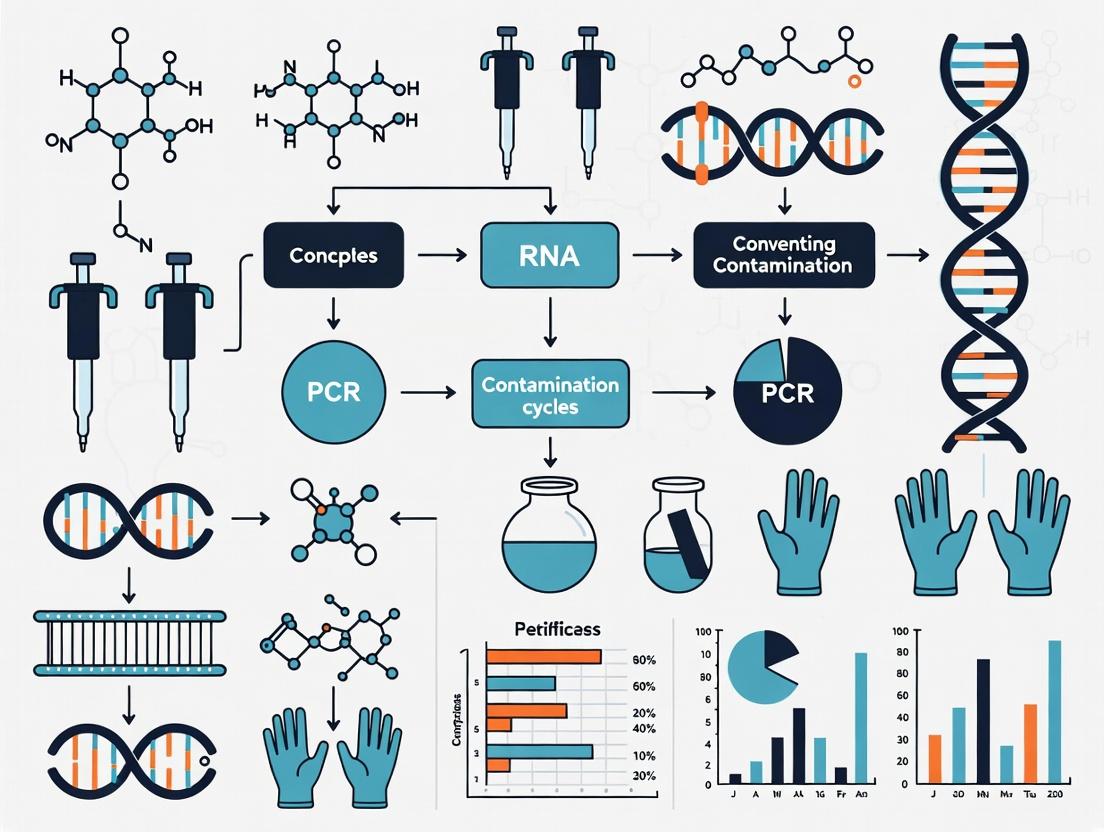
Visualization of Concepts and Workflows
Title: PCR Contamination Cascade Pathway
Title: Physical Separation Workflow for PCR
Title: dUTP/UNG Contamination Control Mechanism
Effective prevention of PCR contamination is foundational to the integrity of molecular diagnostics, genetic research, and drug development. Within a thesis on best practices, three primary culprits are identified: amplicon carryover (contamination from previous PCR products), sample cross-contamination (spillage between samples during handling), and reagent/primer pollution (contamination of master mixes, enzymes, or primers with exogenous nucleic acids). This document provides detailed application notes and protocols to identify, mitigate, and prevent these sources of error, ensuring data fidelity.
A summary of recent studies (2022-2024) on contamination frequency and impact is presented below.
Table 1: Prevalence and Impact of PCR Contamination Sources in Research Laboratories
| Contamination Source | Typical Frequency Range (%) | Average Ct Shift in qPCR | Primary Detection Method |
|---|---|---|---|
| Amplicon Carryover | 5-15% (in labs without UNG/dUTP) | 3-8 cycles earlier | Negative Template Controls (NTCs) |
| Sample Cross-Contamination | 2-10% | Variable, 1-10 cycles earlier | Replicate Sample Discrepancy, Sample Processing Controls |
| Reagent/Primer Pollution | 1-5% | Consistent across all samples | Multiple NTCs from different reagent lots |
| Composite Contamination | Up to 20% in audits | N/A | Systematic QC Panel Analysis |
Detailed Experimental Protocols
Protocol 3.1: Establishing a Contamination Monitoring Workflow
Objective: To systematically detect and identify the source of contamination within a laboratory.
Materials:
- Dedicated, filtered pipettes for pre- and post-PCR work.
- UV-equipped PCR workstation or dead air box.
- qPCR reagents, including separate lots for testing.
- Nuclease-free water (from multiple, sealed sources).
- Pre-aliquoted, single-use primer stocks.
- A full set of controls (see Table 2).
Procedure:
- Spatial Segregation: Physically separate pre-PCR (reaction setup) and post-PCR (analysis) areas. Never open PCR tubes in the setup area.
- Control Setup: For every qPCR run, include the following controls in duplicate:
- Negative Template Control (NTC): Contains all reagents + nuclease-free water instead of sample.
- Positive Extraction Control: A known sample carried through the entire extraction process.
- Negative Extraction Control: A blank (e.g., water) carried through the entire extraction process.
- Reagent-Only Control: Master mix + water, set up in the post-PCR area to test for amplicon aerosol contamination.
- Systematic Testing:
- If NTCs are positive, test each reagent component (polymerase, buffer, primers, water) individually in an NTC format.
- Prepare master mixes in a UV-PCR workstation, irradiating for 10-15 minutes before adding template.
- Use uracil-DNA glycosylase (UNG) and dUTP in all reactions to prevent carryover of prior amplicons.
Table 2: Interpretation of Control Results for Contamination Source Identification
| Control Type Positive | Likely Contamination Source | Immediate Corrective Action |
|---|---|---|
| NTC (in setup area) | Reagent/Primer Pollution or Master Mix Contamination | Test reagent components; use new aliquots. |
| NTC (in post-PCR area) | Amplicon Aerosol Carryover in lab environment | Decontaminate area; relocate setup to clean space. |
| Negative Extraction Control | Cross-Contamination during Sample Processing | Decontaminate extraction workstation; review technique. |
| All Samples & Controls | Widespread Reagent/Primer Pollution | Discard suspect reagent lot; source new materials. |
Protocol 3.2: Decontamination and Prevention Protocol
Objective: To eradicate existing contamination and implement preventive measures.
Part A: Laboratory Surface and Equipment Decontamination
- Clean all surfaces, pipettes, and equipment with 10% (v/v) sodium hypochlorite (bleach) solution. Leave on for 10 minutes, then wipe with nuclease-free water or 70% ethanol to prevent corrosion.
- Irradiate pipettes, tube racks, and workstations with 254 nm UV light for at least 30 minutes.
- Use dedicated, aerosol-barrier pipette tips for all liquid handling.
Part B: Reagent and Workflow Safeguards
- Aliquot all reagents (especially primers and enzymes) into single-use volumes upon validated receipt.
- Implement UNG/dUTP system: Substitute dTTP with dUTP in all PCR mixes. Include UNG enzyme in the master mix, with a 50°C incubation step for 2-10 minutes prior to amplification to degrade any uracil-containing carryover amplicons.
- Establish unidirectional workflow: Personnel should move from pre-PCR areas (clean) to post-PCR areas (contaminated) only, never in reverse.
Visualization of Workflows and Relationships
Title: Contamination Source Identification Decision Tree
Title: Laboratory Spatial Segregation and Workflow
The Scientist's Toolkit: Essential Research Reagent Solutions
Table 3: Key Reagents and Materials for PCR Contamination Prevention
| Item | Primary Function in Contamination Control |
|---|---|
| Uracil-DNA Glycosylase (UNG) + dUTP | Enzymatically degrades PCR products from previous reactions (carryover) by targeting uracil bases, preventing re-amplification. |
| Aerosol-Barrier Pipette Tips | Prevent aerosols and liquids from entering pipette shafts, a major vector for cross-contamination between samples. |
| Pre-aliquoted, Single-Use Reagents | Minimizes the risk of introducing contamination through repeated handling and opening of stock reagent tubes. |
| 10% (v/v) Sodium Hypochlorite | Effective chemical decontaminant for surfaces and equipment; degrades nucleic acids. |
| Multiple Lots of Nuclease-Free Water | Allows for testing and verification that water source is not contaminated with nucleic acids. |
| Dedicated, UV-equipped Laminar Flow Hood | Provides a sterile, amplicon-free environment for setting up PCR reactions. |
| Comprehensive Control Panels | Enables diagnostic identification of the specific contamination source (see Table 2). |
| Quantitative PCR (qPCR) with High-Resolution Melting | Allows detection of low-level contamination in NTCs and can differentiate specific products from non-specific amplification or primer-dimer. |
Aerosols, microscopic liquid or solid particles suspended in air, are a pervasive and potent source of cross-contamination in molecular biology laboratories, particularly those conducting PCR and other nucleic acid amplification techniques. Generated during routine liquid handling—especially pipetting, vortexing, centrifugation, and tube opening—aerosols can carry amplicons or target sequences into reagents, samples, and the laboratory environment. This creates a significant risk for false-positive results, compromising data integrity and derailing research and drug development projects. This application note, framed within a broader thesis on best practices for preventing PCR contamination, details the sources, risks, and mitigation strategies for aerosol-based contamination, providing actionable protocols for researchers and scientists.
The following tables summarize key quantitative data on aerosol generation and contamination potential from current literature.
Table 1: Aerosol Generation During Common Lab Procedures
| Procedure | Estimated Aerosol Droplet Size Range | Particle Travel Distance | Contamination Potential (Relative Risk) | Primary Citation |
|---|---|---|---|---|
| Pipetting (expelling liquid) | 0.5 - 50 µm | Up to 1 meter | High | Shin et al., 2024 |
| Vortex Mixing (with tube open) | 5 - 100 µm | Up to 2 meters | Very High | Mertens et al., 2023 |
| Centrifugation (tube failure) | <1 - >100 µm | Entire rotor compartment | Extreme | Clinical Lab Standards, 2023 |
| Opening Tube (snap-cap) | 1 - 10 µm | Immediate local cloud | Moderate-High | Green & Hughes, 2022 |
| PCR Plate Sealing Removal | 1 - 20 µm | Localized to work surface | Moderate | BioRad Application Note, 2024 |
Table 2: Efficacy of Contamination Control Measures
| Mitigation Strategy | Reduction in Aerosol Transfer (%) | Key Limitation/Consideration |
|---|---|---|
| Use of Filtered Pipette Tips | >99.9% for liquids | Does not protect from external contamination on tip barrel. |
| Physical Segregation (Separate rooms) | ~99% | Cost and space prohibitive for many labs. |
| UNG/dUTP Anti-Carryover System | >90% for amplicons | Only effective against dU-containing amplicons; requires protocol integration. |
| UV Decontamination of Workstations | 95-99% (surface) | Shadowing effects; limited penetration; does not inactivate all nucleic acids. |
| Positive Displacement Pipettes | ~99.5% | Higher cost; requires specific, often more expensive, tips. |
Experimental Protocols
Protocol 1: Assessing Aerosol Generation from Pipetting
Objective: To visualize and semi-quantify aerosols generated during routine pipetting. Materials: Fluorescent dye (e.g., fluorescein), PBS, adjustable-volume micropipettes and tips, a dark room or cabinet, UV lamp, a clean white poster board or filter paper, safety goggles. Procedure:
- Prepare a 1 mg/mL solution of fluorescent dye in PBS.
- In a darkened room, position a clean white poster board vertically 30 cm behind the target tube.
- Pipette 100 µL of the dye solution from a source tube to a target tube using standard forward pipetting. Perform 50 repetitions.
- Repeat step 3 using reverse pipetting technique.
- Illuminate the poster board with a UV lamp. Document the number and spread of fluorescent spots, which represent aerosol deposition.
- Analysis: Compare the density and spread of spots between the two pipetting techniques. Reverse pipetting typically generates significantly fewer aerosols.
Protocol 2: Evaluating Cross-Contamination via Aerosols in a Simulated PCR Setup
Objective: To demonstrate cross-contamination between adjacent tubes during vortexing and opening. Materials: Two distinct plasmid DNA solutions (e.g., Plasmid A at 10^6 copies/µL and Plasmid B at 10^2 copies/µL), water (negative control), PCR master mix with primers specific to Plasmid A, strip tubes or plate, vortex mixer, real-time PCR instrument. Procedure:
- In a tube containing a high concentration of "contaminant" Plasmid A (10^6 copies/µL), add 50 µL of water. Cap tightly.
- Place this tube next to a tube containing 50 µL of the "low-copy sample" Plasmid B (10^2 copies/µL) in a tube rack.
- Vigorously vortex the "contaminant" tube (Plasmid A) for 10 seconds.
- Immediately open both tubes and perform a simulated transfer (pipette up and down in the low-copy tube without actually taking volume).
- Close all tubes. Prepare real-time PCR reactions for Plasmid A from: a) the original high-copy tube (positive control), b) the low-copy Plasmid B tube (test for contamination), c) a fresh water sample (negative control).
- Run the real-time PCR assay.
- Analysis: A positive signal (low Ct value) in the low-copy Plasmid B tube indicates aerosol-mediated transfer of Plasmid A. The negative control should remain negative.
Visualizations
Title: Aerosol Contamination Pathway to PCR False Positives
Title: Integrated Strategy for Aerosol Contamination Control
The Scientist's Toolkit: Essential Research Reagent Solutions
| Item | Function & Relevance to Aerosol Risk Mitigation |
|---|---|
| Aerosol-Barrier Filter Pipette Tips | Contain a hydrophobic filter that prevents aerosols and liquids from entering the pipette barrel, protecting both the sample and the instrument. The primary physical defense against pipette-borne contamination. |
| UNG (Uracil-N-Glycosylase) / dUTP System | A biochemical carryover prevention method. dUTP is incorporated into amplicons, and pre-PCR UNG treatment enzymatically degrades any contaminating dU-containing amplicons from previous reactions before new amplification. |
| PCR Decontamination Reagents (e.g., DNA-ExitusPlus, DNA-Zap) | Chemical solutions (often acidic peroxides or specific nucleases) used to decontaminate work surfaces, equipment, and plasticware by degrading nucleic acids. Crucial for cleaning spills and routine decontamination. |
| Pre-PCR Aliquotable Master Mix | A ready-to-use master mix provided in single-use aliquots or small bottles to minimize repeated openings of a central stock, thereby reducing the risk of introducing aerosols into the bulk reagent. |
| Positive Displacement Pipette & Tips | Utilizes a disposable piston that makes direct contact with the liquid, eliminating the air cushion. Virtually eliminates aerosol generation within the tip and is ideal for handling viscous or volatile samples. |
| UV-C Irradiating Laminar Flow Cabinet | Provides a contained, HEPA-filtered workspace with a built-in UV-C light for nucleic acid decontamination of surfaces and air before and after use. Essential for setting up amplification reactions. |
| Dedicated Lab Coats & Color-Coded Gloves | Personal protective equipment (PPE) that also serves as a contamination control measure. Lab coats should be worn only in designated areas (e.g., pre-PCR room). Color-coded gloves help enforce unidirectional workflow. |
In the context of best practices for preventing PCR contamination in research and diagnostic settings, the persistent nature of PCR amplicons represents a paramount challenge. Amplicons, the double-stranded DNA products of amplification, are prolific (billions of copies per reaction), stable, and are themselves perfect templates for subsequent reactions. Their small size facilitates aerosolization and environmental persistence, making them the primary source of contaminative false positives. This application note details the reasons for their problematic nature and provides validated protocols for mitigation.
Quantitative Data on Amplicon Persistence and Contamination Risk
Table 1: Amplicon Stability and Detectability Under Various Conditions
| Condition | Approximate Persistence Time | Minimum Detectable Copies (in a 50 µL PCR) | Key Risk Factor |
|---|---|---|---|
| Dry on benchtop (20-25°C) | Weeks to months | 1-10 copies | Dust, particulate shedding |
| In aerosol (≤5 µm droplets) | Hours (airborne), indefinite upon settlement | 1-10 copies | Cross-rack contamination during plate sealing/venting |
| In liquid solution (4°C) | Years | 1 copy | Contaminated water, buffer stocks, or shared reagents |
| On skin & gloves | Hours, transferable | 10-100 copies | Direct transfer to tubes, pipettes |
| UV irradiation (254 nm, typical crosslinker dose) | Reduced by 3-4 log10, not eliminated | 10-100 copies* | False sense of security; incomplete inactivation |
Source: Recent studies (2023-2024) on contamination control in NGS and qPCR labs. Data synthesized from current literature on environmental DNA persistence and contamination event analyses.
Table 2: Comparison of Contamination Sources in a PCR Laboratory
| Source | Relative Contribution to False Positives | Ease of Mitigation | Primary Amplicon Load |
|---|---|---|---|
| Post-PCR Amplicons (open tubes, gels) | High (>70%) | Moderate (requires strict discipline) | Very High (10^9 – 10^12 copies) |
| Contaminated Reagents (water, master mix) | Medium (15-20%) | High (use of aliquots, UV treatment) | Low-Medium (distributed) |
| Contaminated Consumables (tips, tubes) | Low (5-10%) | High (use of pre-sterilized, dedicated sets) | Variable |
| Cross-contamination from Samples (high-titer target) | Low (<5%) | Moderate (extraction controls, plate layout) | Sample-dependent |
Experimental Protocol: Monitoring and Decontaminating Amplicon Contamination
Protocol 3.1: Environmental Monitoring for Amplicon Contamination
Purpose: To proactively detect amplicon accumulation on surfaces, equipment, and in reagents. Materials: See "Scientist's Toolkit" below. Procedure:
- Swab Sampling: Moisten a sterile, DNA-free swab with 50 µL of molecular-grade water or a dedicated elution buffer.
- Surface Wipe: Vigorously swab a ~10 cm² area of the test surface (e.g., pipette handle, bench, centrifuge button, tube rack).
- Elution: Place the swab tip in a 1.5 mL microcentrifuge tube with 100 µL of elution buffer. Vortex for 30 seconds.
- Template Preparation: Use 5-10 µL of the eluate directly as a template in a highly sensitive (ideally nested) PCR assay targeting a ubiquitous amplicon sequence used in the lab (e.g., a segment of a common plasmid vector or a human gene target).
- Controls:
- Negative Control: Swab a known clean surface (e.g., inside a UV cabinet post-treatment).
- Positive Control: Diluted, quantified amplicon stock spotted on a surface.
- PCR Negative: No-template control (NTC).
- Amplification & Analysis: Run PCR and analyze by gel electrophoresis. A positive signal indicates contamination.
Protocol 3.2: Chemical Decontamination with Sodium Hypochlorite (Bleach)
Purpose: To effectively degrade amplicons on non-corrosive surfaces and in liquid spills. Principle: Hypochlorite oxidizes nucleic acids, fragmenting them and rendering them non-amplifiable. Procedure:
- Solution Preparation: Prepare a fresh 1% (v/v) solution of household bleach (typically ~5% sodium hypochlorite) in molecular biology grade water. Caution: Do not mix with acids or other cleaners.
- Surface Decontamination:
- Apply the 1% bleach solution liberally to the contaminated surface.
- Allow it to stand for a minimum of 10 minutes.
- Wipe thoroughly with water or ethanol to remove residue.
- Liquid Waste Decontamination:
- For amplicon-containing liquids (gels, post-PCR reactions), add bleach to a final concentration of 1%.
- Incubate for 30 minutes at room temperature in a fume hood.
- Dispose of as standard liquid chemical waste.
- Validation: Post-decontamination, use Protocol 3.1 to verify efficacy.
Visualization: Workflow for Amplicon Contamination Prevention
Title: PCR Lab Contamination Prevention Strategy Workflow
Title: Amplicon Contamination Pathways Leading to False Positives
The Scientist's Toolkit: Key Reagents and Materials for Contamination Control
Table 3: Essential Research Reagent Solutions for Amplicon Contamination Prevention
| Item | Function/Benefit | Application Notes |
|---|---|---|
| Uracil-N-glycosylase (UNG) + dUTP | Enzymatically degrades previous PCR products containing dU, preventing carryover contamination. | Incorporate into master mix. Requires use of dUTP instead of dTTP in all PCRs. Inactivated by high temp (95°C) step. |
| DNA Decontamination Solutions (e.g., DNA Away, 1% Bleach) | Chemically degrades DNA/RNA on non-porous surfaces. | Regular wiping of workspaces, equipment. Bleach must be freshly diluted and rinsed to prevent corrosion. |
| UV Crosslinker (254 nm) | Creates thymine dimers in DNA, inhibiting polymerase extension. | For decontaminating workstations, plasticware, and some reagents (excluding enzymes, dNTPs). Not 100% effective. |
| Aerosol-Barrier Pipette Tips | Prevents aerosols from entering pipette shaft, a common contamination reservoir. | Mandatory for all post-PCR and master mix pipetting. Use in all phases for maximum safety. |
| Pre-sterilized, DNA-free Consumables (Tubes, Plates, Tips) | Eliminates consumables as a source of contaminating DNA. | Purchase certified "DNA-free" or "PCR clean". Autoclaving does not remove DNA. |
| Molecular Biology Grade Water (Nuclease-free) | Free of nucleases and contaminating DNA. | Use for all reagent preparations and sample dilutions. Aliquot into small, single-use volumes. |
| Dedicated Lab Coats & Gloves | Prevents clothing from acting a reservoir for amplicons. | Use different colored coats for pre- and post-PCR areas. Change gloves frequently. |
| PCR Workstation / Dead Air Box | Provides a physically separated, UV-treatable space for master mix assembly. | Ideally equipped with a UV lamp for nightly decontamination. |
Application Notes and Protocols
Effective prevention of PCR contamination is foundational to molecular biology research and diagnostics. Contamination leads to false positives, data invalidation, and significant resource waste. This document outlines integrated strategies focusing on laboratory design, workflow, and specific decontamination protocols, framed within best practices for PCR contamination prevention.
1. Laboratory Zoning and Unidirectional Workflow
The most critical defense is physical separation of PCR preparation, template addition, and amplification/product analysis.
- Zone 1: Pre-PCR (Reagent Preparation). Dedicated room or enclosed cabinet for master mix preparation. Contains dedicated equipment (pipettes, centrifuges, tubes) and reagents. Airflow is positive pressure relative to other zones.
- Zone 2: Template Addition. Separate area or dead-air box for adding nucleic acid template to master mix. Equipment is dedicated to this zone.
- Zone 3: Amplification. Thermal cyclers are housed separately.
- Zone 4: Post-PCR Analysis. Dedicated room for analyzing PCR products (e.g., gel electrophoresis, qPCR plate reading). Airflow is negative pressure relative to cleaner zones.
Protocol 1.1: Implementing a Unidirectional Workflow
- Objective: To prevent amplicon carryover into pre-PCR areas.
- Materials: Dedicated lab coats, gloves, pipettes, filter tips, and waste containers for each zone.
- Methodology:
- Personnel enter Zone 1 first, don a dedicated lab coat.
- Prepare master mix in Zone 1 using UV-irradiated consumables.
- Aliquot master mix into reaction tubes/plates.
- Move to Zone 2. Change gloves. Add template nucleic acids.
- Seal plates/tubes and transfer to Zone 3 for amplification.
- For analysis, proceed to Zone 4. Never return equipment or personnel from Zone 4 to Zones 1 or 2 without rigorous decontamination.
- Discard waste in zone-specific containers; Zone 4 waste must be sealed immediately.
2. Active Decontamination Strategies
Protocol 2.1: Surface and Equipment Decontamination with Sodium Hypochlorite
- Objective: To degrade contaminating nucleic acids on work surfaces and equipment.
- Materials: Freshly prepared 0.5-1% (v/v) sodium hypochlorite (bleach) solution, RNase Away for RNA work, 70% ethanol, DNAZap or similar commercial DNA degradation solution.
- Methodology:
- Before and after each use, clean work surfaces in all zones with 70% ethanol to remove debris.
- For Pre-PCR zones (1 & 2), follow with a 10-minute exposure to a 0.5% bleach solution applied via spray or wipe. Caution: Corrosive to metal.
- Wipe down surfaces with water or ethanol to remove bleach residue.
- For sensitive equipment, use validated commercial nucleic acid degradation solutions.
Protocol 2.2: Enzymatic Decontamination of Reagents using dUTP/UNG
- Objective: To selectively destroy carryover amplicons prior to amplification.
- Principle: Incorporate dUTP in place of dTTP during PCR. In subsequent reactions, Uracil-N-Glycosylase (UNG) cleaves uracil-containing contaminants, which are then degraded at high temperature.
- Methodology:
- Reaction Setup: Include dUTP in the nucleotide mix and add 0.2-1.0 U of UNG per reaction to the master mix.
- Incubation: Perform a UNG incubation step at 25°C for 2-10 minutes prior to thermal cycling.
- Inactivation: Inactivate UNG by a 50°C hold for 2 minutes or by the initial denaturation step (95°C) of the PCR cycle.
3. Quantitative Impact of Contamination Prevention Measures
Table 1: Efficacy of Contamination Prevention Strategies
| Prevention Strategy | Reported Reduction in False-Positive Rate | Key Limitation/Cost |
|---|---|---|
| Physical Separation (3-zone workflow) | 90-99% | High space requirement; operational discipline |
| UV Irradiation of Workstations | 85-95% | Ineffective on shadowed areas; tube/plate opacity |
| UNG/dUTP System | >99% for carryover amplicons | Only destroys uracil-containing DNA; reagent cost |
| Extensive Use of Filter Tips | 95-98% (aerosol prevention) | Increased consumables cost (~30-40% premium) |
| Rigorous Bleach Decontamination | >99.9% (surface nucleic acids) | Corrosive; requires careful handling and rinsing |
Diagram 1: PCR Lab Unidirectional Workflow
Diagram 2: UNG/dUTP Amplicon Inactivation Mechanism
The Scientist's Toolkit: Essential Research Reagent Solutions
Table 2: Key Reagents and Materials for PCR Contamination Prevention
| Item | Function & Role in Prevention |
|---|---|
| UNG Enzyme | Enzymatically cleaves uracil bases in carryover dUTP-containing amplicons, preventing their amplification. |
| dUTP Nucleotide | Substituted for dTTP in PCR mixes, making all amplicons susceptible to subsequent UNG degradation. |
| UV-C Light Source (e.g., Crosslinker) | Irradiates work surfaces and open consumables (tips, tubes) to crosslink and inactivate nucleic acids. |
| Nucleic Acid Degradation Solution (e.g., DNAZap) | Chemical blend that rapidly degrades DNA/RNA on surfaces and equipment without corrosion. |
| Molecular Biology Grade 0.5% Sodium Hypochlorite | Oxidizes and destroys contaminating nucleic acids on non-corrodible surfaces. |
| Aerosol-Resistant Filter Pipette Tips | Physical barrier preventing aerosols and liquids from entering pipette shaft, a major contamination vector. |
| PCR Plates/Tubes with Optically Clear Seals | Provide a secure seal to prevent amplicon aerosol escape during thermal cycling and plate reading. |
| Dedicated Pre-PCR Lab Coats & Gloves | Single-use or zone-specific PPE to prevent clothing-borne contaminant transfer. |
Building Your Defense: Practical Protocols and Workflow Solutions
Contamination of pre-amplification materials with PCR amplicons is the single most critical point of failure in molecular diagnostics and quantitative research. This application note details the non-negotiable protocols for establishing a uni-directional workflow, a foundational best practice within the broader thesis on preventing PCR contamination. Its rigorous implementation is the gold standard for ensuring data integrity in drug development and clinical research.
Foundational Principles & Quantitative Justification
The necessity of physical separation is driven by the extreme sensitivity of PCR and the overwhelming target copy number difference between samples and amplicons.
Table 1: Contamination Risk Assessment - Sample vs. Amplicon Copy Number
| Material Type | Typical Target Copy Number Range | Potential Aerosol/Droplet Volume (µL) | Copies per Droplet (Estimate) | Risk Level |
|---|---|---|---|---|
| Pre-PCR Sample (e.g., cDNA) | 10^0 – 10^6 | 0.001 – 1 | 0.001 – 1,000 | Baseline |
| Post-PCR Amplicon | 10^9 – 10^13 | 0.001 – 1 | 1,000,000 – 10,000,000,000 | Critical |
Protocol: Establishing a Uni-Directional Workflow
Laboratory Design & Spatial Separation
Objective: To create irreversible, dedicated physical zones for pre- and post-PCR activities.
- Pre-PCR Zone ("Clean Area"): Dedicated room or enclosed space for reagent preparation, nucleic acid extraction, and reaction setup. This area must never contain amplified DNA.
- Post-PCR Zone ("Amplification Area"): Separate room for thermal cycling and analysis of amplified products.
- Buffer Zone: An anteroom or hallway for gowning and material staging. This area should contain a dedicated centrifuge and vortex for pre-PCR materials only.
- Unidirectional Flow: Personnel and materials must move from the Pre-PCR zone to the Post-PCR zone only. Reverse movement is prohibited without documented decontamination procedures.
Procedural Workflow & Temporal Separation
Objective: To enforce a sequence of work that prevents carryover.
Detailed Daily Protocol:
- Start of Day: Begin all work in the Pre-PCR zone. Prepare master mixes, aliquot reagents, and handle patient samples or extracted nucleic acids.
- Reaction Setup: Pipette template into prepared master mix or plates in the Pre-PCR zone.
- Sealing & Transition: Seal reaction plates/tubes completely in the Pre-PCR zone. Wipe exteriors with 10% bleach or DNA decontamination solution. Transport sealed vessels to the Post-PCR zone in a dedicated, closed container.
- Amplification & Analysis: Place plates in thermal cyclers located in the Post-PCR zone. Post-amplification, plates remain sealed until analysis (e.g., in a qPCR instrument) within the Post-PCR zone.
- End of Day: All amplicons are discarded within the Post-PCR zone. Equipment (pipettes, benches) in the Pre-PCR zone is cleaned with 10% bleach followed by ethanol or RNase Away.
Diagram 1: Uni-Directional Lab Workflow Path
Contamination Monitoring Protocol
Objective: To routinely audit the cleanliness of the Pre-PCR zone.
- Method: Weekly, run "no-template controls" (NTCs) and "environmental controls" (open tubes left during setup, then filled with water).
- Acceptance Criterion: All controls must show no amplification (Cq > 40 or undetermined). A positive control indicates a breach and mandates shutdown, investigation, and full decontamination of the Pre-PCR zone.
The Scientist's Toolkit: Essential Research Reagent Solutions
Table 2: Key Reagents for Contamination Prevention
| Item | Function & Rationale | Example/Best Practice |
|---|---|---|
| UDG/dUTP System | Enzymatic carryover prevention: Uses uracil-DNA glycosylase (UDG) to degrade prior amplicons containing dUTP, while protecting new dTTP-containing reactions. | Include in master mix for routine qPCR assays. |
| Aerosol-Resistant Tips (ART) | Physical barrier: Prevents pipette aerosol and liquid from contaminating the pipette shaft. | Use for all liquid handling, especially master mix preparation and template addition. |
| Single-Use, Aliquot Reagents | Limits exposure: Prevents repeated sampling from a stock bottle that could become contaminated. | Aliquot all enzymes, primers, probes, and dNTPs into single-experiment volumes. |
| 10% (v/v) Sodium Hypochlorite (Bleach) | Chemical decontamination: Efficiently degrades DNA/RNA on surfaces. | Wipe down benches and equipment daily; immerse contaminated tips/tubes for >1 min. |
| DNA Decontamination Solutions (e.g., DNA-ExitusPlus) | Commercial nucleic acid degrading agents. | Alternative to bleach for surfaces and equipment sensitive to corrosion. |
| Dedicated Labware & Pipettes | Zone fidelity: Ensures no shared equipment between pre- and post-PCR areas. | Color-code pipettes and lab coats for each zone. |
Decontamination Response Protocol
Objective: Standardized action plan following a contamination event.
- Immediate Action: Halt all pre-PCR work. Document the event.
- Decontamination: Discard all open reagents in the Pre-PCR zone. Clean all surfaces, pipettes, racks, and equipment with 10% bleach, followed by ethanol or water to prevent corrosion. UV-irradiate the cabinet if available (30 mins).
- Verification: After cleaning, run an extensive NTC panel (e.g., 8-12 wells). Only resume work if all NTCs are clean.
Diagram 2: Decontamination Response Protocol
Within the context of a comprehensive thesis on best practices for preventing PCR contamination, the implementation of physically separated, dedicated workspaces is the single most critical procedural intervention. Contamination from previously amplified PCR products (amplicons) or sample cross-over can lead to false-positive results, invalidating data and compromising research integrity, particularly in sensitive applications like diagnostic assay development and drug discovery. This document provides detailed application notes and protocols for establishing and maintaining dedicated PCR zones to mitigate these risks.
Core Principles of Physical Separation
Effective contamination control is predicated on the strict unidirectional workflow and spatial segregation of PCR-related activities. The process is divided into three distinct, physically separated zones.
Diagram Title: Unidirectional Workflow for Dedicated PCR Zones
Detailed Zone Design and Operational Protocols
Zone 1: Pre-PCR Area (Clean Zone)
- Location: A positive-pressure, low-traffic area, ideally in a clean room or dedicated laminar flow cabinet (ISO Class 5 or better).
- Function: Preparation of master mixes, aliquoting of reagents, and addition of template nucleic acid (for one-step RT-PCR or qPCR).
- Protocol 3.1: Aseptic Master Mix Preparation in the Clean Zone
- Objective: To assemble reaction mixes free of contaminating amplicons or nucleases.
- Materials: See "The Scientist's Toolkit."
- Method:
- Decontaminate the work surface and equipment (pipettes, tube racks) with a 10% (v/v) sodium hypochlorite solution, followed by 70% ethanol. Irradiate with UV light for 15-30 minutes prior to starting work.
- Use only dedicated equipment (pipettes, tips, centrifuges, lab coats) marked for the Pre-PCR zone. Equipment must never leave this zone.
- Prepare master mixes in a clean, dedicated PCR workstation or biosafety cabinet. Use aerosol-resistant barrier tips for all liquid handling.
- Thaw reagents on ice, vortex briefly, and centrifuge before opening. Always add the template nucleic acid last.
- After adding template, seal tubes/plates, and immediately transfer them to the Post-PCR zone via a designated pass-through or by exiting the lab. The analyst must not re-enter the Pre-PCR zone after this step without a full decontamination protocol (showering, complete change of clothing).
Zone 2: PCR Amplification Area
- Location: A separate room or enclosed space.
- Function: Housing of thermal cyclers.
- Protocol 3.2: Loading and Operating Thermal Cyclers
- Objective: To contain amplified product within sealed reaction vessels.
- Method:
- Transport sealed reaction plates/tubes from the Pre-PCR zone to the amplification area.
- Load the thermal cycler. Avoid opening plates or tubes in this area if possible.
- After the run is complete, carefully remove the sealed plate. Visually inspect for any tube leaks or plate seal failures.
- Wipe the thermal cycler block and surfaces with 10% bleach and 70% ethanol after each run.
Zone 3: Post-PCR Area (Contamination Zone)
- Location: A separate, negatively pressured room, distant from the Pre-PCR area.
- Function: All downstream analyses: gel electrophoresis, qPCR plate reading, sequencing prep, etc.
- Protocol 3.3: Post-Amplification Analysis with Containment
- Objective: To analyze amplicons while preventing their aerosol release and back-migration.
- Method:
- All work with opened reaction tubes must be performed in a dedicated biosafety cabinet or enclosed workstation in the Post-PCR zone.
- Use dedicated pipettes, tips, and lab coats for this zone only.
- Decontaminate all work surfaces daily with 10% bleach. Treat liquid waste with bleach before disposal.
- Never remove any equipment, notebooks, or samples from the Post-PCR zone to the Pre-PCR zone. Data should be transferred electronically.
Quantitative Efficacy Data
The effectiveness of physical separation is supported by empirical data comparing contamination rates under different laboratory configurations.
Table 1: Contamination Incident Frequency vs. Laboratory Zoning Configuration
| Laboratory Configuration | Avg. Contamination Incidents per 1000 PCR Runs (95% CI) | Relative Risk Reduction | Key Study Reference |
|---|---|---|---|
| Single-room, no dedicated equipment | 18.5 (15.2 - 22.1) | Baseline (0%) | Millar et al., 2002 |
| Temporal separation only | 8.2 (6.1 - 10.5) | 56% | Espy et al., 2006 |
| Dedicated rooms with unidirectional workflow | 0.7 (0.2 - 1.5) | 96% | Borst et al., 2004 |
Table 2: Cost-Benefit Analysis of Implementing Dedicated Zones (Initial 5-Year Period)
| Cost Category | Initial Investment (USD) | Annual Maintenance (USD) | Benefit: Estimated Cost Averted* (USD/year) |
|---|---|---|---|
| Facility Modifications | 15,000 - 50,000 | - | - |
| Dedicated Equipment (x2 zones) | 20,000 - 30,000 | 1,000 | - |
| Personnel Training | 2,000 - 5,000 | 500 | - |
| Contaminated Run Mitigation | - | - | 25,000 - 75,000 |
| Net Projected Benefit | - | - | Positive ROI within 2-3 years |
*Based on averting repeat experiments, delayed timelines, and potential diagnostic errors.
Decontamination and Validation Protocols
Protocol 5.1: Routine Surface and Equipment Decontamination
- Reagent: Freshly prepared 10% (v/v) sodium hypochlorite (bleach). Note: Bleach must be prepared weekly as it degrades.
- Method: Apply bleach to all surfaces, pipettes, and tube racks. Leave in contact for 10-15 minutes. Wipe down with water, followed by 70% ethanol to remove residual salt.
Protocol 5.2: UV Irradiation for Nucleic Acid Destruction
- Validation Experiment: To determine effective UV exposure time for your cabinet.
- Spot 10 µL of a high-titer amplicon (e.g., 10^8 copies/µL) onto a clean, dry surface inside the cabinet.
- Expose to 254 nm UV light for varying durations (0, 1, 5, 10, 15, 30 min).
- Post-exposure, resuspend the spot in nuclease-free water and use it as a template in a highly sensitive (40+ cycle) PCR.
- The minimum exposure time that yields a negative PCR result is the validated decontamination time for your setup.
Protocol 5.3: Environmental Monitoring for Contamination
- Objective: Periodic audit of zone integrity.
- Method:
- Prepare "sentinel" PCR tubes containing master mix with water instead of template.
- Place these open tubes in each zone (Pre, Post, Amplification) during normal lab activity for 30-60 minutes.
- Close the tubes and run them through a full PCR protocol.
- A positive signal in a Pre-PCR sentinel tube indicates a critical containment breach.
Diagram Title: Environmental Monitoring Workflow for PCR Zones
The Scientist's Toolkit
Table 3: Essential Research Reagent Solutions for Dedicated PCR Zones
| Item | Function & Rationale |
|---|---|
| Aerosol-Resistant Barrier Pipette Tips | Prevent aerosols from contaminating pipette shafts, the primary vector for cross-contamination. Mandatory for all zones. |
| Single-Use, DNA/RNA Nuclease-Free Tubes & Plates | Prevents carryover from previous experiments. Use low-binding tubes for sensitive applications. |
| Molecular Biology Grade Water | Certified free of nucleases and contaminating nucleic acids for reagent preparation. |
| Aliquoted PCR Reagents (dNTPs, Primers, Enzyme Mix) | Store small, single-use aliquots to limit repeated exposure of stock reagents to potential contamination. |
| Sodium Hypochlorite (Bleach), 10% solution | Effective chemical decontaminant that degrades exposed nucleic acids on surfaces. |
| UV-C Light Source (254 nm) | Installed in cabinets and workstations to degrade nucleic acids on surfaces and in the air between uses. |
| Dedicated Lab Coats & Gloves | A unique color for each zone is highly recommended. Gloves must be changed when exiting any zone. |
| Pass-Through Freezer/Incubator or Sample Transfer Locks | Allows movement of samples between zones without personnel or equipment crossing boundaries. |
In the context of a thesis on best practices for preventing PCR contamination, the implementation of robust procedural controls is foundational. Negative controls are non-negotiable diagnostic tools that validate the integrity of every step in the molecular workflow, from nucleic acid extraction to amplification. Their consistent absence of signal is a critical indicator of a contamination-free process. The failure to include and properly analyze these controls invalidates experimental results and undermines data credibility in research and drug development.
Application Notes: Types and Functions of Critical Negative Controls
Two primary negative controls must be incorporated into every experimental run:
- No-Template Control (NTC): Contains all PCR master mix components (primers, polymerase, dNTPs, buffer) but uses nuclease-free water instead of template DNA. It detects contamination in the master mix, primers, or amplicon carryover.
- Extraction Negative Control (Blank): A sample (e.g., water or sterile swab) taken through the entire nucleic acid extraction and purification process alongside experimental samples. It identifies contamination introduced during extraction reagents, kits, or the extraction environment.
Table 1: Interpretation of Negative Control Results
| Control Result | Experimental Sample Result | Likely Interpretation | Required Action |
|---|---|---|---|
| Negative | Positive | Valid target detection. | Proceed with data analysis. |
| Positive | Positive | Contamination confirmed. Experimental result is unreliable. | Discard run. Decontaminate workspace/reagents. Re-run with fresh aliquots. |
| Negative | Negative | Valid negative result (or assay inhibition). | Check for inhibition using an internal positive control (IPC). |
| Positive | Negative | High-level contamination. All results are suspect. | Cease work. Perform full lab decontamination. Validate with new reagent lots. |
Detailed Experimental Protocols
Protocol 3.1: Comprehensive Negative Control Setup for qPCR/PCR
Objective: To detect contamination in reagent preparation, sample extraction, and amplicon carryover. Materials: See "The Scientist's Toolkit" below. Procedure:
- Prepare Extraction Negative Control: For every batch of samples (max 10-12 samples per batch), include one extraction blank. Use the same volume of sterile, nuclease-free water or buffer as used for sample lysis.
- Extraction: Process the blank through the identical extraction and purification protocol as all other samples. Elute in the same volume.
- Master Mix Preparation: In a clean, amplicon-free zone (ideally a separate room or cabinet), prepare a master mix sufficient for all experimental samples + NTCs + a positive amplification control.
- Plate/Tube Setup: Aliquot the master mix into wells/tubes first.
- Positive Control Well: Add known positive template.
- NTC Wells: Add nuclease-free water (volume equal to template).
- Sample & Extraction Blank Wells: Add extracted nucleic acids (samples and the eluate from step 2).
- Run and Analyze: Execute the PCR/qPCR protocol. The run is valid only if: NTCs and extraction blanks show no amplification (Ct value >40 or undetermined in qPCR). Any amplification in these controls necessitates run rejection.
Protocol 3.2: Decontamination Procedure Following Control Failure
Objective: To eliminate contaminating nucleic acids from surfaces and equipment after a positive negative control. Materials: DNA Away or 10% (v/v) commercial bleach, UV crosslinker (if available), microfuge tubes, nuclease-free water. Procedure:
- Discard all reagents from the failed run.
- Decontaminate Workspaces: Wipe down pipettes, benchtops, and equipment with DNA decontamination solution or 10% bleach, followed by ethanol and nuclease-free water to prevent corrosion.
- UV Irradiation: Expose pipettes, racks, and unsealed plasticware in a PCR workstation to 254 nm UV light for 30 minutes.
- Prepare Fresh Reagents: Use new, never-opened aliquots of critical reagents (primers, dNTPs, water) from a dedicated "clean" stock stored separately from amplified DNA.
- Re-run starting with the extraction step, ensuring all controls are included.
Visualizations
Workflow for Contamination Monitoring
Title: PCR Workflow with Control Decision Points
Contamination Source Identification Logic
Title: Diagnostic Logic for Contamination Source
The Scientist's Toolkit
Table 2: Essential Research Reagent Solutions for Contamination Control
| Item | Function & Rationale |
|---|---|
| Molecular-grade Nuclease-free Water | Used for NTCs and sample reconstitution. Certified free of nucleases and contaminating nucleic acids. |
| DNA/RNA Decontamination Solution (e.g., DNA Away) | For surface decontamination. Degrades contaminating nucleic acids without corrosive damage. |
| dUTP and Uracil-DNA Glycosylase (UDG) | Enzymatic carryover prevention. dUTP incorporates into amplicons; UDG pre-treatment destroys carryover contamination prior to PCR. |
| Separated, Dedicated Workspaces | Physical separation of pre- and post-PCR areas with dedicated equipment (pipettes, racks, coats) is the single most effective control. |
| Aerosol-Resistant Filter Pipette Tips | Prevents sample carryover and protects pipette shafts from contamination. Mandatory for all liquid handling. |
| Validated Nucleic Acid Extraction Kit | Kits with demonstrated high purity and consistent yield. Include carrier RNA for low-copy RNA recovery. |
| Positive Control Template (Synthetic Oligo or Plasmid) | A non-genomic, sequence-specific synthetic control to validate assay performance without risk of sample crossover. |
| UV Crosslinker/Cabinet | 254 nm UV light creates thymine dimers in contaminating DNA, rendering it non-amplifiable for surface decontamination. |
Within PCR-driven research, contamination represents a critical failure point, leading to false positives, erroneous data, and compromised reproducibility. This document details standardized application notes and protocols for the handling of consumables and reagents—specifically aliquoting, UV treatment, and safe handling—to establish robust contamination barriers integral to a comprehensive PCR contamination prevention thesis.
Table 1: Efficacy of Common Contamination Prevention Measures
| Prevention Measure | Reduction in Contaminant Copies (Log10) | Key Application | Key Limitation |
|---|---|---|---|
| Physical Separation (Separate Rooms) | 3-4 | Setup vs. Analysis areas | High resource requirement |
| Aliquotting (Single-use volumes) | 2-3 | All liquid reagents | Does not destroy contaminant |
| UV-C Irradiation (254nm, 10min) | 4-6* | Surfaces, plastics, air in cabinets | Variable efficacy on shaded areas |
| UNG/dUTP System | >6 | PCR mix for carryover prevention | Only effective against dU-containing amplicons |
| Positive Displacement Pipettes | 2-3 | Handling all PCR liquids | Slower workflow vs. air displacement |
| *Dependent on intensity, exposure time, and material translucency. Data synthesized from current laboratory guidelines and instrument manuals. |
Table 2: UV-C Dose Required for 1-Log Reduction of Common Contaminants
| Contaminant Type | Approximate UV Dose (J/m²) for 90% Reduction | Implications for Protocol |
|---|---|---|
| Bacterial Spores (e.g., B. subtilis) | 100 - 200 | Highest dose required for surfaces |
| Viral Particles | 20 - 100 | Moderate dose sufficient |
| Naked DNA/RNA | 10 - 50 | Readily inactivated; shadows are critical |
| Based on recent biosafety and instrumentation literature. Dose = Intensity (W/m²) x Time (s). |
Detailed Protocols
Protocol 3.1: Aliquoting of PCR Reagents for Contamination Prevention
Objective: To minimize repeated freeze-thaw cycles and cross-contamination of master mix components, primers, dNTPs, and templates. Materials: Nuclease-free microcentrifuge tubes, filtered pipette tips, dedicated PCR-area pipettes, personal protective equipment (PPE), -20°C or -80°C freezer.
- Preparation: Clean the workspace with a DNA/RNA decontaminant (e.g., 10% bleach, followed by ethanol and RNase-free water). Use dedicated equipment only for aliquoting.
- Volume Calculation: Calculate aliquot volume based on a single experiment's typical usage (e.g., 25µL x number of reactions + 10% overage). Avoid aliquots larger than needed for 1-2 experiments.
- Process: Thaw the stock reagent briefly on ice. Gently mix by vortexing and quick spin.
- Dispensing: Using sterile, filtered tips, dispense the calculated volume into pre-labeled, nuclease-free tubes. Label each aliquot with: Reagent Name, Concentration, Lot Number, Date, and Initials.
- Storage: Immediately place aliquots at the recommended storage temperature. Discard the original stock vial after aliquoting to avoid its use as a recurring contamination source.
- Usage: Use each aliquot once and discard. Never return unused reagent to the stock aliquot.
Protocol 3.2: UV-C Decontamination of Consumables and Workstations
Objective: To enzymatically degrade contaminating nucleic acids on surfaces of plasticware, instruments, and in laminar flow hoods prior to PCR setup. Materials: UV-C crosslinker or PCR workstation with built-in UV lamp, items for decontamination. Safety Note: UV-C is harmful to eyes and skin. Ensure the UV source is interlocked or only operated when the cabinet is closed.
- Preparation: Ensure all items to be treated are clean and free of dust or droplets that can shield contaminants. Arrange items to ensure maximal, unobstructed exposure (avoid stacking).
- Surface Decontamination (Cabinet): Remove all non-essential items from the PCR workstation. Wipe surfaces with a suitable decontaminant. Close the sash or hood and run the UV cycle for a minimum of 10-15 minutes.
- Consumable Decontamination (Crosslinker): Place consumables (e.g., empty reaction tubes, racks, pipette tip boxes with lids slightly ajar) inside the UV crosslinker chamber. Ensure the lids of tip boxes do not create deep shadows.
- Settings: Use a 254nm wavelength setting. The energy dose is critical. Standard protocol: 10,000 - 30,000 µJ/cm² (equivalent to 100 - 300 J/m²). This typically requires 5-15 minutes in modern crosslinkers.
- Completion: After the cycle is complete, allow a 2-minute pause before opening to let ozone dissipate. Use treated items immediately for sensitive setup.
Protocol 3.3: Safe Handling and Pipetting Technique for PCR
Objective: To prevent aerosol- and droplet-based cross-contamination during liquid handling. Materials: Filtered barrier tips, positive displacement pipettes and tips (for high-risk templates), dedicated pre- and post-PCR pipettes, disposable bench coats, 10% bleach solution, waste containers.
- Workflow Discipline: Maintain a strict unidirectional workflow: Reagent Preparation Area → Template Addition Area → Amplification Area. Never move equipment or samples from a post-PCR area to a pre-PCR area.
- Pipetting: Always use filtered tips. For master mix assembly, use a dedicated set of pipettes. When pipetting templates, change gloves frequently and consider using positive displacement systems for high-concentration stock templates.
- Tube Management: Always open tubes in a brief, careful centrifuge spin before opening. Open tubes away from the body and never directly over other open tubes.
- Decontamination: Keep a beaker of 10% bleach at the workstation. Immerse used pipette tips and disposable tubes before discarding. Wipe down pipettes and surfaces with bleach and ethanol after each session.
Visualization
Title: PCR Contamination Prevention Workflow
The Scientist's Toolkit
Table 3: Essential Research Reagent Solutions for PCR Contamination Control
| Item | Function in Contamination Prevention |
|---|---|
| Nuclease-Free Water | Solvent for all mixes; certified free of nucleases and background DNA/RNA. |
| dUTP/UNG System | Incorporates dUTP into amplicons; pre-PCR UNG treatment degrades carryover contamination from previous PCRs. |
| UV-C Crosslinker | Provides controlled, high-dose UV irradiation to degrade nucleic acids on consumable surfaces. |
| Filtered Barrier Pipette Tips | Prevent aerosol contamination of pipette shafts and cross-contamination between samples. |
| Positive Displacement Tips | Use a piston in direct contact with liquid; eliminates airborne aerosol risk for high-titer templates. |
| DNA/RNA Decontaminant (e.g., 10% Bleach) | Oxidizes and destroys nucleic acids on non-critical surfaces and for waste treatment. |
| Dedicated Pre-PCR Pipettes | Pipettes used only for master mix assembly, never exposed to template or amplicons. |
| Single-Use, UV-Treated Microtubes | Tubes irradiated to degrade ambient DNA; single-use prevents carryover. |
Within the context of best practices for preventing PCR contamination, enzymatic carryover prevention using Uracil-DNA Glycosylase (UDG/UNG) and dUTP represents a cornerstone strategy. This approach systematically prevents the re-amplification of PCR products from previous reactions, a critical concern in sensitive applications like diagnostic assay development and low-copy-number target detection. The method relies on incorporating dUTP in place of dTTP during PCR, generating uracil-containing amplicons. Subsequent reactions are treated with UDG prior to amplification, which excises uracil bases, rendering any carryover template non-amplifiable. Heat inactivation of UDG before the cycling phase allows only the intended, dTTP-containing template to be amplified.
Table 1: Comparison of dUTP Incorporation and UDG Efficiency Across Polymerases
| Polymerase Family | dUTP Incorporation Efficiency* | Recommended [dUTP]:[dTTP] Ratio | UDG Thermolability | Optimal UDG Incubation (Pre-PCR) |
|---|---|---|---|---|
| Taq-based | High | 100% (complete substitution) | No (requires heat) | 37°C for 2-10 min |
| High-Fidelity | Moderate to Low | 20-50% (mixed dNTPs) | Often Yes | 25-37°C for 2-10 min |
| Ultra-Hot Start | High | 100% | Compatible | 37°C for 2-5 min |
*Efficiency relative to dTTP.
Table 2: Impact of dUTP/UDG on PCR Sensitivity and Fidelity
| Parameter | Standard dTTP PCR | dUTP/UDG-treated PCR | Notes |
|---|---|---|---|
| Limit of Detection (LoD) | Baseline | No significant change | Optimized protocols show equivalent sensitivity. |
| Amplification Efficiency | 90-105% | 85-100% | Slight reduction possible; primer design is critical. |
| Error Rate (Substitutions) | Baseline | Comparable | No increase in misincorporation attributed to dUTP. |
| Amplicon Yield (ng/µL) | Baseline | ~90-95% of baseline | Minimal yield reduction acceptable for contamination control. |
Detailed Protocols
Protocol 1: Establishing a dUTP-incorporated Master Mix
Objective: To formulate a robust PCR master mix where dTTP is fully replaced by dUTP. Reagents:
- 10X PCR Buffer (Mg²⁺ plus)
- dATP, dCTP, dGTP (10 mM each)
- dUTP (100 mM stock)
- Forward/Reverse Primers (10 µM each)
- UDG (1 U/µL)
- Thermostable DNA Polymerase (e.g., Taq, 5 U/µL)
- Template DNA (dTTP-containing)
- Nuclease-free Water
Procedure:
- dNTP Mix Preparation: Prepare a 10X dNTP mix containing 2 mM dATP, dCTP, dGTP, and 2 mM dUTP (final concentration in PCR: 200 µM each).
- Master Mix Assembly (for 50 µL reaction):
- Nuclease-free Water: to 50 µL final volume
- 10X PCR Buffer: 5 µL
- 10X dUTP-dNTP Mix: 5 µL
- Forward Primer (10 µM): 1.25 µL
- Reverse Primer (10 µM): 1.25 µL
- DNA Polymerase: 0.25 µL (1.25 U)
- UDG: 0.5 µL (0.5 U)
- Contamination Cleavage: Add template DNA (1-100 ng genomic) to the master mix. Incubate at 25-37°C for 5-10 minutes to allow UDG to cleave any uracil-containing contaminants.
- Enzyme Inactivation & Amplification: Immediately transfer reaction to a thermal cycler. Perform an initial step at 95°C for 2-5 minutes to inactivate UDG and activate hot-start polymerase. Proceed with standard cycling (e.g., 30 cycles of 95°C/15s, 60°C/30s, 72°C/30s).
- Analysis: Analyze PCR products by agarose gel electrophoresis.
Protocol 2: Validating Carryover Prevention
Objective: To test the efficacy of the dUTP/UDG system in preventing amplification of contaminating amplicons. Part A: Generating Uracil-Contaminated Environment
- Perform Protocol 1 to generate a dUTP-containing amplicon (e.g., 500 bp product).
- Purify the amplicon. Serially dilute it in nuclease-free water to create a contamination stock (1e6 copies/µL).
- Spike a set of new, clean PCR reactions (set up with dUTP master mix) with 1 µL of this stock to simulate severe carryover contamination.
Part B: Testing UDG Mediated Prevention
- Prepare two identical master mixes (from Protocol 1) for multiple reactions, omitting the new, intended template.
- To one set, add UDG as per protocol (+UDG set). To the other set, omit UDG or add water (-UDG control set).
- Follow the incubation and cycling steps. The +UDG set should show no amplification, while the -UDG control set will show strong amplification from the contaminant.
- In parallel, run reactions with the intended, native (dTTP-containing) template with both +UDG and -UDG. Both should amplify robustly, demonstrating selective degradation of only uracil-containing DNA.
Visualization
Title: UDG/dUTP Carryover Prevention Workflow
Title: Molecular Mechanism of UDG Action
The Scientist's Toolkit: Research Reagent Solutions
Table 3: Essential Reagents for dUTP/UDG Carryover Prevention
| Reagent | Function & Role in Protocol | Key Selection Criteria |
|---|---|---|
| Thermostable DNA Polymerase | Catalyzes DNA synthesis. Must efficiently incorporate dUTP. | High dUTP incorporation rate; compatibility with UDG; hot-start capability is preferred. |
| dUTP (Deoxyuridine Triphosphate) | Replaces dTTP in the nucleotide mix, generating "marked" amplicons susceptible to UDG. | High purity (PCR-grade); provided at stable pH (7.0-8.0); concentration verified. |
| Uracil-DNA Glycosylase (UDG) | The prevention enzyme. Cleaves uracil bases from DNA backbone, creating non-amplifiable abasic sites. | High specific activity; free of contaminating nucleases; available as thermolabile form for convenience. |
| UDG-compatible Reaction Buffer | Provides optimal ionic and pH conditions for both UDG pre-incubation and subsequent PCR. | Contains no components that inhibit UDG (e.g., high phosphate); often supplied with polymerase. |
| dNTP Mix (dATP, dCTP, dGTP) | Standard nucleotides for DNA synthesis. Used in conjunction with dUTP. | Balanced concentrations; purity to prevent misincorporation; provided in a mix with dUTP for consistency. |
| Nuclease-free Water | Solvent for all reactions. Critical for preventing exogenous nuclease degradation and RNase contamination in RT-PCR. | Certified nuclease-free; low in ions and organics; often DEPC-treated or 0.1 µm filtered. |
| Positive Control Template (dTTP-containing) | Validates PCR efficiency. Must be natural DNA containing thymine (not uracil) to be immune to UDG. | Cloned plasmid or genomic DNA of known concentration; sequence matches primer set. |
| dUTP-containing Amplicon Control | Validates UDG decontamination efficacy. Used as a spike-in contaminant in validation experiments. | Purified amplicon from a previous dUTP-PCR; quantified precisely (copies/µL). |
Personal Protective Equipment (PPE) and Decontamination Routines for Personnel and Surfaces
1.0 Introduction and Thesis Context Within the framework of a thesis on best practices for preventing PCR contamination, establishing stringent PPE and decontamination protocols is paramount. Contamination from personnel, surfaces, or aerosols is a primary source of false-positive results in nucleic acid amplification. This document outlines application notes and detailed protocols for creating effective physical and chemical barriers against contamination in PCR-dedicated laboratories.
2.0 Personal Protective Equipment (PPE) Protocols PPE acts as the first line of defense against personnel-borne contamination (skin, hair, clothing).
2.1 Mandatory PPE for Pre- and Post-PCR Areas Table 1: Mandatory PPE by Laboratory Zone
| Zone | Lab Coat/Gown | Gloves | Eye Protection | Hair Cover | Face Mask | Shoe Covers | Rationale |
|---|---|---|---|---|---|---|---|
| Pre-PCR (Reagent Prep, Sample Handling) | Dedicated, single-use, closed-front. | Nitrile, changed frequently. | Required for splashes. | Mandatory. | Required (see 2.2). | Recommended. | Prevents introduction of contaminants into master mixes and samples. |
| Amplification (Thermocycler) Room | Dedicated, non-crossover. | Required when handling instruments. | As needed. | Recommended. | Not required if room is isolated. | Optional. | Prevents amplicon spread; secondary containment. |
| Post-PCR (Analysis) | Dedicated, never enters pre-PCR areas. | Mandatory. | As needed. | Optional. | Not required for analysis. | No. | Contains amplicons; prevents back-contamination. |
2.2 Detailed Protocol: Donning and Doffing Procedure Objective: To safely don and remove PPE without cross-contaminating oneself or the environment. Materials: PPE items from Table 1, biohazard waste bin, 10% bleach or 70% ethanol spray. Workflow:
- Remove personal items (jewelry, watches) and secure hair.
- Hand Hygiene: Wash hands with soap and water.
- Don in Order: Lab coat → Shoe covers → Hair cover → Face mask → Safety glasses → Gloves (ensure cuffs of coat are covered).
- Entry: Enter the laboratory via appropriate access.
- Work Conduct: Avoid touching face, adjusting PPE, or moving between zones without decontamination.
- Doffing in PCR Zone: Upon completing work within a zone, remove gloves first, then perform hand hygiene.
- Final Doffing at Exit: Remove in order: Gloves → Hand hygiene → Safety glasses → Face mask (untie from back) → Hair cover → Shoe covers → Lab coat. Dispose of single-use items in biohazard bin. Reusable lab coats must be left in zone-specific laundry.
- Final Hand Hygiene: Wash hands thoroughly upon exiting.
Title: PPE Donning and Doffing Workflow
3.0 Surface and Equipment Decontamination Routines Systematic decontamination destroys nucleic acids (including amplicons) on work surfaces and equipment.
3.1 Decontamination Reagents: Efficacy and Preparation Table 2: Common Decontamination Reagents for PCR Laboratories
| Reagent | Recommended Concentration | Contact Time | Primary Mode of Action | Effective Against | Notes |
|---|---|---|---|---|---|
| Sodium Hypochlorite (Bleach) | 10% (v/v) of domestic bleach (~0.6% final NaClO) | 10-30 minutes | Oxidative degradation of nucleic acids. | RNA, DNA, amplicons, pathogens. | Corrosive; must be freshly prepared weekly; inactivated by organics. |
| Ethanol | 70-80% (v/v) | 1-5 minutes | Precipitation and denaturation; fixes contaminants. | Bacteria, enveloped viruses; NOT reliable for nucleic acid degradation. | Evaporates quickly; good for quick wiping but not sole agent for amplicons. |
| DNAZap or RNaseZap | As per manufacturer | 1-2 minutes | Chemical degradation of nucleic acids. | RNA and DNA. | Commercial kits; often less corrosive than bleach. |
| UV Irradiation (254 nm) | N/A | 15-30 min (per surface) | Induction of thymine/cytosine dimers. | Surface nucleic acids, microbes. | Requires direct line-of-sight; effectiveness varies with distance and shadowing. |
3.2 Detailed Protocol: Daily and Weekly Surface Decontamination Objective: To eliminate nucleic acid contamination from all work surfaces, equipment, and common touchpoints. Materials: 10% fresh bleach solution, 70% ethanol, DNA/RNA decontamination spray, UV cabinet (if available), lint-free wipes, dedicated waste container. Pre-PCR Area Daily Protocol:
- Clear Surface: Remove all equipment and reagents.
- Primary Decontamination: Liberally apply 10% bleach solution to the entire surface (bench, pipettes, tube racks, centrifuge handles). Ensure full coverage.
- Contact Time: Allow to sit for 10-15 minutes. Do not allow to dry prematurely.
- Wipe: Use a clean wipe to remove the bleach solution.
- Secondary Decontamination (Optional but Recommended): Wipe surface with 70% ethanol to remove bleach residues and any remaining organics. Allow to evaporate.
- UV Treatment (If Available): Place small items (pipettes, racks) in a UV cabinet for 15-30 minutes.
- Final Setup: Wipe down reagent bottles and equipment with ethanol before returning them to the clean surface.
Weekly Deep Clean: Perform steps 1-5, but extend bleach contact time to 30 minutes. Include walls, doors, fridge handles, and computer peripherals in the cleaning zone.
Post-PCR Area Protocol: Must be performed after every use. Use 10% bleach as the primary agent. Equipment (e.g., gel tanks) require dedicated decontamination per manufacturer instructions.
4.0 The Scientist's Toolkit: Key Research Reagent Solutions Table 3: Essential Materials for PCR Contamination Control
| Item | Function | Key Consideration |
|---|---|---|
| Nuclease-free, Aerosol-Resistant Pipette Tips | Prevents carryover during pipetting and protects pipette shafts from contamination. | Use a fresh tip for every transfer; never reuse. |
| Single-Use, Sterile Microcentrifuge Tubes/Plates | Provides clean, nuclease-free containment for samples and reagents. | Purchase certified nuclease-free; avoid bulk pouring. |
| PCR Cabinet with UV Light & HEPA Filtration | Provides a sterile, amplicon-free environment for master mix preparation. | Decontaminate with bleach before UV treatment. Turn off UV during human use. |
| Dedicated Pre-PCR Labware (Racks, Timers, Coolers) | Physically segregates pre-amplification materials from post-amplification products. | Color-code for easy identification; never leave the designated zone. |
| Uracil-DNA Glycosylase (UDG) and dUTP | Enzymatic contamination control; replaces dTTP with dUTP. UDG cleaves uracil-containing carryover amplicons before PCR. | Effective against PCR product carryover but not genomic contamination. |
| DNA/RNA Decontamination Solution (e.g., DNA Away) | Chemical degradation of nucleic acids on surfaces and equipment. | Less corrosive than bleach for sensitive instruments. |
5.0 Integrated Contamination Prevention Workflow This diagram illustrates the logical relationship between PPE, decontamination, and spatial segregation in a comprehensive contamination control strategy.
Title: Integrated PCR Contamination Control Strategy
Detecting and Eradicating Contamination: A Step-by-Step Response Plan
Within the critical framework of best practices for preventing PCR contamination, the accurate interpretation of aberrant results and low-level signals is paramount. Contamination, often at trace levels, can manifest as atypical amplification curves, elevated baselines, or unexpected low-level fluorescence, jeopardizing data integrity. This application note provides detailed protocols and analytical frameworks to identify, investigate, and mitigate such events, reinforcing robust laboratory practices.
Key Indicators of Potential Contamination
The following table summarizes quantitative thresholds and qualitative signs suggestive of PCR contamination.
Table 1: Quantitative and Qualitative Indicators of Potential PCR Contamination
| Indicator | Typical Quantitative Threshold / Pattern | Possible Contamination Source |
|---|---|---|
| Early Cq Shift | ΔCq > 3 cycles earlier than negative controls | Amplicon (product) carryover, plasmid DNA |
| Non-Specific Amplification | Melting Curve: Multiple peaks outside expected Tm (± 2°C) | Primer-dimers, non-target genomic DNA |
| Elevated Baseline (Fluorescence) | RFU in early cycles > 10-15% of plateau signal | Contaminated reagents, fluorescent contaminants |
| Low-Level Positive in NTC | Cq value > 5 cycles later than sample mean | Aerosolized amplicons, contaminated pipettors |
| High Well-to-Well Variability | %CV of Cq > 10% among replicates | Cross-contamination during plate setup |
Experimental Protocols
Protocol 1: Systematic Investigation of Suspected Low-Level Contamination
Objective: To diagnose the source of low-level signal in No Template Controls (NTCs) or unexpected early amplification. Materials: Fresh aliquots of all PCR reagents, fresh sterile barrier tips, dedicated pre-PCR area, UV decontamination system. Methodology:
- Reagent Interrogation: Set up a grid of reactions testing each reagent component (polymerase, buffer, dNTPs, primers, water) individually as the "sample" against fresh, uncontaminated counterparts.
- Environmental Monitoring: Leave open tubes containing master mix only (no template, no primers) in the suspected contaminated workspace for 1 hour. Subsequently add primers/template and run PCR.
- Surface Testing: Swab key surfaces (pipettors, tube racks, centrifuge lids) with a moistened sterile swab. Elute the swab in nuclease-free water and use 5 µL as a template in a PCR reaction.
- UV Decontamination Efficacy Test: Expose a deliberately contaminated master mix (with trace amplicon) to UV irradiation (254 nm) in a PCR cabinet for 10-30 minutes. Compare Cq shift to a non-exposed control. Data Interpretation: A positive signal pinpointed to a specific reagent or surface identifies the contamination source. A reduction in Cq after UV treatment confirms amplicon contamination.
Protocol 2: Distinguishing Low-Level True Target from Artifact
Objective: To validate whether a late-amplifying signal (e.g., Cq > 35) represents biological target or contamination. Materials: Restriction enzymes targeting the amplicon, probe-based detection chemistry, alternative primer sets. Methodology:
- Post-PCR Digestion: Treat the PCR product from the low-level signal with a restriction enzyme that cuts the expected amplicon. Re-run the digestion product on an agarose gel. A shift in fragment size confirms specific amplification.
- Probe-Based Confirmation: Design a hydrolysis probe (TaqMan) internal to the primer set. Re-test the original sample. Probe specificity provides a higher confidence level than intercalating dye chemistry.
- Alternative Primer Set: Amplify the same sample using a primer set targeting a different region of the same gene. Correlation of Cq values supports true positive identification. Data Interpretation: Consistent detection across orthogonal methods strongly indicates a true, low-abundance target rather than artifact.
Visualizing the Diagnostic Workflow
Title: PCR Anomaly Diagnostic and Mitigation Workflow
The Scientist's Toolkit: Research Reagent Solutions
Table 2: Essential Reagents and Materials for Contamination Prevention and Analysis
| Item | Function in Contamination Control |
|---|---|
| dUTP / Uracil-DNA Glycosylase (UDG) | Incorporates dUTP in place of dTTP in amplicons. Pre-PCR UDG treatment degrades contaminating carryover amplicons, leaving native DNA (with dTTP) intact. |
| AmpErase (UNG) | A proprietary form of UDG, essential for hydrolyzing dU-containing contaminants in many diagnostic and research kits. |
| Plasma-Oriented or Inhibitor-Tested Polymerases | Engineered enzymes resistant to common sample inhibitors, reducing false negatives and the need for excessive template input which can spread contamination. |
| Pre-Aliquoted Master Mixes | Single-use, factory-prepared mixes eliminate cross-contamination risks during manual reagent pooling. |
| RNase/DNase Decontamination Sprays | Chemical solutions for rapid decontamination of non-critical surfaces and equipment outside the PCR cabinet. |
| Molecular Biology Grade Water (Nuclease-Free) | Ultra-pure water certified free of nucleases and nucleic acids, a critical but often overlooked potential contamination vector. |
| dUTP/dNTP Blend | Ready-to-use nucleotide mix containing dUTP for seamless integration of UDG-based carryover prevention protocols. |
| UV-C Decontaminable Pipettors | Pipettors designed with internal components that can be exposed to UV light to degrade nucleic acids within the instrument. |
Within the critical framework of PCR-based research and diagnostic development, contamination represents a primary threat to data integrity, leading to false positives, erroneous conclusions, and costly delays in drug development. This document outlines a systematic diagnostic protocol for identifying the source of PCR contamination, serving as an essential component of a broader thesis on contamination prevention best practices. The process is iterative, moving from general assessment to specific identification.
Initial Assessment & Symptom Analysis
The first step is to characterize the contamination event. The pattern of results provides the initial clues.
Table 1: Contamination Symptom Patterns and Probable Sources
| Contamination Pattern | Description | Probable Source |
|---|---|---|
| High-Level, Widespread | Strong false-positive signals in most/no-template controls (NTCs) and many samples. | Amplicon (PCR product) contamination of mastermix, water, or lab environment. |
| Low-Level, Sporadic | Weak, intermittent false positives in NTCs and random samples. | Carryover contamination from sample-to-sample, or low-level environmental amplicons. |
| Sample-Specific | False positives only in specific sample batches or plates. | Cross-contamination during sample preparation or template addition. |
| Reagent-Only | False positives only in certain reagent lots or preparations. | Contaminated commercial reagents (polymerase, primers, nucleotides) or in-house water. |
Systematic Diagnostic Protocol
Follow this sequential workflow to isolate the contamination source.
Phase 1: Containment & Preliminary Testing
- Action 1: Immediately quarantine all affected reagents and batches. Cease all non-essential PCR work in the affected lab space.
- Action 2: Run an expanded contamination panel. Include multiple NTCs using different water sources, a mastermix-only control, and individual reagent controls (polymerase control, primer control, etc.).
- Protocol 1: Expanded Contamination Panel Setup
- Prepare a standard PCR mastermix, dividing it into aliquots.
- For each critical reagent component (PCR water, 10x buffer, dNTP mix, primer mix, polymerase), create a separate reaction where that component is the only variable.
- Use a fresh, unopened vial of molecular-grade water as the universal diluent.
- Include a positive control template known to be clean, handled in a separate, clean area.
- Run the PCR. Analysis of which specific control shows amplification pinpoints the contaminated component.
Phase 2: Spatial & Procedural Investigation
If Phase 1 implicates carryover or environmental contamination, investigate laboratory workflows.
- Action 3: Audit physical lab layout. Confirm unidirectional workflow (pre-PCR → post-PCR) with separate rooms or dedicated, spaced equipment.
- Action 4: Implement an "imaginary template" test using a dyed solution (e.g., food coloring in water) to visually trace aerosol generation and pipetting accuracy during the simulated template addition step.
Phase 3: Confirmatory & Decontamination Experiments
- Action 5: Apply targeted decontamination.
- For suspect reagents: Replace with new, validated lots.
- For equipment: Decontaminate pipettes and surfaces with DNA-degrading solutions (e.g., 10% bleach, commercial DNA-ExitusPlus, or UV irradiation).
- Protocol 2: UV Irradiation Decontamination of Surfaces & Reagents
- Expose open pipette tip boxes, microcentrifuge tube racks, and bench surfaces in a PCR workstation or crosslinker.
- Use a UV-C wavelength of 254 nm.
- Irradiation Dose: A minimum of 10 J/cm² is effective for degrading DNA. For a typical 30W UV lamp at a distance of 50 cm, this requires approximately 30-60 minutes of exposure.
- Critical: Do not expose heat-labile reagents (polymerase, dNTPs) or primers to UV, as it can damage them. This protocol is for surfaces, water, and buffers only.
- Action 6: Re-test with quarantined and new reagents after decontamination procedures to confirm resolution.
Data Presentation: Quantitative Benchmarks
Table 2: Acceptable Thresholds & Decontamination Efficacy
| Parameter | Acceptable Benchmark | Experimental Data (from recent literature) |
|---|---|---|
| NTC Positivity Rate | < 5% of runs | A 2023 study of 10 clinical labs showed a median rate of 2.1% (Range: 0-8%). |
| UV Efficacy on Amplicons | > 6-log reduction | 10 J/cm² UV-C dose achieved a 7.5-log reduction of 200 bp amplicon in a 2024 study. |
| Enzymatic Cleaner Incubation Time | 5-10 minutes | 2.5% bleach and commercial enzymatic cleaners showed >99.9% degradation of 500 bp plasmid DNA in 5 min. |
| Aerosol Contamination Distance | Critical within 1 meter | Simulated aerosol experiments showed detectable plasmid template 1.2m from source after vortexing. |
Visualizing the Diagnostic Workflow
Diagram 1: Systematic Contamination Diagnosis Pathway (85 chars)
The Scientist's Toolkit: Key Reagent & Material Solutions
Table 3: Essential Research Reagents for Contamination Diagnosis & Prevention
| Item | Function & Rationale |
|---|---|
| Ultra-Pure, Certified Nuclease-Free Water | The universal solvent for all PCR mixes. Using a certified, aliquoted source eliminates it as a variable and is critical for meaningful NTCs. |
| dUTP / UNG System | Incorporates dUTP in place of dTTP during PCR. Prior to amplification, Uracil-N-Glycosylase (UNG) enzymatically degrades any uracil-containing carryover amplicons from previous runs, while leaving natural thymine-containing template intact. |
| Aerosol-Resistant Barrier Pipette Tips | Prevent aerosols and liquids from entering the pipette shaft, a major vector for cross-contamination between samples. |
| Pre-PCR Aliquoting Tubes & Plates | Use low-binding, DNA/RNA-free microtubes and plates for aliquoting all pre-PCR reagents. Pre-aliquoting minimizes freeze-thaw cycles and repeated exposure to potential environmental contamination. |
| Commercial DNA Decontamination Solutions | Ready-to-use enzymatic or chemical sprays (e.g., containing bleach or specialized nucleases) for effective surface and equipment decontamination without corrosion. |
| Dedicated Pre-PCR Labware (Racks, Coolers) | Color-coded or uniquely labeled labware used exclusively in the clean, pre-PCR area to prevent accidental introduction of amplicons. |
| Digital PCR (dPCR) for Absolute Quantification | Enables precise, droplet-based quantification of target molecules without a standard curve. Can distinguish very low-level contamination from true low-copy signals more accurately than qPCR. |
In the context of preventing PCR contamination, rigorous decontamination of laboratory equipment is paramount. Contaminating nucleic acids, particularly amplicons from previous PCR reactions, can lead to false-positive results, compromising research integrity and drug development processes. This application note provides detailed, actionable protocols for decontaminating key equipment: pipettes, benches, and general instruments, framed within a thesis on best practices for contamination-free molecular biology.
The Scientist's Toolkit: Essential Reagent Solutions
| Item | Function in Decontamination |
|---|---|
| 10% (v/v) Sodium Hypochlorite (Bleach) | Oxidizes and destroys contaminating nucleic acids; effective on surfaces and soak solutions. Must be freshly prepared. |
| DNA Away or RNase Away | Commercial surface decontaminants designed to denature and remove nucleic acids from labware and surfaces. |
| UV-C Light (254 nm) | Crosslinks any residual nucleic acids on exposed surfaces (e.g., open benches, instrument interiors). |
| Molecular Grade Ethanol (70-80%) | Does not destroy nucleic acids but is used as a final rinse after bleach to remove residual bleach and for general disinfection. |
| RNAseZap or equivalent | Specifically targets and inactivates ribonucleases, critical for RNA work and pre-PCR setup. |
| PCR Clean Spray or Wipes | Ready-to-use solutions of mild oxidizing agents for quick decontamination of small items and surfaces. |
| Autoclave | Uses pressurized steam (121°C, 15-20 psi) to sterilize heat- and moisture-resistant items, destroying nucleases and organisms. |
| DEPC-Treated Water | Inactivates RNases by covalent modification for preparing RNase-free solutions and final rinses. |
Detailed Decontamination Protocols
Pipette Decontamination Protocol
Objective: To eliminate contaminating DNA/RNA from internal and external surfaces of adjustable and fixed-volume pipettes.
Materials: 10% fresh bleach, DNA decontamination spray, 70% ethanol, DEPC-treated water (for RNA work), plastic bags, lint-free wipes, pipette disinfectant wipes.
Methodology:
- Disassembly: Carefully disassemble the pipette according to the manufacturer's instructions. Remove the tip ejector, shaft, and O-rings.
- Soaking: For metal and plastic parts resistant to corrosion, soak in a freshly prepared 10% bleach solution for 30 minutes. For non-bleach-resistant parts, use a commercial DNA decontamination solution as per instructions.
- Surface Wiping: Wipe the entire external body with a DNA decontamination wipe or lint-free cloth soaked in 10% bleach. Allow a contact time of 10 minutes.
- Rinsing: Rinse all soaked and wiped parts thoroughly with nuclease-free water (DEPC-treated for RNA work) to neutralize the bleach.
- Final Disinfection: Wipe all parts with 70% ethanol and allow to air-dry completely in a clean, dust-free environment.
- Reassembly & UV Exposure: Reassemble the pipette. If possible, place under a UV-C light in a PCR workstation or cabinet for 15-20 minutes before use.
Bench and Worksurface Decontamination Protocol
Objective: To create a nucleic acid-free surface for pre-PCR reagent preparation and sample handling.
Materials: 10% fresh bleach, DNA decontamination spray, RNAse decontamination spray (if doing RNA work), 70% ethanol, dedicated wipes, UV-C lamp.
Methodology:
- Clear Area: Remove all items from the benchtop.
- Initial Clean: Wipe down the surface with 70% ethanol to remove dust and debris.
- Nucleic Acid Destruction: Liberally apply 10% bleach or a commercial DNA/RNA decontaminant to the entire surface. Ensure the surface remains wet for a minimum contact time of 15 minutes.
- Rinsing/Neutralization: Wipe the surface with nuclease-free water or a damp cloth to remove bleach residue (which can corrode surfaces). If using a commercial agent that does not require rinsing, proceed to the next step.
- Final Wipe & Dry: Perform a final wipe with 70% ethanol and allow the bench to air-dry completely.
- UV Irradiation: Shield the area and irradiate the clean, empty bench with UV-C light (254 nm) for at least 30 minutes before commencing work. This crosslinks any residual nucleic acids.
General Instrument Decontamination (Centrifuges, Vortexers, Racks)
Objective: To decontaminate non-autoclavable instrument surfaces and accessories that may harbor aerosols containing amplicons.
Materials: 10% fresh bleach, DNA decontamination spray, 70% ethanol, UV-C light.
Methodology:
- Power Down & Cool: Ensure instruments are turned off and, if applicable, cooled.
- External Surfaces: Wipe all external surfaces, knobs, and touchscreens with a cloth soaked in 10% bleach. After 10 minutes, follow with a wipe of 70% ethanol.
- Internal Chambers: For centrifuge rotors and buckets, wipe interiors with bleach solution, followed by water and ethanol. Allow to dry fully. For instrument interiors (e.g., PCR machine sample block), consult the manufacturer's guidelines; often a dilute bleach or ethanol wipe is approved.
- Small Accessories: Microcentrifuge tube racks, float racks, and cooler blocks should be soaked in 10% bleach for 30 minutes, rinsed with water, dried, and then irradiated with UV-C light before use in a pre-PCR area.
Data Presentation: Efficacy of Common Decontaminants
Table 1: Efficacy of Common Decontaminants Against Nucleic Acids
| Decontaminant | Recommended Concentration | Minimum Contact Time | Mode of Action | Effectiveness vs. DNA | Effectiveness vs. RNA | Notes |
|---|---|---|---|---|---|---|
| Sodium Hypochlorite (Bleach) | 10% (v/v) | 15-30 min | Oxidation/Chlorination | High | High | Corrosive; must be fresh (<24h); requires rinsing. |
| Hydrogen Peroxide | 3-6% | 10-20 min | Oxidation via free radicals | High | High | Less corrosive than bleach; commercial blends available. |
| UV-C Radiation (254 nm) | 0.1-1 J/cm² | 10-30 min | Pyrimidine dimer formation | Moderate | Moderate-High | Surface-only; shadowing reduces efficacy; dose-dependent. |
| Ethanol | 70-80% | N/A | Precipitation/Denaturation | Low | Low | Good for general disinfection but does NOT destroy nucleic acids. |
| Commercial DNA Away | As per manufacturer | 1-5 min | Denaturation & Chelation | High | High | Ready-to-use; often no rinse required. |
| Acidic Solutions (e.g., HCl) | 0.1-1 N | 5-10 min | Depurination | Moderate | High | Hazardous; requires careful handling and neutralization. |
Table 2: Recommended Decontamination Frequency for PCR Laboratories
| Equipment/Area | Decontamination Method | Frequency | Criticality for PCR Prevention |
|---|---|---|---|
| Pre-PCR Bench (Dedicated) | Bleach + UV-C irradiation | Before and after each use | Critical |
| Pipettes (Pre-PCR set) | Bleach soak & UV (Full) | Weekly or after suspected contamination | Critical |
| Pipettes (Pre-PCR set) | Surface decontamination wipe | Before each run | Critical |
| Centrifuge Rotors/Buckets | Bleach wipe | Weekly | High |
| Microcentrifuge Tube Racks | Bleach soak & UV | Before each use in pre-PCR area | High |
| Vortexers, Mini-centrifuges | Surface wipe with decontaminant | Daily if in shared area | Medium |
| PCR Workstation/Cabinet Interior | Ethanol wipe + UV-C | Before each use | Critical |
| General Lab Benches | Decontamination spray | End of each day | Medium |
Visualizing the Decontamination Decision Workflow
Diagram Title: PCR Decontamination Method Decision Tree
Consistent and correct application of these protocols forms the first and most crucial physical barrier against PCR contamination. Integrating these equipment-specific decontamination routines with spatial separation of pre- and post-PCR areas and rigorous procedural controls is essential for maintaining the validity of sensitive molecular assays in research and drug development.
Within the framework of best practices for preventing PCR contamination, the concept of strategic discard is a critical, yet often overlooked, principle. This protocol does not advocate for waste but for the intelligent, pre-emptive sacrifice of potentially compromised reagents to protect the integrity of an entire assay, save time, and preserve more valuable samples. The cost of a single contaminated experiment far exceeds the cost of discarded aliquots of primers, nucleotides, or even master mixes.
Quantitative Risk Assessment: When to Discard
The decision to discard is guided by a risk-benefit analysis. The following table summarizes key risk scenarios and recommended actions.
Table 1: Strategic Discard Decision Matrix
| Reagent / Material | High-Risk Trigger Event | Recommended Action | Rationale |
|---|---|---|---|
| PCR Master Mix Aliquot | Tube touched/opened in a post-PCR area. Aerosol suspected. | Immediate Discard. Sacrifice the entire aliquot. | The cost of a contaminated run (reagents, time, samples) is ~10-100x the cost of the mix aliquot. |
| Primer Stocks (100 µM) | Re-capping after use with potentially contaminated gloves or in a shared space. | Discard working dilution; consider re-aliquoting main stock if high-value. | Primers are a primary vector for amplicon contamination. Low cost, high risk. |
| Nuclease-Free Water | Bottle mouth contacted by a potentially contaminated pipette tip. | Discard the bottle or aliquot remaining volume into single-use portions. | Water is used ubiquitously; contamination here spreads to all downstream reactions. |
| cDNA Synthesis Reaction | Post-RT handling in a non-dedicated pre-PCR area. | Aliquot and store in single-use volumes immediately; discard aliquot after first use. | cDNA is a potent template for qPCR. Single-use aliquots prevent re-introduction of contaminants. |
| Gel Loading Dye / Buffer | Used to load post-PCR gels; then brought back to pre-PCR zone. | Designate dyes as "Post-PCR Only" and discard if cross-zoned. | Dyes are frequent contamination sources as they contact high-copy-number amplicons. |
| Purified Amplicon / Plasmid | Handling of high-copy template in a shared main lab space. | Always aliquot upon arrival; never bring the main stock into a routine PCR setup area. | These are the ultimate contamination hazards. The main stock should be treated as infectious. |
Core Protocols for Strategic Discard Implementation
Protocol 3.1: Pre-emptive Reagent Aliquoting for Contamination Containment
Objective: To minimize the "blast radius" of any potential contamination event by physically segregating reagents into single-use or limited-use aliquots.
Materials:
- Primary reagent stock (e.g., enzyme mix, dNTPs, primers, water).
- Sterile, nuclease-free microcentrifuge tubes.
- Dedicated pre-PCR pipettes and aerosol-barrier tips.
- Labeling system.
Procedure:
- Perform all steps in a dedicated, clean pre-PCR area.
- Centrifuge the primary stock briefly before opening.
- Using sterile technique, aliquot the reagent into volumes sufficient for a single experiment (e.g., one master mix) or a small, defined series of experiments (e.g., for a 96-well plate screen).
- Label each aliquot clearly with contents, concentration, date, and aliquot number.
- Store aliquots at the appropriate temperature (-20°C or -80°C).
- Sacrifice Rule: Once an aliquot is used, it should not be returned to the main storage. If re-used, it must be used only for the same target/assay and with extreme caution. For high-sensitivity applications (e.g., NGS, rare allele detection), implement a strict single-use policy.
Protocol 3.2: The "One-Way Street" Workflow for Critical Reagents
Objective: To enforce unidirectional movement of materials, preventing backtracking of post-PCR materials into pre-PCR spaces.
Materials:
- Color-coded tubes/racks (e.g., white for pre-PCR, red for post-PCR).
- Dedicated equipment and zones.
Procedure:
- Designate Zones: Physically separate lab areas: Pre-PCR (clean, for setup), Amplification (thermocycler location), and Post-PCR (for analysis).
- Color Code: Assign white or clear tubes/racks for all pre-PCR reagents and reactions. Assign a distinct color (e.g., red, yellow) for all post-PCR products.
- Unidirectional Flow: Once a tube containing a reaction mix leaves the pre-PCR area for amplification, it may never re-enter the pre-PCR area.
- Sacrifice Rule: If a critical pre-PCR reagent (e.g., a primer tube) is accidentally carried into a post-PCR area, it must be immediately discarded or permanently relegated to post-PCR use only. Do not attempt to "decontaminate" the outside and return it to stock.
Protocol 3.3: Decontamination and Discard of Surfaces & Equipment
Objective: To define when cleaning is sufficient versus when replacement/discard of equipment is strategically wiser.
Procedure:
- Pipettes: External surfaces should be decontaminated regularly with DNA-off solutions (e.g., 10% bleach, commercial DNA degrading agents). However, if a pipette is used with an contaminated tip to draw up post-PCR product, internal contamination is likely.
- Sacrifice Rule for Pipettes: For high-sensitivity work, maintain a dedicated set of pipettes for pre-PCR use only. If a pre-PCR pipette is suspected of internal contamination (e.g., due to a torn or missing barrier tip), it must be removed for professional decontamination or relegated to post-PCR use immediately. The cost of service is far less than the cost of repeated failed assays.
- Centrifuge Rotors & Racks: Clean with 10% bleach and ethanol. However, microfuge tube racks that are used in post-PCR areas should be sacrificed (discarded or permanently color-coded for post-PCR) and not returned to pre-PCR.
Visualization of Strategic Discard Logic
Diagram 1: The Strategic Discard Decision Tree
Diagram 2: Unidirectional Workflow with Sacrificial Aliquots
The Scientist's Toolkit: Key Reagent Solutions
Table 2: Essential Research Reagent Solutions for Contamination Control
| Item | Function in Contamination Control | Strategic Discard Consideration |
|---|---|---|
| Aerosol-Barrier Pipette Tips | Prevents aerosols from entering pipette shaft, protecting equipment. | Single-use only. Never use for serial pipetting of different templates. |
| UDG (Uracil-DNA Glycosylase) / dUTP System | Enzymatically degrades carryover contaminant amplicons prior to PCR. | Incorporate into master mix. Allows salvage of assays with low-level contamination but is not a substitute for good practice. |
| Pre-Mixed, Aliquot-Ready Master Mixes | Commercial mixes reduce handling steps and contamination points. | Ideal for strategic discard. Purchase small reaction-count tubes to enable low-cost, whole-tube sacrifice per experiment. |
| DNA Decontamination Solution (e.g., 10% Bleach, Commercial Sprays) | Degrades DNA/RNA on surfaces. | Use for routine cleaning. Sacrifice any cloths/wipes used for cleaning; do not move them from post-PCR to pre-PCR areas. |
| Color-Coded Tube System | Provides visual cue for workflow segregation (Pre-PCR vs. Post-PCR). | Enforces the discard rule. A red tube in the white (pre-PCR) rack is an immediate visual trigger for corrective action. |
| Dedicated Pre-PCR Microcentrifuge & Tube Racks | Eliminates a major fomite for contaminant transfer. | Never share with post-PCR. If contaminated, decontaminate thoroughly or relegate to post-PCR use only. |
Following a suspected or confirmed PCR contamination event, a systematic re-optimization and validation protocol is critical to restore laboratory integrity. This document provides Application Notes and Protocols framed within the thesis on Best practices for preventing PCR contamination in research. The goal is to provide a definitive pathway to validate a clean return to normal, contamination-free operations.
Post-Event Decontamination & Re-Optimization Workflow
A logical, phased approach is required.
Title: Post-Contamination Lab Recovery Workflow
Key Validation Experiments & Quantitative Data
Post-decontamination, specific experiments must be performed to validate a clean state. The following protocols and summarized data are essential.
Protocol: Extended No-Template Control (NTC) & Environmental Amplification Control (EAC) Assay
Purpose: To test for residual contaminating nucleic acids in reagents, environment, and equipment. Methodology:
- Prepare a master mix in a new, validated clean hood using new, validated stock reagents.
- Include uracil-DNA glycosylase (UDG) if using dUTP-based systems.
- For NTCs: Use nuclease-free water as template. Run at least 16 replicates per PCR run.
- For EACs: Expose a PCR tube with master mix and water to the lab environment (pre- and post-cleaning) for 30 minutes before closing. Run 8 replicates.
- Use a highly sensitive assay (e.g., a ubiquitous human gene target like RNase P or a previously amplified target).
- Perform qPCR for 45-50 cycles.
- Acceptance Criterion: Zero positive amplifications out of all NTCs and EACs (0/24).
Table 1: Example Validation Data from Post-Decontamination NTC/EAC Runs
| Assay Target | Control Type | Number of Replicates | Number of Positive Signals | Ct of Positives (if any) | Pass/Fail (P/F) |
|---|---|---|---|---|---|
| RNase P (Human) | Pre-Clean EAC | 8 | 7 | 32.1 - 39.8 | Fail |
| RNase P (Human) | Post-Clean NTC | 16 | 0 | N/A | Pass |
| RNase P (Human) | Post-Clean EAC | 8 | 1 | 45.2 | Fail |
| SARS-CoV-2 N | Post-Clean NTC | 16 | 0 | N/A | Pass |
| Previous Project Amplicon | Post-Clean NTC | 16 | 0 | N/A | Pass |
Protocol: Limit of Detection (LoD) Re-Establishment
Purpose: To ensure analytical sensitivity has not been compromised by altered reagent efficacy or new clean protocols. Methodology:
- Create a serial dilution of the target nucleic acid in a clean, dedicated area using new pipettes.
- Dilution range should span from known high concentration down to single-copy levels.
- Run 20 replicates per dilution level.
- Perform qPCR analysis.
- Acceptance Criterion: The 95% LoD (concentration at which 19/20 replicates are positive) must match or exceed the laboratory's pre-event established LoD.
Table 2: LoD Re-Establishment Data for Example Gene X Assay
| Input Copy Number | Replicates (n) | Positive Replicates | Detection Rate | Pre-Event Detection Rate |
|---|---|---|---|---|
| 100 | 20 | 20 | 100% | 100% |
| 10 | 20 | 20 | 100% | 100% |
| 5 | 20 | 19 | 95% | 95% |
| 1 | 20 | 12 | 60% | 65% |
| Calculated 95% LoD | 5.2 copies/rxn | 5.0 copies/rxn |
The Scientist's Toolkit: Essential Research Reagent Solutions
Table 3: Key Reagents & Materials for Post-Contamination Recovery
| Item | Function & Rationale |
|---|---|
| DNA Decontamination Solution (e.g., 10% Bleach, DNA Away, DNA-ExitusPlus) | Chemical degradation of contaminating nucleic acids on surfaces and equipment. Bleach is potent but corrosive; commercial products are often safer and more specific. |
| Uracil-DNA Glycosylase (UDG) / UNG | Enzymatic carryover prevention. Incorporated into master mixes to degrade PCR products from previous reactions containing dUTP before amplification. |
| dUTP (vs. dTTP) | Used in routine PCR to universally label amplicons, making them susceptible to degradation by UDG/UNG, a key preventive best practice. |
| Aerosol-Resistant Barrier Tips (Filter Tips) | Absolute necessity post-event. Prevents cross-contamination via pipette aerosols. Must be used for all liquid handling steps. |
| Dedicated Pre-PCR Reagent Aliquots | Small, single-use aliquots of all core reagents (MgCl2, dNTPs, primers, polymerase) prevent contamination of bulk stocks. |
| Nuclease-Free Water (Certified PCR Clean) | The universal negative control and dilution solvent. Must come from a new, sealed bottle during validation. |
| Plasmid or Synthetic DNA Control (in E. coli host) | A non-human, non-lab-ample target used as a positive control to avoid contamination risk from human or common amplicon sequences. |
| UV-C Crosslinker or Cabinet | Provides physical decontamination via UV irradiation (254 nm) to break down nucleic acids on open surfaces, tubes, and racks. |
Sustained Normal Operations: Ongoing Monitoring Diagram
Returning to normal requires implementing ongoing, rigorous monitoring.
Title: Ongoing PCR Contamination Monitoring Cycle
Application Note AN-001: SARS-CoV-2 PCR Laboratory Cross-Contamination
Event Summary: In a high-throughput diagnostic laboratory, routine SARS-CoV-2 testing reported sporadic false-positive results from pre-operative screening samples. Epidemiological tracing confirmed these patients had no clinical symptoms or exposure risks. The contamination source was traced to aerosolized amplicons from post-amplification handling in a shared processing area.
Quantitative Impact Analysis:
Table 1: Contamination Event Metrics and Resolution Impact
| Metric | Pre-Contamination (Baseline) | During Contamination Event | Post-Implementation of Corrective Actions |
|---|---|---|---|
| False Positive Rate | 0.05% | 1.2% | 0.03% |
| Sample Throughput (daily) | 1,200 | 900 (due to re-testing) | 1,300 |
| Amplicon Concentration in Air (copies/m³) | Not Detected | 15-20 (near opening station) | Not Detected |
| Corrective Action Cost | - | - | $25,000 (equipment) |
Root Cause Protocol: Investigation of Amplicon Aerosolization
- Surface Sampling: Swab workstations (pre-PCR, extraction, amplification, post-amplification) with RNase-free swabs wetted in viral transport media. Extract RNA using automated magnetic-bead protocol.
- Air Sampling: Use portable air samplers with gelatin filters. Place in pre-PCR (negative control), amplification room exit, and post-PCR handling areas. Run for 1 hour at 50L/min. Dissolve filters in PBS, extract RNA.
- Testing: Analyze all extracted samples via the laboratory's standard SARS-CoV-2 RT-qPCR assay targeting N and E genes. Include a no-template control (NTC) and a positive control (synthetic RNA at 5 copies/µL).
- Data Interpretation: A positive signal (Ct < 40) from air or surface samples in post-amplification areas, concurrent with negative pre-PCR controls, confirms amplicon aerosol contamination.
Corrective Action Protocol: Unidirectional Workflow Enforcement
- Physical Separation: Establish distinct, physically separated rooms or enclosed cabinets for: a) Reagent Prep, b) Sample Extraction/Setup, c) Amplification/Drop-off, d) Post-PCR Analysis.
- Pressure Gradients: Implement negative air pressure in post-PCR areas and positive pressure in pre-PCR areas. Verify with anemometer (target differential > 5 Pa).
- Workflow & PPE: Enforce a strict one-way movement of personnel and materials. Dedicated lab coats and color-coded sets of PPE (gloves, sleeves) for each area. Decontamination steps (10% bleach, then 70% ethanol) for any equipment that must travel backward.
- Procedural Controls: Introduce closed-system, pierceable-plate seals for amplification plates. Mandate use of aerosol barrier pipette tips for all liquid handling. Implement mandatory daily decontamination of all surfaces with DNA/RNA degrading solutions (e.g., sodium hypochlorite or commercial nucleic acid degrading agents).
Title: Unidirectional PCR Lab Workflow for Contamination Control
Application Note AN-002: NGS Library Carryover Contamination in Oncology Panels
Event Summary: During a longitudinal circulating tumor DNA (ctDNA) study, low-frequency variant calls (VAF < 0.5%) were detected in patient baseline samples that later disappeared. Investigation revealed index-hopping and carryover contamination from a previous, high-tumor-burden sequencing run due to incomplete cleanup of a shared sequencer flow cell.
Quantitative Impact Analysis:
Table 2: NGS Contamination Metrics Before/After Protocol Optimization
| Metric | Contaminated Run | After Flow Cell Bake-Out | After Unique Dual Indexing |
|---|---|---|---|
| Mean Unexpected Reads per Sample | 1,250 | 400 | 22 |
| Index-Hopping Rate | 8.5% | 8.0% | 0.5% |
| False Positive Low-Frequency Variants (<0.5% VAF) | 15 per run | 6 per run | 0-1 per run |
| Data Loss (Failed Samples) | 3% | 1% | <0.5% |
Corrective Protocol: Post-Run Flow Cell Decontamination & Library Prep
- Flow Cell "Bake-Out": After standard sequencer wash, perform a high-salt wash: Inject 1.5 mL of 0.4 M NaOH into the flow cell. Incubate at 35°C for 15 minutes. Flush with 3 mL of nuclease-free water. Follow with a second wash using 1.5 mL of 0.1 M HCl, incubate 5 min, flush with water.
- Use Unique Dual Indexes (UDIs): Replace combinatorial single indexes with UDIs. Ensure each sample has a unique pair of i5 and i7 indexes, eliminating index-hopping ambiguity.
- In-Silico Decontamination: Implement a bioinformatics filter that flags reads where i5-i7 index pair combinations do not match the sample sheet, removing them from downstream analysis.
Title: Diagnostic Logic for NGS Contamination Sources
The Scientist's Toolkit: Essential Reagents for Contamination Prevention
Table 3: Key Research Reagent Solutions
| Reagent/Material | Function in Contamination Control | Example Use Case |
|---|---|---|
| dUTP with Uracil-DNA Glycosylase (UNG) | Enzymatic degradation of carryover amplicons from previous PCRs by incorporating dUTP, making them susceptible to cleavage by UNG prior to amplification. | Routine qPCR for pathogen detection (e.g., SARS-CoV-2, Mycoplasma). |
| Aerosol-Barrier Pipette Tips | Prevent aerosolized samples or amplicons from entering pipette shafts, a major source of cross-contamination. | All pipetting steps, especially post-amplification handling and master mix preparation. |
| Nucleic Acid Degrading Solutions | Chemical degradation of DNA/RNA on surfaces and equipment (e.g., containing sodium hypochlorite or specific enzymes). | Daily decontamination of benches, equipment, and safety cabinets. |
| Closed-System PCR Tubes/Plates | Feature integrated, pierceable seals that prevent opening of reaction vessels post-amplification, containing amplicons. | Setting up real-time PCR or RT-qPCR reactions. |
| Unique Dual Indexes (UDIs) | Provide a unique pair of molecular barcodes for each NGS library, enabling bioinformatic identification and removal of index-hopped reads. | Multiplexing samples in next-generation sequencing applications. |
| PCR Decontamination Beads/Drops | Inert additives that increase density, physically preventing opening of tubes or evaporation, reducing aerosol risk. | For long-term storage or transport of amplified DNA. |
| Pre-PCR Aliquoted Master Mixes | Single-use, small-volume aliquots of PCR reagents prevent contamination of bulk stocks with template or amplicons. | Preparing reaction mixes in a clean reagent preparation area. |
Ensuring Ongoing Integrity: Monitoring, Validation, and Advanced Techniques
Within the context of establishing best practices for preventing PCR contamination in a research laboratory, environmental monitoring for latent amplicons is a critical quality control procedure. Contaminating nucleic acids, especially high-copy amplicons from previous PCR experiments, can lead to false-positive results, compromising data integrity. This document outlines application notes and detailed protocols for the systematic swabbing, processing, and testing of laboratory surfaces to detect and mitigate such contamination.
Application Notes
- Purpose: Proactive detection of nucleic acid contamination on laboratory surfaces, equipment, and air handling systems to prevent carryover into new experiments.
- Frequency: Monitoring should be performed routinely (e.g., weekly or monthly) and always following high-throughput PCR work or the use of legacy plasmids.
- Critical Control Points: Include PCR workbench surfaces, pipettors, centrifuge lids, door handles, water baths, and nucleic acid extraction stations. Positive (contaminated) and negative (clean) control swabs are essential for validation.
- Action Limits: Establish threshold cycle (Ct) values indicative of significant contamination. A result below a pre-defined Ct (e.g., Ct < 35-37) should trigger immediate decontamination and a review of workflow practices.
| Location/Surface | Contamination Risk Level (High/Med/Low) | Typical Contaminant | Recommended Monitoring Frequency |
|---|---|---|---|
| Real-time PCR Instrument Block | High | Amplicons, genomic DNA | Weekly |
| Micropipettors (Post-PCR) | High | Amplicons | After each use |
| Bench Top in Pre-PCR Area | Low-Med | Genomic DNA, amplicons | Weekly |
| Centrifuge Handles & Lids | Medium | Cross-contamination aerosols | Monthly |
| Door and Freezer Handles | Low | General lab contamination | Monthly |
| Air Vents in PCR Suite | Low-Med | Airborne particulates | Quarterly |
Detailed Protocol: Swab Sampling and Analysis
Materials and Equipment (The Scientist's Toolkit)
Table 2: Essential Research Reagent Solutions for Environmental Monitoring
| Item | Function | Example Product/Brand |
|---|---|---|
| Sterile, Nuclease-Free Swabs | To collect samples from surfaces without introducing new nucleases or DNA. | Puritan HydraFlock or similar polyester-flocked swabs. |
| Nucleic Acid Stabilization Buffer | To preserve collected nucleic acids and inhibit nuclease activity during transport. | DNA/RNA Shield or TE Buffer with carrier RNA. |
| Nuclease-Free Water | For eluting nucleic acids from swabs and as a negative control. | Invitrogen UltraPure DNase/RNase-Free Water. |
| Commercial Nucleic Acid Extraction Kit | To purify and concentrate trace DNA from the swab eluate. | QIAamp DNA Micro Kit, ZymoBIOMICS DNA Miniprep Kit. |
| qPCR Master Mix (with UDG) | For sensitive detection; Uracil-DNA Glycosylase (UDG) helps control carryover of dU-containing amplicons. | TaqMan Environmental Master Mix 2.0, qPCRBIO Probe Mix No-ROX. |
| Primers/Probes for Ubiquitous or Target Sequences | To detect general mammalian DNA (human Alu, β-actin) or specific high-risk amplicons used in the lab. | Custom or pre-designed assays. |
| Positive Control Template | Diluted amplicon or plasmid to validate assay sensitivity. | Known target sequence at 10-100 copies/µL. |
Swab Collection Procedure
- Preparation: Don clean gloves. Moisten a sterile, nuclease-free swab with 50-100 µL of nucleic acid stabilization buffer or nuclease-free water.
- Sampling: Swab a defined area (e.g., 10 cm x 10 cm) using a consistent, firm pressure and a rolling motion. For irregular surfaces, focus on high-contact points.
- Storage: Immediately place the swab tip into a sterile, labeled microcentrifuge tube containing 200-500 µL of stabilization buffer. Snap the shaft at the score line.
- Controls: Process a negative control swab (exposed to air but not touched to a surface) and a positive control swab (spiked with a known low-copy DNA fragment) in parallel.
- Transport: Store samples at 4°C and process within 24 hours, or freeze at -20°C for longer storage.
Nucleic Acid Extraction and Purification
- Vortex the swab in buffer for 1 minute. Incubate at room temperature for 10 minutes.
- Remove the swab, squeezing the liquid from the tip against the tube wall.
- Extract DNA from the entire eluate volume using a commercial micro-elution column kit, following the manufacturer's protocol for low-biomass samples. Elute in 20-50 µL of nuclease-free water or the provided elution buffer.
qPCR Testing for Latent Amplicons
- Assay Design: Use a qPCR assay targeting a common contaminant (e.g., human Alu repeats: Forward 5’-CACCTGTAATCCCAGCACTTT-3’, Reverse 5’-CGAGGCAGGAGAATTGCTT-3’, Probe: [FAM]ATCTCGGCTCACTGCAACCTCCGC[TAMRA]) or the specific amplicon sequence of concern.
- Reaction Setup:
- Total Volume: 20-25 µL.
- Components: 1X qPCR Master Mix (with UDG), 0.2-0.9 µM each primer, 0.1-0.25 µM probe, 5 µL of extracted environmental DNA template.
- Controls: Include a no-template control (NTC), negative extraction control, and a positive control dilution series (e.g., 10^1, 10^2, 10^3 copies/reaction).
- Thermal Cycling:
- UDG Incubation: 50°C for 2 minutes.
- Polymerase Activation: 95°C for 2-10 minutes.
- Amplification (45 cycles): 95°C for 15 sec (denaturation), 60°C for 1 min (annealing/extension).
- Data Analysis: Determine Ct values. Compare environmental sample Cts to the standard curve and negative controls. A sample with a Ct value significantly lower than the NTC (e.g., >5 cycles) indicates detectable contamination.
Experimental Workflow and Decision Pathway
Title: Environmental Swab Testing and Response Workflow
Decontamination Protocol Following Positive Detection
If contamination is confirmed:
- Prepare 10% (v/v) Fresh Sodium Hypochlorite (Bleach) Solution: This is effective for degrading nucleic acids. Allow a 1-5 minute contact time.
- Wipe Down Contaminated Surfaces Thoroughly: Follow with 70% ethanol to remove bleach residue.
- For Equipment: Use dedicated commercial DNA/RNA decontamination solutions (e.g., DNA Away, RNase Away) according to manufacturer instructions.
- UV Irradiation: If available, expose the PCR workstation and pipettors to 254 nm UV light for 20-30 minutes.
- Resample: After decontamination, repeat the swabbing and testing protocol to verify efficacy.
Application Notes
Within a comprehensive thesis on best practices for preventing PCR contamination, the selection of appropriate contamination control methods is a foundational consideration. Contaminants, primarily in the form of amplicons from previous PCRs, can lead to false-positive results, compromising data integrity and drug development pipelines. This analysis compares two principal barrier strategies: physical separation methods and enzymatic decontamination using Uracil-DNA Glycosylase (UDG).
Physical Barrier Methods rely on spatial and procedural separation to prevent amplicon ingress into PCR setup areas. Key practices include dedicated pre- and post-PCR laboratories, the use of closed-system positive displacement pipettes, and physical containment devices like PCR workstations or dead-air boxes with UV sterilization. The efficacy is high but is fundamentally dependent on rigorous adherence to strict uni-directional workflow protocols. A primary limitation is the requirement for significant laboratory infrastructure and space, which may not be feasible in all settings.
Enzymatic Barrier Methods (UDG) provide a biochemical defense. This approach incorporates dUTP in place of dTTP during PCR, generating uracil-containing amplicons. In subsequent reactions, pre-incubation with UDG selectively cleaves these contaminating uracil-incorporated DNA strands, while native thymidine-containing templates remain intact. The enzyme is then inactivated prior to the thermal cycling. This method is highly effective against carryover contamination and can be implemented within a single laboratory space. However, it is ineffective against contamination from non-uracil-containing DNA (e.g., genomic DNA, plasmid preps) and requires the universal adoption of dUTP in all assays, which can sometimes affect PCR efficiency for certain targets.
Integrated Approach: Best-practice guidelines from current literature strongly advocate for a layered defense. The most robust contamination control strategy integrates both methodologies: utilizing physical separation to minimize the introduction of contaminants and employing UDG/dUTP as a chemical "backstop" to degrade any amplicons that inadvertently enter the reaction setup. This dual-barrier approach maximizes protection across diverse laboratory environments and assay types.
Table 1: Comparative Efficacy and Practicality of Contamination Barrier Methods
| Parameter | Physical Barrier Methods | Enzymatic (UDG) Barrier Method |
|---|---|---|
| Primary Mechanism | Spatial separation, UV irradiation, aerosol prevention | Biochemical cleavage of uracil-containing DNA |
| Typical Reduction in Contamination Events* | >90% with strict protocol adherence | >99% for dUTP-containing amplicons |
| Infrastructure Requirement | High (separate rooms, dedicated equipment) | Low (single tube chemistry) |
| Protocol Complexity | High (workflow discipline) | Low (simple pre-incubation step) |
| Cost Basis | High capital investment | Moderate reagent cost |
| Susceptibility to Human Error | High | Low |
| Compatibility with All Assays | Universal | Requires dUTP incorporation; may not suit all multiplex or specialized assays |
| Time Added to Workflow | Significant (room/bench change) | Minimal (15-30 min pre-incubation) |
*Data synthesized from recent publications and manufacturer application notes. Efficacy is contingent on correct implementation.
Experimental Protocols
Protocol 1: Establishing a Uni-directional Physical Workflow
Objective: To prevent amplicon carryover by enforcing a linear, non-return workflow from pre-PCR to post-PCR areas.
Materials:
- Three physically separated areas: Pre-PCR (Reagent Prep), PCR Setup, and Post-PCR (Analysis).
- Dedicated lab coats, gloves, and equipment for each area.
- Closed-system positive displacement pipettes or filtered tips for setup.
- PCR workstation or dead-air box with UV lamp (for Setup area).
- Reagents: Master mix, primers, template, nuclease-free water.
Methodology:
- Pre-PCR Area (Clean Reagent Preparation): Prepare bulk master mixes and aliquots of primers/nucleotides. Use only sterile, certified nuclease-free reagents and consumables.
- PCR Setup Area (Template Addition): In a contained workstation (preferably UV-irradiated before use), assemble individual reactions. Add template DNA last. Keep tubes closed whenever possible. Never bring amplicons or post-PCR materials into this area.
- Thermal Cycler Location: Place cyclers in a separate location or within the Post-PCR area. Transfer sealed reaction plates/tubes to the cycler.
- Post-PCR Area (Analysis): Open tubes and analyze products (e.g., gel electrophoresis, qPCR analysis) only in this designated contaminated area.
- Decontamination: Regularly clean all areas with 10% bleach or DNA-specific decontamination solutions. UV-irradiate workstations for >15 minutes between uses.
Protocol 2: Implementing UDG (dUTP) Contamination Control
Objective: To enzymatically degrade contaminating uracil-containing PCR amplicons from previous reactions.
Materials:
- PCR master mix containing UDG (e.g., Thermostable UDG or UNG).
- dNTP mix where dTTP is fully replaced by dUTP.
- Forward and Reverse PCR Primers.
- Template DNA.
- Thermal cycler.
Methodology:
- Reaction Assembly: Prepare the PCR master mix on ice. A standard 25 µL reaction contains:
- 1X PCR Buffer
- 200 µM each dATP, dCTP, dGTP, dUTP
- 0.5-1.0 U of UDG/UNG enzyme
- 0.2-0.5 µM each forward and reverse primer
- 1-2.5 U DNA Polymerase
- Nuclease-free water to volume
- Add template DNA last.
- UDG Incubation Step: Place reactions in a thermal cycler and incubate at 25°C or 37°C (per enzyme specification) for 5-10 minutes. During this step, UDG actively cleaves the glycosidic bond of uracil bases in any contaminating DNA, creating abasic sites.
- UDG Inactivation & PCR Activation: Increase the cycler temperature to 95°C for 2-5 minutes. This step simultaneously inactivates the UDG enzyme (it is typically heat-labile) and denatures the DNA, causing strand breakage at abasic sites and rendering contaminants non-amplifiable. It also activates the hot-start DNA polymerase.
- Standard PCR Cycling: Proceed with the optimized cycling protocol for the target of interest (e.g., 35 cycles of 95°C denaturation, 55-60°C annealing, 72°C extension).
Visualizations
Title: Uni-directional Physical Workflow for PCR Contamination Prevention
Title: Enzymatic Decontamination Pathway Using UDG/dUTP
The Scientist's Toolkit: Essential Research Reagent Solutions
Table 2: Key Materials for Implementing Contamination Barriers
| Item | Category | Primary Function |
|---|---|---|
| UDG/UNG Enzyme | Enzymatic Barrier | Catalyzes the excision of uracil bases from DNA, forming the basis for chemical carryover prevention. |
| dUTP Nucleotide | Enzymatic Barrier | Replaces dTTP in PCR, generating amplicons susceptible to subsequent UDG cleavage. |
| dUTP-Compatible DNA Polymerase | Enzymatic Barrier | A thermostable polymerase with equivalent efficiency and processivity when incorporating dUTP. |
| PCR Workstation with UV Lamp | Physical Barrier | Creates a dead-air/HEPA-filtered enclosure for reaction setup, with UV light to degrade nucleic acids on surfaces. |
| Closed-System Positive Displacement Pipettes | Physical Barrier | Physically prevents aerosol transfer from pipette shaft to sample, superior to filtered tips for high-risk applications. |
| Nuclease-Decontamination Solution (e.g., 10% Bleach, Commercial DNA-ExitusPlus) | Physical Barrier | Used for surface and equipment decontamination to hydrolyze contaminating DNA. |
| Molecular Biology Grade Water & Reagents | Foundational | Certified nuclease-free to avoid introducing degradative enzymes or background DNA. |
| Single-Use, Sterile Filtered Pipette Tips | Foundational | Prevents liquid and aerosol carryover from cross-contaminating samples during pipetting. |
Within the critical framework of best practices for preventing PCR contamination, the post-implementation validation of clean workflows is paramount. This document details application notes and protocols for conducting protocol-specific sensitivity testing. Such testing quantifies the resilience of an established workflow to contamination events, providing empirical data to support ongoing quality assurance in PCR-based research and diagnostics, crucial for drug development and clinical research.
Core Concepts & Rationale
Every molecular biology workflow possesses an inherent, protocol-specific contamination sensitivity threshold. This threshold is defined as the minimum number of contaminating target molecules that reliably lead to a false-positive result under standard operating conditions. Post-implementation sensitivity testing moves beyond theoretical safeguards (e.g., unidirectional workflow, UV irradiation) to empirically challenge the entire integrated system—including human operators, equipment, and reagent handling—with controlled contamination spikes. The goal is not to prove the impossibility of contamination, but to define the operational limits of the workflow and identify potential weak points.
The following table summarizes typical outcomes from a sensitivity test on a standard qPCR assay for a mid-copy number gene target (e.g., 100-200 bp amplicon). Data is synthesized from current literature and empirical studies.
Table 1: Protocol Sensitivity Test Results for a Model qPCR Assay
| Contamination Spike (Copies per Reaction) | % of Replicates Testing Positive (n=12 per level) | Mean Cq (Cycle Threshold) of Positives | CV of Cq (%) | Workflow Vulnerability Assessment |
|---|---|---|---|---|
| 0 (Negative Control) | 0% | Undetermined | N/A | Pass |
| 1 | 8% | 38.5 | 2.1 | Very Low |
| 5 | 33% | 36.2 | 1.8 | Low |
| 10 | 92% | 35.1 | 1.5 | Moderate |
| 50 | 100% | 32.7 | 0.9 | High |
| 100 | 100% | 31.5 | 0.7 | Critical Failure |
CV: Coefficient of Variation; Assumption: Assay Limit of Detection (LoD) ≈ 5 copies for pure template.
Experimental Protocols
Protocol 1: Preparation of Quantitative Contamination Spike Material
Objective: To generate a serial dilution of target DNA for use as a controlled contamination source. Materials: Purified PCR amplicon or plasmid containing target sequence, DNA quantification kit (e.g., Qubit dsDNA HS Assay), nuclease-free water, low-binding microcentrifuge tubes. Methodology:
- Quantify: Precisely measure the concentration of the stock DNA solution (in ng/μL).
- Calculate: Convert concentration to copy number/μL using the formula: Copies/μL = (Concentration (g/μL) * 6.022x10^23) / (Amplicon Length (bp) * 660 g/mol).
- Dilution Series: Perform a 10-fold serial dilution in nuclease-free water to create a working stock of 10^5 copies/μL.
- Working Spikes: From the working stock, perform a log-phase dilution series (e.g., 10^4, 10^3, 10^2, 50, 10, 5, 1 copies/μL) in a dedicated pre-PCR area. Use a fresh pipette tip for each transfer.
- Aliquot & Store: Aliquot each spike concentration into single-use volumes and store at -20°C.
Protocol 2: Protocol-Specific Sensitivity Challenge Test
Objective: To challenge the clean workflow at a critical point (e.g., master mix preparation) with defined copy numbers of contaminant. Materials: Contamination spike dilutions (Protocol 1), all standard reagents for the target PCR/qPCR assay, dedicated equipment for pre- and post-PCR. Methodology:
- Experimental Design: Define the contamination point (e.g., empty reaction tube, water bottle, master mix tube). Test a minimum of 5 spike levels (e.g., 0, 1, 5, 10, 50 copies) in replicates of 12.
- Spike Introduction: In the post-PCR laboratory area, introduce the contamination spike directly into the vessel defined in Step 1. Allow the droplet to dry completely under a laminar flow hood to simulate an aerosol-derived contaminant.
- Workflow Execution: Transfer the contaminated vessel to the pre-PCR clean area. A trained operator, blinded to the spike locations/levels, then performs the entire target assay protocol (master mix assembly, sample addition, PCR setup) as normal, using this vessel.
- Amplification & Analysis: Run the PCR/qPCR. For qPCR, a Cq ≤ 40 is typically considered positive.
- Data Interpretation: Determine the proportion of positive replicates at each spike level. The Contamination Threshold (CT) is defined as the lowest spike level yielding ≥95% positive replicates. The Vulnerability Point (VP) is defined as the lowest spike level yielding any positive replicate.
Visualization of Workflows and Pathways
Title: Sensitivity Testing Contamination Workflow Pathway
Title: PCR Contamination Cascade & Mitigation
The Scientist's Toolkit: Research Reagent Solutions
Table 2: Essential Materials for Contamination-Sensitive Work
| Item | Function & Rationale |
|---|---|
| UDG (Uracil-DNA Glycosylase) / dUTP System | Enzymatic carryover prevention. Incorporation of dUTP allows subsequent degradation of amplicons by UDG prior to PCR, preventing re-amplification. |
| AmpErase (UNG) | Commercial formulation of UDG for robust pre-PCR destruction of dU-containing contaminants. |
| Dedicated Pre-PCR Reagent Kits | Master mixes and nucleotides aliquoted for single-use or dedicated clean room use, minimizing handling. |
| Low-Binding/Aerosol-Resistant Tips | Reduce nucleic acid adherence to pipette tips and prevent aerosol generation during dispensing. |
| PCR Clean Decontamination Spray | Ready-to-use solution (e.g., 10% bleach, DNA-ExitusPlus, DNA-Off) for surface decontamination. Validated to hydrolyze DNA. |
| UV-C Crosslinker (254 nm) | For decontaminating surfaces and non-volatile reagents (e.g., master mixes). Damages nucleic acids via pyrimidine dimer formation. |
| Nuclease-Free Water (Certified) | Water treated to remove nucleases and tested for absence of contaminating DNA/RNA. Critical for all reagent preparation. |
| Positive Control Template (Non-Target Sequence) | A control plasmid or synthetic oligonucleotide with a unique sequence, used to monitor amplification efficiency without risking contamination of the primary target assay. |
The Role of Digital PCR (dPCR) and Next-Generation Sequencing (NGS) in Contamination Assessment
Within the framework of a thesis on best practices for preventing PCR contamination in research, the assessment and detection of contamination are paramount. Traditional qPCR methods, while sensitive, can struggle with absolute quantification and detecting rare variants in a background of wild-type sequences. Digital PCR (dPCR) and Next-Generation Sequencing (NGS) have emerged as powerful orthogonal technologies for contamination assessment, offering unparalleled sensitivity, precision, and specificity. These techniques are critical for validating cleanroom environments, monitoring cross-contamination in multiplexed assays, and ensuring the integrity of reagents and templates in sensitive applications like liquid biopsy, microbial diagnostics, and cell and gene therapy manufacturing.
Application Notes
Absolute Quantification of Contaminating Nucleic Acids with dPCR
dPCR partitions a sample into thousands of individual reactions, enabling absolute quantification without the need for a standard curve. This is invaluable for quantifying low-level contaminating DNA or RNA, such as residual plasmid DNA in recombinant protein preps or carryover amplicon contamination.
Key Data: Sensitivity Comparison of Techniques
| Technique | Limit of Detection (LOD) | Quantitative Capability | Ability to Detect Rare Variants (<1%) |
|---|---|---|---|
| Traditional qPCR | ~10 copies/µL | Relative (requires standard curve) | Limited |
| Digital PCR (dPCR) | ~1-3 copies/µL | Absolute (copy number/µL) | Moderate (via partitioning) |
| Next-Generation Sequencing (NGS) | Variable; can be <0.1% VAF* | Semi-quantitative (depends on depth) | Excellent |
*Variant Allele Frequency
High-Resolution Characterization of Contaminants with NGS
NGS provides a comprehensive view of contaminating sequences, identifying their source (e.g., human, bacterial, viral, or previous amplicon) and revealing exact sequence identities. This is crucial for root-cause analysis during contamination events.
Key Data: NGS Panel for Environmental Monitoring
| Target Contaminant Class | Recommended NGS Approach | Key Informational Output |
|---|---|---|
| Broad Microbial | 16S rRNA (bacteria) / ITS (fungal) amplicon sequencing | Taxonomic identification of bacterial/fungal contaminants. |
| Viral | Metagenomic NGS (mNGS) or targeted viral panels | Detection of known and novel viral sequences. |
| Human DNA | Whole Genome Sequencing (low-pass) or targeted SNP panels | Identification of individual human sources (genotyping). |
| Specific Amplicon Carryover | Targeted sequencing of the amplicon region | Confirmation of amplicon sequence identity and quantification. |
Experimental Protocols
Protocol 1: dPCR-Based Quantification of Residual Plasmid DNA Contamination
Objective: To absolutely quantify trace levels of a specific plasmid DNA sequence in a purified protein solution.
Materials (Research Reagent Solutions Toolkit):
| Item | Function |
|---|---|
| dPCR Supermix (for probes) | Provides optimized reagents for partition generation and PCR amplification. |
| FAM-labeled TaqMan Probe Assay | Target-specific primers and probe for the plasmid sequence of interest. |
| Droplet Generator/Oil | Creates thousands of nanoliter-sized water-in-oil droplets (droplet dPCR) or chips (chip-based dPCR). |
| Droplet Reader/PCR Thermocycler | Fluorescently reads each partition post-PCR to determine positivity. |
| Nuclease-free Water | Serves as negative control and dilution matrix. |
| Quantified Plasmid DNA Standard | Used for assay validation, not for the standard curve in dPCR. |
Method:
- Sample Prep: Serially dilute the protein solution (1:10, 1:100) in nuclease-free water to reduce potential PCR inhibitors.
- Reaction Mix: Prepare a 20µL master mix per reaction: 10µL of 2x dPCR supermix, 1µL of 20x primer/probe assay, and 9µL of diluted sample. Include a no-template control (NTC) with water.
- Partitioning: Transfer the entire 20µL reaction mix to the droplet generator. Follow manufacturer's instructions to generate approximately 20,000 droplets per sample.
- PCR Amplification: Transfer droplets to a 96-well PCR plate. Seal and run on a standard thermocycler with the optimized TaqMan cycling conditions (e.g., 95°C for 10 min, then 40 cycles of 95°C for 15 sec and 60°C for 1 min).
- Reading & Analysis: Place the plate in the droplet reader. Software will count fluorescent positive and negative droplets. The concentration (copies/µL) is calculated using the Poisson distribution: c = –ln(1 – p) / V, where p is the fraction of positive partitions and V is the partition volume.
- Result Interpretation: A clear positive cluster in the sample with zero positives in the NTC confirms specific contamination. Report copies/µL in the original sample, factoring in the dilution.
Protocol 2: NGS-Based Metagenomic Screening for Environmental Contaminants
Objective: To identify unknown bacterial and viral nucleic acid contaminants in a laboratory water sample.
Materials (Research Reagent Solutions Toolkit):
| Item | Function |
|---|---|
| Nuclease-free Water (negative control) | Critical process control to assess background in the workflow. |
| Broad-Range Nucleic Acid Extraction Kit | Efficiently co-extracts DNA and RNA of varying fragment sizes. |
| DNase/RNase Treatment Reagents | Allows for specific isolation of DNA or RNA if separate analysis is needed. |
| Reverse Transcription Kit (if targeting RNA) | Converts RNA to cDNA for library preparation. |
| Metagenomic Library Prep Kit | Fragments and adds sequencing adapters to all nucleic acids in a sample. |
| Dual Indexing Barcodes | Allows multiplexing of multiple samples in one sequencing run. |
| Sequence Purification Beads | For size selection and cleanup of libraries post-amplification. |
| High-Sensitivity DNA Assay Kit | For accurate quantification of final libraries prior to sequencing. |
Method:
- Nucleic Acid Extraction: Extract total nucleic acids from 1-2 mL of the water sample using the broad-range kit. Process the nuclease-free water control in parallel.
- Library Preparation: Following the metagenomic library prep kit protocol: a. Fragment DNA (and cDNA if applicable) to ~300bp. b. Perform end-repair, A-tailing, and ligation of indexed adapters. c. Amplify the library with limited PCR cycles (typically 8-12). d. Clean up and size-select libraries using beads.
- Quality Control & Quantification: Assess library fragment size on a Bioanalyzer/TapeStation and quantify using a fluorescence-based assay.
- Sequencing: Pool libraries at equimolar ratios. Sequence on an Illumina platform (e.g., MiSeq, NextSeq) with paired-end 150bp reads to achieve a minimum of 10-20 million reads per sample.
- Bioinformatic Analysis: a. Quality Trimming: Remove low-quality reads and adapters. b. Host Depletion: Optionally, subtract reads aligning to a host genome (e.g., human, mouse). c. Taxonomic Classification: Align reads to comprehensive microbial databases (e.g., NCBI nr, RefSeq) using tools like Kraken2 or MetaPhlAn. d. Assembly & Annotation: For deeper analysis, de novo assemble reads and annotate contigs for functional genes.
- Result Interpretation: Compare identified species and their read counts in the sample versus the negative control. Contaminants are indicated by taxa with significantly higher read counts in the sample. Consider potential kitome/biome background.
Visualizations
Title: Digital PCR (dPCR) Contamination Assessment Workflow
Title: NGS-Based Metagenomic Contamination Analysis Workflow
Title: Decision Guide: Choosing dPCR or NGS for Contamination Assessment
Adopting ISO 17025 and CLSI Guidelines for Quality Management in PCR Testing
Within the broader thesis on best practices for preventing PCR contamination, implementing a robust Quality Management System (QMS) is foundational. Adopting the international standard ISO/IEC 17025:2017, "General requirements for the competence of testing and calibration laboratories," alongside guidelines from the Clinical and Laboratory Standards Institute (CLSI), provides a structured framework to manage pre-analytical, analytical, and post-analytical processes. This synergy systematically addresses contamination sources—a critical challenge in PCR testing that can lead to false-positive results and compromise research integrity and drug development pipelines.
Foundational Guidelines: ISO 17025 and CLSI
ISO/IEC 17025:2017 focuses on competence, impartiality, and consistent operational management. Key clauses relevant to PCR contamination prevention include:
- Clause 6: Structural Requirements: Demands clear segregation of pre-PCR, PCR amplification, and post-PCR areas.
- Clause 7: Process Requirements: Mandates validation of methods (including limit of detection, specificity, robustness), equipment calibration, and handling of reagents.
- Clause 8: Management System Requirements: Drives continuous improvement through corrective actions for nonconformities, such as contamination events.
CLSI Guidelines offer practical, procedure-level detail. The most pertinent documents include:
- MM03-A2: Molecular Diagnostic Methods for Infectious Diseases.
- EP12-A2: User Protocol for Evaluation of Qualitative Test Performance.
- GP05-A3: Implementation of a Laboratory Quality Management System.
- M29-A4: Protection of Laboratory Workers From Occupationally Acquired Infections.
Application Notes: Implementing the Framework
Note 1: Physical Segregation and Unidirectional Workflow A core contamination control strategy is the physical separation of laboratory areas and the establishment of a unidirectional workflow for personnel, samples, and materials. This prevents amplicon (PCR product) carryover into pre-amplification areas.
Note 2: Validation of Contamination Control Procedures All procedures for contamination control (e.g., decontamination, use of uracil-DNA glycosylase (UDG), UV irradiation of workstations) must be validated under the laboratory's specific conditions, as required by ISO 17025 (Clause 7.2).
Note 3: Environmental Monitoring Routine monitoring of laboratory surfaces, equipment, and air for nucleic acid contamination is essential. CLSI MM03 provides guidance on establishing an environmental monitoring plan, including frequency and acceptance criteria.
Note 4: Personnel Competency and Training ISO 17025 Clause 6.2 requires laboratories to define competence requirements for all staff performing PCR testing. Training must include specific contamination prevention protocols and be documented.
Note 5: Management of Nonconformities and Corrective Action Any suspected or confirmed contamination event must be documented as a nonconformity (ISO 17025 Clause 8.7). A root-cause analysis must be performed, and effective corrective actions (e.g., process change, retraining) implemented and monitored.
Table 1: Environmental Monitoring Action Limits Based on CLSI Guidance
| Monitoring Location | Sample Method | Frequency | Acceptable Threshold (Copies/µL) | Corrective Action Trigger |
|---|---|---|---|---|
| Pre-PCR Laminar Flow Hood | Surface Swab (RT-qPCR) | Daily (post-decontamination) | Not Detected (Cq > 40) | Cq < 40; re-clean, halt work until cleared |
| PCR Pipettes (Pre-PCR area) | Surface Swab (RT-qPCR) | Weekly | Not Detected (Cq > 40) | Cq < 40; decontaminate, re-train user |
| Post-PCR Analysis Bench | Surface Swab (RT-qPCR) | Post-Use | Detected (Expected) | N/A (Routine decontamination required) |
| Reagent Storage Refrigerator | Surface Swab (RT-qPCR) | Monthly | Not Detected (Cq > 40) | Cq < 40; decontaminate, audit reagent handling |
Table 2: Key Validation Parameters for a Qualitative PCR Test (ISO 17025 & CLSI EP12)
| Parameter | Objective | Recommended Performance Standard | Typical Study Outcome |
|---|---|---|---|
| Analytical Specificity | Assess cross-reactivity with non-target organisms. | No amplification from a panel of near-neighbor/commensal organisms. | 100% exclusivity (0/20 non-targets amplified). |
| Analytical Sensitivity (LoD) | Determine the lowest concentration detected ≥95% of the time. | Probit analysis with 20 replicates at 3-5 concentrations near expected LoD. | LoD = 10 copies/µL (95% hit rate: 19/20). |
| Robustness (Contamination Resistance) | Evaluate impact of common variables on false positives. | Introduce known amplicon into pre-PCR area during routine testing. | UDG system prevented 100% (10/10) of carryover events. |
| Precision (Repeatability) | Assess agreement under identical, within-run conditions. | Run 20 replicates of low-positive sample (2x LoD) in one batch. | 100% detection (20/20 positive). |
Detailed Experimental Protocols
Protocol 1: Validation of Environmental Decontamination Procedure
- Objective: To validate the efficacy of a 10% bleach (sodium hypochlorite) followed by 70% ethanol decontamination protocol for destroying contaminating DNA on laboratory surfaces.
- Materials: See "Research Reagent Solutions" table.
- Method:
- Spiking: Apply 10 µL of a synthetic DNA amplicon (10^6 copies/µL) to a 100 cm² area of a benchtop in the pre-PCR area.
- Air-Dry: Allow the spot to air-dry for 30 minutes.
- Decontaminate: Apply 10% bleach solution, ensuring the surface remains wet for a 1-minute contact time. Wipe with a clean lint-free cloth.
- Neutralize/Rinse: Apply 70% ethanol to the same area and wipe dry with a new cloth.
- Sample: Swab the entire treated area and a 100 cm² adjacent control (non-spiked, non-cleaned) area using a pre-moistened swab. Elute nucleic acid into 1 mL of nuclease-free water.
- Analyze: Perform RT-qPCR targeting the spiked amplicon sequence on the eluates (5 µL template in 20 µL reaction). Include a standard curve for quantification.
- Acceptance Criterion: The decontaminated area must show no detectable amplification (Cq > 40 or below the assay's validated LoD), while the control area must show positive amplification.
Protocol 2: Routine Environmental Monitoring for Nucleic Acid Contamination
- Objective: To routinely monitor designated critical control points in the PCR workflow for nucleic acid contamination.
- Method:
- Site Selection: Swab pre-defined locations (Table 1): inside pipettes, cabinet surfaces, centrifuges, reagent freezer handles.
- Sampling: Use a sterile, DNA-free swab moistened with molecular-grade water or a validated buffer. Swab a standardized area (e.g., 10x10 cm) using a rotating motion.
- Processing: Break the swab tip into a microcentrifuge tube containing 500 µL of elution buffer. Vortex vigorously.
- Analysis: Perform a highly sensitive broad-range PCR (e.g., targeting 16S rRNA gene for bacterial or a conserved human gene if testing for human DNA). Use a real-time PCR assay with a sensitivity of ≤10 copies/reaction.
- Interpretation: Compare Cq values to the established threshold (Table 1). Any detectable signal above the LoD in pre-PCR areas triggers a documented corrective action.
Diagrams for Workflows and Relationships
PCR Lab Zoning and Unidirectional Workflow
ISO 17025 Nonconformity Management Process
The Scientist's Toolkit: Research Reagent Solutions
Table 3: Essential Materials for PCR Contamination Control Protocols
| Item | Function & Relevance to ISO/CLSI |
|---|---|
| UDG/dUTP System | Incorporation of dUTP in place of dTTP during PCR generates amplicons susceptible to cleavage by UDG. Pre-PCR treatment with UDG destroys carryover contamination from previous reactions, a CLSI-recommended procedural control. |
| PCR Grade Water (Nuclease-Free) | Essential for reagent preparation. Must be certified free of nucleases and contaminating DNA/RNA. ISO 17025 requires critical reagents to be traceable and qualified for use. |
| Validated Surface Decontaminant (e.g., 10% Bleach, DNA Away) | Used in environmental cleaning protocols. Must be validated (Protocol 1) for efficacy in degrading nucleic acids on surfaces, as per the laboratory's documented procedures. |
| Molecular-Grade Swabs for Environmental Monitoring | Sterile, DNA-free swabs used for routine surface monitoring. Their low DNA background is critical for obtaining accurate contamination surveillance data. |
| Positive Control Template (Synthetic DNA/RNA) | A non-infectious, synthetic nucleic acid construct used as a positive control and for validation studies (e.g., LoD, contamination simulations). Reduces biohazard risk when testing for pathogen detection. |
| Dedicated Pipettes with Aerosol-Resistant Filters | Physical containment equipment. Filter tips prevent aerosol contamination of pipette shafts, a key source of cross-contamination. ISO 17025 requires equipment to be fit for purpose. |
| Real-Time PCR Master Mix with Robust Inhibitor Tolerance | For environmental monitoring and diagnostic assays. Reduces false negatives from inhibitors collected during swabbing, ensuring reliable monitoring data for management review. |
Application Notes
Contamination-free nucleic acid amplification remains the cornerstone of reliable molecular diagnostics and life science research. Modern strategies integrate physical separation, enzymatic degradation, and novel chemistry to combat amplicon and cross-sample contamination. The following notes detail the current technological landscape.
1. Spatial Segregation and Closed Systems: The adoption of physically partitioned laboratories (pre-PCR, post-PCR) remains a best practice. Emerging trends involve modular, disposable lab-in-a-box systems and fully integrated, closed-tube digital PCR platforms that eliminate post-amplification handling.
2. Enzymatic Decontamination: Uracil-DNA Glycosylase (UDG/dUTP) systems are standard. Next-generation approaches include the use of Double-Strand-Specific DNases (dsDNases) like HaloSHIELD, which are active at room temperature and rapidly inactivated by heat, offering pre-amplification decontamination of equipment and reagents.
3. Chemical Decontamination: Novel nucleic acid crosslinking agents, such as Psoralen derivatives activated by visible light (e.g., LiteSHIELD), permanently crosslink and inactivate contaminating amplicons on surfaces and in liquid reagents without leaving PCR-inhibitory residues.
4. CRISPR-Based Guard RNAs (gRNAs): An emerging in-silico concept involves designing guide RNAs (gRNAs) complementary to common amplicon sequences. Co-delivered with Cas12a/Cas13 proteins into pre-PCR mixes, these could theoretically cleave any contaminating amplicon present, with the Cas enzyme being heat-inactivated prior to cycling.
5. Modified Nucleotides & Asymmetric Primers: The use of chemically modified primers (e.g., RNA bases, 5' blocking groups) that require a specific pre-PCR enzymatic step (e.g., RNase H2 cleavage, enzyme activation) to become active. Only intended targets in the sample are amplified, while contaminating standard amplicons cannot prime.
Table 1: Comparison of Contamination Prevention Technologies
| Technology | Mechanism of Action | Effective Against | Time to Inactivation | Key Advantage |
|---|---|---|---|---|
| UDG/dUTP | Enzymatic degrades uracil-containing DNA | dUTP-incorporated amplicons | ~5-10 min at 50°C | Well-established, cheap |
| HaloSHIELD dsDNase | Enzymatic digests dsDNA | All dsDNA amplicons | <1 min at 95°C | Room-temp activity, fast heat kill |
| LiteSHIELD Psoralen | Photo-chemical crosslinks DNA | All nucleic acids | Instant upon light exposure | Permanent inactivation, no inhibition |
| Closed-Tube dPCR | Physical partitioning | All contaminants | N/A (never opened) | Absolute containment, digital quantification |
| Asymmetric Primers | Requires enzymatic activation | Contaminating amplicons | N/A (pre-cycling step) | Pre-emptive prevention |
Experimental Protocols
Protocol 1: Pre-PCR Workspace Decontamination using HaloSHIELD dsDNase
Purpose: To treat pipettes, work surfaces, and non-single-use equipment to degrade contaminating dsDNA amplicons.
Materials:
- HaloSHIELD dsDNase (10X solution)
- Nuclease-free water
- Spray bottle or wipe
- Microfuge tubes
Procedure:
- Prepare a 1X working solution by diluting HaloSHIELD 10X stock in nuclease-free water.
- Apply the solution generously to all surfaces (bench, pipette exteriors, tube racks) using a spray bottle or saturated wipe.
- Allow the solution to sit at room temperature for 5 minutes. The enzyme actively digests any dsDNA present.
- Wipe surfaces dry with a clean paper towel. Residual enzyme will be denatured and inactivated upon subsequent cleaning or heat exposure.
- For internal pipette decontamination, flush the shaft with 1X solution, let stand for 5 min, then flush with nuclease-free water and air dry.
Protocol 2: Incorporation of UDG/dUTP System in Routine PCR
Purpose: To establish a standard workflow for preventing carryover contamination from previous PCRs.
Materials:
- PCR Master Mix containing dUTP in place of dTTP
- Uracil-DNA Glycosylase (UDG, also known as UNG)
- Target-specific primers
- Template DNA
Procedure:
- Reaction Setup (on ice): Prepare a master mix containing: PCR buffer, dATP, dCTP, dGTP, dUTP, primers, DNA polymerase, and 0.2 U/µL UDG.
- Add template DNA to individual reaction tubes, then add master mix.
- UDG Incubation: Run the thermal cycler with a hold at 25°C for 5-10 minutes. UDG will excise uracil from any contaminating dUTP-containing amplicon.
- UDG Inactivation & PCR: Immediately initiate the PCR protocol with a 5-minute hold at 50°C (optional, enhances UDG activity) followed by a 10-minute hold at 95°C. The 95°C step inactivates UDG and activates the hot-start polymerase.
- Proceed with standard PCR cycling.
Protocol 3: Validation of Decontamination Efficacy via Spiked Contamination Test
Purpose: To quantitatively assess the effectiveness of a decontamination protocol.
Materials:
- High-concentration amplicon stock (10^8 copies/µL)
- qPCR master mix (dUTP-based)
- Primers for the amplicon target
- Nuclease-free water (negative control)
- qPCR instrument
Procedure:
- Contamination Spike: Apply 5 µL of the amplicon stock (5x10^8 copies) to a clean surface area. Let air dry.
- Decontamination Treatment: Apply the test decontamination method (e.g., HaloSHIELD 1X solution, 10% bleach, water control) as per Protocol 1.
- Sample Recovery: Swab the treated area thoroughly with a nuclease-free swab pre-wet in 50 µL of nuclease-free water. Elute the swab in 200 µL of water.
- qPCR Analysis: Perform qPCR (using a dUTP/UDG system to prevent in-process contamination) on serial dilutions of the eluate.
- Data Analysis: Compare Cq values between decontamination treatments and the water control. Effective decontamination should yield Cq values similar to or only marginally earlier than the no-template control.
Table 2: Expected qPCR Results from Spiked Contamination Test
| Decontamination Agent | Contact Time | Mean Cq (Eluate) | Log10 Reduction* |
|---|---|---|---|
| Nuclease-free Water (Control) | 5 min | 18.5 | 0 |
| 10% Bleach | 5 min | Undetected (≥40) | ≥6 |
| 1X HaloSHIELD | 5 min | Undetected (≥40) | ≥6 |
| 70% Ethanol | 5 min | 22.1 | ~2 |
*Calculated relative to water control recovery.
Visualization
Title: Unidirectional PCR Workflow with Barriers
Title: dUTP/UDG Contamination Control Workflow
The Scientist's Toolkit: Research Reagent Solutions
| Item | Function in Contamination Prevention |
|---|---|
| dNTP Mix with dUTP | Replaces dTTP in PCR. New amplicons incorporate dUTP, making them susceptible to degradation by UDG in subsequent reactions. |
| Uracil-DNA Glycosylase (UDG/UNG) | Enzyme added to pre-PCR mixes. Excises uracil bases, fragmenting any contaminating dUTP-containing amplicon from previous runs. |
| HaloSHIELD (dsDNase) | A recombinant, thermolabile double-strand-specific DNase. Used to decontaminate surfaces/reagents pre-PCR; rapidly inactivated at 95°C. |
| LiteSHIELD (Psoralen) | A visible-light activated DNA crosslinker. Permanently modifies contaminating nucleic acids on surfaces/in solutions, preventing amplification. |
| AmpErase UNG | A proprietary, recombinant form of UDG optimized for stability and efficiency in PCR buffer systems. |
| PCR Clean Wipes | Pre-saturated with DNA-degrading solutions (often containing bleach or specific enzymes) for rapid surface decontamination. |
| UDG-Containing Master Mix | A ready-to-use PCR mix incorporating dUTP and a pre-optimized concentration of UDG for streamlined contamination-safe setup. |
| RNase H2-dependent PCR Primers | Primers containing a single RNA base. Require RNase H2 cleavage to become active, preventing extension of contaminating amplicons. |
| Single-Use, Sterile Barrier Pipette Tips | Physical barrier to prevent aerosol and liquid contamination of pipette shafts, a major source of cross-contamination. |
Conclusion
Effective PCR contamination prevention is not a single action but a comprehensive, layered culture of vigilance, grounded in a solid understanding of contamination mechanisms and enforced through meticulous procedural discipline. By integrating the foundational knowledge of sources, applying strict unidirectional workflows, developing robust troubleshooting protocols, and validating environments with modern monitoring techniques, laboratories can achieve the reliability required for high-stakes research, clinical diagnostics, and drug development. As molecular techniques evolve towards higher sensitivity and automation, the principles outlined here will remain paramount. The future lies in designing contamination prevention into assays and laboratory informatics from the outset, ensuring that the pursuit of scientific truth is never compromised by preventable artifact.