Mastering ChIP for Transcription Factors: A Complete Protocol Guide from Basics to Advanced Applications
This comprehensive guide provides researchers, scientists, and drug development professionals with a complete framework for Chromatin Immunoprecipitation (ChIP) targeting transcription factors.
Mastering ChIP for Transcription Factors: A Complete Protocol Guide from Basics to Advanced Applications
Abstract
This comprehensive guide provides researchers, scientists, and drug development professionals with a complete framework for Chromatin Immunoprecipitation (ChIP) targeting transcription factors. Covering foundational principles through advanced applications, the article details optimized protocols, critical troubleshooting steps, and validation strategies essential for obtaining publication-quality data. We address key challenges in TF-ChIP including antibody selection, chromatin preparation, low-abundance target detection, and appropriate controls, while highlighting cutting-edge variations like CUT&RUN and CUT&Tag that are revolutionizing the field.
Understanding Transcription Factor ChIP: Core Principles and Critical Design Considerations
Within the broader thesis on optimizing Chromatin Immunoprecipitation (ChIP) protocols for transcription factor (TF) research, this application note delineates the distinct challenges and solutions specific to TF-ChIP. Unlike histone modification ChIP, TF-ChIP contends with transient, low-abundance DNA-protein interactions, necessitating refined biological understanding and technical precision.
Key Biological Distinctions of Transcription Factors
Transcription factors are characterized by their dynamic binding, often at low occupancy sites, and their interactions are highly context-dependent on cell state and signaling pathways. Their binding is typically of lower affinity and shorter duration compared to structural proteins like histones.
Table 1: Biological Comparison: TF-ChIP vs. Histone Modification ChIP
| Feature | Transcription Factor (TF) ChIP | Histone Modification ChIP |
|---|---|---|
| Binding Dynamics | Transient, rapid turnover (minutes) | Stable, slow turnover (hours to days) |
| Occupancy at Target Sites | Low to moderate (often <10% of alleles) | High (often >90% of alleles) |
| Cross-linking Requirement | Mandatory (typically formaldehyde) | Optional (often performed natively) |
| Primary Challenge | Capturing brief, low-affinity interactions | Shearing chromatin effectively |
| Signal-to-Noise Ratio | Inherently lower | Inherently higher |
Key Technical Distinctions and Optimized Protocols
The technical workflow for TF-ChIP requires stringent optimization at multiple steps to overcome biological challenges.
Cell Fixation and Cross-linking Protocol
- Objective: To capture transient TF-DNA interactions without over-fixing, which masks epitopes.
- Detailed Protocol:
- Culture & Treatment: Grow adherent or suspension cells under appropriate conditions. Apply any stimulatory/inhibitory treatment to activate TFs of interest.
- Fixation: Add 37% formaldehyde directly to culture medium to a final concentration of 1%. Incubate for 8-10 minutes at room temperature with gentle agitation. Critical: Time must be empirically determined for each TF; over-fixation (>15 min) reduces antibody accessibility.
- Quenching: Add glycine to a final concentration of 0.125 M. Incubate for 5 minutes at room temperature to halt cross-linking.
- Wash & Harvest: Rinse cells twice with ice-cold PBS. Scrape adherent cells in PBS containing protease inhibitors. Pellet cells (500 x g, 5 min, 4°C). Flash-freeze pellet in liquid N₂ or proceed immediately to lysis.
Chromatin Shearing by Sonication
- Objective: To fragment chromatin to 200-500 bp pieces, ensuring the TF epitope remains intact and accessible.
- Detailed Protocol:
- Cell Lysis: Resuspend cell pellet in Lysis Buffer 1 (e.g., 50 mM HEPES-KOH pH 7.5, 140 mM NaCl, 1 mM EDTA, 10% Glycerol, 0.5% NP-40, 0.25% Triton X-100) with protease inhibitors. Incubate 10 min on ice. Pellet nuclei.
- Nuclear Lysis: Resuspend nuclei in Lysis Buffer 2 (e.g., 10 mM Tris-HCl pH 8.0, 200 mM NaCl, 1 mM EDTA, 0.5 mM EGTA) with protease inhibitors. Incubate 10 min on ice.
- Sonication: Pellet nuclei and resuspend in Sonication Buffer (e.g., 10 mM Tris-HCl pH 8.0, 100 mM NaCl, 1 mM EDTA, 0.5 mM EGTA, 0.1% Na-Deoxycholate, 0.5% N-Lauroylsarcosine). Aliquot into 1.5 mL tubes. Sonicate using a focused ultrasonicator (e.g., Covaris) or tip sonicator. Critical: Optimize time/cycles for each cell type to achieve 200-500 bp fragments. Avoid overheating.
- Clearing: Centrifuge sonicated lysate at 20,000 x g for 10 min at 4°C. Transfer supernatant (sheared chromatin) to a new tube. Take a 50 µL aliquot for fragment size analysis via agarose gel electrophoresis.
Immunoprecipitation with High-Quality Antibodies
- Objective: To specifically capture the cross-linked TF-DNA complex from a background of non-specific chromatin.
- Detailed Protocol:
- Pre-clearing (Optional): Incubate chromatin with Protein A/G magnetic beads for 1 hour at 4°C to reduce non-specific binding. Remove beads.
- Antibody Binding: Divide chromatin into IP and Input samples. Add validated, ChIP-grade antibody against the TF to the IP sample (typically 1-5 µg per 10⁶ cells). For control, use species-matched IgG. Incubate overnight at 4°C with rotation.
- Bead Capture: Add pre-blocked Protein A/G magnetic beads. Incubate for 2-4 hours at 4°C with rotation.
- Washing: Wash beads sequentially with low-salt, high-salt, LiCl, and TE buffers (5 minutes per wash at 4°C).
- Elution & Reversal: Elute complexes in Elution Buffer (1% SDS, 100 mM NaHCO₃). Add NaCl to a final concentration of 200 mM. Reverse cross-links by heating at 65°C for 4-6 hours (or overnight).
- DNA Purification: Treat with Proteinase K, then RNase A. Purify DNA using silica-membrane columns or phenol-chloroform extraction.
Table 2: Technical Parameter Optimization for TF-ChIP
| Parameter | TF-ChIP Recommendation | Rationale |
|---|---|---|
| Cross-link Duration | 8-10 min (1% formaldehyde) | Balances capture efficiency with epitope availability |
| Sonication Goal | 200-500 bp fragments | Increases resolution and access to compact regions |
| Antibody Specificity | ChIP-grade, validated for cross-linked material | Highest single point of failure; non-specific antibodies yield high background |
| Cell Number per IP | 1x10⁶ to 5x10⁶ | Compensates for low TF abundance |
| Wash Stringency | Includes high-salt (500 mM NaCl) and LiCl washes | Reduces non-specific background interactions |
| Detection Method | qPCR (for known sites) or sequencing (ChIP-seq) | qPCR offers sensitivity; sequencing provides genome-wide discovery |
The Scientist's Toolkit: Research Reagent Solutions
Table 3: Essential Materials for TF-ChIP
| Item | Function in TF-ChIP |
|---|---|
| High-Purity Formaldehyde (37%) | Reversible cross-linker to covalently link TFs to DNA. |
| ChIP-Validated Primary Antibody | Specifically immunoprecipitates the target TF from cross-linked chromatin. |
| Protein A/G Magnetic Beads | Efficient capture and washing of antibody-TF-DNA complexes. |
| Broad-Spectrum Protease Inhibitors | Prevents proteolytic degradation of TFs during cell lysis and processing. |
| Focused Ultrasonicator (e.g., Covaris) | Provides consistent, cool, and controllable chromatin shearing to desired size range. |
| Silica-Membrane DNA Purification Columns | Efficient recovery of low-abundance, short ChIP-DNA fragments post-reversal. |
| ChIP-seq Library Prep Kit (NGS) | For preparing immunoprecipitated DNA for next-generation sequencing. |
| Control IgG (Species-Matched) | Critical negative control to establish baseline non-specific signal. |
| Primers for Positive & Negative Genomic Loci | Essential qPCR controls to validate successful IP (positive locus) and assess background (negative locus). |
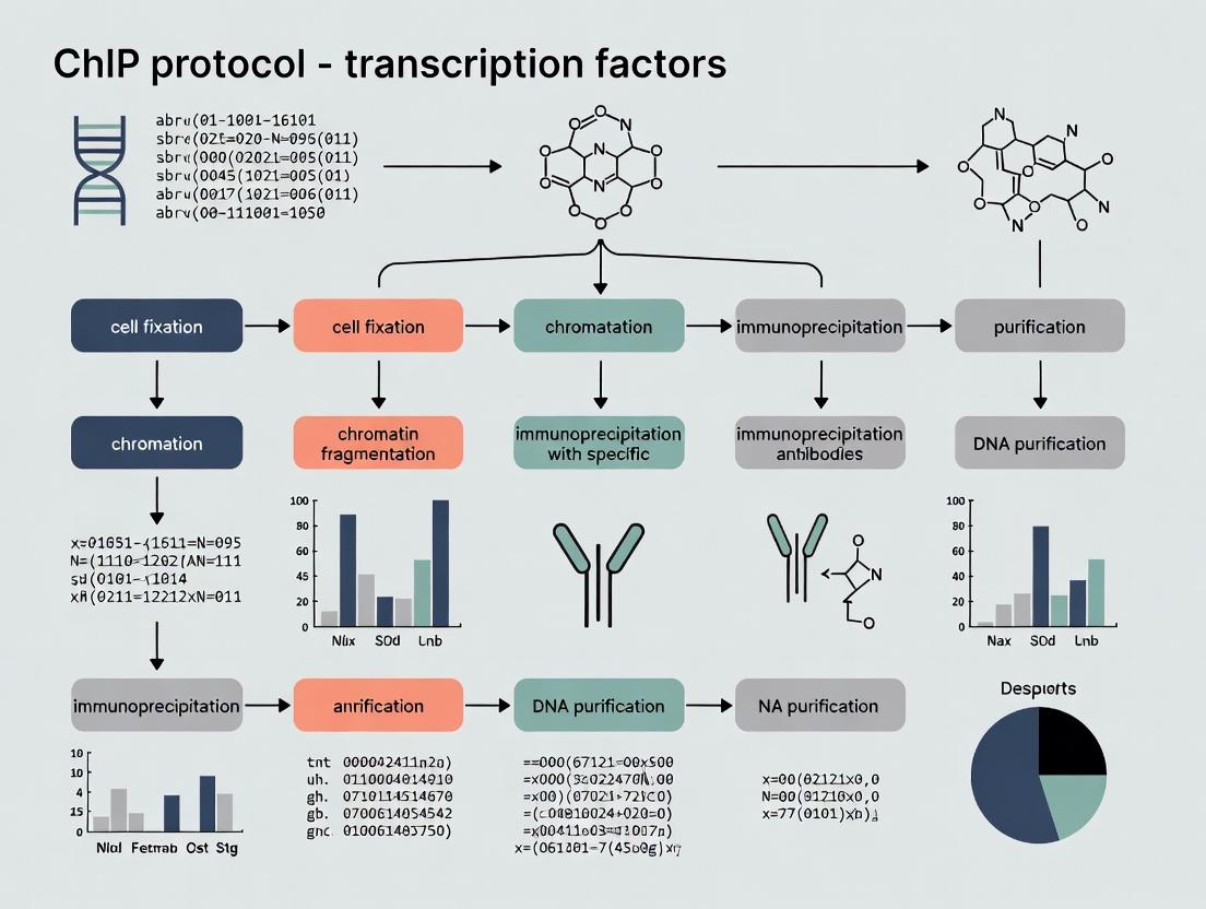
Visualizing Key Concepts
TF-ChIP Experimental Workflow
TF Activation and DNA Binding Pathway
The Chromatin Immunoprecipitation (ChIP) assay is a cornerstone technique for mapping in vivo protein-DNA interactions, particularly for transcription factors (TFs). Within the broader thesis of standardizing and optimizing ChIP for TFs, three components are paramount: selection of a high-specificity antibody, optimization of crosslinking conditions to capture transient TF-DNA interactions, and controlled shearing of chromatin to an ideal fragment size. This document provides detailed application notes and protocols to address these critical points, ensuring robust, reproducible data for research and drug development targeting transcriptional regulation.
Key Components: Quantitative Data and Application Notes
Antibody Selection and Validation
The specificity of the ChIP antibody is the single greatest determinant of success. Non-specific antibodies yield high background and false-positive signals.
Table 1: Antibody Selection Criteria for Transcription Factor ChIP
| Criterion | Recommended Standard | Quantitative Benchmark | Validation Protocol |
|---|---|---|---|
| Immunogen | Recombinant full-length protein or epitope-containing domain. | N/A | Check vendor datasheet. |
| Application Citation | Must list "ChIP" or "ChIP-seq" specifically. | ≥3 peer-reviewed publications using it for ChIP. | Literature search using PubMed. |
| Species Reactivity | Must match the model organism of the experiment. | N/A | Confirm via vendor specification. |
| Validation (Knockout/Down) | Loss of ChIP signal in KO/KD cells is gold standard. | ≥90% reduction in ChIP signal in KO control. | Perform ChIP-qPCR on a positive locus in WT vs. KO cell lines. |
| IgG Type | Prefer monoclonal for consistency; high-quality polyclonals are acceptable. | Lot-to-lot consistency data provided. | Compare new lot to old lot using a standard sample. |
Protocol: Antibody Validation via Knockout Cell Line
- Prepare Cells: Harvest wild-type (WT) and transcription factor knockout (KO) isogenic cell lines (e.g., generated via CRISPR-Cas9).
- Perform Parallel ChIP: Conduct ChIP for the target TF on both cell lines following the protocol in Section 3, using the same antibody lot.
- Quantitative PCR (qPCR): Analyze immunoprecipitated DNA with primers for a known, strong binding site (positive locus) and a non-binding genomic region (negative locus).
- Analysis: Calculate % Input for each sample. The KO signal at the positive locus should be reduced to near-background levels (comparable to the negative locus).
Crosslinking Optimization for Transcription Factors
Crosslinking captures transient TF-DNA interactions. Under-crosslinking leads to loss of signal; over-crosslinking masks epitopes and reduces shearing efficiency.
Table 2: Crosslinking Conditions for Common Transcription Factors
| TF Class / Stability | Recommended Fixative | Typical Concentration | Incubation Time & Temp | Key Consideration |
|---|---|---|---|---|
| Strong, Stable Binders (e.g., CTCF) | Formaldehyde (FA) | 1% | 10 min, RT | Standard condition; often sufficient. |
| Weak/Transient Binders (e.g., NF-κB, GR) | Formaldehyde (FA) | 1% | 15-20 min, RT OR Dual-crosslink with EGS/DSG | Longer FA or dual-crosslink enhances capture. |
| Pioneer Factors (e.g., FOXA1) | Dual: DSG + FA | 2 mM DSG (45 min), then 1% FA (15 min) | 45 min (DSG), then 15 min (FA), RT | DSG, a reversible amine crosslinker, improves efficiency for challenging TFs. |
| General Starting Point | Formaldehyde (FA) | 1% | 12 min, RT | Optimize around this point via time course. |
Protocol: Crosslinking Time-Course Optimization
- Cell Preparation: Grow cells in 15-cm dishes to 80-90% confluence. Prepare 1% formaldehyde solution in culture medium pre-warmed to 37°C.
- Variable Crosslinking: For each dish, add formaldehyde directly to medium. Incubate at room temperature with gentle shaking for 5, 10, 15, and 20 minutes.
- Quenching: Add glycine to a final concentration of 125 mM and incubate for 5 min.
- Harvest & Wash: Wash cells twice with ice-cold PBS.
- Parallel Processing: Lyse cells and shear chromatin from all time points identically (see Section 2.3).
- Analysis: Perform parallel ChIP-qPCR for a positive binding locus. The time point yielding the highest signal-to-noise ratio is optimal.
Chromatin Shearing for Transcription Factors
Shearing must fragment chromatin to 200-500 bp to achieve sufficient resolution while preserving the TF-DNA complex. Sonication is most common.
Table 3: Chromatin Shearing Parameters and Outcomes
| Shearing Method | Optimal Fragment Size (bp) | Typical Settings (Covaris S2) | Critical Quality Control Step |
|---|---|---|---|
| Sonication (Covaris) | 200-500 (peak ~300) | Duty Cycle: 10%, Intensity: 5, Cycles/Burst: 200, Time: 10-15 min (varies by cell type). | Bioanalyzer/TapeStation analysis post-reversal. |
| Sonication (Bioruptor) | 200-1000 (broader distribution) | 30 sec ON / 30 sec OFF, 10-15 cycles, High power setting, 4°C water bath. | Agarose gel electrophoresis. |
| Enzymatic (MNase) | Mainly mononucleosomes (~147 bp + linker). | Titration required; typically 0.5-5 units per 10^6 cells, 37°C, 5-20 min. | Less ideal for TFs as it may displace some factors. |
Protocol: Shearing Optimization and QC with a Covaris S2
- Prepare Lysate: After crosslinking and lysis (from 5-10 million cells), pellet nuclei. Resuspend in 1 mL of shearing buffer (1% SDS, 10 mM EDTA, 50 mM Tris-HCl, pH 8.1 with protease inhibitors).
- Shearing Setup: Transfer lysate to a Covaris microTUBE. Place in the S2 filled with degassed, chilled water.
- Initial Test Run: Use the settings in Table 3 as a start. Run for 8 minutes.
- Test Fragment Size: Reverse crosslinks for a 50 µL aliquot (65°C overnight with 200 mM NaCl), then purify DNA with a PCR purification kit. Analyze 1 µL on a High Sensitivity DNA Bioanalyzer chip or by agarose gel.
- Iterate: If fragments are too large (>500 bp), increase time or intensity incrementally. If fragments are too small (<200 bp), decrease time or intensity.
- Finalize: Once optimal conditions are found, use them consistently for all experiments.
Integrated ChIP Protocol for Transcription Factors
Materials: See "The Scientist's Toolkit" below.
Day 1: Crosslinking, Lysis, and Shearing
- Crosslink: Adherent cells (one 15-cm dish) are crosslinked per optimized conditions (e.g., 1% formaldehyde, 12 min, RT). Quench with 125 mM glycine.
- Harvest: Wash cells 2x with ice-cold PBS. Scrape into PBS with protease inhibitors, pellet.
- Lyse: Resuspend pellet in 1 mL Cell Lysis Buffer, incubate 10 min on ice. Pellet nuclei.
- Nuclear Lysis: Resuspend nuclei in 1 mL Nuclear Lysis Buffer, incubate 10 min on ice.
- Shear Chromatin: Sonicate using optimized Covaris/Bioruptor settings. Pellet debris (20,000 x g, 10 min, 4°C). Transfer supernatant (sheared chromatin) to a new tube. Take a 50 µL aliquot as "Input" and store at -20°C.
- Dilution & Pre-clear: Dilute sheared chromatin 10-fold with ChIP Dilution Buffer. Add 50 µL of Protein A/G beads (blocked with BSA/sheared salmon sperm DNA). Rotate for 1-2 hr at 4°C. Pellet beads, keep supernatant.
Day 2: Immunoprecipitation and Washes
- IP: Split chromatin into two tubes: one for Specific Antibody (2-5 µg), one for Species-Matched IgG control. Rotate overnight at 4°C.
- Capture: Add 60 µL blocked Protein A/G beads. Rotate for 2 hr at 4°C.
- Wash: Pellet beads and perform sequential 5-min washes on a rotator at 4°C:
- Wash once with Low Salt Immune Complex Wash Buffer.
- Wash once with High Salt Immune Complex Wash Buffer.
- Wash once with LiCl Immune Complex Wash Buffer.
- Wash twice with TE Buffer.
- Elute: Prepare fresh Elution Buffer (1% SDS, 100 mM NaHCO3). Add 250 µL to beads and input sample. Vortex, incubate 15 min at RT with agitation. Pellet beads, transfer supernatant. Repeat elution, combine eluates (~500 µL total).
Day 3: Reverse Crosslinks and DNA Purification
- Reverse Crosslinks: Add 20 µL of 5 M NaCl to each eluate and input. Heat at 65°C overnight.
- Digest Proteins: Add 10 µL of 0.5 M EDTA, 20 µL of 1 M Tris-HCl (pH 6.5), and 2 µL of Proteinase K (20 mg/mL). Incubate at 45°C for 2 hr.
- Purify DNA: Use a PCR purification kit. Elute DNA in 30-50 µL of EB buffer or nuclease-free water.
- Analysis: Proceed to qPCR (primers for positive and negative control loci) or library preparation for sequencing.
Diagrams of Workflows and Relationships
Title: ChIP-seq Workflow for Transcription Factors
Title: Core ChIP Component Interdependence
The Scientist's Toolkit: Essential Research Reagents and Materials
Table 4: Key Reagent Solutions for TF ChIP
| Reagent/Material | Function & Purpose | Example/Notes |
|---|---|---|
| Formaldehyde (37%) | Primary crosslinker; creates protein-DNA and protein-protein bridges. | Use molecular biology grade. Prepare 1% solution in medium/PBS fresh. |
| Disuccinimidyl Glutarate (DSG) | Amine-reactive reversible crosslinker; used for dual-crosslinking of challenging TFs. | Prepare fresh in DMSO. Use prior to FA crosslinking. |
| Protease Inhibitor Cocktail | Prevents degradation of TFs and chromatin during processing. | Use EDTA-free if subsequent steps require divalent cations. |
| Protein A/G Magnetic Beads | Efficient capture of antibody-bound complexes; easier washing than agarose beads. | Pre-block with BSA and sheared salmon sperm DNA to reduce non-specific binding. |
| TF-specific Validated Antibody | Specifically immunoprecipitates the target transcription factor. | Must be validated for ChIP (see Table 1). Critical investment. |
| Control IgG | Species/isotype-matched non-specific antibody for negative control IP. | Essential for determining background signal. |
| Covaris microTUBE | Specific tube for focused ultrasonication; ensures consistent shearing. | AFA fiber ensures correct energy transfer. |
| DNA HS Bioanalyzer Kit | High-sensitivity analysis of sheared chromatin fragment size distribution. | Chip-based electrophoresis; superior to agarose gels for QC. |
| ChIP-Seq Library Prep Kit | Prepares immunoprecipitated DNA for next-generation sequencing. | Select kits optimized for low-input DNA. |
| qPCR Primers | Validate ChIP efficiency at known binding (positive) and non-binding (negative) loci. | Design amplicons 80-150 bp within known binding sites. |
1. Introduction: Within the Context of Transcription Factor ChIP Research Chromatin Immunoprecipitation (ChIP) is the definitive method for mapping protein-DNA interactions in vivo. Within a broader thesis on ChIP protocol development for transcription factors (TFs), rigorous experimental design is paramount. This document outlines the framework for formulating a testable hypothesis and selecting the appropriate biological and methodological system, complete with application notes and detailed protocols.
2. Formulating a Testable Hypothesis A valid hypothesis in TF-ChIP research must be specific, measurable, and grounded in preliminary data.
- Core Structure: "If [MANIPULATION] of [INDEPENDENT VARIABLE] is performed in [BIOLOGICAL SYSTEM], then [MEASURABLE CHANGE] in [DEPENDENT VARIABLE] will be observed, due to [MECHANISTIC RATIONALE]."
- Example: "If TNF-α stimulation (manipulation) of NF-κB p65 (independent variable) is performed in primary human umbilical vein endothelial cells (HUVECs, system), then a ≥2-fold increase (measurable change) in p65 occupancy at the ICAM1 promoter (dependent variable) will be detected by qPCR-ChIP, due to stimulus-induced nuclear translocation and DNA binding (rationale)."
- Falsifiability: The hypothesis must allow for an experiment whose outcome could disprove it.
3. Choosing the Right System: Critical Considerations The choice of system dictates the validity and relevance of ChIP outcomes.
Table 1: Quantitative Comparison of Model Systems for TF-ChIP
| System | Typical TF ChIP-qPCR Signal (Fold over IgG) | Endogenous Tagging Feasibility | Genetic Manipulation Ease | Physiological Relevance | Key Limitations |
|---|---|---|---|---|---|
| Immortalized Cell Lines (e.g., HEK293) | 10-50 | Low | High (transfection) | Moderate | Aneuploidy, adapted phenotype |
| Primary Cells (e.g., HUVECs) | 5-20 | Very Low | Very Low | High | Finite lifespan, donor variability |
| Cancer Cell Lines (e.g., MCF-7) | Variable (5-100) | Low | Moderate | Context-specific | Genomic instability, high background |
| Engineered Cell Lines (e.g., CRISPR/dCas9-FP fusions) | 50-200 (via epitope tag) | High (via knock-in) | High | Can be high | Engineering artifacts, clonal variation |
| Murine Tissue (e.g., liver homogenate) | 3-15 | Possible (transgenic) | Low (in vivo) | Very High | Cellular heterogeneity, fixation challenges |
4. Featured Protocol: Optimized Crosslinking ChIP for a Nuclear Transcription Factor This protocol is designed for a hypothesis testing NF-κB p65 binding in TNF-α stimulated adherent cells.
A. Reagents & Materials: The Scientist's Toolkit Table 2: Essential Research Reagent Solutions
| Item | Function & Critical Detail |
|---|---|
| 37% Formaldehyde | Crosslinks proteins to DNA; quality is critical. Use fresh, methanol-free. |
| 2.5M Glycine | Quenches formaldehyde to stop crosslinking. |
| ChIP-Validated Antibody | Must be validated for IP; check target specificity (knockout/knockdown controls). |
| Protein G Magnetic Beads | Bind antibody-antigen complex; magnetic separation minimizes background. |
| Cell Lysis Buffer (10 mM HEPES pH 7.9, 85 mM KCl, 1% NP-40, protease inhibitors) | Lyses plasma membrane, isolates intact nuclei. |
| Nuclear Lysis/Sonication Buffer (50 mM Tris-HCl pH 8.0, 10 mM EDTA, 1% SDS, protease inhibitors) | Lyses nuclei and prepares chromatin for fragmentation. |
| Covaris S220 Focused-Ultrasonicator | Provides consistent, tunable shearing to desired fragment size (200-500 bp). |
| ChIP Elution Buffer (1% SDS, 0.1M NaHCO3) | Reverses crosslinks and elutes protein-DNA complexes from beads. |
| RNAse A & Proteinase K | Digest RNA and protein post-elution for clean DNA recovery. |
| qPCR Primers | Target positive control site (known binding), negative control site (non-bound genomic region), and test sites. |
B. Step-by-Step Workflow
Cell Stimulation & Crosslinking:
- Culture 2-4 x 10^6 cells per condition/IP.
- Stimulate with TNF-α (e.g., 10 ng/mL, 30 min). Include an unstimulated control.
- Add 37% formaldehyde directly to culture medium to 1% final concentration. Incubate 10 min at room temperature with gentle agitation.
- Quench with 2.5M glycine to 0.125M final concentration. Incubate 5 min at RT.
- Wash cells 2x with ice-cold PBS. Pellet cells and freeze at -80°C or proceed.
Chromatin Preparation & Shearing:
- Resuspend cell pellet in 1 mL Cell Lysis Buffer. Incubate 15 min on ice.
- Pellet nuclei (5,000 x g, 5 min, 4°C). Discard supernatant.
- Resuspend nuclei in 1 mL Nuclear Lysis/Sonication Buffer. Incubate 10 min on ice.
- Critical Step – Sonication: Transfer lysate to a Covaris milliTUBE. Shear using focused ultrasonication (e.g., Covaris S220: Peak Intensity 140, Duty Factor 5%, Cycles/Burst 200, Time 180 seconds). Goal: fragment size of 200-500 bp.
- Centrifuge sheared lysate (16,000 x g, 10 min, 4°C). Transfer supernatant (chromatin) to a new tube.
Immunoprecipitation:
- Pre-clear chromatin with Protein G beads for 1 hour at 4°C.
- Take 1% input sample and store at -20°C.
- Incubate pre-cleared chromatin with 1-5 µg of ChIP-validated anti-p65 antibody (or species-matched IgG control) overnight at 4°C with rotation.
- Add pre-washed Protein G magnetic beads. Incubate 2 hours at 4°C.
- Wash beads sequentially (with rotation, 5 min per wash, 4°C):
- 2x with Low Salt Wash Buffer
- 1x with High Salt Wash Buffer
- 1x with LiCl Wash Buffer
- 2x with TE Buffer
Elution & DNA Recovery:
- Elute complexes from beads in 150 µL ChIP Elution Buffer by incubating at 65°C for 30 min with gentle shaking. Collect supernatant.
- Reverse crosslinks by adding 5 µL of 5M NaCl and incubating at 65°C overnight.
- Add 2 µL RNAse A (10 mg/mL), incubate 30 min at 37°C.
- Add 2 µL Proteinase K (20 mg/mL), incubate 2 hours at 55°C.
- Purify DNA using a silica-membrane spin column. Elute in 30-50 µL elution buffer.
Analysis – Quantitative PCR:
- Analyze 1-2 µL of purified DNA by qPCR using SYBR Green.
- Primers: Include known positive control (e.g., ICAM1 promoter), negative control (gene desert), and test loci.
- Calculation: Calculate % Input for each sample: % Input = 2^(Ct[Input] - Ct[IP]) x 100. Enrichment is reported as Fold over IgG control: (% Input IP / % Input IgG).
5. Mandatory Visualizations
Title: Hypothesis-Driven ChIP Experimental Design Flow
Title: NF-κB Signaling Pathway Leading to ChIP Detection
Title: Step-by-Step Chromatin Immunoprecipitation Workflow
This document provides detailed application notes and protocols for Chromatin Immunoprecipitation (ChIP), framed within a broader thesis investigating transcription factor dynamics in gene regulation. The reproducibility and precision of ChIP are paramount for generating high-quality data that can inform mechanistic models in basic research and identify novel therapeutic targets in drug development.
Crosslinking: Capturing Protein-DNA Interactions
Crosslinking covalently stabilizes transient transcription factor-DNA interactions. Formaldehyde is the predominant reagent due to its reversible, short-range crosslinks.
Protocol: Formaldehyde Crosslinking for Adherent Cells
- Grow cells to 70-80% confluence in a 15 cm dish.
- Add 1/10 volume of fresh 37% formaldehyde directly to the culture medium to a final concentration of 1%. Mix gently.
- Incubate at room temperature for 10 minutes with gentle rocking.
- Quench the reaction by adding glycine to a final concentration of 0.125 M. Rock for 5 minutes.
- Aspirate medium, wash cells twice with ice-cold PBS.
- Scrape cells into PBS with protease inhibitors. Pellet cells (800 x g, 5 min, 4°C) and proceed to lysis or flash-freeze pellet at -80°C.
Critical Consideration: Over-crosslinking (e.g., >15 min or using >1% formaldehyde) can mask epitopes and reduce sonication efficiency, compromising IP success.
Chromatin Preparation and Sonication
Cells are lysed, and chromatin is sheared to fragments of 200-1000 bp, optimizing resolution and antibody accessibility.
Protocol: Cell Lysis and Sonication
- Resuspend cell pellet in 1 mL Cell Lysis Buffer (10 mM Tris-HCl pH 8.0, 10 mM NaCl, 0.2% NP-40/Igepal, plus protease inhibitors). Incubate on ice for 15 min. Spin (2000 x g, 5 min, 4°C). Discard supernatant (cytoplasmic fraction).
- Resuspend nuclear pellet in 1 mL Nuclei Lysis/Sonication Buffer (50 mM Tris-HCl pH 8.0, 10 mM EDTA, 1% SDS, plus protease inhibitors). Incubate on ice for 10 min.
- Sonication: Transfer lysate to a microtube. Using a focused ultrasonicator (e.g., Covaris M220, Qsonica), shear chromatin. A typical program for 1 mL in a Covaris milliTUBE is:
- Peak Incident Power: 75W
- Duty Factor: 10%
- Cycles per Burst: 200
- Treatment Time: 7-12 minutes (must be optimized per cell type).
- Pellet debris (16,000 x g, 10 min, 4°C). Transfer supernatant (sheared chromatin) to a new tube. Keep a 50 µL aliquot as "Input" control.
Table 1: Sonication Optimization Parameters and Outcomes
| Cell Type | Sonication Instrument | Optimal Time | Average Fragment Size | Key Note |
|---|---|---|---|---|
| HEK293 (Adherent) | Covaris M220 | 8 min | 250-500 bp | Consistent, low heat generation. |
| Jurkat (Suspension) | Bioruptor Pico | 6 cycles (30s ON/30s OFF) | 300-600 bp | Water bath system; keep ice-water full. |
| Mouse Tissue | Q800R3 Sonicator | 4 x 15s pulses, 50% amplitude | 400-1000 bp | Use large tip; cool extensively between pulses. |
Immunoprecipitation (IP)
Sheared chromatin is incubated with a validated antibody specific to the transcription factor of interest to immunoprecipitate the protein-DNA complex.
Protocol: Magnetic Bead-Based IP
- Pre-clear & Dilution: Dilute sheared chromatin 10-fold in ChIP Dilution Buffer (16.7 mM Tris-HCl pH 8.0, 167 mM NaCl, 1.2 mM EDTA, 1.1% Triton X-100, 0.01% SDS). Add 20 µL of pre-washed Protein A/G magnetic beads per sample. Rotate for 1 hour at 4°C. Magnetize and transfer supernatant to a new tube.
- Antibody Incubation: Add the specific antibody to the pre-cleared chromatin. Use 1-5 µg of antibody per 25-50 µg of chromatin. For a negative control, use species-matched IgG. Rotate overnight at 4°C.
- Bead Capture: The next day, add 30 µL of pre-washed Protein A/G magnetic beads. Rotate for 2 hours at 4°C.
- Washes: Place tube on a magnet. Discard supernatant. Perform sequential 5-minute rotations with cold wash buffers:
- Low Salt Wash Buffer: (20 mM Tris-HCl pH 8.0, 150 mM NaCl, 2 mM EDTA, 1% Triton X-100, 0.1% SDS).
- High Salt Wash Buffer: (20 mM Tris-HCl pH 8.0, 500 mM NaCl, 2 mM EDTA, 1% Triton X-100, 0.1% SDS).
- LiCl Wash Buffer: (10 mM Tris-HCl pH 8.0, 250 mM LiCl, 1 mM EDTA, 1% NP-40, 1% Sodium Deoxycholate).
- TE Buffer: (10 mM Tris-HCl pH 8.0, 1 mM EDTA). Perform twice.
Elution, Reversal, and Analysis
Crosslinks are reversed, proteins are digested, and DNA is purified for quantitative analysis.
Protocol: Elution and DNA Purification
- Elution: Add 150 µL of Fresh Elution Buffer (100 mM NaHCO₃, 1% SDS) to beads. Vortex and incubate at 65°C for 15 minutes with shaking. Magnetize, transfer eluate to new tube. Repeat elution and combine eluates (~300 µL total).
- Reverse Crosslinks & Purify: Add 12 µL of 5M NaCl to eluate and Input sample. Incubate at 65°C overnight. Add 10 µL of 0.5M EDTA, 20 µL of 1M Tris-HCl pH 6.5, and 2 µL of Proteinase K (20 mg/mL). Incubate at 45°C for 2 hours.
- Purify DNA using a silica-membrane PCR purification kit. Elute in 30-50 µL of nuclease-free water or TE buffer.
Analysis: Purified DNA is analyzed via qPCR (for candidate regions) or next-generation sequencing (ChIP-seq) for genome-wide mapping. Data is normalized to Input and expressed as %Input or Fold Enrichment over IgG control.
The Scientist's Toolkit: Key Reagent Solutions
Table 2: Essential Materials for ChIP Experiments
| Reagent/Material | Function & Critical Notes |
|---|---|
| 37% Formaldehyde | Crosslinking agent. Must be fresh (<3 months old) for efficient, reversible crosslinking. |
| Protease Inhibitor Cocktail (PIC) | Prevents degradation of transcription factors and chromatin during preparation. Add fresh to all buffers. |
| Magnetic Beads (Protein A/G) | Solid support for antibody-antigen capture. More consistent and easier to handle than agarose beads. |
| Validated ChIP-Grade Antibody | The most critical reagent. Must be validated for immunoprecipitation of crosslinked chromatin. |
| Sodium Dodecyl Sulfate (SDS) | Denaturing detergent in lysis/sonication buffer; aids in chromatin shearing but must be diluted for IP. |
| Covaris milliTUBE | AFA-fiber tubes designed for focused ultrasonication, ensuring consistent and efficient shearing. |
| RNA Polymerase II Antibody (Positive Control) | Control antibody for successful workflow in every experiment, as Pol II is universally present. |
| PCR Purification Kit | For efficient recovery of low-abundance ChIP DNA. Low-elution-volume kits increase final DNA concentration. |
Visualization: ChIP Experimental Workflow
Diagram Title: Step-by-step ChIP protocol workflow for transcription factor mapping.
Diagram Title: ChIP data analysis pathway from samples to results.
Application Notes
This protocol is designed for the precise mapping of transcription factor (TF) binding sites and the study of associated chromatin dynamics. It is integral to a broader thesis investigating TF-driven gene regulatory networks in disease models, with direct implications for identifying novel therapeutic targets.
Application 1: Mapping Binding Sites with High Resolution Chromatin Immunoprecipitation (ChIP) followed by high-throughput sequencing (ChIP-seq) remains the gold standard for genome-wide TF binding site identification. Recent advancements in library preparation and sequencing depth allow for single-nucleotide resolution mapping when paired with appropriate peak-calling algorithms.
Application 2: Studying Protein-DNA Dynamics Combining ChIP with kinetic assays or sequential ChIP (Re-ChIP) enables the study of TF binding dynamics, co-occupancy, and turnover in response to stimuli. This is critical for understanding transient regulatory events.
Application 3: Investigating Epigenetic Regulation TF binding is intimately linked with chromatin state. Integrative analysis of ChIP-seq data for TFs alongside histone modifications (e.g., H3K27ac, H3K4me3) and chromatin accessibility assays (e.g., ATAC-seq) elucidates the epigenetic framework of gene regulation.
Quantitative Data Summary (Typical ChIP-seq Experiment Output)
Table 1: Key Sequencing and Analysis Metrics
| Metric | Target Value | Purpose |
|---|---|---|
| Sequencing Depth | 20-40 million reads (mammalian genome) | Ensures sufficient coverage for peak calling. |
| Percentage of Reads in Peaks (FRiP) | >1% (TF ChIP), >5% (Histone ChIP) | Primary indicator of ChIP enrichment success. |
| Peak Number | Varies by TF (1,000 - 50,000) | Reflects TF specificity and cellular context. |
| Peak Width (TF) | 100 - 500 bp | Defines binding region resolution. |
| Replicate Correlation (Pearson's R) | R > 0.9 | Indicates high reproducibility between biological replicates. |
Detailed Protocols
Protocol 1: Standard ChIP-seq for Transcription Factors Materials: Formaldehyde, Glycine, Cell Lysis Buffer, Sonication Device, Protein A/G Magnetic Beads, Target-specific TF Antibody, DNA Clean-up Kit, Library Prep Kit, High-fidelity DNA Polymerase.
Method:
- Crosslinking: Treat cells with 1% formaldehyde for 10 min at room temperature. Quench with 125mM glycine.
- Cell Lysis & Chromatin Shearing: Lyse cells. Isolate nuclei and shear chromatin via sonication to 200-500 bp fragments. Verify fragment size by agarose gel electrophoresis.
- Immunoprecipitation: Pre-clear lysate with beads. Incubate with antibody against target TF overnight at 4°C. Add beads, incubate, and wash stringently.
- Elution & Decrosslinking: Elute chromatin in Elution Buffer (1% SDS, 100mM NaHCO3). Add NaCl and incubate at 65°C overnight to reverse crosslinks.
- DNA Purification: Treat with Proteinase K and RNase A. Purify DNA using a spin column.
- Library Preparation & Sequencing: Prepare sequencing library from enriched DNA (end-repair, A-tailing, adapter ligation, PCR amplification). Perform QC and sequence on an appropriate platform.
Protocol 2: Sequential ChIP (Re-ChIP) for Co-occupancy Materials: As for Protocol 1, with two distinct antibodies.
Method:
- Perform first ChIP as in Protocol 1, steps 1-4.
- First Elution: Elute bound complexes from the beads using 10mM DTT at 37°C for 30 min.
- Second Immunoprecipitation: Dilute eluate 1:50 with ChIP Dilution Buffer. Perform a second ChIP with an antibody against a different TF or chromatin mark.
- Proceed with decrosslinking and purification (Protocol 1, steps 4-5). Analyze by qPCR or sequencing.
Visualizations
Title: Standard ChIP-seq Experimental Workflow
Title: TF Binding Drives Epigenetic Regulation & Expression
The Scientist's Toolkit: Key Research Reagent Solutions
Table 2: Essential Materials for ChIP Experiments
| Reagent/Material | Function & Importance |
|---|---|
| High-Specificity, ChIP-Validated Antibody | The critical reagent for specific immunoprecipitation. Validated for use in ChIP ensures success. |
| Protein A/G Magnetic Beads | Facilitate efficient antibody-antigen complex capture and separation during washes. |
| Controlled Sonication System | Ensures consistent and optimal chromatin fragmentation, crucial for resolution and sensitivity. |
| Crosslinking Reagents (Formaldehyde, DSG) | Preserves transient protein-DNA interactions in vivo. |
| ChIP-seq Grade Library Prep Kit | Optimized for converting low-input, sheared chromatin DNA into sequencing libraries. |
| SPRI Beads | For precise size selection and clean-up of DNA fragments during library prep. |
| qPCR Primers for Positive/Negative Loci | Essential for quantitative validation of ChIP enrichment prior to sequencing. |
Step-by-Step Optimized ChIP Protocol for Transcription Factors: From Cells to Sequencing
Within the broader thesis investigating Chromatin Immunoprecipitation (ChIP) protocols for transcription factor research, the initial phase of cell fixation is critical. Transcription factors (TFs) often exhibit transient or weak chromatin interactions, making crosslinking optimization paramount for capturing authentic in vivo binding events. This application note details protocols comparing standard formaldehyde fixation with a dual crosslinker approach, focusing on cell culture, crosslinking optimization, and harvesting.
Comparative Analysis of Crosslinking Strategies
Table 1: Quantitative Comparison of Formaldehyde vs. Dual Crosslinker Fixation
| Parameter | Formaldehyde (FA) Only | Dual Crosslinker (FA + EGS/DSP) |
|---|---|---|
| Primary Target | Protein-DNA, Protein-Protein (short-range) | Protein-DNA (FA) + Protein-Protein (long-range, EGS/DSP) |
| Crosslink Reversibility | Reversible with heat | EGS/DSP: Reversible with DTT. FA: Reversible with heat. |
| Optimal Conc. & Time | 1% FA, 8-10 min at RT | 1% FA, 8-10 min, then 1-2 mM EGS, 30-45 min |
| Chromatin Shearability | Generally good | Can be more challenging; requires optimization |
| Best For | Strong, stable TF-DNA interactions | Fragile TFs, complexes distal from DNA, histone modifications |
| Key Drawback | May miss weak or indirect interactions | Increased background, more complex reversal |
| Typical TF Yield (vs. Input) | Variable; 0.5-5% for stable TFs | Can increase yield 2-5 fold for difficult TFs |
Table 2: Harvesting & Lysis Buffer Formulations
| Buffer Component | Standard FA Lysis Buffer | Dual X-Link Lysis Buffer | Function |
|---|---|---|---|
| SDS | 0.1% | 0.3-0.5% | Denatures proteins, aids lysis |
| Triton X-100 | 1% | 1% | Solubilizes membranes |
| Sodium Deoxycholate | 0.1% | 0.1% | Disrupts membranes |
| Tris-HCl (pH 8.0) | 50 mM | 50 mM | Buffer capacity |
| NaCl | 150 mM | 150 mM | Controls ionic strength |
| EDTA | 1 mM | 2-5 mM | Chelates Mg2+, inhibits nucleases |
| Protease Inhibitors | Yes (1x) | Yes (2x) | Prevents protein degradation |
Detailed Experimental Protocols
Protocol 1: Cell Culture and Formaldehyde Crosslinking
Materials: Adherent or suspension cells, growth medium, 37% formaldehyde, 2.5M glycine, PBS, cell scrapers. Procedure:
- Culture: Grow cells to 70-80% confluence in appropriate medium.
- Crosslink: Add 37% formaldehyde directly to culture medium to a final concentration of 1%. Mix gently. Incubate for 8-10 minutes at room temperature with gentle agitation.
- Quench: Add 2.5M glycine to a final concentration of 0.125M. Incubate for 5 minutes at room temperature.
- Harvest: For adherent cells, aspirate medium, wash twice with cold PBS, and scrape into cold PBS. Pellet cells at 800 x g for 5 min at 4°C.
- Storage: Flash-freeze cell pellet in liquid nitrogen. Store at -80°C or proceed to lysis.
Protocol 2: Dual Crosslinking with Formaldehyde and EGS
Materials: As in Protocol 1, plus Ethylene glycol bis(succinimidyl succinate) (EGS) dissolved in DMSO, PBS. Procedure:
- Culture & Formaldehyde Fixation: Perform steps 1-3 from Protocol 1.
- EGS Crosslinking: Wash cells once with cold PBS. Resuspend cell pellet in PBS. Add EGS from a fresh stock to a final concentration of 1-2 mM. Incubate for 30-45 minutes at room temperature with gentle agitation.
- Quench EGS: Add Tris-HCl (pH 7.5) to a final concentration of 10 mM and incubate for 5 minutes.
- Harvest & Storage: Wash cells twice with cold PBS. Pellet and flash-freeze as in Protocol 1.
Protocol 3: Cell Lysis and Chromatin Preparation
Materials: Lysis Buffer (see Table 2), protease inhibitors, sonicator. Procedure:
- Thaw & Lyse: Thaw cell pellet on ice. Resuspend in 1 mL of appropriate cold Lysis Buffer per 10^7 cells. Incubate on ice for 10-15 minutes.
- Pellet Nuclei: Centrifuge at 2000 x g for 5 minutes at 4°C. Discard supernatant.
- Shear Chromatin: Resuspend nuclear pellet in 0.5-1 mL Lysis Buffer. Sonicate on ice to achieve DNA fragments of 200-1000 bp. Optimize conditions (power, time, pulses).
- Clarify Lysate: Centrifuge at 16,000 x g for 10 minutes at 4°C. Transfer supernatant (chromatin lysate) to a new tube. Proceed to ChIP or store at -80°C.
Visualizations
Title: Cell Fixation and Harvesting Workflow
Title: Crosslinking Mechanism: FA vs. Dual
The Scientist's Toolkit: Research Reagent Solutions
Table 3: Essential Materials for Crosslinking and Harvesting
| Item | Function & Rationale | Example/Catalog Consideration |
|---|---|---|
| Formaldehyde (37%), Molecular Biology Grade | Primary crosslinker for protein-nucleic acid and proximal protein-protein interactions. High purity minimizes side reactions. | Thermo Fisher Scientific (28906) or Sigma-Aldrich (F8775). |
| Ethylene Glycol Bis(succinimidyl succinate) (EGS) | Homobifunctional, amine-reactive, reversible crosslinker. Stabilizes protein complexes distal from DNA. | Thermo Fisher Scientific (21565). Prepare fresh in DMSO. |
| Dithiothreitol (DTT) | Reduces disulfide bonds in EGS, reversing protein-protein crosslinks after immunoprecipitation. | Included in most elution buffers. |
| Complete Protease Inhibitor Cocktail | Prevents proteolytic degradation of transcription factors and complexes during lysis. | Roche (11836170001) or equivalent EDTA-free version for Mg2+-dependent processes. |
| Glycine (2.5M Solution) | Quenches formaldehyde crosslinking by reacting with excess aldehydes, preventing over-crosslinking. | Sterile-filtered stock solution. |
| Cell Scrapers (Sterile) | For gentle detachment of adherent crosslinked cells without disrupting nuclei. | Corning (3010) or similar, non-pyrogenic. |
| Covaris S-series Sonicator or equivalent | Provides consistent, controlled acoustic shearing of crosslinked chromatin to desired fragment size. | Covaris S220. Settings must be optimized per cell type and crosslink. |
| Bradford or BCA Assay Kit | Quantifies protein concentration in chromatin lysate to normalize input across samples. | Bio-Rad (5000001) or Pierce (23225). |
Within the broader thesis on optimizing Chromatin Immunoprecipitation (ChIP) for transcription factor research, Phase 2 is critical. The goal is to isolate and shear chromatin to an ideal size range of 200-500 base pairs (bp). This size range represents a single nucleosome plus associated linker DNA, ensuring that transcription factor binding sites remain in close proximity to the core histone particle for efficient immunoprecipitation. Inadequate shearing can lead to high background or loss of signal, compromising downstream sequencing or PCR analysis.
Key Parameters Influencing Sonication Efficiency
The shearing efficiency is influenced by multiple variables. The following table summarizes the key parameters and their optimal ranges based on current literature and protocols.
Table 1: Key Sonication Parameters and Optimal Ranges for Transcription Factor ChIP
| Parameter | Optimal Range/Type | Impact on Fragment Size |
|---|---|---|
| Cell Fixation | 1% Formaldehyde, 8-12 min | Under-fixation: poor cross-linking; Over-fixation: difficult shearing. |
| Lysis Buffer Ionic Strength | Low to Moderate (150-200 mM NaCl) | High salt can dissociate transcription factors; low salt aids nuclear integrity. |
| Covaris Duty Factor | 5-10% (for focused ultrasonicator) | Higher % increases shear force, reducing fragment size. |
| Covaris Peak Incident Power | 105-140 W | Higher power increases energy, reducing fragment size. |
| Covaris Cycles per Burst | 200-400 | More cycles per burst increase shear events per unit time. |
| Processing Time | 4-8 cycles of 30-60 sec (Bioruptor) | Total energy input; must be optimized empirically. |
| Sample Volume | 100-200 µL per tube (Covaris microTUBE) | Consistent volume ensures reproducible cavitation. |
| Sample Temperature | 2-6°C (maintained by chilled water bath or chiller) | Prevents sample heating and degradation. |
| Chromatin Concentration | 5-20 million cells per 100 µL sonication | Too dense: inefficient shearing; too dilute: low yield. |
Detailed Protocol: Chromatin Preparation and Sonication
A. Materials & Reagents (The Scientist's Toolkit)
Table 2: Research Reagent Solutions for Chromatin Preparation & Sonication
| Item | Function |
|---|---|
| Formaldehyde (37%) | Cross-links proteins (e.g., transcription factors) to DNA. |
| 2.5M Glycine | Quenches formaldehyde to stop cross-linking reaction. |
| Cell Lysis Buffer (10 mM Tris-HCl pH 8.0, 10 mM NaCl, 0.2% NP-40/Igepal) | Lyses cell membrane while leaving nuclei intact. |
| Nuclear Lysis Buffer (50 mM Tris-HCl pH 8.0, 10 mM EDTA, 1% SDS) | Lyses nuclear membrane and solubilizes cross-linked chromatin. |
| Protease Inhibitor Cocktail (PIC) | Prevents proteolytic degradation of proteins/chromatin. |
| PMSF (Phenylmethylsulfonyl fluoride) | Serine protease inhibitor, added fresh to buffers. |
| Covaris microTUBE AFA Fiber Screw-Cap | Specialized tube for consistent acoustic shearing. |
| Bioruptor Pico Sonication Device | Alternative water bath-based sonicator for shearing. |
| DynaMag-2 Magnet | For magnetic bead-based cleanup and size selection. |
| AMPure XP or SPRIselect Beads | Solid-phase reversible immobilization (SPRI) beads for DNA fragment size selection. |
| Tris-EDTA (TE) Buffer (10 mM Tris-HCl, 1 mM EDTA, pH 8.0) | Elution and storage buffer for sheared chromatin/DNA. |
| Agilent High Sensitivity DNA Kit | For analyzing fragment size distribution on a Bioanalyzer. |
B. Step-by-Step Methodology
Day 1: Cross-linking & Chromatin Preparation
- Cell Harvesting: Harvest approximately 1x10^7 cells per ChIP assay. Pellet cells by centrifugation.
- Cross-linking: Resuspend cell pellet in 10 mL of room temperature PBS. Add 270 µL of 37% formaldehyde (1% final concentration). Incubate for 10 minutes at room temperature with gentle rotation.
- Quenching: Add 1 mL of 2.5M glycine (0.125M final) to quench. Incubate for 5 minutes at room temperature with rotation.
- Washing: Pellet cells (4°C, 5 min, 800 x g). Wash twice with 10 mL of ice-cold PBS containing 1x PIC.
- Cell Lysis: Resuspend pellet in 1 mL of ice-cold Cell Lysis Buffer + 1x PIC. Incubate on ice for 15 minutes.
- Nuclear Pellet: Centrifuge (4°C, 5 min, 2000 x g). Discard supernatant. Pellet contains nuclei.
- Nuclear Lysis: Resuspend nuclear pellet in 500 µL of Nuclear Lysis Buffer + 1x PIC. Incubate on ice for 10 minutes. The lysate is now ready for sonication.
Day 1: Sonication (Using a Covaris S220/S2)
- Pre-cool: Ensure the water bath is at 5-6°C and degassed.
- Transfer: Aliquot 130 µL of the chromatin lysate into a Covaris microTUBE.
- Program Settings: Input the following parameters into the Covaris software:
- Peak Incident Power (W): 140
- Duty Factor: 5%
- Cycles per Burst: 200
- Treatment Time (seconds): 300 (5 minutes)
- Shearing: Place the microTUBE in the holder and start the run.
- Collection: Pool sheared samples if multiple tubes were used. Centrifuge briefly to collect droplets.
- Clarification: Centrifuge the sheared chromatin at 20,000 x g for 10 minutes at 4°C to pellet insoluble debris. Transfer the supernatant (sheared chromatin) to a new tube.
Day 1: Fragment Size Analysis & Cleanup
- Reverse Cross-linking (Test Sample): Take a 50 µL aliquot of sheared chromatin. Add 200 µL of TE buffer, 10 µL of 5M NaCl, and 2 µL of RNase A (10 mg/mL). Incubate at 65°C for 4-6 hours or overnight.
- DNA Purification: Purify the DNA using a QIAquick PCR Purification Kit. Elute in 30 µL of TE buffer.
- Fragment Analysis: Analyze 1 µL of the purified DNA on an Agilent Bioanalyzer using the High Sensitivity DNA chip.
- Interpretation: The bulk of the DNA smear should center between 200-500 bp (see Diagram 1). If fragments are too large, increase sonication time or power. If fragments are too small, reduce time or power.
- Chromatin Storage: The main sheared chromatin sample can be stored at -80°C until use for immunoprecipitation (Phase 3).
Visualizing the Process and Logic
Diagram 1: Chromatin Shearing Optimization Workflow
Diagram 2: Key Parameters for Ideal Fragment Size
Within the framework of a comprehensive ChIP protocol for transcription factor (TF) research, the selection and validation of an antibody for immunoprecipitation (IP) is the single most critical determinant of experimental success. A poorly characterized antibody can lead to false-positive signals, lack of specificity, and irreproducible data, undermining subsequent analyses. This application note provides a structured approach for choosing and rigorously validating TF antibodies for ChIP, ensuring the reliability of results in drug discovery and mechanistic studies.
Key Criteria for Antibody Selection
The following table summarizes the primary factors to consider when selecting an antibody for ChIP.
Table 1: Critical Selection Criteria for ChIP-Grade Transcription Factor Antibodies
| Criterion | Description & Rationale | Optimal Specification/Check |
|---|---|---|
| Immunogen | The specific peptide or protein fragment used to generate the antibody. | Antibody raised against the full-length protein or a known functional domain of the TF. Epitope should be accessible in crosslinked, sheared chromatin. |
| Host Species & Clonality | Determines compatibility with secondary reagents and consistency. | Monoclonal antibodies offer superior lot-to-lot consistency. Rabbit host is common for high-affinity monoclonal/polyclonal options. |
| Application Validation | Evidence provided by the vendor that the antibody works in ChIP. | Explicit "ChIP," "ChIP-seq," or "ChIP-grade" validation listed. Review published data in vendor's product sheet. |
| Species Reactivity | Confirms the antibody recognizes the TF in your experimental model system. | Must match your model organism (e.g., human, mouse, rat). Check for cross-reactivity if using non-standard models. |
| Validation in Knockout/Down Systems (Gold Standard) | Data showing loss of ChIP signal in cells where the target TF is absent. | Vendor or independent data showing abolished signal in TF knockout/knockdown cells is highly persuasive. |
| Citation Record | Peer-reviewed publications using the antibody for ChIP. | Multiple citations, preferably in reputable journals, using the same catalog number for ChIP. |
Validation Protocols for ChIP Antibodies
Relying solely on vendor claims is insufficient. In-house validation is mandatory. Below are detailed protocols for key validation experiments.
Protocol: Validation by Immunoblotting (Pre-ChIP Specificity Check)
Objective: Confirm antibody specificity and appropriate cross-reactivity in your cell lysate before proceeding to ChIP. Materials: Cell lysate, SDS-PAGE system, transfer apparatus, candidate antibody, appropriate controls. Procedure:
- Prepare Whole-Cell Lysates: Harvest cells of interest. Include a positive control (cells known to express the TF) and a negative control (TF knockout cells or cells known not to express the TF, if available).
- Perform SDS-PAGE and Western Blot: Resolve 20-50 µg of total protein per lane. Transfer to PVDF membrane.
- Immunoblot: Probe with the candidate TF antibody (following manufacturer's recommended dilution). Use appropriate loading control (e.g., Histone H3, GAPDH).
- Analysis: The antibody should detect a single band at the expected molecular weight for the TF. Signal should be absent or drastically reduced in the negative control lane. Non-specific bands indicate potential for off-target IP in ChIP.
Protocol: Knockout/Knockdown Validation (The Gold Standard)
Objective: Provide definitive evidence of antibody specificity by demonstrating loss of ChIP signal in the absence of the target TF. Materials: Isogenic wild-type and TF knockout cell lines, or reagents for RNAi/CRISPR-mediated knockdown. Procedure:
- Generate Paired Cell Lines: Create a TF knockout (KO) or stable knockdown (KD) line from your parental cell line using CRISPR-Cas9 or RNAi. Validate loss of TF protein by western blot.
- Perform Parallel ChIP Experiments: Conduct the full ChIP protocol simultaneously on the parental (WT) and TF KO/KD cells using the candidate antibody and an appropriate IgG control.
- Quantitative Analysis: Analyze enrichment at a known, high-affinity binding site for the TF by qPCR.
- Interpretation: Specific ChIP-qPCR signal should be present in WT cells and be abolished or significantly reduced (>70-80%) in the KO/KD cells. This confirms the antibody's specificity for the target TF in the ChIP context.
Protocol: Peptide Competition Assay
Objective: Confirm that the ChIP signal is specifically mediated by antibody binding to its intended epitope. Materials: Candidate antibody, immunizing peptide (or a scrambled control peptide), standard ChIP reagents. Procedure:
- Pre-block the Antibody: Prior to adding the antibody to the chromatin, incubate the typical amount of antibody with a 5-10X molar excess of the immunizing peptide in ChIP dilution buffer for 30-60 minutes on ice. A control sample is incubated with a scrambled peptide or buffer alone.
- Proceed with ChIP: Add the pre-blocked antibody mixture to the divided chromatin samples and complete the standard ChIP protocol.
- Analysis by qPCR: Enrichment at target sites in the peptide-blocked sample should be significantly reduced compared to the control sample, indicating competition for the antigen-binding site.
The Scientist's Toolkit: Research Reagent Solutions
Table 2: Essential Materials for Antibody Validation and ChIP
| Item | Function/Application | Example/Notes |
|---|---|---|
| ChIP-Validated Primary Antibody | Specific immunoprecipitation of the protein-DNA complex. | Target-specific (e.g., Anti-STAT3, Cat# ab12345). Must be validated for ChIP. |
| Species-Matched Normal IgG | Negative control for non-specific antibody binding. | Critical for background determination. Must match host species of primary antibody. |
| Protein A/G Magnetic Beads | Efficient capture of antibody-antigen complexes. | Preferred over agarose beads for reduced background and easier handling. |
| PCR/QPCR System | Quantification of enriched DNA fragments. | SYBR Green chemistry is standard for target validation. |
| Validated Positive Control Primers | Amplify a known, strong binding site for your TF. | Essential for validating the ChIP experiment itself. |
| Validated Negative Control Primers | Amplify a genomic region devoid of TF binding. | Typically in a gene desert or inactive promoter. Used to assess background. |
| TF Knockout Cell Line | Definitive specificity control for antibody validation. | Can be generated via CRISPR-Cas9 or obtained from commercial repositories. |
| Immunizing Peptide | For peptide competition assays. | Often available from the antibody manufacturer. |
Visualized Workflows and Relationships
Diagram 1: Antibody Validation Decision Workflow for ChIP
Diagram 2: Core IP Step in ChIP Protocol Workflow
Within the context of a broader thesis on Chromatin Immunoprecipitation (ChIP) for transcription factor research, Phase 4 is critical for achieving high signal-to-noise ratios. This phase directly impacts the specificity of the assay by removing non-specifically bound chromatin and efficiently recovering the target protein-DNA complexes. Insufficient washing leads to high background, while overly stringent washing can elute specific interactions. Subsequent elution and crosslink reversal must be complete to ensure optimal yield and integrity for downstream analysis (e.g., qPCR, sequencing). This protocol details optimized steps to maximize specificity and minimize background.
Key Principles for Minimizing Background
Background in ChIP originates from non-specific antibody binding, bead adherence of chromatin, and incomplete removal of reagents. Key strategies include:
- Stringent, Buffered Washes: Sequential use of buffers with increasing ionic strength and detergent composition.
- Temperature-Controlled Elution: Efficient release of complexes from beads under denaturing conditions.
- Complete Reversal of Crosslinks: Ensures DNA is free from proteins for purification.
- RNase A and Proteinase K Treatment: Degrades contaminating RNA and proteins, respectively, preventing interference.
Detailed Protocols
Washing Protocol (Low Salt to High Stringency)
Objective: To remove non-specifically bound chromatin while retaining the antibody-target transcription factor complex.
Materials:
- Magnetic rack for 1.5 mL tubes
- Low Salt Wash Buffer (See Table 1)
- High Salt Wash Buffer (See Table 1)
- LiCl Wash Buffer (See Table 1)
- TE Buffer (10 mM Tris-HCl, 1 mM EDTA, pH 8.0)
Method:
- After Phase 3 (Antibody Incubation and Bead Capture), place the tube on a magnetic rack for 2 minutes. Carefully remove and discard the supernatant without disturbing the bead pellet.
- Wash 1 (Low Salt): Resuspend beads in 1 mL of Low Salt Wash Buffer. Rotate at 4°C for 5 minutes. Place on magnet, discard supernatant.
- Wash 2 (High Salt): Resuspend beads in 1 mL of High Salt Wash Buffer. Rotate at 4°C for 5 minutes. Place on magnet, discard supernatant.
- Wash 3 (LiCl Wash): Resuspend beads in 1 mL of LiCl Wash Buffer. Rotate at 4°C for 5 minutes. Place on magnet, discard supernatant.
- Wash 4 (TE Buffer): Resuspend beads in 1 mL of TE Buffer. Rotate at 4°C for 5 minutes. Place on magnet, discard supernatant. Perform this wash twice.
- After the final TE wash, briefly spin the tube, place on magnet, and use a fine pipette tip to remove all residual buffer. Proceed immediately to elution.
Elution & Crosslink Reversal Protocol
Objective: To release immunoprecipitated complexes from the beads and reverse formaldehyde crosslinks to free DNA.
Materials:
- Elution Buffer (See Table 1)
- Thermonixer or water bath (65°C, 95°C)
- 5M NaCl
- 0.5M EDTA
- 1M Tris-HCl, pH 6.8
- Proteinase K (20 mg/mL stock)
- RNase A (10 mg/mL stock)
Method:
- Elution: To the washed bead pellet, add 100 µL of Elution Buffer. Vortex briefly to mix.
- Incubate at 65°C for 15 minutes with gentle shaking (e.g., 1000 rpm in a thermomixer). This denatures the antibody-protein-DNA interaction.
- Briefly spin and place on magnet. Transfer the supernatant (eluate) to a new 1.5 mL tube. This contains your target chromatin.
- Crosslink Reversal: To the 100 µL eluate, add:
- 2 µL of 5M NaCl (Final: ~0.1M)
- 1 µL of 0.5M EDTA (Final: ~5mM)
- 4 µL of 1M Tris-HCl, pH 6.8 (Final: ~40mM)
- Mix and incubate at 65°C overnight (12-16 hours).
- Post-Reversal Treatment: The next day, add to the sample:
- 2 µL of RNase A (10 mg/mL). Incubate at 37°C for 30 minutes.
- 2 µL of Proteinase K (20 mg/mL). Incubate at 55°C for 2 hours.
- The DNA is now ready for purification using a standard phenol-chloroform extraction or a silica-membrane based PCR purification kit.
Data Presentation
Table 1: Wash and Elution Buffer Compositions for Transcription Factor ChIP
| Buffer Name | Core Components & Typical Concentrations | Function & Rationale |
|---|---|---|
| Low Salt Wash | 20 mM Tris-HCl (pH 8.0), 150 mM NaCl, 2 mM EDTA, 1% Triton X-100, 0.1% SDS | Removes non-specifically bound proteins/chromatin with mild ionic strength. High detergent helps solubilize membranes. |
| High Salt Wash | 20 mM Tris-HCl (pH 8.0), 500 mM NaCl, 2 mM EDTA, 1% Triton X-100, 0.1% SDS | Disrupts weak electrostatic interactions and non-specific DNA-protein binding. Critical for reducing background from loosely associated chromatin. |
| LiCl Wash | 10 mM Tris-HCl (pH 8.0), 250 mM LiCl, 1 mM EDTA, 1% NP-40, 1% Sodium Deoxycholate | Removes proteins bound via hydrophobic interactions. The chaotropic salt (LiCl) and different detergents (NP-40, DOC) provide a distinct chemical environment for stringent washing. |
| TE Buffer | 10 mM Tris-HCl (pH 8.0), 1 mM EDTA | Final rinse to remove salts and detergents that could interfere with downstream enzymatic steps (elution, Proteinase K). |
| Elution Buffer | 50 mM Tris-HCl (pH 8.0), 10 mM EDTA, 1% SDS | Denaturing conditions (SDS, high temperature) disrupt antibody-antigen and bead-protein bonds, releasing the entire immunoprecipitated complex. |
Table 2: Optimized Incubation Parameters for Phase 4 Steps
| Step | Temperature | Duration | Key Parameter for Optimization |
|---|---|---|---|
| Individual Washes | 4°C | 5 minutes each | Ensure complete resuspension of beads. Time can be reduced to 3 min for robust TFs if background is low. |
| Elution | 65°C | 15 minutes | Must be performed with shaking/agitation. Increasing time to 20-25 min may improve yield for some antibodies. |
| Crosslink Reversal | 65°C | 12-16 hours (O/N) | Shorter times (4-6h) can be used for histone marks but are not recommended for transcription factors due to incomplete reversal. |
| RNase A Treatment | 37°C | 30 minutes | Essential for removing RNA that can co-purify and affect qPCR or library prep metrics. |
| Proteinase K Treatment | 55°C | 2 hours | Complete proteolysis is necessary for clean DNA recovery. |
Mandatory Visualizations
Title: Workflow for Washing, Elution, and Crosslink Reversal
Title: Logic of Background Reduction in ChIP Phase 4
The Scientist's Toolkit
Table 3: Essential Research Reagent Solutions for Phase 4
| Item / Reagent | Function & Critical Role in Minimizing Background |
|---|---|
| Magnetic Protein A/G Beads | Solid support for antibody capture. Quality (uniform size, low non-specific binding) is paramount to prevent background chromatin adherence. |
| High-Salt Wash Buffer | The single most critical buffer for TF-ChIP. Disrupts non-specific ionic interactions between proteins and non-cognate DNA sequences. |
| LiCl Wash Buffer | Removes proteins bound via hydrophobic or non-ionic interactions, which are not eliminated by salt alone. Complements high-salt wash. |
| Elution Buffer (1% SDS) | The denaturant (SDS) is essential for efficient elution. Incomplete elution leads to massive yield loss. Must be fresh and at correct pH. |
| Proteinase K | Digests all proteins, including antibodies, histones, and the transcription factor itself, freeing crosslinked DNA. Incomplete digestion traps DNA. |
| RNase A (DNase-free) | Eliminates RNA that can co-purify, which can artificially inflate DNA concentration measurements and interfere with library preparation for sequencing. |
| PCR Purification Kit | For final DNA clean-up after reversal. Silica-membrane columns effectively remove salts, detergents, and proteinase K, which are PCR inhibitors. |
Within the broader thesis on Chromatin Immunoprecipitation (ChIP) for transcription factor research, Phase 5 is critical for converting immunoprecipitated chromatin into analyzable data. This phase encompasses the purification of ChIP-enriched DNA from protein and other contaminants, followed by quantitative PCR (qPCR) for target validation and next-generation sequencing (NGS) library preparation for genome-wide analysis. The quality of this phase directly impacts the accuracy and reliability of conclusions regarding transcription factor binding sites.
DNA Purification from ChIP Eluates
Following cross-link reversal and proteinase K digestion, the sample contains fragmented DNA in a complex mixture. Purification removes proteins, salts, detergents, and other enzymatic inhibitors.
Detailed Protocol: Silica-Membrane Column-Based Purification
- Binding: Add 5 volumes of a binding buffer (e.g., containing guanidine hydrochloride) to 1 volume of your ChIP DNA sample (typically 50-100 µL). Mix thoroughly by pipetting. Transfer the entire volume to a silica-membrane spin column.
- Washing: Centrifuge the column at ≥12,000 x g for 30 seconds. Discard the flow-through. Apply 700 µL of a wash buffer (often containing ethanol). Centrifuge again and discard flow-through. Repeat the wash step with 500 µL of wash buffer. Centrifuge the empty column for 1 minute to dry the membrane completely.
- Elution: Place the column in a clean 1.5 mL microcentrifuge tube. Apply 20-50 µL of nuclease-free water or a low-EDTA TE buffer (10 mM Tris-HCl, 0.1 mM EDTA, pH 8.5) directly to the center of the membrane. Incubate at room temperature for 2 minutes. Centrifuge at maximum speed for 1 minute to elute the purified DNA.
- Quality Assessment: Measure DNA concentration using a fluorometric assay (e.g., Qubit dsDNA HS Assay). Typical yields for a successful transcription factor ChIP range from 0.5 ng to 20 ng total DNA.
Table 1: Comparison of DNA Purification Methods
| Method | Principle | Elution Volume | Recovery Efficiency (%) | Suitability for Low Yield ChIP |
|---|---|---|---|---|
| Silica-Membrane Column | DNA binding to silica in high salt | 10-50 µL | 70-90% | Excellent (with carrier RNA) |
| SPRI Beads | Size-selective binding to magnetic beads | 15-30 µL | 80-95% | Excellent (optimized bead:sample ratio critical) |
| Phenol-Chloroform | Liquid-phase separation & ethanol ppt. | 20-100 µL | 50-80% | Poor (losses during precipitation) |
Quantitative PCR (qPCR) Analysis
qPCR validates ChIP experiments by quantifying the enrichment of specific genomic regions relative to control regions.
Detailed Protocol: SYBR Green qPCR for ChIP DNA
- Primer Design: Design primers flanking the suspected transcription factor binding site (peak region) and a control non-enriched region (e.g., gene desert or active gene body). Amplicons should be 70-150 bp.
- Reaction Setup: Prepare a master mix for each primer set containing: 10 µL of 2X SYBR Green Master Mix, 0.8 µL of forward primer (10 µM), 0.8 µL of reverse primer (10 µM), and 6.4 µL of nuclease-free water per reaction. Aliquot 18 µL of master mix into each well of a 96-well plate. Add 2 µL of template DNA (purified ChIP sample, Input DNA diluted 10-100 fold, or No-Template Control). Run in triplicate.
- qPCR Program: Run on a real-time PCR instrument: Stage 1: 95°C for 3 min (polymerase activation). Stage 2 (40 cycles): 95°C for 15 sec (denaturation), 60°C for 1 min (annealing/extension). Stage 3: Melt curve analysis.
- Data Analysis: Calculate the Percent Input for each sample: % Input = 100 * 2^(Ct[Input] - Ct[IP] - Log2(Input Dilution Factor)). Enrichment is expressed as fold-change over a negative control region or IgG control IP.
Table 2: Example qPCR Data for a Hypothetical Transcription Factor (TF-X)
| Sample Type | Target Region | Mean Ct | % Input | Fold-Enrichment vs. Control Region |
|---|---|---|---|---|
| Anti-TF-X ChIP | Promoter of Target Gene A | 24.5 | 2.5% | 45.0 |
| Anti-TF-X ChIP | Negative Control Region | 32.1 | 0.055% | 1.0 (reference) |
| IgG Control ChIP | Promoter of Target Gene A | 31.8 | 0.063% | 1.1 |
| 10% Input DNA | Promoter of Target Gene A | 21.8 | 10% | N/A |
Sequencing Library Preparation for ChIP-seq
This protocol converts nanogram quantities of purified ChIP DNA into a library compatible with Illumina sequencers.
Detailed Protocol: End-Repair, dA-Tailing, and Adapter Ligation for Low-Input DNA
- End Repair: Combine up to 30 µL of ChIP DNA (1-20 ng) with 3 µL of End Repair Enzyme Mix and 3.5 µL of End Repair Buffer. Incubate at 20°C for 30 minutes. Purify using SPRI beads at a 1.8X ratio (e.g., 65.7 µL of beads to 36.5 µL reaction).
- dA-Tailing: Elute DNA from beads in 25 µL. Add 3 µL of dA-Tailing Buffer and 2 µL of Klenow Fragment (3'→5' exo-). Incubate at 37°C for 30 minutes. Purify with SPRI beads at a 1.8X ratio. Elute in 17 µL.
- Adapter Ligation: To the eluted DNA, add 2.5 µL of DNA Ligase, 2.5 µL of Ligation Buffer, and 3 µL of a uniquely indexed Adapter (diluted 1:20). Incubate at 20°C for 15 minutes. Purify with SPRI beads at a 1.8X ratio to remove excess adapters. Elute in 22 µL.
- Library Amplification: Perform a limited-cycle PCR to enrich for adapter-ligated fragments. Combine 20 µL of ligated DNA with 5 µL of a Universal PCR Primer, 5 µL of an Indexed PCR Primer, and 25 µL of 2X High-Fidelity PCR Master Mix. Cycle: 98°C 30s; 10-14 cycles of [98°C 10s, 60°C 30s, 72°C 30s]; 72°C 5 min. Purify final library with SPRI beads at a 1.2X ratio. Assess size distribution (expected peak ~250-350 bp) and concentration via capillary electrophoresis (e.g., Bioanalyzer).
The Scientist's Toolkit
Table 3: Research Reagent Solutions for ChIP Downstream Analysis
| Item | Function in Protocol | Example Product/Kit |
|---|---|---|
| DNA Clean-up Columns | Purifies DNA from enzymatic reactions; removes salts, proteins, and inhibitors. | MinElute PCR Purification Kit (Qiagen), DNA Clean & Concentrator-5 (Zymo) |
| SPRI Magnetic Beads | Size-selective purification and concentration of DNA; used for clean-up and library size selection. | AMPure XP Beads (Beckman Coulter), SPRIselect (Beckman Coulter) |
| Fluorometric DNA Assay | Accurate quantitation of low-concentration, double-stranded DNA. Critical for ChIP DNA and libraries. | Qubit dsDNA HS Assay Kit (Thermo Fisher) |
| SYBR Green qPCR Master Mix | Contains all components for robust, sensitive qPCR with intercalating dye detection. | PowerUp SYBR Green Master Mix (Thermo Fisher), SsoAdvanced Universal SYBR Green Supermix (Bio-Rad) |
| Low-Input Library Prep Kit | Optimized enzymatic mixes and buffers for constructing sequencing libraries from ≤10 ng DNA. | NEBNext Ultra II DNA Library Prep Kit (NEB), KAPA HyperPrep Kit (Roche) |
| Dual-Indexed Adapters | Provide unique molecular identifiers for multiplexing samples on a single sequencing run. | IDT for Illumina UD Indexes, TruSeq DNA UD Indexes (Illumina) |
Visualizations
Workflow for ChIP DNA Downstream Analysis
Steps in Low-Input Sequencing Library Prep
qPCR Data Processing for ChIP Enrichment
Solving Common TF-ChIP Problems: Troubleshooting Guide and Protocol Optimization
Within the context of optimizing Chromatin Immunoprecipitation (ChIP) for transcription factor research, obtaining low signal-to-noise ratios is a critical bottleneck. These Application Notes detail systematic troubleshooting approaches, focusing on antibody performance and immunoprecipitation (IP) efficiency as primary failure points.
Table 1: Diagnostic Metrics for IP and Antibody Performance
| Parameter | Optimal Range/Result | Problematic Indicator | Primary Diagnostic Assay |
|---|---|---|---|
| Antibody Specificity (Pre-IP) | Single band at expected MW in WB | Multiple bands or smear | Western Blot (Whole Cell Lysate) |
| Antibody Affinity (KD) | < 10 nM | > 100 nM | Bio-Layer Interferometry (BLI) / ELISA |
| IP Efficiency | >5% recovery of target protein | <1% recovery | Input vs. IP Flow-Through Western Blot |
| Chromatin Fragmentation Size | 200-500 bp (TF ChIP) | >1000 bp or <150 bp | Agarose Gel Electrophoresis |
| DNA Yield Post-ChIP (qPCR) | Ct(ChIP) within 8-12 cycles of Input | Ct(ChIP) >15 cycles from Input | qPCR at Positive Control Locus |
| Signal-to-Noise (ChIP-qPCR) | >10-fold over IgG/Negative Region | <3-fold over control | qPCR at Negative Control Locus |
Table 2: Reagent Impact on Immunoprecipitation Efficiency
| Reagent Component | Concentration Effect on Efficiency | Typical Problem | Suggested Adjustment |
|---|---|---|---|
| Antibody Amount | Saturation curve; excess increases background. | Non-linear yield, high background. | Titrate (1-10 µg per IP). |
| Bead Type/Amount | Protein A/G capacity ~10-20 µg IgG/mg beads. | Bead saturation, poor recovery. | Increase bead volume 1.5-2x. |
| Salt Concentration (NaCl) | Optimal 150 mM for most. | >250 mM reduces affinity; <100 mM increases non-specific binding. | Adjust to 120-150 mM. |
| Detergent (SDS/Triton) | Triton X-100 (0.1-1%) critical for accessibility. | High SDS (>0.1%) disrupts antibody-antigen binding. | Use optimized ChIP lysis buffers. |
| Protease Inhibitors | Essential; omission leads to degradation. | Degraded target, epitope loss. | Use fresh, broad-spectrum cocktails. |
Detailed Experimental Protocols
Protocol 1: Pre-Validation of Antibody for ChIP (Essential Pre-IP Check) Objective: Confirm antibody specificity and affinity before committing to ChIP.
- Cell Lysate Preparation: Lyse 1x10^6 cells in 100 µL RIPA buffer with protease inhibitors.
- Western Blot: Run 20-30 µg lysate on SDS-PAGE, transfer to PVDF membrane.
- Antibody Validation: Probe with ChIP candidate antibody (1:1000). Check for a single band at the expected molecular weight. Parallel blots with knockout cell lines are ideal.
- Immunoprecipitation-Western (IP-WB): Perform a small-scale IP using 500 µg lysate and 2 µg antibody with protein A/G beads. Analyze eluate and flow-through by WB. A valid antibody should deplete the target from the flow-through.
Protocol 2: Quantitative IP Efficiency Assay Objective: Measure the percentage of target protein successfully immunoprecipitated.
- Prepare Input Sample: Reserve 5% (by volume) of your pre-cleared chromatin or cell lysate as "Input."
- Perform IP: Carry out standard IP with remaining sample.
- Collect Flow-Through: Save the supernatant post-bead binding.
- Elution: Elute bound material from beads.
- Western Blot Analysis: Run Input, Flow-Through, and Eluate samples on the same gel. Probe for your target protein.
- Quantification: Use densitometry. Calculate efficiency as: (Signal(Eluate) / [Signal(Input) + Signal(Flow-Through) + Signal(Eluate)]) x 100%. Efficiency <1% indicates a failing antibody or IP condition.
Protocol 3: Chromatin Integrity and Fragmentation Check Objective: Ensure chromatin is properly sheared for transcription factor ChIP.
- Reverse Cross-linking: Take 50 µL of sheared chromatin sample. Add 200 µL of Elution Buffer (TE + 1% SDS) and 2 µL of Proteinase K (20 mg/mL). Incubate at 65°C for 2 hours.
- DNA Purification: Purify using a PCR purification kit. Elute in 30 µL TE buffer.
- Electrophoresis: Run the entire sample on a 1.5-2% agarose gel. Ideal fragmentation for TF ChIP appears as a smear centered between 200-500 bp.
Visualization Diagrams
Title: Diagnostic Workflow for Poor ChIP Results
Title: Specific vs. Non-Specific Antibody Binding in IP
The Scientist's Toolkit: Research Reagent Solutions
Table 3: Essential Reagents for Robust ChIP
| Reagent | Function | Critical Note for Transcription Factor ChIP |
|---|---|---|
| Validated ChIP-Grade Antibody | Specifically binds the target transcription factor. | Must be validated for IP/ChIP. Check vendor citations. Knockout cell line validation is gold standard. |
| Protein A/G Magnetic Beads | Solid-phase matrix to immobilize antibody-antigen complexes. | Choose based on antibody species/isotype. Magnetic beads reduce background vs. agarose. |
| Formaldehyde (1%) | Reversible cross-linker fixing protein-protein/DNA interactions. | Over-crosslinking (>10 min) masks TF epitopes; requires titration. |
| Protease Inhibitor Cocktail (EDTA-free) | Prevents proteolytic degradation of TFs during isolation. | Use EDTA-free if subsequent enzymatic steps (e.g., MNase) are needed. |
| Micrococcal Nuclease (MNase) | Enzyme for chromatin digestion; gives precise mononucleosome fragments. | Preferred for histone ChIP; may be combined with sonication for "native" TF ChIP. |
| Ultrasonic Sonicator (Cup Horn or Probe) | Shears chromatin via physical cavitation. | Critical for TF ChIP. Must be optimized to yield 200-500 bp fragments. Over-sonication destroys epitopes. |
| ChIP-Seq Grade Proteinase K | Digests proteins post-IP to release cross-linked DNA. | Essential for complete reversal of crosslinks and high DNA yield. |
| SPRI Beads (for DNA Cleanup) | Solid-phase reversible immobilization beads for post-ChIP DNA purification. | Faster, more efficient recovery of small DNA fragments vs. column-based kits. |
| Control Primer Sets (qPCR) | Validate successful ChIP at known binding/negative sites. | Positive control locus is non-negotiable for assay validation. |
In chromatin immunoprecipitation (ChIP) for transcription factor (TF) research, formaldehyde crosslinking is a critical step that captures transient, protein-DNA interactions. The thesis of this broader work posits that suboptimal crosslinking is a primary source of data irreproducibility in TF-ChIP, leading to both false-positive and false-negative binding calls. Optimizing fixation is therefore not a mere procedural detail but a foundational requirement for accurate mechanistic insights in gene regulation and drug target validation.
The Impact of Fixation Artifacts
- Under-Fixation: Results in poor recovery of chromatin, loss of weak or transient TF-DNA interactions, and increased background noise. This can lead to an underestimation of a TF's regulatory network.
- Over-Fixation: Causes epitope masking (reducing antibody accessibility), chromatin fragmentation difficulties, and increased non-specific background. This creates false negatives and compromises resolution.
Table 1: Effects of Formaldehyde Concentration and Duration on ChIP Outcomes
| Formaldehyde Concentration | Fixation Time | Chromatin Yield | TF Signal-to-Noise Ratio | DNA Fragment Size (post-sonication) | Risk Artifact |
|---|---|---|---|---|---|
| 0.5% | 5 min | Low | Very Low | >1000 bp | Severe Under-fixation |
| 1% | 10 min | Moderate | Optimal | 200-500 bp | Low |
| 1% | 30 min | High | Reduced | 150-300 bp | Epitope Masking |
| 2% | 10 min | High | Low | <150 bp | Severe Over-fixation |
Table 2: Troubleshooting Crosslinking Artifacts
| Observed Problem | Potential Cause | Recommended Solution |
|---|---|---|
| Low DNA yield post-IP | Under-fixation | Increase formaldehyde to 1% or time to 15 min. |
| High background in control IgG | Over-fixation | Reduce formaldehyde to 0.75% or time to 5-8 min. |
| Poor antibody efficiency | Over-fixation (epitope mask) | Titrate antibody; use antigen retrieval step. |
| Large fragment size | Under-fixation or poor sonication | Verify crosslinking; optimize sonication power/time. |
Detailed Protocol: Optimization of Crosslinking for TF-ChIP
Protocol 1: Titration of Crosslinking Conditions
Objective: To empirically determine the optimal formaldehyde concentration and fixation time for a specific transcription factor and cell type.
Materials: See "The Scientist's Toolkit" below.
Method:
- Cell Preparation: Grow adherent cells to 70-80% confluence in 10cm dishes. Prepare one dish per condition.
- Fixation Titration:
- Prepare fresh formaldehyde solutions in 1X PBS at 0.5%, 1%, and 2%.
- For each concentration, treat separate dishes for 5, 10, and 30 minutes at room temperature with gentle rocking.
- Quenching: Add glycine to a final concentration of 0.125 M. Incubate for 5 minutes with rocking.
- Harvesting: Aspirate medium, wash cells twice with ice-cold PBS. Scrape cells into PBS with protease inhibitors. Pellet cells (800 x g, 5 min, 4°C).
- Cell Lysis & Sonication: Lyse cell pellets in ChIP lysis buffer (with protease inhibitors) for 15 min on ice. Sonicate using optimized conditions (e.g., 5 cycles of 30 sec ON/30 sec OFF, high power). Centrifuge to clear debris.
- Analysis: Run an aliquot of sheared chromatin on an agarose gel to assess fragment size (target: 200-500 bp). Measure DNA concentration. Proceed with parallel ChIP assays using a validated antibody for your TF and an IgG control.
- Evaluation: Determine optimal condition by qPCR analysis of a known positive genomic target, calculating the Signal (TF-IP) to Noise (IgG-IP) ratio.
Protocol 2: Standardized ChIP Protocol with Optimized Fixation
Objective: To perform a robust ChIP-seq/qPCR experiment using the determined optimal crosslinking condition.
Method:
- Crosslink: Treat cells with your optimized condition (e.g., 1% formaldehyde, 10 min).
- Quench, Harvest, and Lysate as in Protocol 1.
- Chromatin Shearing: Sonicate lysate to achieve 200-500 bp fragments. Verify size on gel.
- Immunoprecipitation: Dilute chromatin in ChIP dilution buffer. Take a 1% input sample. Incubate the remainder with pre-cleared protein A/G beads and target-specific antibody overnight at 4°C.
- Wash & Elute: Wash beads sequentially with low salt, high salt, LiCl, and TE buffers. Elute complexes in fresh elution buffer (1% SDS, 0.1M NaHCO3).
- Reverse Crosslinks: Combine eluates with input samples. Add NaCl to 200 mM and incubate at 65°C overnight.
- DNA Purification: Treat with RNase A and Proteinase K. Purify DNA using phenol-chloroform extraction or spin columns.
- Analysis: Analyze by qPCR (for candidate loci) or prepare libraries for next-generation sequencing (ChIP-seq).
Visualization of Workflow and Decision Logic
Title: Crosslinking Optimization Decision Workflow
Title: Mechanism of Formaldehyde Crosslinking
The Scientist's Toolkit: Essential Reagents & Materials
Table 3: Key Research Reagent Solutions for Crosslinking Optimization
| Reagent/Material | Function & Importance in TF-ChIP |
|---|---|
| Formaldehyde (37%), Methanol-free | Crosslinking agent. Methanol-free grade prevents confounding inhibition of fixation. Must be fresh. |
| Glycine (2.5M stock) | Quenches formaldehyde to stop the crosslinking reaction, preventing over-fixation. |
| ChIP-Validated Primary Antibody | High-specificity antibody against the target TF. Must be validated for ChIP application. |
| Protein A/G Magnetic Beads | For efficient immunoprecipitation of antibody-bound complexes. Reduce background vs. agarose beads. |
| Protease Inhibitor Cocktail (PIC) | Prevents proteolytic degradation of TFs and histones during cell lysis. |
| Sonicator (Ultrasonic Processor) | For chromatin shearing. Consistent power output is critical for reproducible fragment size. |
| ChIP Lysis & Wash Buffers | Specific buffers (with SDS, Triton, salts) maintain complex integrity while reducing non-specific binding. |
| RNase A & Proteinase K | Essential for removing RNA and proteins during DNA purification post-reversal. |
| DNA Purification Spin Columns | For efficient cleanup and concentration of low-abundance ChIP DNA prior to qPCR or sequencing. |
Introduction Within the broader thesis on optimizing Chromatin Immunoprecipitation (ChIP) for transcription factor (TF) research, chromatin shearing represents a critical, yet often problematic, foundational step. The core challenge lies in simultaneously achieving two competing objectives: generating chromatin fragments of an ideal size (200–500 bp) for high-resolution mapping while preserving the structural integrity of TF epitopes for subsequent immunoprecipitation. Excessive mechanical or enzymatic shearing can denature or dislodge the TF from its binding site, leading to false-negative results. This application note details current protocols and reagent solutions to navigate this balance, ensuring reliable and reproducible TF-ChIP data.
Quantitative Comparison of Shearing Methods Table 1: Comparison of Chromatin Shearing Methodologies for TF-ChIP
| Method | Principle | Typical Fragment Size | TF Epitope Preservation | Key Considerations for TFs |
|---|---|---|---|---|
| Bath Sonicator | Cavitation from sound waves in water bath. | 200-1000 bp (variable) | Moderate to Low. Risk of sample heating. | High batch variability. Requires extensive optimization for each cell type. |
| Probe Sonicator | Direct probe transmits ultrasonic energy. | 200-500 bp (precise) | Low. High local heat/foaming can denature epitopes. | Fast but harsh. Not ideal for labile TFs. Short, pulsed cycles on ice are mandatory. |
| Covaris (Focused Acoustics) | Targeted, adaptive focused acoustic energy. | 150-500 bp (highly consistent) | High. Non-contact, isothermal cooling. | Gold standard for reproducibility. Optimal for preserving protein-DNA interactions. |
| MNase Digestion | Enzymatic cleavage of linker DNA. | Mononucleosomal (~147 bp + linker) | Variable. Gentle mechanically but may disrupt some TF complexes. | Reveals nucleosome positioning; may digest TF-bound regions. Requires titration. |
| Hybrid (e.g., MNase + Sonication) | Enzymatic pre-digestion followed by mild sonication. | 100-300 bp | High for well-protected complexes. | Can improve accessibility for compact chromatin but adds steps. |
Table 2: Optimization Parameters and Their Impact on TF Recovery
| Parameter | Typical Range | Effect on Fragment Size | Effect on TF Epitope | Recommendation for TFs |
|---|---|---|---|---|
| Sonication Time | 5-30 min (varies by device) | Longer time = smaller fragments. | Increased risk of denaturation. | Use shortest effective time; determine via time-course assay. |
| Amplitude/Peak Incident Power | 10-75% (Bath); 5-20W (Covaris) | Higher power = smaller fragments. | Dramatically increases heat/denaturation risk. | Start low, increase incrementally. |
| MNase Concentration | 0.5-20 U/1e6 cells | Higher [ ] = more digestion. | Over-digestion can disrupt protein-DNA interactions. | Critical to titrate for each cell type; stop with EGTA. |
| Fixation Time | 5-15 min (1% FA) | Longer fixation = harder to shear. | Over-fixation can mask epitopes. | Use minimal effective fixation (e.g., 8-10 min for many TFs). |
| Cell Lysis Stringency | Low to High (Salt detergents) | Affects chromatin accessibility to shearing. | Harsh lysis can strip TFs from chromatin. | Use gentle lysis buffers with protease/phosphatase inhibitors. |
Detailed Protocols
Protocol 1: Optimized Focused Acoustics Shearing for TF-ChIP (Covaris) Objective: Generate 200-500 bp chromatin fragments while maximizing TF epitope integrity. Materials: Covaris S220 or equivalent, AFA Fiber Snap-Cap microTUBEs, ChIP-validated cell lysis buffer, protease inhibitors, 1x PBS. Procedure:
- Cell Preparation & Crosslinking: Harvest 1x10^6 to 1x10^7 cells. Crosslink with 1% formaldehyde for 10 min at RT. Quench with 125 mM glycine.
- Cell Lysis: Pellet cells. Wash twice with cold PBS. Resuspend in 1 mL cold Lysis Buffer (e.g., 50 mM HEPES pH 7.5, 140 mM NaCl, 1 mM EDTA, 10% Glycerol, 0.5% NP-40, 0.25% Triton X-100 + inhibitors). Incubate 10 min on ice. Pellet nuclei.
- Nuclear Wash & Resuspension: Wash pellet once with Shearing Buffer (10 mM Tris-HCl pH 8.0, 1 mM EDTA, 0.1% SDS). Pellet nuclei. Carefully resuspend in 130 µL Shearing Buffer.
- Shearing Setup: Transfer suspension to a Covaris microTUBE. Place in pre-cooled (4°C) water bath of Covaris.
- Shearing Parameters: Run with following settings (for ~500 bp target): Peak Incident Power: 105W, Duty Factor: 5%, Cycles per Burst: 200, Time: 4 min 30 sec. Adjust based on validation.
- Post-Shearing: Transfer sheared chromatin to a clean tube. Pellet debris at 16,000 x g for 10 min at 4°C. Collect supernatant. Analyze 10 µL on a 2% agarose gel or Bioanalyzer for fragment size distribution.
- Immunoprecipitation: Proceed immediately to IP with pre-blocked magnetic beads and TF-specific antibody.
Protocol 2: Micrococcal Nuclease (MNase) Assisted Shearing for Dense Chromatin Objective: Gently shear compact, heterochromatic regions where TFs may bind. Materials: MNase (e.g., Worthington), Chromatin Prep Buffer (20 mM Tris pH 7.5, 70 mM NaCl, 20 mM KCl, 5 mM MgCl2, 1 mM CaCl2), 0.5 M EGTA. Procedure:
- Nuclei Preparation: Prepare nuclei from crosslinked cells as in Protocol 1, steps 1-3, using non-ionic detergent buffers.
- MNase Digestion: Resuspend nuclear pellet in 100 µL Chromatin Prep Buffer. Aliquot 50 µL per titration point. Add MNase to different tubes (e.g., 0.5, 2, 5, 10 U). Incubate at 37°C for 5 min.
- Reaction Stop: Immediately stop digestion by adding 5 µL of 0.5 M EGTA (final 25 mM) and placing on ice.
- Mild Sonication (Optional): To further reduce fragment size and ensure epitope accessibility, subject MNase-digested chromatin to a brief, low-power Covaris cycle (e.g., 2 min, 10% DF, 75W PIP).
- Clarification & Analysis: Pellet debris. Analyze supernatant for fragment size. Select optimal digestion condition yielding majority of fragments between 150-400 bp.
Diagrams
Title: TF-ChIP Shearing Method Decision Workflow
Title: Shearing Impact on TF-ChIP Outcomes
The Scientist's Toolkit: Research Reagent Solutions Table 3: Essential Materials for Chromatin Shearing in TF-ChIP
| Item | Function & Importance for TF Studies |
|---|---|
| Focused Acoustics Shearer (Covaris) | Provides reproducible, isothermal shearing critical for maintaining labile TF-chromatin interactions. Minimizes heating denaturation. |
| AFA Fiber & Snap-Cap Tubes | Specialized tubes for focused acoustics. Ensure consistent energy transfer and prevent sample cross-contamination. |
| Chromatin Shearing Buffer with 0.1% SDS | SDS aids in chromatin unraveling for efficient shearing. Concentration is critical—too high can denature TFs, too low impedes shearing. |
| MNase (Micrococcal Nuclease) | Enzymatic shearing alternative. Useful for digesting linker DNA in compact chromatin, potentially exposing TF-bound regions. |
| TF-Validated ChIP Antibody | The core reagent. Must be validated for IP of crosslinked, sheared chromatin. Poor antibody performance negates optimal shearing. |
| Magnetic Protein A/G Beads | For efficient immunoprecipitation. Pre-blocking with BSA/sheared salmon sperm DNA is essential to reduce non-specific background. |
| Formaldehyde (1% final) | Reversible crosslinker. Short incubation times (8-12 min) are often sufficient for TFs to preserve epitopes while fixing interactions. |
| Dual-Stranded DNA HS Assay (Bioanalyzer/TapeStation) | Accurate quantification and sizing of sheared chromatin fragments is non-negotiable for optimization and QC. |
| Protease/Phosphatase Inhibitor Cocktails | Preserve TF integrity and post-translational modifications during lysis and shearing processes. |
Managing Low-Abundance Transcription Factors and Rare Cell Populations
The fundamental thesis of advanced Chromatin Immunoprecipitation (ChIP) protocol development posits that standard methodologies are intrinsically biased toward abundant targets and homogeneous populations. This application note addresses two critical, interrelated challenges within this thesis: the inefficient capture of low-abundance transcription factors (TFs) and the genomic analysis of rare cell types (<5% of total population). Success here requires specialized pre-analytical and enrichment strategies integrated into a robust, ultra-sensitive ChIP workflow.
Table 1: Quantitative Challenges in Low-Abundance/Rare Cell ChIP
| Parameter | Standard ChIP Challenge | Target Requirement for Rare Studies | Typical Impact on Yield/Noise |
|---|---|---|---|
| TF Abundance | Low copy number per cell (<1000 molecules) | Detect binding sites from <10,000 cells | Signal obscured by non-specific background. |
| Cell Population Rarity | Input dominated by majority population | Isolate & analyze 0.1% - 5.0% target population | Majority cell chromatin dilutes specific signals. |
| Chromatin Input | Requires 10^6 - 10^7 cells | Must function with 10^3 - 10^4 target cells | Low DNA yield risks PCR/sequencing bias. |
| Antibody Efficiency | <10% capture efficiency common | Requires high specificity (low off-target) | Poor efficiency catastrophic with low input. |
| Signal-to-Noise Ratio | Moderate for abundant TFs | Must be dramatically enhanced | Critical for identifying true binding sites. |
Pre-Enrichment Strategies for Rare Cell Populations
Effective analysis requires physical or molecular isolation of the target population prior to ChIP.
Protocol 3.1: Fluorescence-Activated Cell Sorting (FACS) for Rare Cell ChIP
- Objective: To isolate a pure population of rare cells based on specific surface/intracellular markers for subsequent chromatin preparation.
- Materials: Single-cell suspension, fixation reagent (e.g., 1% formaldehyde for 5 min for intracellular TFs), staining antibodies, viability dye, sorter with biocontainment.
- Method:
- Prepare & Fix Cells: Generate a single-cell suspension. For TF targets, a gentle fixation step prior to sorting may stabilize complexes but requires optimization to avoid epitope masking.
- Stain & Sort: Stain cells with conjugated antibodies against lineage-defining markers. Include a viability dye. Use stringent gating and a "purity" sort mode into collection tubes containing lysis buffer or PBS with 1% BSA.
- Post-Sort Processing: Pellet sorted cells (~5,000-50,000). Proceed directly to chromatin shearing. Include a crosslinking reversal step if pre-fixed.
- Critical Notes: Sort directly into ChIP lysis buffer for best results. Expect significant cell loss; oversample starting material. Use high-sensitivity stream-in-air sorters for maximal recovery.
Protocol 3.2: Magnetic-Activated Cell Sorting (MACS) for Sequential Enrichment
- Objective: To rapidly deplete abundant lineages, enriching for the rare population before FACS or direct ChIP.
- Materials: MACS depletion kit(s) for non-target lineages, LS columns, magnetic separator.
- Method:
- Label & Deplete: Incubate single-cell suspension with biotinylated antibody cocktails against lineage markers of abundant cells. Then incubate with anti-biotin microbeads.
- Column Separation: Pass cell mixture through a MACS LS column placed in the magnetic field. The flow-through contains the lineage-negative (Lin-) enriched population.
- Concentrate: Centrifuge the flow-through to pellet the enriched rare cells. Proceed to FACS for final purification or to ChIP if purity is sufficient (>90%).
Enhanced ChIP Protocol for Low-Abundance Targets
This protocol integrates modifications to maximize signal from limited input material.
Protocol 4.1: Micrococcal Nuclease (MNase)-Based ChIP for Precise Fragmentation
- Rationale: MNase digests linker DNA, producing nucleosome-sized fragments (~150 bp). This reduces background from protein-free DNA compared to sonication, improving resolution and specificity for TF binding sites.
- Materials: MNase enzyme, Chromatin Prep Buffer, EDTA, SDS lysis buffer.
- Method (after cell isolation):
- Nuclei Isolation: Lyse sorted cells in ice-cold cytoplasmic lysis buffer. Pellet nuclei.
- MNase Digestion: Resuspend nuclei in MNase digestion buffer. Titrate MNase concentration/time on test samples to achieve majority of fragments between 100-300 bp. Stop reaction with EDTA.
- Chromatin Release: Lyse nuclei with SDS-based lysis buffer. The solubilized chromatin is now ready for immunoprecipitation.
- Critical Notes: Over-digestion destroys epitopes. Optimization with test cells is mandatory.
Protocol 4.2: Carrier-Enabled Immunoprecipitation
- Rationale: Adding inert, chromatin-free "carrier" cells (e.g., Drosophila S2, yeast) during IP increases total protein/DNA mass, improving antibody capture kinetics and reducing tube-surface losses.
- Materials: Fixed carrier cells (different species), species-specific ChIP buffer.
- Method:
- Prepare Carrier: Fix 5x10^5 to 1x10^6 carrier cells from a divergent species (e.g., Drosophila). Sonicate or MNase-treat to generate chromatin fragments.
- Combine: Mix sheared chromatin from your rare target cells (~10,000) with the prepared carrier chromatin.
- Proceed with IP: Perform standard IP steps. The antibody specific to your human/mouse TF will not recognize carrier chromatin.
- Bioinformatic Subtraction: During sequencing analysis, map reads to a combined reference genome; subsequently filter out all reads mapping to the carrier genome.
Signal Amplification & Detection
Table 2: Downstream Analysis Methods for Low-Input ChIP
| Method | Input Requirement (IP'd DNA) | Advantage for Rare Studies | Primary Application |
|---|---|---|---|
| qPCR (Locus-Specific) | 0.1 - 1 pg | Highly sensitive; quantitative for known sites. | Validation of suspected binding sites. |
| ChIP-seq (Library Amplification) | 1 - 10 pg | Genome-wide; uses PCR to amplify library. | Discovery & mapping of binding sites. |
| ChIP-exo/nexus | 5 - 50 pg | Single-base-pair resolution; reduces background. | Precise TF footprint mapping from complex samples. |
Protocol 4.3: Library Preparation for Ultra-Low Input ChIP-seq
- Objective: To generate sequencing libraries from picogram quantities of ChIP-enriched DNA.
- Materials: Ultra-low input DNA library prep kit (e.g., ThruPLEX, SMARTer), size selection beads, high-fidelity polymerase.
- Method:
- End Repair & A-Tailing: Perform on total IP eluate in minimal reaction volumes.
- Adapter Ligation: Use diluted, pre-complexed adapters to maximize efficiency.
- Limited-Cycle PCR Amplification: Use 12-18 cycles of PCR with bead-based cleanups between steps. Always include a "no antibody" control library.
- Size Selection: Perform double-sided bead-based size selection (e.g., 150-300 bp) to remove adapter dimers and large fragments.
The Scientist's Toolkit: Research Reagent Solutions
Table 3: Essential Materials for Managing Low-Abundance TFs & Rare Cells
| Item | Function & Rationale |
|---|---|
| High-Affinity, ChIP-Grade Antibodies | Mouse monoclonal or rabbit recombinant antibodies with validated ChIP-seq performance are critical for low-abundance TFs due to high specificity and low background. |
| Crosslinking Reagents (e.g., DSG, EGS) | Used for reversible protein-protein crosslinking prior to standard formaldehyde fixation. Stabilizes weak or transient TF-cofactor interactions, improving yield. |
| Protein A/G Magnetic Beads | Provide efficient, clean capture of antibody complexes with low non-specific binding, essential when working with scarce material. |
| Spike-In Control Chromatin (e.g., Drosophila, S. pombe*) | A defined amount of chromatin from a different species added pre-IP. Allows normalization for technical variability (IP efficiency, fragmentation, PCR bias) across samples. |
| Cell Preservation/Cryopreservation Media | Enables batch collection and storage of rare cell samples over time until sufficient numbers are accrued for a ChIP experiment. |
| Ultra-Low DNA Binding Tubes & Tips | Minimizes loss of picogram quantities of DNA during library preparation and purification steps. |
Visualizations
Title: Workflow for Rare Cell ChIP Analysis
Title: Crosslinking Strategy for Low-Abundance TFs
In Chromatin Immunoprecipitation (ChIP) protocols for transcription factor research, the reliability of data hinges on the implementation of robust experimental controls. Controls account for background noise, antibody specificity, and protocol artifacts, enabling accurate interpretation of protein-DNA interactions. This document details the application and protocols for four critical control types within the framework of a thesis focused on optimizing ChIP for transcription factor binding site mapping.
The Critical Controls: Purpose and Protocol
Input DNA Control
Purpose: The Input control consists of sheared, crosslinked chromatin that is set aside prior to the immunoprecipitation step. It serves as a reference for the total amount and fragmentation quality of chromatin used in the experiment. It is essential for normalizing ChIP-seq/ChIP-qPCR signals and identifying regions of open chromatin or repetitive elements that may bind non-specifically.
Detailed Protocol:
- After cell fixation and chromatin shearing (via sonication or enzymatic digestion), remove a volume equivalent to 1-10% of the total sheared chromatin.
- Reverse crosslinks by adding NaCl to a final concentration of 200 mM and incubating at 65°C for 4-6 hours (or overnight).
- Add Proteinase K and incubate at 45°C for 1-2 hours to digest proteins.
- Purify DNA using a standard column-based PCR purification kit.
- Elute in nuclease-free water or TE buffer. This is the Input DNA stock.
- Analyze alongside immunoprecipitated samples via qPCR or next-generation sequencing library preparation.
IgG Isotype Control
Purpose: A non-specific immunoglobulin G (IgG) from the same host species as the ChIP antibody. It controls for non-specific binding of antibodies to protein complexes or chromatin. Any enrichment over IgG indicates specific antibody binding.
Detailed Protocol:
- Use normal rabbit or mouse IgG (depending on your specific antibody host species) at the same concentration as your specific antibody (typically 1-5 µg per IP).
- Follow the identical ChIP protocol used for the target antibody, replacing the specific antibody with the IgG.
- Process the precipitated DNA identically (crosslink reversal, purification).
- Compare enrichment at target loci from the specific antibody sample to enrichment from the IgG sample. A common metric is the signal-to-noise ratio (Specific Ab / IgG).
No-Antibody Control (Bead-Only Control)
Purpose: Chromatin is incubated with Protein A/G magnetic beads without any antibody. This control identifies background chromatin that sticks non-specifically to the beads or the agarose/sepharose matrix.
Detailed Protocol:
- After pre-clearing (if performed), take an aliquot of sheared chromatin equivalent to your ChIP samples.
- Incubate with the same amount of washed Protein A/G beads used in your ChIP protocol, but omit the antibody addition.
- Follow all subsequent wash, elution, and reverse crosslinking steps.
- The resulting DNA represents non-specific bead-binding background.
Positive & Negative Genomic Loci Controls (by qPCR)
Purpose: These are genomic regions validated to be bound (positive) or not bound (negative) by the transcription factor under study. They are essential for validating the success of each individual ChIP experiment using qPCR before proceeding to sequencing.
Detailed Protocol:
- Selection: Positive loci are often identified from literature (e.g., known promoter targets from previous studies). Negative loci are typically gene deserts, inactive gene bodies, or regions not expected to bind the factor (e.g., GAPDH coding region for most TFs).
- qPCR Analysis:
- Design and validate specific primer pairs for 2-3 positive and 2-3 negative control loci.
- Perform qPCR on Input DNA (diluted 1:10 to 1:100), specific ChIP DNA, IgG control DNA, and No-Antibody control DNA.
- Calculate % Input for each sample at each locus:
% Input = 2^(Ct[Input] - Ct[ChIP]) * Dilution Factor * 100. - Success Criteria: Positive loci should show significant enrichment in the specific ChIP sample compared to IgG and No-Antibody controls. Negative loci should show minimal enrichment across all samples.
Table 1: Expected qPCR Enrichment Patterns for Critical Controls
| Control Type | Purpose | Expected Result at Positive Locus | Acceptable Threshold (Typical) |
|---|---|---|---|
| Specific Antibody ChIP | Primary experimental sample | High, specific enrichment | >10-fold over IgG; >1% Input |
| Input DNA | Normalization & chromatin quality | Ct value 4-8 cycles earlier than ChIP samples (for 1-10% aliquot) | N/A (Reference) |
| IgG Control | Non-specific antibody binding | Low background signal | Enrichment < 0.1% Input; Target/IgG ratio > 10 |
| No-Antibody Control | Non-specific bead binding | Very low or undetectable signal | Enrichment << IgG; Ct near/at water control |
| Positive Locus | Assay validity | Specific Ab >> IgG & No-Ab | Statistically significant enrichment (p < 0.01) over IgG |
| Negative Locus | Assay specificity | Specific Ab ≈ IgG ≈ No-Ab (no enrichment) | No statistically significant difference between Specific Ab/IgG |
Table 2: Common Positive & Negative Control Loci for Human/Mouse Transcription Factors
| Transcription Factor | Example Positive Control Locus (Gene) | Example Negative Control Locus (Region) | Notes |
|---|---|---|---|
| RNA Polymerase II | GAPDH Promoter or ACTB Promoter | Gene desert (e.g., Chr5:55,100,000-55,150,000 in hg19) | Pol II binds active promoters. |
| H3K4me3 | ACTB Promoter | MYOD1 Promoter (in non-muscle cells) | Mark of active promoters; use a tissue-inactive promoter as negative. |
| H3K27ac | Super-enhancer region | Inactive heterochromatic region (e.g., Satellite repeat) | Mark of active enhancers/promoters. |
| c-MYC | NCL or CAD Promoter | GAPDH Coding Region | Well-characterized target genes. |
| p53 | CDKN1A (p21) Promoter | GAPDH Coding Region | Induced upon DNA damage. |
Experimental Workflow Diagram
Title: ChIP-seq Workflow with Critical Controls
Control Relationships & Decision Logic Diagram
Title: Logic Flow for Interpreting ChIP Controls
The Scientist's Toolkit: Research Reagent Solutions
Table 3: Essential Materials for ChIP Controls
| Item/Category | Example Product/Source | Function in Control Experiments |
|---|---|---|
| Crosslinking Agent | Formaldehyde (37%), DSG (Disuccinimidyl glutarate) | Stabilizes protein-DNA interactions. Concentration and time are critical for TF ChIP (e.g., 1% formaldehyde for 10 min). |
| Chromatin Shearing Kit | Covaris microTUBES & AFA system; Enzymatic Shearing Kits (e.g., MNase, Fragmentase) | Produces optimally sized chromatin fragments (200-500 bp). Shearing efficiency must be checked via agarose gel using the Input DNA control. |
| Protein A/G Magnetic Beads | Dynabeads, Magna ChIP Protein A/G Beads | Capture antibody-chromatin complexes. The No-Antibody Control tests their non-specific binding. |
| Control IgG | Species-matched Normal IgG (e.g., Rabbit IgG, Mouse IgG) | Isotype control for the IgG Control. Must be the same host species, isotope, and concentration as the specific antibody. |
| Validated Antibody for Positive Control | Anti-RNA Polymerase II (phospho S5), Anti-H3K4me3, Anti-H3K27ac | Provides a positive control for the overall ChIP procedure when used with known positive loci. Ensures protocol is working. |
| Validated qPCR Primers | Commercial Control Loci Primer Sets (e.g., for GAPDH promoter, gene desert) or custom-designed primers from UCSC/Primer3. | Essential for evaluating Positive & Negative Control Loci. Must be verified for specificity and efficiency. |
| DNA Purification Kit | Column-based PCR purification kits, SPRI beads | Purifies DNA after reverse crosslinking. Consistent purification across Input, IgG, No-Ab, and Specific IP samples is crucial. |
| qPCR Master Mix | SYBR Green or TaqMan-based kits | Quantifies DNA enrichment at control loci. Enables calculation of % Input for all samples. |
| High-Sensitivity DNA Assay | Agilent Bioanalyzer High Sensitivity DNA Kit, Fragment Analyzer, Qubit dsDNA HS Assay | Assesses size distribution and concentration of sequencing libraries prepared from Input and Specific IP samples post-qPCR validation. |
Validating Your ChIP Data and Comparing Modern Alternatives for TF Binding Studies
Within a thesis on Chromatin Immunoprecipitation (ChIP) protocols for transcription factor (TF) research, validation of primary ChIP-seq data is paramount. Initial findings—such as TF binding sites, co-localizing factors, or putative target genes—require rigorous secondary validation to confirm functional relevance and biological significance. This document details three critical validation methodologies: Re-ChIP for probing protein-protein interactions on chromatin, motif analysis for verifying direct DNA binding specificity, and integrative analysis with RNA-seq and ATAC-seq to establish functional transcriptional outcomes and chromatin accessibility dynamics.
Re-ChIP (Sequential Chromatin Immunoprecipitation)
Application Note
Re-ChIP is used to determine whether two or more proteins co-occupy the same genomic region simultaneously, providing evidence for direct interaction on chromatin. This is crucial for validating hypotheses about TF complexes, co-activators, or histone modifications at specific loci identified in initial ChIP-seq experiments.
Protocol: Sequential Chromatin Immunoprecipitation
Materials & Buffers:
- Crosslinking: 1% formaldehyde, 2.5 M Glycine.
- Lysis Buffers: LB1 (50 mM HEPES-KOH pH 7.5, 140 mM NaCl, 1 mM EDTA, 10% Glycerol, 0.5% NP-40, 0.25% Triton X-100), LB2 (10 mM Tris-HCl pH 8.0, 200 mM NaCl, 1 mM EDTA, 0.5 mM EGTA), LB3 (10 mM Tris-HCl pH 8.0, 100 mM NaCl, 1 mM EDTA, 0.5 mM EGTA, 0.1% Na-Deoxycholate, 0.5% N-lauroylsarcosine).
- Immunoprecipitation (IP) Buffer: 16.7 mM Tris-HCl pH 8.0, 167 mM NaCl, 1.2 mM EDTA, 0.01% SDS, 1.1% Triton X-100.
- Elution Buffer: 50 mM Tris-HCl pH 8.0, 10 mM EDTA, 1% SDS.
- Antibodies: Highly specific, validated antibodies for the first target (Ab1) and the second target (Ab2).
- Magnetic Beads: Protein A/G magnetic beads.
- DNA Purification: PCR purification kit or Phenol-Chloroform extraction.
Detailed Methodology:
- Crosslink & Sonicate: Prepare chromatin from ~10^7 cells as per standard ChIP protocol (crosslink with 1% formaldehyde for 8-12 min, quench with glycine, sonicate to 200-500 bp fragments).
- First IP: Pre-clear chromatin with beads for 1 hr at 4°C. Incubate supernatant with Ab1 overnight at 4°C. Add beads and incubate for 2 hr. Wash beads 5x with IP Buffer.
- Elution for Re-IP: Elute bound complexes from Ab1 beads using 100 µL of freshly prepared Re-ChIP Elution Buffer (10 mM DTT in IP Buffer). Incubate at 37°C for 30 min with gentle shaking. Centrifuge and collect supernatant.
- Dilution & Second IP: Dilute eluate 1:40 with IP Buffer (containing fresh protease inhibitors). Perform second IP with Ab2 overnight, followed by bead capture as in step 2.
- Final Wash, Elution & Reverse Crosslinks: Wash beads stringently (High Salt Wash: IP Buffer + 500 mM NaCl). Perform final elution in Elution Buffer. Reverse crosslinks at 65°C overnight.
- DNA Recovery: Treat with RNase A and Proteinase K. Purify DNA using a PCR purification kit.
- Analysis: Analyze recovered DNA by qPCR at loci of interest identified from the primary ChIP-seq data.
The Scientist's Toolkit: Re-ChIP Essentials
| Item | Function |
|---|---|
| High-Specificity Antibodies (Ab1 & Ab2) | Critical for successful sequential pull-down; must be validated for IP under the same buffer conditions. |
| Protein A/G Magnetic Beads | Facilitate rapid washing and buffer changes between IP steps, reducing background. |
| DTT (Dithiothreitol) | Breaks the antibody-antigen bond from the first IP by reducing disulfide bonds, releasing the chromatin complex for the second IP. |
| PCR Purification Kit | For efficient recovery of low-abundance DNA after the two sequential IPs. |
| Control Primer Sets | For positive control (known co-occupied region) and negative control (non-bound genomic region) loci. |
Diagram 1: Re-ChIP Experimental Workflow
Motif Analysis
Application Note
Motif analysis validates whether DNA sequences from ChIP-seq peaks contain statistically enriched binding motifs for the immunoprecipitated TF or its known partners. This confirms the specificity of the ChIP assay and can reveal novel cooperating factors.
Protocol:De NovoMotif Discovery & Enrichment Analysis
Materials & Software:
- Input Data: FASTA files of sequences from ChIP-seq peak summits (±50-100 bp).
- Control Data: FASTA files from background genomic regions (e.g., random genomic regions, input DNA).
- Software Tools: HOMER, MEME-ChIP, STREME.
- Motif Databases: JASPAR, CIS-BP, TRANSFAC.
Detailed Methodology:
- Peak Data Preparation: Using BEDTools, extract genomic sequences from your reference genome based on the coordinates of high-confidence ChIP-seq peaks. Focus on summit regions for highest signal-to-noise.
- Background Selection: Generate a matched background set (e.g., for GC content, genome accessibility).
- De Novo Discovery: Run a tool like HOMER (
findMotifsGenome.pl). The algorithm scans peak sequences for overrepresented k-mers, building Position Weight Matrices (PWMs). - Motif Enrichment Testing: The tool statistically compares motif frequency in peaks vs. background (e.g., binomial test). Output includes P-values, % of targets with motif, and motif logos.
- Database Matching: Compare discovered PWMs to known motifs in databases. A top match to the expected TF's motif validates the experiment.
- Functional Annotation: Associate motifs with potential co-factor binding sites to generate hypotheses about cooperation.
Table 1: Representative Motif Analysis Output for a Hypothetical TF 'X'
| Motif Rank | Logo (Top Sequence) | P-value | % of Targets | Best Match in JASPAR | Match P-value |
|---|---|---|---|---|---|
| 1 | CCATATTAGG |
1e-50 | 45.2% | TF-X (MA####) | 1e-12 |
| 2 | TTGANTTCA |
1e-25 | 22.1% | AP-1 family (MA####) | 1e-08 |
| 3 | GGGCGGG |
1e-15 | 18.5% | SP1 (MA####) | 1e-06 |
The Scientist's Toolkit: Motif Analysis Essentials
| Item | Function |
|---|---|
| HOMER Suite | Integrated tool for de novo discovery, enrichment analysis, and annotation. |
| MEME-ChIP Web Server | User-friendly web service for comprehensive motif analysis on peak sets. |
| BEDTools | Critical for manipulating genomic intervals (peaks) and extracting sequences. |
| JASPAR Database | Curated, non-redundant collection of TF binding profiles for motif matching. |
| UCSC Genome Browser | Visualize peak locations relative to gene features for annotation context. |
Diagram 2: Motif Analysis Validation Pipeline
Correlative RNA-seq/ATAC-seq Analysis
Application Note
Integrating ChIP-seq data with RNA-seq (gene expression) and ATAC-seq (chromatin accessibility) validates the functional transcriptional consequences of TF binding and its role in modulating chromatin landscape.
Protocol: Integrative Multi-omics Analysis Workflow
Materials & Data:
- Datasets: ChIP-seq peaks (BED), RNA-seq differential expression (gene list with log2FC, p-value), ATAC-seq peaks (BED). All from comparable biological conditions.
- Software: BEDTools, R/Bioconductor (ChIPseeker, clusterProfiler, GenomicRanges), Integrative Genomics Viewer (IGV).
Detailed Methodology:
- Data Generation: Perform RNA-seq and ATAC-seq on the same or genetically matched samples (e.g., TF knockout/overexpression vs. control).
- Peak-Gene Association: Annotate ChIP-seq peaks to nearest transcriptional start site (TSS) or using chromatin interaction data. Link each peak to a potential target gene.
- Accessibility Correlation: Use BEDTools to intersect ChIP-seq peaks with ATAC-seq peaks. Calculate the overlap statistically (Fisher's exact test). A significant overlap suggests the TF binds in accessible regions.
- Expression Correlation: For genes associated with TF peaks, compare their expression change (from RNA-seq) between conditions. Functional target genes are expected to be differentially expressed.
- Pathway Enrichment: Perform Gene Ontology (GO) or KEGG pathway analysis on the set of genes bound by the TF and differentially expressed to infer biological function.
Table 2: Example Integrative Analysis for TF-X Knockdown
| Gene | Has TF-X ChIP Peak | ATAC-seq Signal Change (log2FC) | RNA-seq Expression (log2FC) | Interpretation |
|---|---|---|---|---|
| Gene A | Yes (Promoter) | -1.8 | -2.5 | Direct activated target; binding lost, accessibility & expression decrease. |
| Gene B | Yes (Enhancer) | +0.5 | +1.2 | Possible indirect repression; binding lost, derepression occurs. |
| Gene C | No | +0.1 | -0.3 | Unlikely direct target; expression change is indirect. |
The Scientist's Toolkit: Integrative Analysis Essentials
| Item | Function |
|---|---|
| BEDTools | Computes overlaps between genomic interval files (ChIP, ATAC peaks). |
| ChIPseeker (R) | Annotates peaks genomic features (promoter, intron, etc.) and visualizes distributions. |
| clusterProfiler (R) | Performs functional enrichment analysis on gene lists from integrated data. |
| Integrative Genomics Viewer (IGV) | Visualizes aligned read tracks for ChIP, ATAC, and RNA-seq simultaneously at specific loci. |
| DESeq2 / edgeR (R) | Standard packages for differential expression analysis from RNA-seq count data. |
Diagram 3: Multi-Omics Data Integration Logic
Within the broader thesis on optimizing Chromatin Immunoprecipitation (ChIP) protocols for transcription factor research, a fundamental challenge lies in distinguishing between qualitative and quantitative data interpretation. Traditional ChIP provides a snapshot of protein-DNA interactions but is inherently semi-quantitative. Quantitative ChIP (qChIP), coupled with spike-in controls and rigorous normalization, transforms the assay into a tool for measuring absolute changes in occupancy across conditions, which is critical for drug development research assessing compound efficacy on transcriptional regulation.
Key Concepts and Comparative Analysis
Qualitative vs. Quantitative ChIP Outputs
| Aspect | Qualitative (Traditional) ChIP | Quantitative (q)ChIP |
|---|---|---|
| Primary Goal | Identify presence/absence of binding at a genomic locus. | Measure absolute or relative enrichment levels at loci across samples. |
| Typical Readout | Agarose gel, simple PCR (presence/absence). | qPCR (Ct values) or sequencing library metrics (counts). |
| Data Normalization | Often limited to Input DNA reference; vulnerable to technical variability. | Multi-step: Input, spike-in controls, reference genomic regions. |
| Suitability for TFs | Yes, for mapping binding sites preliminarily. | Essential for comparing occupancy changes (e.g., drug treatment vs. control). |
| Inter-Sample Comparison | Not reliable; differences in cell count, lysis, and IP efficiency confound results. | Reliable when using spike-in controls to correct for technical variation. |
Normalization Strategies in qChIP
| Method | Description | Advantage | Limitation |
|---|---|---|---|
| Input DNA % | Enrichment expressed as % of input sample. | Accounts for chromatin accessibility and DNA recovery. | Does not normalize for IP efficiency or sample-to-sample variation. |
| Reference Locus | Normalize target locus to a control genomic region (e.g., non-enriched). | Simple for within-sample comparison. | Assumes control region is invariant, which may not hold under all treatments. |
| Spike-In Controls | Add fixed amount of exogenous chromatin (e.g., from Drosophila, yeast) or synthetic DNA to each sample prior to IP. | Directly normalizes for IP efficiency, cell number, and technical losses. | Requires species-specific antibodies and qPCR primers/ bioinformatic separation. |
Protocols
Protocol 1: qChIP withDrosophila melanogasterSpike-in Controls for Mammalian Cells
Application: Precisely comparing transcription factor occupancy in drug-treated vs. untreated mammalian cell lines.
Materials: Fixed mammalian cells, fixed Drosophila S2 cells (commercially available), species-specific antibody for the TF, Protein A/G beads, cross-link reversal buffer, DNA purification kit, species-specific qPCR primers.
Procedure:
- Spike-in Addition: For each ChIP reaction, mix 1 x 10^6 fixed mammalian cells with a fixed amount (e.g., 5 x 10^4) of fixed Drosophila S2 cells. Critical: The ratio must be kept constant across all samples in an experiment.
- Co-Immunoprecipitation: Perform standard ChIP protocol (sonication, incubation with antibody, washing). The antibody should be validated for specificity to the mammalian TF and not cross-react with Drosophila material.
- DNA Elution & Purification: Co-elute DNA from both species.
- Quantitative PCR: Perform duplex qPCR or separate qPCR reactions using:
- Target Primers: Specific to mammalian genomic regions of interest.
- Spike-in Control Primers: Specific to a constitutively bound factor (e.g., histone H3) or an inert genomic region in the Drosophila genome.
- Data Calculation:
- Calculate % Input for both mammalian target and Drosophila spike-in signals separately.
- Normalized Occupancy = (Mammalian Target % Input) / (Drosophila Spike-in % Input).
- This normalized value can be compared directly across samples.
Protocol 2: Data Normalization Workflow for qChIP-Seq with Spike-ins
Application: Normalizing sequencing read counts for differential occupancy analysis.
Procedure:
- Sequencing & Alignment: Sequence the ChIP library. Map reads to a combined reference genome (e.g., hg38 + dm6) using an aligner like Bowtie2 or BWA. Flag reads by species of origin.
- Read Counting: Count reads mapping to the mammalian regions of interest (peaks) and to the entire Drosophila genome.
- Scale Factor Calculation: Compute a scaling factor for each sample based on the total read count from the spike-in genome. A common method is to use the median-of-ratios method (like DESeq2) applied only to spike-in derived reads.
- Normalization: Divide the mammalian read counts (in peaks or bins) by the sample-specific scaling factor derived from step 3. These normalized counts are used for downstream comparative analysis (e.g., differential binding with tools like
csaworDiffBind).
Visualizations
Title: qChIP Experimental Workflow with External Spike-in Controls
Title: Decision Pathway for ChIP Normalization Strategy
The Scientist's Toolkit: Key Reagent Solutions
| Reagent/Material | Function & Rationale |
|---|---|
| Species-Matched Fixed Chromatin (e.g., D. melanogaster S2) | Provides exogenous, invariant chromatin for spike-in controls. Normalizes for variability in cell number, lysis efficiency, and IP kinetics. |
| Magna ChIP Protein A/G Magnetic Beads | Uniform magnetic beads for efficient antibody capture and wash steps, improving reproducibility over slurry beads. |
| Validated Transcription Factor-Specific Antibody | The critical reagent. Must be validated for ChIP (ChIP-grade) and demonstrate specificity in the species of interest without cross-reacting with spike-in chromatin. |
| Dual-Sequence Specific qPCR Probe Master Mix | Enables simultaneous duplex qPCR of target and spike-in amplicons in a single well, reducing pipetting error and well-to-well variation. |
| Universal Kits for Crosslink Reversal & DNA Cleanup | Standardized columns or beads for consistent DNA recovery post-IP, crucial for both qPCR and sequencing library prep. |
| Indexed Adapter Kits for NGS | For preparing sequencing libraries from low-input ChIP DNA. Unique dual indices allow multiplexing of many samples, reducing batch effects. |
| Bioinformatics Pipelines (e.g., nf-core/chipseq) | Reproducible, containerized workflows that include steps for spike-in genome alignment, scaling factor calculation, and peak calling. |
This application note is framed within a broader thesis on Chromatin Immunoprecipitation (ChIP) protocols for transcription factor (TF) research. The evolution from ChIP-seq to cleavage-based techniques like CUT&RUN and CUT&Tag represents a significant shift in how researchers map protein-DNA interactions. For professionals investigating TFs—key targets in drug development—selecting the appropriate method is critical for data quality, efficiency, and biological relevance.
Table 1: Core Method Comparison for Transcription Factor Studies
| Parameter | ChIP-seq | CUT&RUN | CUT&Tag |
|---|---|---|---|
| Cells Required | 0.5-10 million | 10,000 - 500,000 | 1,000 - 60,000 |
| Hands-on Time | 2-4 days | ~1 day | ~1 day |
| Sequencing Depth | High (20-50M reads) | Low-Medium (3-10M reads) | Very Low (1-5M reads) |
| Signal-to-Noise Ratio | Moderate | High | Very High |
| Resolution | 100-300 bp | ~Single nucleosome | ~Single nucleosome |
| Crosslinking Required | Yes (X-ChIP) | No | No |
| Primary Consumable Cost | High | Medium | Low |
| Suitability for Rare Cells | Poor | Good | Excellent |
Table 2: Performance Metrics for Transcription Factor Mapping
| Metric | ChIP-seq | CUT&RUN | CUT&Tag |
|---|---|---|---|
| Peak Concordance (vs ChIP-seq) | Benchmark | ~90% | ~85% |
| Background Reads | 70-90% | 10-30% | <20% |
| Success Rate with Low-affinity Antibodies | Low | Moderate | High |
| Multiplexing Potential | Low | Moderate | High (hashtag barcoding) |
| Compatibility with Fixed Tissue | High (standard) | Low | Moderate (optimized protocols exist) |
Detailed Protocols
Protocol 1: Native ChIP-seq for Transcription Factors
This protocol avoids crosslinking to preserve epitopes sensitive to formaldehyde.
Key Materials & Reagents:
- Micrococcal Nuclease (MNase): Digests chromatin to mononucleosomes.
- MAGnify Protein A/G Beads: Magnetic beads for immunoprecipitation.
- TF-specific antibody (validated for ChIP): Critical for specificity.
- Protease Inhibitors: Prevent TF degradation during isolation.
- Glycogen (molecular biology grade): Carrier for ethanol precipitation of DNA.
Procedure:
- Cell Harvest & Lysis: Harvest 1 million cells, wash in PBS, and lyse in hypotonic buffer (10 mM Tris-HCl pH 7.5, 10 mM NaCl, 3 mM MgCl2, 0.5% NP-40) on ice for 10 min. Pellet nuclei.
- Micrococcal Nuclease Digestion: Resuspend nuclei in digestion buffer (50 mM Tris-HCl pH 7.5, 5 mM CaCl2). Add 2-5 U MNase per 1 million cells. Incubate 10 min at 37°C. Stop with 10 mM EDTA.
- Chromatin Extraction: Pellet nuclei, extract chromatin in low-salt buffer (10 mM Tris-HCl pH 7.5, 1 mM EDTA, 0.2 mM PMSF). Centrifuge to remove debris.
- Immunoprecipitation: Incubate soluble chromatin (from ~0.5M cells) with 2-5 µg TF antibody overnight at 4°C. Add 50 µl pre-washed magnetic beads for 2 hours.
- Washes: Wash beads sequentially for 5 min each with: Low Salt Wash Buffer (20 mM Tris pH 8, 150 mM NaCl, 2 mM EDTA, 1% Triton X-100), High Salt Wash Buffer (same, but 500 mM NaCl), LiCl Wash Buffer (10 mM Tris pH 8, 250 mM LiCl, 1% NP-40, 1% Na-deoxycholate, 1 mM EDTA), and TE Buffer (10 mM Tris pH 8, 1 mM EDTA).
- Elution & Decrosslinking: Elute DNA in Elution Buffer (50 mM NaHCO3, 1% SDS) at 65°C for 30 min with shaking. Add Proteinase K (final 0.2 µg/µl) and incubate at 55°C for 1 hour.
- DNA Purification: Purify using spin columns or phenol-chloroform. Quantify via Qubit.
Protocol 2: CUT&RUN for Transcription Factors
A cleavage-under-target-and-release-using-nuclease approach for high-resolution TF mapping.
Key Materials & Reagents:
- Concanavalin A-coated Magnetic Beads: Bind to cell membrane glycoproteins, immobilizing cells.
- Protein A-Micrococcal Nuclease (pA-MN) Fusion Protein: Key reagent that binds antibody and cleaves DNA.
- Digitonin: Permeabilizes cell membrane without disrupting the nucleus.
- Calcium Chloride (100 mM): Activates MNase for targeted cleavage.
- EGTA: Chelates calcium to stop MNase activity.
Procedure:
- Cell Binding: Wash 100,000 cells in Wash Buffer (20 mM HEPES pH 7.5, 150 mM NaCl, 0.5 mM Spermidine, protease inhibitors). Bind to 10 µl ConA beads for 10 min at RT.
- Permeabilization & Antibody Binding: Resuspend bead-cell complex in 100 µl Antibody Buffer (Wash Buffer + 0.01% Digitonin). Add primary TF antibody (0.5-2 µg). Incubate overnight at 4°C.
- pA-MN Binding: Wash twice in 1 ml Digitonin Wash Buffer. Resuspend in 100 µl Digitonin Wash Buffer with 1:100 dilution of pA-MN. Incubate 1 hour at 4°C.
- Washes: Wash twice in 1 ml Digitonin Wash Buffer to remove unbound pA-MN.
- Targeted Cleavage: Resuspend in 150 µl Digitonin Wash Buffer. Place tube on a magnetic stand to clear, then transfer supernatant to a PCR tube. Pre-warm to 0°C, then add 3 µl 100 mM CaCl2 to activate MNase. Incubate exactly 30 min at 0°C.
- Reaction Stop & Release: Add 150 µl Stop Buffer (340 mM NaCl, 20 mM EDTA, 4 mM EGTA, 0.05% Digitonin, 50 µg/ml RNase A, 50 µg/ml Glycogen). Incubate 10 min at 37°C to release cleaved fragments.
- DNA Purification: Spin down, transfer supernatant. Add 1 µl 10% SDS and 2.5 µl 20 mg/ml Proteinase K. Incubate 10 min at 70°C. Purify DNA with spin columns.
Protocol 3: CUT&Tag for Transcription Factors
Cleavage under targets and tagmentation, integrating tagmentation into the in-situ assay.
Key Materials & Reagents:
- Protein A-Tn5 Transposase (pA-Tn5): Pre-loaded with sequencing adapters, the core enzyme.
- Digitonin: For permeabilization.
- Activation Buffer (10 mM MgCl2): Provides Mg2+ to activate Tn5 tagmentation.
- SDS & Proteinase K: For stopping reaction and digesting protein.
- NET-seq or THS-seq Adapter-loaded Tn5: Commercially available (e.g., EZ-Tn5).
Procedure:
- Cell Binding & Permeabilization: Bind 10,000-60,000 cells to ConA beads as in CUT&RUN. Permeabilize in 100 µl Digitonin Wash Buffer for 10 min on ice.
- Primary Antibody Incubation: Resuspend in 50 µl Antibody Buffer (Digitonin Wash Buffer + 2 mM EDTA) with primary TF antibody (1:50-1:100 dilution). Incubate overnight at 4°C.
- Secondary Antibody Incubation (Optional): Wash twice. If using a secondary antibody (recommended for rabbit primary), incubate in 50 µl Digitonin Wash Buffer with secondary antibody (1:100) for 30 min at RT. Wash twice.
- pA-Tn5 Binding: Dilute pA-Tn5 in Digitonin Wash Buffer (1:100). Resuspend cells in 100 µl of this solution. Incubate for 1 hour at RT.
- Washes: Wash 3 times in 1 ml Digitonin Wash Buffer to remove unbound pA-Tn5.
- Tagmentation: Resuspend in 300 µl Tagmentation Buffer (Digitonin Wash Buffer with 10 mM MgCl2). Incubate for 1 hour at 37°C.
- Reaction Stop & DNA Extraction: Add 10 µl 0.5 M EDTA, 3 µl 10% SDS, and 2.5 µl 20 mg/ml Proteinase K. Mix and incubate 1 hour at 55°C. Purify DNA directly using a DNA clean-up kit. Amplify library with 12-15 cycles of PCR.
Visualizing Workflows and Logic
Title: Comparative Workflow of ChIP-seq, CUT&RUN, and CUT&Tag
Title: Decision Tree for Choosing a TF Mapping Method
The Scientist's Toolkit: Key Reagent Solutions
Table 3: Essential Reagents for TF Chromatin Profiling
| Reagent | Primary Function | Key Consideration for TFs |
|---|---|---|
| ChIP-validated Antibody | Specifically immunoprecipitates the target transcription factor. | Must recognize epitope in native (CUT&RUN/Tag) or crosslinked (ChIP) state. Validation with knockout cells is ideal. |
| Magnetic Beads (Protein A/G) | Solid support for antibody-antigen complex retrieval in ChIP. | Binding efficiency varies by antibody host species/isotype. |
| Concanavalin A Beads | Binds cell surface glycans to immobilize intact cells/nuclei for CUT&RUN/Tag. | Critical for handling low cell numbers; maintains cellular architecture. |
| pA-MNase Fusion Protein | Binds antibody via Protein A and cleaves adjacent DNA via MNase in CUT&RUN. | Enzyme-to-antibody ratio and storage conditions affect cleavage efficiency. |
| pA-Tn5 Transposase | Binds antibody and simultaneously fragments/adapters DNA in CUT&Tag. | Must be pre-loaded with sequencing adapters. Lot consistency is crucial. |
| Digitonin | Mild detergent that permeabilizes the cell membrane without nuclear lysis. | Concentration optimization (typically 0.01-0.1%) is key for antibody/enzyme access. |
| Micrococcal Nuclease (MNase) | Digests unprotected DNA for native ChIP or is the engine of CUT&RUN. | Activity is calcium-dependent; requires careful titration for mononucleosome yield. |
| Formaldehyde (37%) | Crosslinks proteins to DNA and to each other for X-ChIP-seq. | Crosslinking time (usually 5-15 min) is TF-dependent; over-crosslinking masks epitopes. |
| Protease Inhibitor Cocktail | Prevents degradation of TFs and chromatin-associated proteins during processing. | Essential for native protocols (Native ChIP, CUT&RUN/Tag) as no crosslinking is used. |
| SPRIselect Beads | Solid-phase reversible immobilization beads for DNA size selection and cleanup. | Ratio adjustment allows selection of mononucleosomal (~150-300 bp) fragments. |
Within the context of Chromatin Immunoprecipitation (ChIP) for transcription factor research, the specificity of the antibody is the single most critical determinant of data validity. Non-specific or cross-reactive antibodies generate false-positive signals, fundamentally compromising the interpretation of transcription factor binding sites. This application note details a mandatory multi-pronged strategy for antibody benchmarking, integrating commercial validation data, genetic controls, and peptide competition assays to establish antibody reliability for ChIP protocols.
Commercial Validation Data Assessment
Before purchasing an antibody for ChIP, a thorough review of the manufacturer's validation data is essential. Key performance indicators must be scrutinized and compared.
Table 1: Quantitative Metrics for Commercial Antibody Validation Data
| Validation Method | Ideal Outcome for ChIP | Common Reported Data | Interpretation Caveats |
|---|---|---|---|
| Western Blot | Single band at expected molecular weight (kDa). | Band intensity, molecular weight. | Does not guarantee ChIP specificity; confirms target recognition in denatured state. |
| Knockout (KO) Validation | Complete loss of signal in KO cell lysate. | % signal reduction in KO vs. WT. | Gold standard for specificity. Look for data from relevant cell types. |
| Knockdown (KD) Validation | Significant reduction of signal proportional to mRNA/protein knockdown. | Correlation with siRNA/shRNA efficiency. | Useful if KO is lethal; confirms target specificity. |
| Immunofluorescence (IF) | Correct subcellular localization (nuclear for TFs). | Co-localization with markers. | Supports antibody specificity in fixed, native conformation. |
| Peptide Blocking | Dose-dependent reduction in signal. | IC50 or % inhibition at given peptide concentration. | Strong evidence for epitope specificity. |
| ChIP-seq/QPCR Data | Enrichment at known positive control genomic loci. | Fold-enrichment over IgG; peak profiles. | Most directly relevant validation for ChIP application. |
Experimental Protocols for In-House Benchmarking
Protocol: Knockout/Knockdown Control Validation for ChIP
Objective: To confirm antibody specificity by using genetically engineered cells lacking or expressing reduced levels of the target transcription factor.
Materials:
- Wild-type (WT) and target transcription factor Knockout (KO) cell lines (e.g., CRISPR-Cas9 generated). Alternatively, siRNA/shRNA for knockdown (KD).
- Standard ChIP reagents: crosslinking agent (formaldehyde), lysis buffers, sonication device, Protein A/G magnetic beads, ChIP-grade antibody, IgG control, DNA purification kit.
- qPCR primers for a known positive binding site and a negative control genomic region.
Procedure:
- Cell Preparation: Culture WT and KO/KD cells in parallel. For KD, transfert cells with target-specific siRNA and a non-targeting control siRNA 48-72 hours before ChIP.
- Chromatin Preparation: Crosslink, harvest, and lyse cells from each population identically. Sonicate chromatin to ~200-500 bp fragments. Verify fragment size by agarose gel electrophoresis.
- Parallel Immunoprecipitation: Split each chromatin sample (WT and KO/KD) into two aliquots. To one aliquot, add the target antibody. To the other, add species-matched non-specific IgG. Incubate overnight at 4°C.
- Bead Capture & Washes: Add Protein A/G beads, capture immune complexes, and perform stringent washes.
- DNA Elution & Clean-up: Reverse crosslinks, treat with RNase A and Proteinase K, and purify DNA.
- Analysis: Perform qPCR on all samples (Target Ab ChIP and IgG ChIP from both WT and KO cells) using positive and negative control primer sets.
Expected Result: Specific enrichment (Target Ab vs. IgG) at the positive control locus in WT cells should be abolished or drastically reduced (>70-80%) in the KO/KD sample. Signal at the negative control region should be low across all conditions.
Workflow for KO/KD Antibody Validation in ChIP
Protocol: Peptide Competition Assay for ChIP
Objective: To confirm that the ChIP signal is specifically due to antibody binding to the intended epitope.
Materials:
- The antibody to be tested.
- Immunizing peptide (specific) and a non-relevant control peptide (same length, scrambled sequence or from unrelated protein).
- Standard ChIP reagents (as in Protocol 3.1).
- Chromatin from a cell line known to express the target transcription factor.
Procedure:
- Pre-absorption of Antibody: Set up three pre-absorption tubes:
- Tube 1 (No Comp): Antibody + buffer only.
- Tube 2 (Specific Comp): Antibody + 5-10 fold molar excess of specific immunizing peptide.
- Tube 3 (Control Comp): Antibody + 5-10 fold molar excess of control peptide. Incubate tubes for 1-2 hours at 4°C with rotation.
- ChIP Procedure: Perform ChIP using the pre-absorbed antibody mixtures from Step 1 on identical aliquots of pre-prepared chromatin. Include a standard IgG control ChIP.
- DNA Analysis: Purify DNA and analyze by qPCR for positive and negative control genomic regions.
Expected Result: Enrichment with the antibody pre-absorbed with the specific peptide (Tube 2) should be significantly reduced (>80%) compared to the no-competition (Tube 1) and control peptide (Tube 3) conditions. The latter two should show similar levels of enrichment.
Logic of Peptide Competition Assay for Specificity
The Scientist's Toolkit: Key Research Reagent Solutions
Table 2: Essential Materials for Antibody Benchmarking in ChIP
| Item | Function & Importance in Benchmarking |
|---|---|
| CRISPR-Cas9 KO Cell Lines | Provides definitive genetic negative control. Isogenic background is critical for clean comparison. |
| Validated siRNA/shRNA Pools | Alternative to KO when gene essentiality is a concern. Requires confirmation of knockdown efficiency (qPCR/WB). |
| Immunizing Peptide | Used for competition assays. Must be available from the antibody vendor or synthesized to match the exact epitope. |
| ChIP-Quality Antibody | Primary reagent. Must have application-specific validation data (ChIP-seq/ChIP-qPCR). |
| Species-Matched IgG | Essential negative control for IP. Should be used in all benchmarking experiments. |
| Magnetic Protein A/G Beads | Ensure efficient and consistent capture of antibody complexes. Reduce non-specific background. |
| qPCR Primers (Positive/Negative Loci) | Quantify ChIP enrichment. Positive control must be a well-established binding site; negative control should be a gene desert or inactive region. |
| Sonication Device (Ultrasonicator) | Generates appropriately sized chromatin fragments. Consistency between samples is vital for comparative benchmarking. |
Integrating TF Binding Data with Functional Assays (CRISPR, Reporter Assays) for Biological Insight
Transcription factor (TF) binding data from chromatin immunoprecipitation (ChIP) assays, including ChIP-seq and ChIP-qPCR, provides a static snapshot of protein-DNA interactions. However, these interactions do not always equate to functional regulatory activity. To move from correlation to causation, it is essential to integrate TF binding maps with functional perturbation and validation assays. This application note details protocols for synergizing ChIP-derived TF binding data with CRISPR-based gene editing and transcriptional reporter assays, framed within a broader thesis on advancing ChIP methodologies for TF research. This integrated approach is critical for validating regulatory targets, deciphering gene regulatory networks, and identifying novel therapeutic targets in drug development.
Foundational Data: ChIP Protocol for Transcription Factors
A robust ChIP protocol is the critical first step. The following optimized protocol is designed for TFs, which often exhibit transient or weak chromatin binding.
Detailed ChIP Protocol for Transcription Factors
- Cell Fixation: Treat cells with 1% formaldehyde for 10 minutes at room temperature to crosslink proteins to DNA. Quench with 125mM glycine.
- Cell Lysis & Sonication: Lyse cells in SDS lysis buffer. Sonicate chromatin to shear DNA to an average fragment size of 200-500 bp. Critical: Perform optimization for each cell type.
- Immunoprecipitation: Pre-clear lysate with protein A/G beads. Incubate with 2-5 µg of high-specificity, validated anti-TF antibody overnight at 4°C. Include an IgG control.
- Washing & Elution: Wash beads sequentially with Low Salt, High Salt, LiCl, and TE buffers. Elute complexes in freshly prepared elution buffer (1% SDS, 100mM NaHCO3).
- Reverse Crosslinking & Purification: Add 200mM NaCl and incubate at 65°C for 4 hours to reverse crosslinks. Treat with Proteinase K. Purify DNA using silica membrane columns.
- Analysis: Analyze enriched DNA by qPCR (for specific loci) or prepare libraries for next-generation sequencing (ChIP-seq).
Table 1: Key Reagents for ChIP-Seq of Transcription Factors
| Reagent | Function & Critical Specification |
|---|---|
| High-Affinity TF Antibody | Must be validated for ChIP; determines specificity and signal-to-noise ratio. |
| Protein A/G Magnetic Beads | Efficient capture of antibody-antigen complexes; reduce non-specific binding. |
| Formaldehyde (1%) | Reversible crosslinking agent to preserve transient TF-DNA interactions. |
| Sonicator (Covaris or Bioruptor) | Provides consistent, controlled chromatin shearing to appropriate fragment size. |
| ChIP-Seq Library Prep Kit | For preparing sequencing libraries from low-input, enriched DNA. |
| SPRI Beads | For size selection and clean-up of DNA fragments during library prep. |
Integration with CRISPR Functional Assays
CRISPR tools enable targeted perturbation of TF binding sites (cis-regulatory elements) or the TF gene itself to assess functional consequences.
Protocol 3.1: CRISPRi/a to Perturb TF Binding Sites
Objective: Repress (CRISPRi) or activate (CRISPRa) a putative enhancer/promoter region identified by ChIP-seq.
- Design gRNAs: Design 2-3 gRNAs targeting within ±50 bp of the ChIP-seq peak summit of the cis-element. Use design tools (e.g., CRISPick).
- Clone gRNAs: Clone gRNAs into lentiviral dCas9-KRAB (for CRISPRi) or dCas9-VPR (for CRISPRa) backbone plasmids.
- Generate Stable Cell Lines: Produce lentivirus and transduce target cells. Select with puromycin (2 µg/mL) for 7 days.
- Functional Readout:
- qRT-PCR: Measure expression changes of putative target gene(s) within 100-500 kb of the perturbed element.
- Reporter Assay: Clone the wild-type and gRNA-targeted element into a minimal promoter reporter vector (see Section 4).
Protocol 3.2: CRISPR Knockout of the Transcription Factor
Objective: Validate global downstream targets by ablating the TF.
- Design gRNAs: Design exonic gRNAs targeting the TF gene.
- Deliver RNP Complexes: Form ribonucleoprotein (RNP) complexes using purified Cas9 protein and synthetic gRNA. Transfect via nucleofection for high efficiency.
- Validate Knockout: After 72-96 hours, assess knockout by Western blot (protein) and T7E1 or Sanger sequencing (genomic).
- Molecular Phenotyping: Perform RNA-seq to identify differentially expressed genes. Integrate with ChIP-seq data.
Table 2: Integration Analysis of ChIP-seq and CRISPR-KO RNA-seq Data
| Gene Category | Definition | Functional Implication |
|---|---|---|
| Direct Functional Targets | Genes with TF ChIP peak and significant expression change in KO. | High-confidence, validated regulatory targets. |
| Bound, Non-Functional | Genes with TF ChIP peak but no expression change in KO. | Redundant regulation, poised state, or false-positive binding. |
| Non-Bound, Functional | Genes without TF ChIP peak but significant expression change in KO. | Indirect effects, secondary targets, or missed binding events. |
Integration with Reporter Assays (Luciferase)
Reporter assays provide a direct, quantitative measure of the transcriptional activity of a DNA element bound by the TF.
Protocol 4.1: Cloning and Testing TF Binding Elements
Objective: Validate the enhancer activity of genomic regions identified by ChIP-seq.
- Amplify Genomic Regions: PCR-amplify genomic regions (typically 300-1000 bp) centered on ChIP-seq peaks from genomic DNA.
- Clone into Reporter Vector: Clone fragments upstream of a minimal promoter (e.g., TK) driving a firefly luciferase gene in a plasmid like pGL4.23.
- Co-transfection: Co-transfect the reporter construct (100 ng) with a TF expression plasmid (or siRNA against the TF) and a Renilla luciferase control plasmid (pRL-SV40, 10 ng) into relevant cells.
- Dual-Luciferase Assay: Assay after 48 hours using a Dual-Luciferase Reporter Assay System. Normalize firefly luminescence to Renilla.
The Scientist's Toolkit: Research Reagent Solutions
Table 3: Essential Toolkit for Integrated TF Studies
| Item | Function/Application |
|---|---|
| Validated ChIP-Grade Antibodies | Essential for specific TF enrichment in ChIP. Critical for data quality. |
| dCas9-KRAB & dCas9-VPR Lentiviral Systems | For robust, stable CRISPRi/a perturbation of regulatory elements. |
| RNP Complex Components (Cas9 Nuclease, synthetic gRNA) | For high-efficiency, rapid TF gene knockout with reduced off-target effects. |
| Dual-Luciferase Reporter Assay Systems | Gold-standard for quantitatively measuring transcriptional activity of cloned elements. |
| Next-Gen Sequencing Library Prep Kits (ChIP-seq & RNA-seq) | For genome-wide profiling of binding and expression changes. |
| SPRIselect Beads | For reproducible size selection and clean-up in NGS library preparation. |
| High-Sensitivity DNA/RNA Bioanalyzers | For accurate quantification and quality control of NGS libraries and total RNA. |
Visualized Workflows & Pathways
Workflow: Integrating ChIP-seq with Functional Validation
Pathway: From Signal to Validation via Integrated Assays
Conclusion
Successful ChIP for transcription factors hinges on a meticulous, optimized protocol tailored to the target's specific biology, combined with rigorous experimental design and validation. From mastering crosslinking and shearing to selecting a high-specificity antibody, each step requires careful consideration to map TF binding accurately. While traditional ChIP remains a cornerstone, emerging techniques like CUT&Tag offer compelling alternatives for low-input or high-throughput studies. As the field advances, integrating multi-omics validation and functional assays will be crucial for translating TF binding maps into mechanistic understanding. For biomedical and clinical research, robust TF-ChIP protocols are indispensable for elucidating gene regulatory networks in development, disease, and drug response, paving the way for novel therapeutic targets and biomarker discovery.