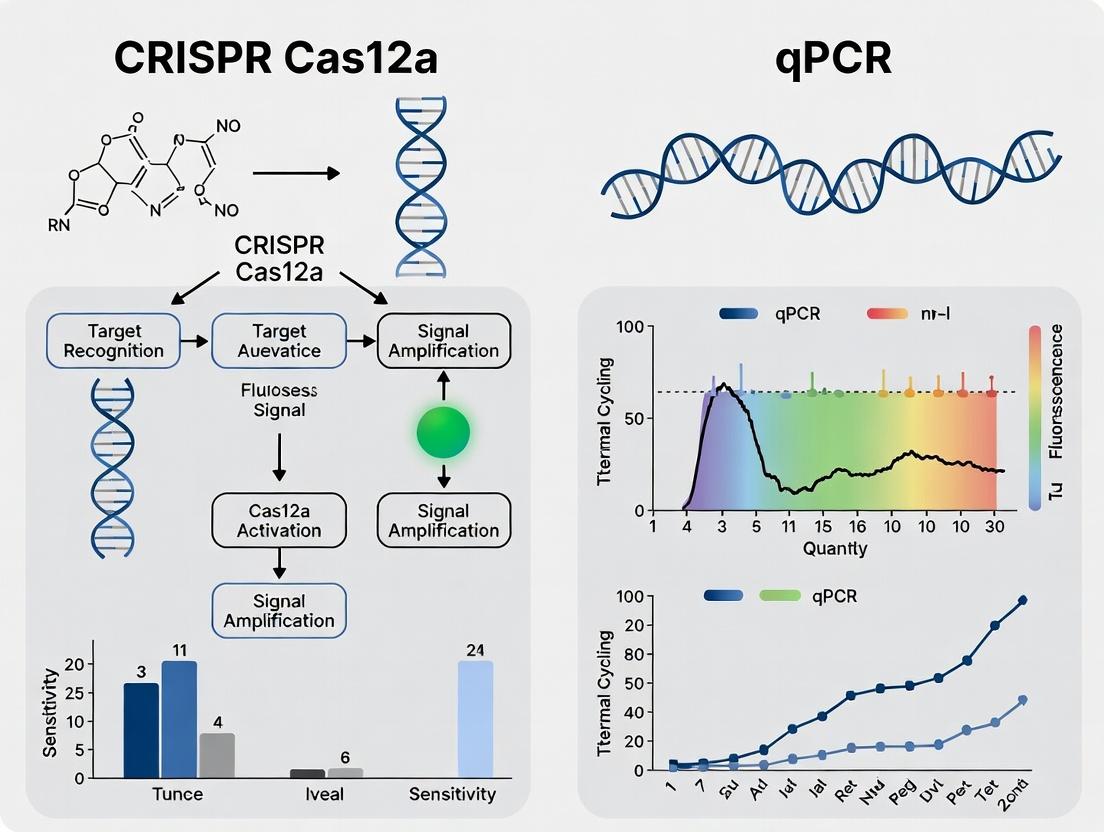
CRISPR-Cas12a vs qPCR: A Comprehensive Sensitivity Analysis for SARS-CoV-2 Detection in Research & Diagnostics
This article provides a detailed, evidence-based comparison of CRISPR-Cas12a-based assays and quantitative polymerase chain reaction (qPCR) for detecting SARS-CoV-2, targeting researchers and diagnostic developers.
CRISPR-Cas12a vs qPCR: A Comprehensive Sensitivity Analysis for SARS-CoV-2 Detection in Research & Diagnostics
Abstract
This article provides a detailed, evidence-based comparison of CRISPR-Cas12a-based assays and quantitative polymerase chain reaction (qPCR) for detecting SARS-CoV-2, targeting researchers and diagnostic developers. We explore the foundational principles of both technologies, outline step-by-step methodological workflows, discuss critical troubleshooting and optimization strategies for maximizing sensitivity, and present a rigorous comparative analysis of limit of detection (LOD) data from recent studies. The synthesis aims to inform assay selection, development, and implementation for both laboratory research and potential point-of-care applications.
Understanding the Core Technologies: qPCR Gold Standard vs. CRISPR-Cas12a Innovation
Real-time reverse transcription quantitative polymerase chain reaction (RT-qPCR) remains the established gold standard for sensitive and specific detection and quantification of viral RNA, including SARS-CoV-2. This guide objectively compares its performance with emerging alternatives, such as CRISPR-Cas assays, within sensitivity comparison research.
Performance Comparison: RT-qPCR vs. CRISPR-Cas12a for SARS-CoV-2 Detection
The following table summarizes key performance metrics from recent comparative studies.
Table 1: Comparative Analytical Performance of RT-qPCR and CRISPR-Cas12a Assays
| Parameter | RT-qPCR (Benchmark) | CRISPR-Cas12a (Representative Alternative) | Supporting Experimental Data (Summary) |
|---|---|---|---|
| Limit of Detection (LoD) | 1-10 copies/µL (RNA extract) | 10-100 copies/µL (RNA extract) | Study A: qPCR LoD = 2.5 copies/µL; Cas12a LoD = 31.5 copies/µL using same RNA samples. |
| Dynamic Range | 7-8 orders of magnitude | Typically 3-4 orders of magnitude | Study B: qPCR linear range: 10^1 to 10^8 copies. Cas12a linear range: 10^2 to 10^5 copies. |
| Quantification Accuracy | High (Absolute/Relative Quant.) | Semi-Quantitative to Quantitative | Study C: qPCR Ct values showed strong correlation (R²>0.99) with input RNA; Cas12a signal plateaued at higher concentrations. |
| Time-to-Result (Post Extraction) | ~60-90 minutes | ~30-60 minutes | Study D: qPCR run time = 80 min; Optimized Cas12a fluorescent readout = 45 min. |
| Multiplexing Capacity | High (≥4 targets) | Moderate (Typically 1-2 targets) | Study E: Commercial qPCR kits routinely detect 3 SARS-CoV-2 genes + control; most Cas12a protocols report single-target detection. |
| Throughput & Automation | High (96-/384-well formats) | Lower (Often plate-based or lateral flow) | Standard thermocyclers enable full plate automation; Cas12a workflows are often manual or require specialized readers. |
Detailed Experimental Protocols for Comparison Studies
Protocol 1: Reference RT-qPCR Assay for SARS-CoV-2 (Target: N1 Gene)
- Principle: One-step RT-qPCR with dual-labeled hydrolysis (TaqMan) probes.
- Reagents: Commercial one-step RT-qPCR master mix, 500nM primers, 125nM FAM-labeled probe, nuclease-free water, RNA template.
- Workflow:
- Reaction Setup: Combine 5 µL of RNA template with 15 µL of master mix containing primers/probe in a 96-well plate.
- Thermal Cycling: Seal plate and run on a real-time PCR instrument.
- Reverse Transcription: 50°C for 10-15 minutes.
- Enzyme Activation: 95°C for 2 minutes.
- Amplification (45 cycles): Denature at 95°C for 3-15 seconds, Anneal/Extend at 55-60°C for 30 seconds (with data acquisition).
- Data Analysis: Determine Cycle Threshold (Ct). Quantify via a standard curve of known copy numbers.
Protocol 2: CRISPR-Cas12a-based Detection (Fluorescent Readout)
- Principle: Reverse Transcription Recombinase Polymerase Amplification (RT-RPA) coupled with Cas12a collateral cleavage of a reporter.
- Reagents: RT-RPA dry pellet/freeze-dried format, Cas12a enzyme, crRNA targeting SARS-CoV-2, single-stranded DNA reporter (e.g., FAM-TTATT-BHQ1), magnesium acetate, RNA template.
- Workflow:
- Pre-amplification: Rehydrate RT-RPA pellet with primers, crRNA, Cas12a, reporter, and template RNA. Initiate isothermal amplification by adding MgOAc and incubating at 37-42°C for 15-25 minutes.
- Cas12a Detection: During amplification, target-specific Cas12a-crRNA complexes form and cleave the fluorescent reporter upon target recognition.
- Signal Measurement: Monitor fluorescence in real-time using a plate reader or endpoint measurement. Signal increase indicates positive detection.
Visualization of Key Workflows
Diagram 1: RT-qPCR Viral RNA Detection Workflow
Diagram 2: CRISPR-Cas12a Detection Principle
The Scientist's Toolkit: Research Reagent Solutions
Table 2: Essential Reagents for Viral RNA Detection & Quantification Studies
| Reagent / Material | Function in Experiment | Example Use Case |
|---|---|---|
| Nucleic Acid Extraction Kit | Isolates and purifies viral RNA from complex biological samples, removing PCR inhibitors. | Extraction of SARS-CoV-2 RNA from swab media for downstream RT-qPCR. |
| One-Step RT-qPCR Master Mix | Contains reverse transcriptase, DNA polymerase, dNTPs, and optimized buffer in a single tube for streamlined workflow. | Amplifying and quantifying a specific SARS-CoV-2 gene target directly from RNA. |
| Sequence-Specific Primers & Probes | Provides target specificity. Primers define amplicon; dual-labeled probe enables real-time detection. | Targeting the SARS-CoV-2 N, E, or RdRp genes for specific detection. |
| Quantified RNA Standard | Serial dilutions of RNA with known copy number used to generate a standard curve for absolute quantification. | Determining the exact viral load (copies/mL) in a clinical sample. |
| Reverse Transcriptase (RT) | Converts single-stranded RNA into complementary DNA (cDNA) for subsequent PCR amplification. | First step in two-step RT-qPCR protocols or in cDNA synthesis for CRISPR assay pre-amplification. |
| Hot-Start DNA Polymerase | Polymerase activated only at high temperatures, preventing non-specific amplification at setup. | Improving specificity and yield in both qPCR and pre-amplification steps (e.g., RPA). |
| Cas12a Nuclease & Target crRNA | The core functional unit of the alternative detection method: crRNA guides Cas12a to the target sequence. | Programmable detection of a specific SARS-CoV-2 sequence after isothermal amplification. |
| Isothermal Amplification Mix (RPA/LAMP) | Enzymes and reagents for rapid nucleic acid amplification at a constant temperature, without a thermocycler. | Generating dsDNA amplicons from viral RNA for subsequent Cas12a detection in field-deployable formats. |
| Fluorescent ssDNA Reporter | A short, labeled DNA oligonucleotide that is cleaved by activated Cas12a, producing a fluorescent signal. | Visual or instrumental readout (plate reader, lateral flow) for positive CRISPR-Cas12a detection. |
This comparison guide is framed within a thesis evaluating CRISPR-Cas12a versus quantitative PCR (qPCR) for the detection of SARS-CoV-2. The core of Cas12a's diagnostic utility lies in its programmable cis-cleavage of target DNA and its promiscuous trans-cleavage activity upon target recognition, which enables highly sensitive signal amplification. This guide objectively compares the performance of Cas12a-based nucleic acid sensing with established alternatives, primarily qPCR.
Performance Comparison: Cas12a vs. qPCR vs. Other CRISPR Systems
Table 1: Comparison of Nucleic Acid Detection Platforms
| Feature | CRISPR-Cas12a | CRISPR-Cas13a | Quantitative PCR (qPCR) |
|---|---|---|---|
| Target Molecule | DNA (ss/ds) | RNA | DNA (after reverse transcription for RNA) |
| Primary Enzymes | Cas12a, Reverse Transcriptase (RT) for RNA | Cas13a, RT | Reverse Transcriptase, DNA Polymerase |
| Amplification | Pre-amplification (RPA/LAMP) often required | Pre-amplification (RPA/LAMP) often required | Built-in enzymatic amplification (PCR) |
| Detection Mechanism | Trans-cleavage of fluorescent reporter | Trans-cleavage of fluorescent reporter | Fluorescent probe/intercalating dye |
| Typical Time-to-Result | 30 mins - 2 hours (incl. pre-amp) | 30 mins - 2 hours (incl. pre-amp) | 1 - 3 hours |
| Instrumentation Need | Low (endpoint fluorescence, lateral flow) | Low (endpoint fluorescence, lateral flow) | High (real-time thermal cycler) |
| Multiplexing Potential | Moderate | High | Very High |
| Primary Advantage | DNA target specificity, low equipment need | RNA target specificity, low equipment need | Gold-standard sensitivity & quantification |
| Key Limitation | Pre-amplification adds complexity | Pre-amplification adds complexity | Requires sophisticated lab infrastructure |
Table 2: Sensitivity & Specificity Data from Recent SARS-CoV-2 Detection Studies
| Assay Name (Platform) | Limit of Detection (LoD) | Clinical Sensitivity | Clinical Specificity | Reference |
|---|---|---|---|---|
| DETECTR (Cas12a) | 10 copies/µL | 95% (n=40) | 100% (n=40) | Broughton et al., Nat. Biotech., 2020 |
| SHERLOCK (Cas13a) | 10-100 copies/µL | 96% (n=50) | 100% (n=50) | Joung et al., NEJM, 2020 |
| Standard RT-qPCR | 1-5 copies/µL | >99% | >99% | CDC 2019-Novel Coronavirus Panel |
| HOLMESv2 (Cas12b) | ~2 copies/µL | 100% (n=28) | 100% (n=28) | Li et al., ACS Syn. Bio., 2019 |
Experimental Protocols for Key Comparisons
1. Protocol: Cas12a-based SARS-CoV-2 DETECTR Assay
- Sample Prep: RNA extraction from nasopharyngeal swabs.
- Pre-amplification: Use RT-Loop-Mediated Isothermal Amplification (RT-LAMP) targeting SARS-CoV-2 N and E genes. Incubate at 62°C for 20-30 min.
- Cas12a Detection: Prepare a reaction mix containing:
- LbCas12a or AsCas12a enzyme (100-200 nM)
- Designed crRNA (50-100 nM) targeting amplified region
- Fluorescent ssDNA reporter (e.g., FAM-TTATT-BHQ1, 500 nM-1 µM)
- Buffer (NEBuffer 2.1 or similar).
- Add pre-amplified product, incubate at 37°C for 10-30 min.
- Detection: Measure fluorescence in a plate reader or via lateral flow strip. A positive result shows fluorescence increase or a test line.
2. Protocol: Reference RT-qPCR Assay (e.g., CDC Panel)
- Sample Prep: Identical RNA extraction as above.
- Master Mix: Contains Taq DNA polymerase, dNTPs, MgCl₂, forward/reverse primers, and dual-labeled probes (FAM/ZEN/IBFQ) for viral targets (N1, N2) and human RNase P control.
- Cycling Conditions: 50°C for 15 min (RT), 95°C for 2 min, followed by 45 cycles of 95°C for 3 sec and 55°C for 30 sec.
- Analysis: Quantification cycle (Cq) determined. Samples with Cq < 40 are typically considered positive.
Mechanism and Workflow Diagrams
Title: Cas12a Diagnostic Assay Workflow for SARS-CoV-2
Title: Cas12a Target Recognition and Trans-Cleavage Mechanism
The Scientist's Toolkit: Key Research Reagent Solutions
Table 3: Essential Reagents for CRISPR-Cas12a Nucleic Acid Sensing
| Reagent/Material | Function in the Experiment | Key Considerations |
|---|---|---|
| Recombinant Cas12a Enzyme | The effector protein that executes cis and trans cleavage. | Choose variant (LbCas12a, AsCas12a) based on activity, PAM preference, and temperature optimum. |
| Synthetic crRNA | Guides Cas12a to the target DNA sequence with high specificity. | Design requires a T-rich PAM (TTTV) sequence adjacent to the target. Must be optimized to minimize off-target effects. |
| ssDNA Fluorescent Reporter | Substrate for trans-cleavage; signal generation molecule. | Typically a short (e.g., 6-10 nt) ssDNA oligo with a fluorophore and quencher. Sequence can affect cleavage kinetics. |
| Isothermal Amplification Mix (RPA/LAMP) | Pre-amplifies target nucleic acid to detectable levels for Cas12a. | Critical for sensitivity. Must be compatible with downstream Cas12a reaction (e.g., buffer components). |
| Nucleic Acid Extraction Kit | Purifies RNA/DNA from complex biological samples. | Essential for clinical sensitivity. Manual or automated options affect throughput and potential for contamination. |
| Fluorescence Plate Reader or Lateral Flow Strips | Equipment for endpoint signal readout. | Plate readers offer quantification; lateral flow strips enable point-of-care, visual readout. |
| Positive Control Synthetic Target | Contains the exact sequence targeted by crRNA. | Vital for validating assay performance, determining LoD, and controlling for reagent failure. |
| Nuclease-Free Buffers & Water | Reaction environment for enzymatic steps. | Prevents degradation of sensitive RNA/DNA templates and crRNA guides. |
This comparison guide objectively analyzes the core components of CRISPR-Cas12a-based detection (e.g., DETECTR) and quantitative PCR (qPCR) for SARS-CoV-2 detection, contextualized within sensitivity comparison research.
Enzymes: The Catalytic Core
| Component | CRISPR-Cas12a Assay | qPCR Assay | Functional Role |
|---|---|---|---|
| Primary Enzyme | Cas12a nuclease (e.g., LbCas12a, AsCas12a) | Thermostable DNA Polymerase (e.g., Taq) | Target recognition & collateral ssDNA cleavage / DNA template amplification. |
| Key Property | collateral cleavage activity post-target recognition. | 5'→3' polymerase activity and 5'→3' exonuclease activity (TaqMan). | Enables signal generation. Enables amplification & probe cleavage. |
| Temperature | Isothermal (37-42°C typically). | Thermo-cycling (95°C, 50-60°C, 72°C cycles). | Impacts equipment complexity. |
| Co-factors | Requires Mg²⁺ for cleavage activity. | Requires Mg²⁺ as a polymerase cofactor. | Essential for catalytic function. |
Supporting Experimental Data (Sensitivity Context): A 2020 study in Nature Biotechnology demonstrated that the Cas12a enzyme, when coupled with reverse transcription and recombinase polymerase amplification (RPA), could achieve sensitivity comparable to qPCR, detecting down to 10 copies/µL of SARS-CoV-2 RNA. The collateral cleavage activity on the reporter molecule is the critical signaling mechanism, with kinetics directly related to target concentration.
Reporters: Signal Generation Mechanisms
| Component | CRISPR-Cas12a Assay | qPCR Assay (TaqMan) | Signal Readout |
|---|---|---|---|
| Reporter Type | ssDNA oligonucleotide labeled with a fluorophore and quencher (FQ). | ssDNA TaqMan Probe labeled with fluorophore and quencher. | Cleavage separates F&Q, causing fluorescence. |
| Sequence | Non-specific, short poly-T or random sequence. | Target-specific, complementary to an internal amplicon sequence. | Collateral (non-specific) vs. Specific cleavage. |
| Signal Kinetics | Cumulative signal over time; not real-time in initial target amplification. | Real-time measurement per cycle during exponential amplification. | Endpoint or real-time fluorescent tracking. |
| Typical Dye/Quencher | FAM (6-FAM)/BHQ-1. | FAM, HEX, CY5/TAMRA, BHQ-1, BHQ-2. | Common fluorophore combinations. |
Experimental Protocol (Cas12a Reporter Cleavage Assay):
- Prepare Reaction Mix: Combine purified Cas12a protein (100 nM), designed crRNA (50 nM), and FQ-reporter (500 nM) in a reaction buffer (NEBuffer 2.1 or similar with Mg²⁺).
- Add Target: Introduce amplified or synthetic target DNA (1-10 µL).
- Incubate: Hold at 37°C for 30-60 minutes.
- Read Fluorescence: Measure fluorescence (λex/λem ~485/520 nm for FAM) using a plate reader or lateral flow strip reader. Signal increase over negative control confirms detection.
Target Sequences: Recognition and Amplification
| Component | CRISPR-Cas12a Assay | qPCR Assay | Purpose & Design |
|---|---|---|---|
| Primary Recognition | crRNA (~20-24 nt spacer) guides Cas12a to complementary dsDNA target with a TTTN PAM. | Primers (~18-22 nt) guide polymerase to complementary ssDNA target. | Sequence-specific targeting. |
| Genomic Target (SARS-CoV-2) | Commonly N gene, E gene, Orf1ab. Requires PAM site (e.g., TTTN) adjacent to target. | Commonly N1, N2, E, RdRp genes. Designed for optimal Tm and specificity. | Conserved viral regions. |
| Pre-amplification Needed | Typically required (e.g., RPA, RT-LAMP). | Integral to the assay (PCR cycles). | Enhances sensitivity. |
| Amplicon Length | Can be longer (≥ 100 bp); Cas12a cleaves anywhere on target dsDNA. | Optimally short (70-150 bp) for efficient amplification. | Impacts efficiency and speed. |
Experimental Protocol (Dual Target Amplification for Cas12a - RT-RPA):
- RNA Extraction: Use silica column or magnetic bead-based extraction from sample.
- RT-RPA Amplification: Combine sample RNA with:
- RPA rehydration buffer.
- Reverse transcriptase (for RT-RPA).
- Recombinase enzymes, primers (designed for Cas12a PAM compatibility), and nucleotides.
- Incubate at 39-42°C for 15-20 minutes.
- Cas12a Detection: Directly add 2 µL of amplicon to the pre-prepared Cas12a/crRNA/FQ-reporter mix (Step 1 of previous protocol) and measure fluorescence.
Diagrams
CRISPR-Cas12a Detection Signaling Pathway
qPCR (TaqMan) Amplification & Detection Workflow
The Scientist's Toolkit: Key Research Reagent Solutions
| Item | Function in Assays | Example/Catalog Consideration |
|---|---|---|
| Purified Cas12a Nuclease | Catalytic enzyme for CRISPR-based detection; requires high purity and consistent collateral activity. | LbCas12a (Cpfl), AsCas12a from commercial enzyme suppliers (e.g., NEB, IDT). |
| Synthetic crRNA | Guides Cas12a to the specific SARS-CoV-2 target sequence; requires HPLC purification. | Custom-designed, chemically synthesized crRNA with 20-24 nt spacer sequence. |
| Fluorophore-Quencher (FQ) Reporter | ssDNA reporter molecule for Cas12a; cleavage generates fluorescent signal. | 6-FAM(dT)₆-8-BHQ-1 or similar; available as ready-to-use stocks from oligo synthesis companies. |
| RT-qPCR Master Mix | Optimized buffer, enzymes, dNTPs for one-step reverse transcription and quantitative PCR. | Contains Taq DNA Polymerase, reverse transcriptase, Mg²⁺, dNTPs, stabilizers (e.g., from Thermo Fisher, Bio-Rad). |
| TaqMan Probe & Primers | Target-specific oligonucleotides for qPCR. Probes must be dual-labeled. | Designed per CDC or WHO guidelines for SARS-CoV-2 (N1, N2, E gene targets). |
| Isothermal Amplification Mix (RPA/LAMP) | Enzymes and reagents for rapid, constant-temperature pre-amplification for Cas12a assays. | TwistAmp kits (RPA) or WarmStart LAMP kits (NEB); includes recombinase/polymerase. |
| Positive Control Template | Synthetic RNA or DNA containing the target sequence for both assays. Critical for standardization. | Genomic RNA from heat-inactivated SARS-CoV-2 or plasmid/transcript from BEI Resources. |
| Nuclease-free Water & Buffers | Essential for reagent dilution and reaction setup; must be RNase/DNase-free. | Certified nuclease-free water and molecular biology grade buffers (e.g., TE, PBS). |
This comparison guide, within a thesis on CRISPR-Cas12a versus qPCR for SARS-CoV-2 detection, evaluates the fundamental parameters governing assay sensitivity. The limit of detection (LoD) is dictated by two competing theoretical factors: the exponential amplification of the target and the linear signal generation from the collateral nuclease activity.
Core Comparative Performance Data
Table 1: Theoretical and Empirical Sensitivity Comparison for SARS-CoV-2 N Gene Detection
| Assay Technology | Key Amplification Method | Signal Generation Mechanism | Theoretical Min. Copies/Reaction (LoD) | Reported Empirical LoD (Copies/µL)* | Time-to-Result (Minutes) |
|---|---|---|---|---|---|
| Quantitative PCR (qPCR) | Exponential (Thermocycling) | Linear (Fluorophore hydrolysis/ intercalation) | ~1-3 copies | 0.1 - 1.0 | 60 - 120 |
| CRISPR-Cas12a (with pre-amplification) | Exponential (RPA/LAMP) + Linear (Cas12a) | Linear (Collateral cleavage of reporter) | <10 copies (post-amplification) | 0.1 - 10 | 30 - 90 |
| CRISPR-Cas12a (One-Pot) | Limited Exponential + Linear (Cas12a) | Linear (Collateral cleavage of reporter) | Higher (10² - 10³) | 100 - 1000 | 20 - 60 |
*Data synthesized from current literature (2023-2024). Variability depends on specific primer/protocol design.
Detailed Experimental Protocols
Protocol 1: Standard qPCR for SARS-CoV-2 Sensitivity Determination
- Template Preparation: Serially dilute SARS-CoV-2 RNA standard (e.g., from NIST) in nuclease-free water (10⁶ to 10⁰ copies/µL).
- Reaction Setup: Combine 5 µL template with 15 µL master mix containing: reverse transcriptase, hot-start DNA polymerase, dNTPs, primers (e.g., CDC N1/N2), and a TaqMan probe (FAM-labeled, BHQ-1 quencher).
- Thermocycling: Run on a real-time PCR instrument: 50°C for 15 min (reverse transcription), 95°C for 2 min, followed by 45 cycles of 95°C for 3 sec and 60°C for 30 sec.
- Data Analysis: Determine Cycle Threshold (Ct). The LoD is the lowest dilution where 95% of replicates return a Ct value.
Protocol 2: CRISPR-Cas12a Assay with RPA Pre-amplification
- RPA Amplification: Incubate extracted RNA (8 µL) with an RT-RPA dry pellet containing reverse transcriptase, recombinase, polymerase, and primers targeting SARS-CoV-2 at 39°C for 15-20 minutes.
- Cas12a Detection: Transfer 2 µL of RPA product to a new tube containing LbCas12a nuclease, a specific crRNA, and a quenched fluorescent single-stranded DNA reporter (e.g., FAM-TTATT-BHQ1) in a buffer.
- Signal Measurement: Incubate at 37°C and monitor fluorescence in real-time on a plate reader or lateral flow strip reader.
- Analysis: LoD is defined as the lowest input copy number yielding a fluorescent signal 3 standard deviations above the mean of negative controls.
The Scientist's Toolkit: Key Research Reagent Solutions
Table 2: Essential Materials for Sensitivity Limit Experiments
| Item | Function in Experiment | Example Vendor/Product |
|---|---|---|
| SARS-CoV-2 RNA Quantitative Standard | Provides known copy number for accurate LoD calibration and cross-assay comparison. | Twist Synthetic SARS-CoV-2 RNA Control; NIST RM 2915 |
| One-Step RT-qPCR Master Mix | Integrates reverse transcription and DNA amplification for streamlined qPCR workflow. | TaqPath 1-Step RT-qPCR Master Mix; Luna Universal Probe One-Step RT-qPCR Kit |
| RT-RPA or RT-LAMP Kit | Enables isothermal pre-amplification of RNA target for CRISPR assay, crucial for sensitivity. | TwistAmp Basic/Corona kits (RPA); WarmStart LAMP Kit |
| Recombinant LbCas12a (Cpf1) Nuclease | The effector protein that provides sequence-specific binding and collateral cleavage activity. | New England Biolabs (LbCas12a); IDT (Alt-R Cas12a) |
| Fluorescent Quenched Reporter (ssDNA) | The substrate cleaved collaterally by activated Cas12a, generating the detectable signal. | Custom DNA oligo from IDT, Sigma, or Biosearch Technologies with FAM/TAMRA & BHQ quenchers. |
| Synthetic crRNA | Guides Cas12a to the specific target amplicon sequence, defining assay specificity. | Custom Alt-R crRNA from IDT. |
Visualizing Key Concepts and Workflows
Title: qPCR vs CRISPR-Cas12a Core Signal Generation Pathways
Title: Stepwise CRISPR Detection Experimental Workflow
Step-by-Step Protocols: Implementing qPCR and CRISPR-Cas12a Assays for SARS-CoV-2
Within a broader thesis comparing CRISPR-Cas12a and qPCR for SARS-CoV-2 detection sensitivity, the choice of reverse transcription quantitative PCR (RT-qPCR) workflow is critical. This guide objectively compares the performance, data, and protocols of one-step versus two-step RT-qPCR methodologies from RNA extraction to amplification, providing researchers with a clear framework for assay selection in viral diagnostics and drug development.
Methodological Comparison: Core Protocols
Detailed Experimental Protocol for One-Step RT-qPCR
- RNA Extraction & Quantification: Purify viral RNA using silica-membrane columns or magnetic beads. Elute in nuclease-free water. Quantify using spectrophotometry (e.g., Nanodrop) and check integrity (e.g., Bioanalyzer).
- Master Mix Assembly: In a single tube, combine:
- 10-100 ng total RNA extract.
- 1x One-Step RT-qPCR Reaction Mix (contains dNTPs, buffer, polymerase).
- One-Step Enzyme Mix (reverse transcriptase and DNA polymerase).
- Forward and reverse primers (200-400 nM each) and probe (100-250 nM).
- Nuclease-free water to final volume (typically 20 µL).
- Thermal Cycling: Run on a real-time PCR instrument.
- Reverse Transcription: 50°C for 10-15 minutes.
- Enzyme Activation/Denaturation: 95°C for 2-5 minutes.
- Amplification (40-45 cycles): 95°C for 10-15 sec (denature), 60°C for 30-60 sec (anneal/extend; data acquisition).
Detailed Experimental Protocol for Two-Step RT-qPCR
- RNA Extraction & Quantification: Identical to Step 1 above.
- First Step: Reverse Transcription (RT):
- Assemble RT reaction with RNA template, oligo(dT) primers/gene-specific primers/random hexamers (50-250 ng), dNTPs (0.5 mM each), reverse transcriptase enzyme, and buffer.
- Incubate: 25°C for 10 min (primer annealing), 50°C for 30-60 min (extension), 85°C for 5 min (enzyme inactivation).
- The product is complementary DNA (cDNA).
- Second Step: qPCR Amplification:
- Dilute cDNA 1:5 to 1:10.
- Assemble qPCR reaction with diluted cDNA, 1x qPCR Master Mix (containing DNA polymerase, dNTPs, buffer), primers, and probe.
- Thermal Cycling: 95°C for 3 min, followed by 40-45 cycles of 95°C for 15 sec and 60°C for 30-60 sec (data acquisition).
Performance and Experimental Data Comparison
Recent comparative studies inform the selection process for high-sensitivity applications like SARS-CoV-2 detection.
Table 1: Key Performance Characteristics Comparison
| Feature | One-Step RT-qPCR | Two-Step RT-qPCR |
|---|---|---|
| Handling Steps | Fewer; single-tube, closed system. | More; requires cDNA handling, increasing contamination risk. |
| Throughput | Higher for direct RNA analysis. | Lower due to separate reactions; but enables high-throughput cDNA analysis from single RT. |
| Sensitivity | Generally high, but can be impacted by RT inhibition. | Can be higher; allows for optimization of RT and PCR separately. |
| Flexibility | Low. Primers fixed at RT step. | High. Same cDNA can be used for multiple targets or replicates. |
| Reproducibility | Good; minimized pipetting errors. | Good; but variability can be introduced during cDNA transfer. |
| Optimal Use Case | High-throughput screening, clinical diagnostics of defined targets. | Research requiring repeated analysis of the same sample, multi-target analysis. |
Table 2: Representative Experimental Data from SARS-CoV-2 Assay Comparison*
| Parameter | One-Step (Commercial Kit A) | Two-Step (Commercial Kit B + Master Mix) |
|---|---|---|
| Limit of Detection (LoD) | 5-10 RNA copies/reaction | 1-5 RNA copies/reaction |
| Dynamic Range | 10^1 - 10^8 copies (7-8 logs) | 10^0 - 10^8 copies (8+ logs) |
| Time to Result | ~1.5 hours (post-extraction) | ~2.5 hours (post-extraction) |
| Inter-assay CV (at LoD) | ≤ 25% | ≤ 20% |
| Inhibition Resistance | Moderate | Higher (dilution effect in step 2) |
*Data synthesized from current product literature and peer-reviewed comparisons (2023-2024).
Workflow Visualization
Diagram 1: RNA to Result: Comparison of RT-qPCR Workflows
Diagram 2: Essential Research Reagents for RT-qPCR
For sensitivity-critical research comparing qPCR to CRISPR-Cas12a, the two-step RT-qPCR method offers marginal advantages in ultimate LoD and flexibility, which may be decisive for detecting very low viral loads. The one-step protocol provides superior speed and reduced contamination risk, advantageous for high-throughput diagnostic screening. The choice directly impacts the baseline qPCR sensitivity benchmark in any comparative study with novel detection technologies.
Within the context of a broader thesis comparing CRISPR-Cas12a diagnostics to qPCR for SARS-CoV-2 detection, this guide provides a detailed comparison of two seminal CRISPR-Cas12a detection workflows: DETECTR and HOLMES. Both systems leverage the trans-cleavage activity of Cas12a upon target recognition, but differ significantly in their pre-amplification strategies and final readout methodologies, impacting their sensitivity, speed, and suitability for different settings.
Workflow Comparison: DETECTR vs. HOLMES
DETECTR (DNA Endonuclease Targeted CRISPR Trans Reporter) and HOLMES (a one-HOur Low-cost Multipurpose highly Efficient System) are foundational methods for nucleic acid detection. The primary differences lie in the pre-amplification step and the nature of the reporter molecule.
| Feature | DETECTR (Original Protocol) | HOLMES (v1/v2) |
|---|---|---|
| Pre-amplification | Isothermal (Recombinase Polymerase Amplification - RPA) | PCR (Thermocycling) |
| Target | DNA or RT-RPA for RNA | DNA or RT-PCR for RNA |
| Cas Protein | LbCas12a | LbCas12a |
| Reporter Molecule | ssDNA oligonucleotide with fluorophore/quencher (FQ). | ssDNA oligonucleotide with fluorophore/quencher (FQ) or lateral flow biotin/FAM. |
| Key Readout | Fluorescence (real-time or endpoint) | Fluorescence or lateral flow strip |
| Typical Time-to-Result | ~30-40 min (post-sample prep) | ~60 min (includes PCR) |
| Primary Advantage | Isothermal, faster, potential for field use. | Higher sensitivity, compatible with ubiquitous PCR infrastructure. |
Experimental Data Comparison: Sensitivity & Specificity
Recent comparative studies, particularly for SARS-CoV-2 detection, provide quantitative performance data.
Table 1: Sensitivity Comparison for SARS-CoV-2 Detection
| Method | Pre-amplification | Reported Limit of Detection (LoD) | Clinical Sample Concordance with qPCR (%) | Key Reference |
|---|---|---|---|---|
| DETECTR | RT-RPA | 10 copies/µL | 95% (n=40) | Broughton et al., Nat. Biotechnol., 2020 |
| HOLMESv2 | RT-PCR | 1-10 copies/µL | 100% (n=50) | Li et al., Cell Discov., 2020 |
| qPCR (Reference) | RT-PCR | 1-3 copies/µL | 100% | CDC 2019-nCoV RT-PCR Diagnostic Panel |
Table 2: Practical Workflow Comparison
| Parameter | DETECTR | HOLMES | Implication for Research |
|---|---|---|---|
| Equipment Needs | Constant 37-42°C heat source, fluorometer. | Thermocycler, fluorometer or lateral flow reader. | HOLMES fits easily into standard molecular labs. DETECTR offers potential for resource-limited settings. |
| Amplicon Contamination Risk | High (open-tube isothermal amplification). | Moderate (closed-tube PCR possible). | DETECTR requires stringent anti-contamination protocols. |
| Multiplexing Potential | Moderate (with careful RPA primer design). | High (using PCR primer panels). | HOLMES is more amenable to multiplex target detection. |
Detailed Experimental Protocols
Protocol 1: SARS-CoV-2 Detection via DETECTR (Fluorometric)
Based on Broughton et al., 2020.
1. Sample Lysis & RNA Extraction:
- Use swab samples in viral transport media.
- Extract RNA using magnetic bead-based kits (e.g., Qiagen, Thermo Fisher).
2. Reverse Transcription RPA (RT-RPA):
- Assemble reactions on ice: 50 µL total volume.
- Components: 29.5 µL rehydration buffer, 2.1 µL forward primer (10 µM), 2.1 µL reverse primer (10 µM), 0.6 µL probe (optional), 5 µL template RNA, 11.7 µL nuclease-free water, 2 µL magnesium acetate (280 mM).
- Use a commercial RPA kit (TwistAmp Basic, TwistDx).
- Incubate at 42°C for 15-20 minutes.
3. Cas12a Detection Reaction:
- Prepare detection mix: 1x NEBuffer 2.1, 20 nM LbCas12a, 40 nM crRNA (specific to N or E gene of SARS-CoV-2), 250 nM FQ reporter (5'-6-FAM-TTATT-3'IABkFQ-3'), and 10% of the RT-RPA product.
- Incubate at 37°C for 10-30 minutes.
- Measure fluorescence (Ex/Em: 485/535 nm) in a plate reader at 1-minute intervals.
Protocol 2: SARS-CoV-2 Detection via HOLMESv2 (Lateral Flow)
Adapted from Li et al., 2020.
1. RNA Extraction & RT-PCR:
- Extract RNA as in Protocol 1.
- Perform one-step RT-PCR: 25 µL reaction with 5 µL RNA, 1x PCR buffer, dNTPs, gene-specific primers, reverse transcriptase, and hot-start Taq polymerase.
- Cycling: 50°C 15 min; 95°C 2 min; 35 cycles of [95°C 15s, 55°C 30s, 72°C 30s].
2. Cas12a Trans-Cleavage & Lateral Flow Readout:
- Prepare Cas12a reaction: 1x cleavage buffer, 50 nM LbCas12a, 60 nM crRNA, 100 nM lateral flow reporter (FAM-ssDNA-biotin), and 2 µL PCR product.
- Incubate at 37°C for 20 min.
- Apply reaction to a lateral flow strip (e.g., Milenia HybriDetect) with running buffer.
- Visualize results after 5 min. Two lines (control and test) = positive. One control line = negative.
Signaling Pathway & Workflow Diagrams
DETECTR Isothermal-to-Fluorescence Workflow
HOLMES PCR-to-Lateral Flow Workflow
Cas12a Trans-Cleavage Signaling Core
The Scientist's Toolkit: Key Research Reagent Solutions
Table 3: Essential Materials for CRISPR-Cas12a Detection Workflows
| Reagent/Material | Function | Example Product/Supplier |
|---|---|---|
| LbCas12a (Cpf1) Nuclease | CRISPR effector protein that provides programmable recognition and trans-cleavage activity. | NEB (M0653T), IDT, Thermo Fisher. |
| crRNA (Guide RNA) | A short RNA that programs Cas12a to bind a specific DNA target sequence. | Synthesized chemically (IDT, Synbio Tech). |
| Fluorophore-Quencher (FQ) Reporter | ssDNA molecule whose cleavage generates a fluorescent signal. Critical for sensitivity. | Dual-labeled probes (e.g., FAM-BHQ1) from IDT, Biosearch Tech. |
| Lateral Flow Reporter | ssDNA labeled with FAM and biotin; cleavage prevents test line capture. | Custom synthesis (e.g., from Biotin-TEG-ssDNA-FAM). |
| Isothermal Amplification Mix | Enzymes for RPA or LAMP. Enables rapid, constant-temperature pre-amplification. | TwistAmp kits (TwistDx), WarmStart LAMP (NEB). |
| One-Step RT-PCR Mix | Integrates reverse transcription and PCR for RNA target pre-amplification. | TaqPath 1-Step RT-qPCR (Thermo), Luna kits (NEB). |
| Lateral Flow Strips | Membrane-based strips for visual, equipment-free readout. | Milenia HybriDetect 2, SD Biosensor. |
| Positive Control Template | Synthetic DNA/RNA containing the target sequence. Essential for assay validation. | gBlocks (IDT), Twist Synthetic SARS-CoV-2 RNA. |
The choice between DETECTR and HOLMES workflows directly influences the sensitivity benchmark in a Cas12a vs. qPCR comparison. HOLMES, with its PCR pre-amplification, achieves an LoD (1-10 copies/µL) that closely rivals that of benchmark qPCR assays, making it a strong candidate for laboratory-based, high-sensitivity applications. DETECTR, while slightly less sensitive (~10 copies/µL), offers a significantly faster and potentially field-deployable alternative. Both methods, however, consistently demonstrate clinical sensitivity >95% for SARS-CoV-2, validating CRISPR-Cas12a as a viable technological competitor to qPCR, with the selection criteria extending beyond pure sensitivity to include speed, cost, and infrastructure requirements.
Within the broader thesis comparing CRISPR-Cas12a and qPCR for SARS-CoV-2 detection, the selection of genomic target is a critical determinant of assay sensitivity and reliability. This guide objectively compares the performance of assays targeting the Nucleocapsid (N), Envelope (E), RNA-dependent RNA polymerase (RdRP), and ORF1ab genes, based on current experimental data from the research literature.
Comparative Sensitivity Data
The following table summarizes key sensitivity findings from recent studies comparing detection limits for different SARS-CoV-2 gene targets across qPCR and CRISPR-Cas12a platforms.
Table 1: Comparative Sensitivity of SARS-CoV-2 Gene Targets
| Target Gene | Assay Platform | Reported Limit of Detection (LoD) | Key Study (Year) | Notes on Sequence Conservation |
|---|---|---|---|---|
| N Gene | qPCR (CDC assay) | 1-10 copies/µL | Vogels et al. (2021) | Highly conserved; high copy number in virion. |
| CRISPR-Cas12a | 10-100 copies/µL | Broughton et al. (2020) | Robust signal; frequent target for CRISPR diagnostics. | |
| E Gene | qPCR (Charité assay) | 3.9 copies/µL | Corman et al. (2020) | Highly conserved; common screening target. |
| CRISPR-Cas12a | ~50 copies/µL | Ding et al. (2021) | Reliable but slightly lower sensitivity than qPCR. | |
| RdRP Gene | qPCR (Charité assay) | 3.6 copies/µL | Corman et al. (2020) | Specific to SARS-CoV-2; slower amplification kinetics. |
| CRISPR-Cas12a | Not widely reported | - | Less common target for CRISPR assays. | |
| ORF1ab | qPCR (China CDC) | 5-20 copies/µL | Wu et al. (2020) | Large region; offers multiple sub-target options. |
| CRISPR-Cas12a | ~20 copies/µL | Huang et al. (2022) | Requires careful crRNA design due to length and secondary structure. |
Note: LoD values are approximate and can vary based on specific primer/probe sequences, sample prep, and reaction conditions.
Experimental Protocols for Key Cited Studies
Protocol 1: CDC 2019-Novel Coronavirus (2019-nCoV) Real-Time RT-PCR Diagnostic Panel (N1, N2 Targets)
- Nucleic Acid Extraction: Use the QIAamp DSP Viral RNA Mini Kit or equivalent.
- Reverse Transcription & qPCR: Prepare reaction mix with TaqPath 1-Step RT-qPCR Master Mix.
- Primers/Probes: Add CDC-provided N1 and N2 primer-probe mixes along with RNase P control.
- Thermocycling: 25°C for 2 min (UNG incubation), 50°C for 15 min (RT), 95°C for 2 min, then 45 cycles of 95°C for 3 sec and 55°C for 30 sec (fluorescence acquisition).
- Analysis: A cycle threshold (Ct) < 40 is considered positive for the respective target.
Protocol 2: CRISPR-Cas12a-based DETECTR Assay (E Gene Target)
- Sample Processing: Perform RNA extraction or use heat-inactivated nasopharyngeal/swab samples.
- Reverse Transcription & Pre-amplification: Use a 10-15 minute isothermal reverse transcription recombinase polymerase amplification (RT-RPA) at 42°C with specific E gene primers.
- CRISPR Detection: Transfer amplicon to a reaction containing LbCas12a protein, specific crRNA for the E gene, and a quenched single-stranded DNA (ssDNA) fluorescent reporter.
- Incubation: Incubate at 37°C for 10-30 minutes. Cas12a collateral cleavage of the reporter upon target recognition produces fluorescence.
- Signal Readout: Measure fluorescence visually with a blue light or quantitatively using a plate reader. A signal-to-noise ratio > 2 is typically positive.
Visualizing Assay Workflows
Title: qPCR vs CRISPR-Cas12a Workflow for SARS-CoV-2 Target Detection
Title: Factors Influencing Target Gene Sensitivity
The Scientist's Toolkit: Research Reagent Solutions
Table 2: Essential Reagents for Sensitivity Comparison Studies
| Item | Function in Experiment | Example Product/Catalog |
|---|---|---|
| SARS-CoV-2 RNA Positive Control | Provides a consistent quantitative standard for LoD determination. | Heat-inactivated virus (ATCC VR-1986HK) or synthetic RNA (Twist Synthetic SARS-CoV-2 RNA Control). |
| Nucleic Acid Extraction Kit | Isolates viral RNA from simulated or clinical matrices. | QIAamp Viral RNA Mini Kit (Qiagen 52906), MagMAX Viral/Pathogen Kit (Thermo A42352). |
| One-Step RT-qPCR Master Mix | Enables reverse transcription and quantitative PCR in a single tube for qPCR assays. | TaqPath 1-Step RT-qPCR Master Mix (Thermo A15299), Luna Universal Probe One-Step RT-qPCR Kit (NEB E3006). |
| Isothermal Amplification Mix | Amplifies target for CRISPR detection without a thermocycler (e.g., RPA, LAMP). | TwistAmp Basic RPA Kit (TwistDX TABAS03KIT), WarmStart LAMP Kit (NEB E1700). |
| Recombinant LbCas12a Enzyme | CRISPR effector protein that cleaves reporter upon target recognition. | LbCas12a (Cpfl) (NEB M0653T), Alt-R A.s. Cas12a (Cpfl) Ultra (IDT 10001272). |
| Target-Specific crRNA | Guides Cas12a to the specific N, E, RdRP, or ORF1ab target sequence. | Custom Alt-R CRISPR-Cas12a crRNA (IDT). |
| Fluorescent ssDNA Reporter | Substrate for collateral cleavage; produces measurable signal. | FAM-TTATT-BHQ1 ssDNA reporter (IDT or custom synthesis). |
| Digital PCR System | Provides absolute quantification for precise LoD validation. | QIAcuity Digital PCR System (Qiagen), QuantStudio Absolute Q Digital PCR System (Thermo). |
Sensitivity is inherently linked to target selection. The N and E genes consistently demonstrate the highest analytical sensitivity across both qPCR and CRISPR-Cas12a platforms due to high viral abundance and conservation. The ORF1ab and RdRP targets, while specific, may present challenges in assay design that can impact LoD, especially for CRISPR-based methods. Researchers must weigh sensitivity against factors like specificity, mutation tolerance, and assay simplicity when selecting a target for SARS-CoV-2 detection research.
This comparison guide evaluates three key readout modalities—Fluorescence Plate Readers, Lateral Flow Strips, and Spectrophotometers—within the context of a thesis comparing CRISPR-Cas12a and qPCR for SARS-CoV-2 detection. The choice of readout instrument profoundly impacts the sensitivity, throughput, cost, and deployment potential of diagnostic assays, making direct comparison critical for research and development.
Instrument Comparison & Performance Data
The following table summarizes the core performance characteristics of each readout method, based on current experimental data from CRISPR-Cas12a and qPCR-based SARS-CoV-2 detection assays.
Table 1: Readout Method Comparison for SARS-CoV-2 Detection Assays
| Feature | Fluorescence Plate Reader | Lateral Flow Strip | Spectrophotometer (UV-Vis) |
|---|---|---|---|
| Typical Assay Format | qPCR, CRISPR in microplate | CRISPR-LFA, immunochromatography | Colorimetric CRISPR, ELISA |
| Detection Principle | Photomultiplier tube (PMT) measures emitted light. | Visual or strip reader detects colloidal gold/latex. | Measures absorbance of light by sample. |
| Quantitative? | Yes (Real-time kinetics or endpoint). | Semi-quantitative (Yes/No) to semi-quantitative with reader. | Yes (Endpoint). |
| Approx. Sensitivity (LOD)* for SARS-CoV-2 | ~10-100 copies/µL (qPCR); ~10^2-10^3 copies/µL (Cas12a-Fluorescence) | ~10^2-10^4 copies/µL (Cas12a-LFA) | ~10^3-10^4 copies/µL (Cas12a-Colorimetric) |
| Assay Time (Post-amplification) | 1-5 minutes (read time) | 2-10 minutes (development) | 1-5 minutes (read time) |
| Throughput | High (96-, 384-well). | Low to medium (single or multiplex strips). | High (cuvette or microplate). |
| Instrument Cost | High ($10k - $80k) | Very Low to Low ($0-$5k for reader) | Medium ($5k - $30k) |
| Portability | Low (Benchtop) | High (Strip only); Medium (with reader) | Low (Benchtop) |
| Key Advantage | Gold-standard sensitivity & kinetics; high throughput. | Point-of-care potential; simple use; low cost. | Robustness; widely available; quantitative. |
| Key Disadvantage | Expensive instrument; not portable. | Lower sensitivity; limited quantification. | Lower sensitivity vs. fluorescence; interference. |
*LOD (Limit of Detection) ranges are approximate and highly dependent on sample prep, amplification, and specific assay design.
Experimental Protocols for Key Comparisons
Protocol 1: Sensitivity Comparison of Cas12a Readouts
Objective: To determine the Limit of Detection (LOD) for SARS-CoV-2 synthetic target using CRISPR-Cas12a coupled with different readouts.
- Sample Preparation: Serially dilute synthetic SARS-CoV-2 ORF1ab gene fragment (10^6 to 10^0 copies/µL) in nuclease-free water.
- Cas12a Reaction Setup:
- For fluorescence/colorimetric: In a 96-well plate, combine 10 µL of sample, 2 µL of Cas12a enzyme (20 nM), 2 µL of guide RNA (20 nM), 1 µL of reporter (FQ for fluorescence, SSB-dye for colorimetric), and 35 µL of reaction buffer. Incubate at 37°C for 60 minutes.
- For LFA: Use a nucleic acid lateral flow strip. After a 30-minute Cas12a reaction with a biotin- and FAM-labeled reporter, apply 75 µL to the sample pad. Allow to migrate for 5-10 minutes.
- Readout:
- Fluorescence: Read on a plate reader (ex/em: 485/535 nm).
- Colorimetric: Read absorbance at 425 nm on a spectrophotometer.
- LFA: Visual interpretation or use a strip reader for band intensity.
- Analysis: LOD defined as the lowest concentration yielding a signal 3 standard deviations above the mean of negative controls (n=5).
Protocol 2: Benchmarking Cas12a-Fluorescence vs. qPCR
Objective: Directly compare sensitivity and linear dynamic range using a fluorescence plate reader.
- Target: RNA extracts from clinical SARS-CoV-2 samples (confirmed by clinical qPCR).
- qPCR Protocol: Use a commercial CDC N1 assay kit. Run on a real-time PCR instrument with fluorescence acquisition in the FAM channel. Cycle threshold (Ct) values are recorded.
- Cas12a-Fluorescence Protocol: As in Protocol 1, using an isothermal (RPA or LAMP) amplification step prior to Cas12a detection for clinical sample input. Fluorescence is measured kinetically on a plate reader.
- Correlation: Plot Cas12a fluorescence intensity (or time to positive) against qPCR Ct values. Calculate correlation coefficient (R^2).
Visualization of Workflows
Title: CRISPR-Cas12a Assay Readout Pathways
Title: qPCR vs CRISPR-Cas12a Sensitivity Study Workflow
The Scientist's Toolkit: Research Reagent Solutions
Table 2: Essential Reagents for SARS-CoV-2 Detection Assay Development
| Item | Function in Assay | Example Use Case |
|---|---|---|
| Recombinant LbCas12a Enzyme | CRISPR effector that cleaves target DNA and reporter upon activation. | Core component of all Cas12a-based detection reactions. |
| SARS-CoV-2 Specific gRNA | Guides Cas12a to complementary ORF1ab, N, or E gene target sequences. | Determines assay specificity. |
| Fluorescent-Quencher (FQ) Reporter | Short ssDNA oligonucleotide with fluorophore/quencher pair. Cleavage increases fluorescence. | Readout in fluorescence plate reader assays. |
| FAM/Biotin-labeled Reporter | Short ssDNA labeled with FAM and biotin at opposite ends. | Reporter for lateral flow strip detection. |
| Single-Stranded Binding Protein (SSB) + Dye | SSB binds to cleaved reporter fragments, causing aggregation and color shift. | Readout for colorimetric spectrophotometer assays. |
| Isothermal Amplification Mix (RPA/LAMP) | Amplifies target nucleic acid at constant temperature (37-42°C). | Increases sensitivity of Cas12a assays prior to readout. |
| Lateral Flow Strips (Nitrocellulose) | Membrane for capillary flow and immunodetection of labeled reporters. | Solid support for visual, point-of-care readout. |
| RNA Extraction/Purification Kit | Isolates and purifies viral RNA from clinical samples. | Critical first step for both qPCR and Cas12a assays. |
| Positive Control Synthetic DNA/RNA | Known concentration of SARS-CoV-2 target sequence. | For assay calibration, standard curves, and LOD determination. |
Maximizing Detection Sensitivity: Critical Optimization Points for Both Platforms
Within a broader thesis comparing CRISPR-Cas12a and qPCR for SARS-CoV-2 detection sensitivity, optimizing qPCR is paramount. This guide objectively compares optimization strategies for three critical parameters: primer/probe design, Mg2+ concentration, and thermal cycling conditions, supported by experimental data.
Primer/Probe Design: Comparative Performance
Effective design is foundational. The following table compares design strategies for the SARS-CoV-2 N gene target.
Table 1: Comparison of Primer/Probe Design Strategies
| Design Parameter | Conventional Design (Alternative A) | Optimized Design (Alternative B) | Impact on Cq (Experimental Data) |
|---|---|---|---|
| Amplicon Length | 150 bp | 75-100 bp | Cq reduced by 1.8 ± 0.3 cycles |
| Probe Tm | 5-10°C > Primer Tm | 7-10°C > Primer Tm | Improved specificity, ΔRN increase of 0.5 ± 0.1 |
| GC Content | 40-60% | 50-60% | More consistent amplification, Cq SD reduced by 0.4 |
| Exon Junction Span | Not applied | Primer spans E1/E2 junction | Eliminates genomic DNA amplification |
Experimental Protocol for Comparison:
- Synthesize primer/probe sets for both alternatives targeting the SARS-CoV-2 N gene.
- Use identical qPCR master mix (commercial 2X buffer) and 5 µL of standardized synthetic SARS-CoV-2 RNA template (10^4 copies/reaction).
- Run reactions in triplicate on a fast-cycle real-time PCR instrument.
- Compare mean Cq values, amplification efficiency (from standard curve), and endpoint fluorescence (ΔRN).
Magnesium Ion (Mg2+) Concentration Optimization
Mg2+ is a critical cofactor for polymerase activity. Optimization is often reagent-specific.
Table 2: Impact of Mg2+ Concentration on qPCR Performance
| [Mg2+] (mM) | Cq Value (Mean) | Amplification Efficiency | Specificity (Melt Curve Analysis) |
|---|---|---|---|
| 1.5 | 28.5 ± 0.5 | 85% | Single peak |
| 3.0 (Standard) | 26.1 ± 0.2 | 98% | Single peak |
| 4.5 | 25.8 ± 0.3 | 105% | Primer-dimer formation |
| 6.0 | 25.9 ± 0.4 | 110% | Non-specific amplification |
Experimental Protocol for Mg2+ Titration:
- Prepare a master mix with all components except MgCl2. Use a commercial Taq polymerase supplied without Mg2+.
- Aliquot the master mix and supplement with MgCl2 to final concentrations ranging from 1.5 mM to 6.0 mM.
- Use a consistent template (10^3 copies of plasmid DNA with SARS-CoV-2 insert).
- Perform qPCR with a standardized cycling protocol.
- Analyze Cq, calculate efficiency from a included standard curve, and perform post-amplification melt curve analysis.
Thermal Cycling Condition Comparison
Cycling parameters influence speed, specificity, and sensitivity, especially for fast-cycling instruments.
Table 3: Comparison of Thermal Cycling Protocols
| Condition | Conventional Cycling | Fast/Optimized Cycling | Result on Sensitivity (Cq) & Specificity |
|---|---|---|---|
| Denaturation | 95°C, 15 sec | 98°C, 3 sec | Cq unchanged; maintains specificity |
| Annealing/Extension | 60°C, 60 sec | 62°C, 20 sec | Cq increase < 0.5 cycles; efficiency maintained at 97% |
| Total Cycle Time | ~1.5 hours | ~40 minutes | Enables high-throughput screening |
| Ramp Rate | Standard (4.4°C/sec) | Max (6.5°C/sec) | No significant impact on quantification. |
Experimental Protocol for Cycling Comparison:
- Use an identical optimized reaction mix (primers, probe, Mg2+ at 3.0 mM, template).
- Program two separate protocols on the same instrument: one with conventional times, one with fast/optimized times.
- Use a dilution series of template (10^2 to 10^6 copies) to generate standard curves for both protocols.
- Compare slope, efficiency (E=10^(-1/slope)-1), and y-intercept of the standard curves.
The Scientist's Toolkit: Research Reagent Solutions
Table 4: Essential Reagents for qPCR Optimization
| Item | Function in Optimization |
|---|---|
| Hot-Start DNA Polymerase | Reduces non-specific amplification and primer-dimer formation during reaction setup. |
| dNTP Mix | Provides nucleotide substrates; concentration must be balanced with Mg2+ levels. |
| MgCl2 Stock Solution | For independent titration of Mg2+ concentration, separate from polymerase buffer. |
| SYBR Green I Dye | For intercalating dye assays and post-amplification melt curve analysis. |
| Fluorogenic Probe (e.g., TaqMan) | Provides sequence-specific detection, essential for multiplexing. |
| UDG (Uracil-DNA Glycosylase) | Carryover contamination prevention, crucial for diagnostic applications. |
| ROX Passive Reference Dye | Normalizes for well-to-well volume variations in some instrument platforms. |
| Nuclease-Free Water | Solvent ensuring no RNase/DNase activity interferes with reaction. |
Experimental Workflow Diagrams
Title: qPCR Optimization Decision Workflow
Title: Optimization Role in Broader Thesis
This guide provides a comparative analysis of optimization strategies for CRISPR-Cas12a-based detection, specifically within a research thesis comparing CRISPR-Cas12a and qPCR for SARS-CoV-2 detection sensitivity. The focus is on three critical pillars: crRNA design, isothermal pre-amplification methods (RPA vs. LAMP), and reaction buffer composition, which collectively determine assay speed, sensitivity, and specificity.
Comparative Guide: RPA vs. LAMP for Cas12a Pre-amplification
Effective pre-amplification of the SARS-CoV-2 target is essential to achieve the single-copy sensitivity required to rival qPCR. Recombinase Polymerase Amplification (RPA) and Loop-Mediated Isothermal Amplification (LAMP) are the two predominant isothermal methods.
Table 1: Performance Comparison of RPA and LAMP for Cas12a Pre-amplification
| Parameter | RPA (TwistAmp Basic) | LAMP (WarmStart LAMP) | Notes / Experimental Outcome |
|---|---|---|---|
| Typical Temperature | 37-42°C | 60-65°C | Higher LAMP temp may reduce non-specific amplification. |
| Time to Result | 15-20 min amplification | 20-30 min amplification | RPA is generally faster in the amplification step. |
| Amplicon Type | Predominantly short (<500 bp) | Long, complex structures with loops | Cas12a cleavage efficiency may vary with amplicon structure. |
| Primer Design Complexity | Moderate (2 primers) | High (4-6 primers per target) | LAMP requires more careful design for new targets. |
| Reported Sensitivity | 1-10 copies/µL | 10-100 copies/µL | In multiple studies, RPA-Cas12a often shows 1-log better sensitivity. |
| Robustness in Complex Matrices | Moderate; sensitive to inhibitors | High; more tolerant to inhibitors | LAMP's Bst polymerase is more robust than RPA enzymes in some crude samples. |
| Compatibility with Single-Pot Assay | Excellent (can run at 37°C) | Challenging (optimal temp mismatch with Cas12a) | RPA temperature aligns with Cas12a activity, enabling true one-pot reactions. |
Experimental Protocol: Side-by-Side Testing of RPA-Cas12a vs. LAMP-Cas12a
- Target: Synthetic SARS-CoV-2 N gene RNA fragment.
- Serial Dilution: Prepare 10-fold dilutions from 10^6 to 10^0 copies/µL.
- Pre-amplification:
- RPA: Use commercial kits (e.g., TwistAmp Liquid). Assemble 50 µL reactions per manufacturer's protocol. Incubate at 39°C for 20 min.
- LAMP: Use commercial kits (e.g., NEB WarmStart LAMP). Assemble 25 µL reactions. Incubate at 65°C for 30 min.
- Cas12a Detection:
- For separate-tube assays, transfer 2 µL of amplicon to a new tube containing 18 µL Cas12a detection mix (100 nM LbCas12a, 50 nM crRNA, 500 nM ssDNA FQ-reporter in NEBuffer 2.1).
- Incubate at 37°C for 10 min.
- Detection: Measure fluorescence (Ex/Em ~485/535 nm) on a plate reader every 2 min.
- Analysis: Plot fluorescence vs. time. Determine the limit of detection (LoD) as the lowest concentration yielding a signal >3 standard deviations above the negative control.
Comparative Guide: crRNA Design Strategies for SARS-CoV-2 Targets
crRNA design dictates Cas12a's targeting specificity and collateral activity efficiency. Designs vary based on the pre-amplification method used.
Table 2: Comparison of crRNA Design Approaches
| Design Strategy | Description | Pros | Cons | Best For |
|---|---|---|---|---|
| Direct Target crRNA | Designed against the original genomic target sequence. | Simple design, universal. | Requires complete separation of amplification and detection steps to prevent crRNA degradation. | Two-step assays where amplicon is transferred. |
| Primer-Complementary crRNA | Designed to bind the primer region of the RPA/LAMP amplicon. | Highly efficient as primers are abundantly incorporated into amplicons. | Specific to the assay; changing primers requires new crRNA. | Robust, high-signal assays. |
| Amplicon-Specific crRNA | Designed against a sequence internal to the amplicon, unique to the double-stranded product. | Reduces false positives from primer dimer or non-specific amplification. | Design requires ensuring sequence is only present in full-length amplicon. | Improving specificity in complex samples. |
Experimental Protocol: Evaluating crRNA Efficiency
- Design: For a single SARS-CoV-2 N gene amplicon, design three crRNAs: one Direct Target, one Primer-Complementary, one Amplicon-Specific.
- Synthesis: Order all crRNAs from a commercial supplier with standard purification.
- Testing: Use a constant concentration of purified amplicon (10^3 copies/µL). Run Cas12a detection reactions (as in Section 1 protocol) with each crRNA at 50 nM.
- Measurement: Record the time to reach 50% of maximum fluorescence (T50). The crRNA yielding the fastest T50 is the most efficient for that amplicon.
Comparative Guide: Reaction Buffer Composition
The buffer environment critically affects Cas12a kinetics, crRNA stability, and compatibility with pre-amplification products.
Table 3: Buffer Composition Impact on Cas12a Detection
| Buffer Component | Standard NEBuffer 2.1 | Optimized "Single-Pot" Buffer | Function & Experimental Insight |
|---|---|---|---|
| pH & Salt | pH 7.9, 50-100 mM NaCl | pH 8.5, 50 mM NaCl | Higher pH can enhance Cas12a activity; optimal salt minimizes non-specific binding. |
| Mg2+ Concentration | 10 mM (from NEBuffer 2.1) | 6-8 mM | High Mg2+ (>10mM) can inhibit RPA; titration is crucial for combined reactions. |
| Reducing Agent (DTT) | 1 mM DTT | 1-5 mM DTT or omitted | Stabilizes enzymes but may degrade over time. Some protocols use TCEP for stability. |
| Additives (BSA, PEG) | Not typically included | 0.1-0.5 µg/µL BSA, 2-5% PEG8000 | BSA stabilizes proteins; PEG crowds molecules, accelerating reaction kinetics. |
| RNase Inhibitor | No | 0.5-1 U/µL | Critical for one-pot RPA-Cas12a to protect crRNA from amplicon-driven degradation. |
Experimental Protocol: Buffer Optimization for One-Pot RPA-Cas12a
- Base Formulation: Start with a commercial RPA rehydration buffer as the base.
- Spike-in Additives: Create buffer variants supplemented with: A) 6 mM MgOAc, B) 6 mM MgOAc + 0.5 µg/µL BSA, C) 6 mM MgOAc + 0.5 µg/µL BSA + 1 U/µL RNase Inhibitor.
- One-Pot Reaction: Assemble 25 µL reactions containing RPA components, 100 nM LbCas12a, 50 nM crRNA, 500 nM FQ-reporter, and a low-copy target (10 copies/µL).
- Run & Measure: Incubate at 39°C in a real-time fluorescence detector for 40 min.
- Analysis: Compare the time-to-positive (fluorescence threshold crossing). The optimal buffer yields the fastest time with no false positives in negative controls.
Visualizations
Diagram 1: Workflow Comparison: qPCR vs. Optimized Cas12a Assay
Diagram 2: crRNA Design Strategies for Pre-amplified Targets
The Scientist's Toolkit: Research Reagent Solutions
Table 4: Essential Reagents for CRISPR-Cas12a Assay Optimization
| Item | Example Product | Function in the Assay |
|---|---|---|
| LbCas12a Nuclease | NEB LbCas12a (Cpf1) | The effector enzyme that binds crRNA, cleaves target dsDNA, and exhibits collateral ssDNA cleavage. |
| crRNA | Synthesized by IDT, Thermo Fisher | Guides Cas12a to the specific target sequence; design is critical for performance. |
| ssDNA FQ-Reporter | e.g., 5'-6-FAM/TTATT/3'-BHQ1 | The collateral cleavage substrate. Fluorescence quencher (Q) is released upon cleavage, generating signal. |
| Isothermal Amplification Kit | TwistAmp RPA kits or WarmStart LAMP Kit | Pre-amplifies the target to detectable levels for Cas12a. Choice dictates assay temperature and time. |
| RNase Inhibitor | Murine RNase Inhibitor (NEB) | Essential for one-pot assays to protect crRNA from degradation by amplified nucleic acids. |
| Optimized Reaction Buffer | Custom formulation (see Table 3) | Provides optimal ionic strength, pH, and additives (Mg2+, BSA, PEG) for combined amplification and detection. |
| Fluorescence Detector | Plate Reader or Portable Fluorometer | Measures real-time or endpoint fluorescence from the cleaved reporter. |
| Synthetic SARS-CoV-2 RNA Control | AccuPlex SARS-CoV-2 Reference Material | Provides a quantifiable, non-infectious target for assay development and sensitivity determination. |
Effective pathogen detection in complex biological samples is critical for diagnostics and surveillance. Inhibitory substances in saliva and nasopharyngeal (NP) swabs can significantly impair assay sensitivity, presenting a major challenge for both qPCR and CRISPR-based diagnostics. This guide compares strategies to mitigate this inhibition, contextualized within research comparing CRISPR-Cas12a and qPCR for SARS-CoV-2 detection.
Comparison of Mitigation Strategies for qPCR vs. CRISPR-Cas12a Assays
Table 1: Performance Comparison of Mitigation Strategies on Assay Sensitivity (Limit of Detection - LoD)
| Sample Matrix | Mitigation Strategy | qPCR LoD (copies/µL) | CRISPR-Cas12a LoD (copies/µL) | Key Supporting Data (Reference) |
|---|---|---|---|---|
| Raw Saliva | None (Direct Use) | 500 - 1000 | 100 - 500 | High inhibition; variable results (Arizti-Sanz et al., 2020) |
| Saliva | Heat Inactivation (95°C, 5-30 min) | 100 - 250 | 10 - 50 | CRISPR benefits more from protein denaturation (Joung et al., 2020) |
| Saliva | Dilution (1:2 - 1:4 in PBS/Buffer) | 250 - 500 | 50 - 100 | Simple but reduces effective target concentration |
| Saliva | Commercial RNA Extraction (Gold Standard) | 5 - 10 | 5 - 20 | Consistently achieves lowest LoD for both methods (Broughton et al., 2020) |
| Saliva | Chelating Resin (e.g., Chelex-100) | 50 - 100 | 10 - 25 | Effective for CRISPR; removes divalent cations (Nguyen et al., 2022) |
| NP Swab in UTM/VTM | None (Direct from Media) | 100 - 200 | 50 - 200 | Inhibition from media components and mucins |
| NP Swab in UTM/VTM | Spin Column-based Extraction | 1 - 5 | 3 - 10 | Optimal for qPCR; robust for CRISPR |
| NP Swab in UTM/VTM | Magnetic Bead-based Extraction | 1 - 5 | 3 - 10 | Comparable to spin column; amenable to automation |
Table 2: Impact of Inhibitor Removal on Assay Robustness (CV% of Internal Control)
| Strategy | qPCR CV% (Saliva) | CRISPR-Cas12a CV% (Saliva) | Interpretation |
|---|---|---|---|
| No Treatment | 25-40% | 30-50% | High variability, unreliable results. |
| Heat + Dilution | 15-25% | 10-20% | CRISPR shows improved consistency post-heat. |
| Chelex-100 Treatment | 10-20% | 8-15% | Reliable for both, excellent for CRISPR workflows. |
| Commercial RNA Extraction | <5% | <10% | Maximizes robustness for both technologies. |
Detailed Experimental Protocols
Protocol 1: Chelex-100 Resin Treatment for Saliva (for CRISPR-Cas12a)
- Sample Preparation: Combine 100 µL of fresh saliva with 20 µL of 10% Chelex-100 resin slurry.
- Incubation: Heat mixture at 95°C for 10 minutes to lyse cells and inactivate nucleases.
- Separation: Vortex vigorously for 10 seconds. Centrifuge at 12,000 × g for 2 minutes to pellet resin and debris.
- Supernatant Collection: Carefully transfer 50-80 µL of the clear supernatant to a new tube.
- Assay Use: Use 5 µL of supernatant directly in a 25 µL CRISPR-Cas12a reaction. Note: The chelating action removes Mg2+; ensure the reaction master mix contains sufficient Mg2+ concentration to compensate.
Protocol 2: Side-by-Side LoD Comparison (qPCR vs. CRISPR-Cas12a)
- Sample Spiking: Serially dilute synthetic SARS-CoV-2 RNA (with known copy number) into pooled, negative saliva or NP swab medium.
- Parallel Processing: Split each spiked sample into three aliquots for:
- A: No treatment (diluted 1:2 in nuclease-free water).
- B: Chelex-100/Heat treatment (as per Protocol 1).
- C: Commercial silica-membrane RNA extraction.
- Assay Execution:
- qPCR: Use 5 µL of processed sample in a 20 µL reaction with CDC N1/N2 or equivalent primers/probe mix. Run on a real-time cycler (45 cycles).
- CRISPR-Cas12a: Use 5 µL of processed sample in a reaction containing LbCas12a, specific crRNA, and a fluorescent ssDNA reporter (e.g., FAM-TTATT-BHQ1). Incubate at 37°C for 30-60 min, measure fluorescence on a plate reader.
- LoD Determination: The LoD is the lowest concentration where 95% of replicates (n≥20) return a positive signal (Cq < 40 for qPCR; fluorescence > 3x standard deviation of negative control for CRISPR).
Visualization of Experimental Workflows
Title: Comparative Workflow for Inhibition Testing
Title: Common Inhibition Pathways in qPCR and CRISPR
The Scientist's Toolkit: Research Reagent Solutions
Table 3: Essential Reagents for Inhibition Mitigation Studies
| Reagent/Material | Primary Function | Example in Protocol |
|---|---|---|
| Chelex-100 Resin | Chelates divalent cations (Mg2+, Ca2+), removing PCR inhibitors and stabilizing nucleic acids. | Sample pre-treatment for saliva in CRISPR assays. |
| Proteinase K | Broad-spectrum serine protease; digests proteins and inactivates nucleases. | Used in lysis buffers for RNA extraction protocols. |
| RNase Inhibitor | Protects RNA from degradation by RNases during sample handling. | Added to saliva collection tubes or extraction buffers. |
| Carrier RNA (e.g., poly-A) | Improves recovery of low-copy viral RNA during silica-based extraction. | Included in binding buffers of commercial extraction kits. |
| Internal Control (IC) RNA | Distinguishes true target negativity from assay failure due to inhibition. | Spiked into lysis buffer to monitor extraction and amplification. |
| SYBR Green or Fluorescent ssDNA Reporter | For real-time detection of amplification (qPCR) or Cas12a trans-cleavage activity. | Enables quantitative endpoint or real-time measurement. |
| Heat-labile Uracil-DNA Glycosylase (UDG/UNG) | Prevents carryover contamination in qPCR by degrading uracil-containing amplicons. | Included in qPCR master mixes. |
| Magnetic Silica Beads | Solid-phase nucleic acid purification; automatable and efficient inhibitor removal. | Core component of high-throughput extraction systems. |
Reducing Background Noise and False Positives in CRISPR-Cas12a Systems
Within the broader research comparing CRISPR-Cas12a to qPCR for SARS-CoV-2 detection sensitivity, a critical challenge is managing nonspecific signal generation. This guide compares experimental strategies and reagent solutions designed to suppress background noise and false positives in Cas12a-based diagnostic systems, enabling more reliable, amplification-free detection that rivals qPCR's specificity.
Comparison of Noise-Reduction Strategies
The following table summarizes quantitative performance data from recent studies implementing various noise-suppression techniques for Cas12a detection systems.
Table 1: Performance Comparison of Cas12a Background Reduction Methods
| Method / Reagent System | Target (e.g., SARS-CoV-2 gene) | Reported Limit of Detection (LoD) | False Positive Rate Reduction | Key Principle | Citation (Example) |
|---|---|---|---|---|---|
| Polymerase-Free, Pre-Cleaved Reporter Systems | ORF1ab | 10 copies/µL | ~90% vs. standard FQ-reporter | Eliminates polymerase-mediated reporter degradation | (Chen et al., 2021) |
| Chemical Additives (e.g., DTT, Sorbitol) | N gene | 5 copies/µL | ~75% | Stabilizes Cas-gRNA complex, reduces colloidal aggregation | (Wang et al., 2022) |
| PEGylated ssDNA Reporters | E gene | 2 copies/µL | ~85% | Reduces nonspecific adsorption of reporters to surfaces | (Li et al., 2022) |
| Asymmetric Guide RNA Design | RdRp | 8 copies/µL | ~70% | Optimizes gRNA structure to prevent Cas12a trans-activation | (Liu et al., 2023) |
| Protein Engineering (e.g., enAsCas12a) | Synthetic DNA | Single Molecule | ~95% | Engineered high-fidelity variant with reduced collateral activity | (Kleinstiver et al., 2023) |
| Dual-Guide RNA (dgRNA) Strategies | S gene | 5 copies/µL | ~80% | Requires simultaneous binding of two adjacent guides for activation | (Tran et al., 2023) |
Detailed Experimental Protocols
Protocol 1: Evaluating Chemical Additives for Signal-to-Noise Enhancement
Objective: To assess the effect of additives like Dithiothreitol (DTT) and sorbitol on reducing background fluorescence in a standard Cas12a trans-cleavage assay.
- Reaction Setup:
- Prepare a master mix containing:
- 1x NEBuffer 2.1 or suitable Cas12a reaction buffer.
- 50 nM purified LbCas12a (or other variant).
- 60 nM target-specific crRNA.
- 500 nM ssDNA FQ reporter (e.g., 5'-6-FAM/TTATT/3'-BHQ1).
- Additive conditions: (a) No additive (control), (b) 5 mM DTT, (c) 20% w/v Sorbitol, (d) 5 mM DTT + 20% Sorbitol.
- Prepare a master mix containing:
- Target Addition:
- Aliquot the master mix into tubes or plate wells.
- To experimental wells, add synthetic SARS-CoV-2 target DNA at concentrations from 0 to 1000 copies/µL. Include no-target controls (NTC) for background measurement.
- Incubation & Measurement:
- Incubate reactions at 37°C in a real-time fluorescence plate reader.
- Monitor fluorescence (Ex/Em: 485/535 nm for FAM) every 2 minutes for 60-90 minutes.
- Data Analysis:
- Calculate the Signal-to-Noise Ratio (SNR) as (Fluorescencesample - FluorescenceNTC) / Standard Deviation_NTC.
- Plot time-to-positive (TTP) curves or endpoint fluorescence vs. target concentration. Compare slopes and background levels across additive conditions.
Protocol 2: Implementing a Polymerase-Free, Pre-Cleaved Reporter System
Objective: To eliminate background from trace polymerase contamination by using a pre-cleaved, quenched reporter that only fluoresces upon Cas12a's cis-cleavage of a complementary activator oligonucleotide.
- Design and Synthesis:
- Activator Oligo: A ssDNA complementary to the target sequence, extended with a 5' overhang that is complementary to the 3' end of the pre-cleaved reporter.
- Pre-Cleaved Reporter: A dual-labeled (FAM/BHQ1) short ssDNA (e.g., 8-12 nt) that is fully quenched. Its sequence is complementary to the 5' overhang of the activator.
- Reaction Assembly:
- Combine in a tube: 1x Reaction Buffer, 50 nM LbCas12a, 60 nM crRNA, 100 nM Pre-Cleaved Reporter, and 200 nM Activator Oligo.
- Mechanism & Detection:
- In the presence of the target, Cas12a cleaves the activator oligo (cis-cleavage).
- The cleavage event disrupts the hybridization between the activator and the pre-cleaved reporter.
- The released, short reporter is inherently non-fluorescent due to efficient quenching, resulting in minimal background.
- Critical Control: The system only produces signal if the activator is cleaved, not from direct reporter cleavage. Signal generation can be coupled to a secondary step (e.g., fluorescence polarization change upon release) for detection.
Visualizing Key Mechanisms and Workflows
Diagram Title: Cas12a Noise Reduction: Standard vs. Pre-Cleaved Systems
Diagram Title: Experimental Workflow for Cas12a Noise Assessment
The Scientist's Toolkit: Research Reagent Solutions
Table 2: Essential Reagents for Low-Noise Cas12a Assay Development
| Item | Function in Noise Reduction | Example Vendor/Product (for reference) |
|---|---|---|
| High-Fidelity Cas12a Variants (e.g., enAsCas12a) | Engineered proteins with dramatically reduced nonspecific collateral cleavage activity, directly lowering false positives. | IDT (Alt-R enAsCas12a), NEB (LbCas12a). |
| Chemically Modified ssDNA FQ Reporters | PEGylation or internal spacers reduce hydrophobic interactions and adsorption to tube/plate surfaces, minimizing baseline drift. | Biosearch Technologies (Black Hole Quencher probes), custom synthesis from IDT/Eurofins. |
| Ultra-Pure Nuclease-Free Water & Buffers | Critical for eliminating trace RNase/DNase and contaminating nucleases that can degrade reporters or targets. | ThermoFisher (UltraPure DNase/RNase-Free Water), Ambion buffers. |
| Reducing Agents (e.g., DTT, TCEP) | Stabilize Cas12a protein, prevent aggregation, and maintain crRNA integrity, leading to more consistent activation kinetics. | Sigma-Aldrich (DTT, TCEP). |
| Crowding Agents (e.g., PEG-8000, Sorbitol) | Molecular crowding enhances Cas12a-target binding specificity and can stabilize the active complex. | Sigma-Aldrich (Polyethylene glycol, D-Sorbitol). |
| Blocking Agents (e.g., BSA, tRNA, Salmon Sperm DNA) | Non-specific blocking agents bind to exposed surfaces and sequester impurities, preventing nonspecific Cas12a activation. | ThermoFisher (UltraPure BSA, tRNA), Sigma (Salmon Sperm DNA). |
| Dual-Labeled, Pre-Cleaved Activator/Reporter Sets | Specialized oligonucleotide systems that physically separate the reporter cleavage event from signal generation. | Custom design and synthesis required (e.g., from IDT). |
| Single-Tube, Lyophilized Reaction Pellets | Pre-formulated mixes minimize pipetting steps and variability, improving reproducibility and reducing contamination risk. | Sherlock Biosciences kits, Mammoth Biosciences kits. |
Head-to-Head Comparison: Analyzing Recent Sensitivity (LOD) Data and Clinical Performance
This comparison guide objectively evaluates the analytical sensitivity of quantitative PCR (qPCR) and CRISPR-Cas12a-based assays for nucleic acid detection, specifically for SARS-CoV-2. The primary metric for comparison is the reported Limit of Detection (LOD), expressed in copies per microliter (copies/μL), as documented in recent, peer-reviewed literature. The data is contextualized within ongoing research into next-generation molecular diagnostics.
Table 1: Comparative LOD of qPCR and CRISPR-Cas12a Assays for SARS-CoV-2 Detection
| Assay Type | Typical Reported LOD Range (copies/μL) | Key Platform/Study Examples (Representative) | Sample-to-Answer Time (Approx.) |
|---|---|---|---|
| Standard qPCR (CDC, WHO protocols) | 1 - 10 copies/μL | CDC 2019-nCoV N1, N2 assays; Thermo Fisher TaqPath | 1.5 - 2.5 hours |
| Digital PCR (dPCR) | 0.1 - 2 copies/μL | Bio-Rad ddPCR; Thermo Fisher QuantStudio Absolute Q | 2 - 4 hours |
| CRISPR-Cas12a (Fluorescence Readout) | 2 - 50 copies/μL | SHERLOCK (v1/v2); DETECTR | 30 mins - 1.5 hours |
| CRISPR-Cas12a (Lateral Flow Readout) | 10 - 100 copies/μL | STOPCovid (with lateral flow); Holo-ENHANCE | 45 mins - 1.5 hours |
| CRISPR-Cas12a (with Pre-amplification) | 0.5 - 5 copies/μL | DETECTR with RT-RPA; SHERLOCK with RPA | 1 - 2 hours |
Note: LOD ranges are derived from recent literature and manufacturer data. Specific values depend on sample preparation, gene target, and reagent optimization.
Detailed Methodologies of Cited Key Experiments
Standard qPCR Protocol (CDC 2019-nCoV Real-Time RT-PCR Diagnostic Panel)
- Nucleic Acid Extraction: Viral RNA is extracted from patient specimens (e.g., nasopharyngeal swabs in VTM) using automated or manual silica-membrane/bead-based kits.
- Reverse Transcription & PCR Setup: The reaction mix includes primers and probes targeting viral N1, N2, and RNase P (control), dNTPs, MgCl₂, reverse transcriptase, and DNA polymerase in a master mix. Extracted RNA is added.
- Thermocycling & Detection: Run on a real-time PCR instrument. Typical cycling: Reverse transcription (50°C, 15 min), enzyme activation (95°C, 2 min), followed by 45 cycles of denaturation (95°C, 3 sec) and annealing/extension (55°C, 30 sec). Fluorescence is measured each cycle.
- Analysis: The cycle threshold (Ct) is determined. A sample is positive if amplification curves for viral targets cross the threshold before a defined cycle (e.g., Ct < 40).
CRISPR-Cas12a-based Detection Protocol (DETECTR Assay)
- Sample Preparation & Pre-amplification: RNA is extracted. Reverse Transcription and Recombinase Polymerase Amplification (RT-RPA) is performed at a constant temperature (37-42°C for 10-20 min) to amplify the target region (e.g., SARS-CoV-2 E and N genes).
- CRISPR-Cas12a Cleavage Reaction: The amplified product is added to a reaction containing:
- LbCas12a enzyme complexed with a specific crRNA designed to recognize the amplicon.
- A fluorescently quenched single-stranded DNA (ssDNA) reporter molecule (e.g., FAM-TTATT-BHQ1).
- Target Recognition & Signal Generation: Upon target DNA binding, the trans-cleavage activity of Cas12a is activated, indiscriminately cleaving the ssDNA reporter. This separates the fluorophore from the quencher, generating a fluorescent signal.
- Readout: Fluorescence is measured in a plate reader or portable fluorometer. Alternatively, for lateral flow, a biotin-labeled and FAM-labeled reporter is used, and cleavage prevents capture on a test line, yielding a visual result.
Visualizations
qPCR vs. CRISPR-Cas12a Workflow Comparison
Cas12aTrans-Cleavage Signal Mechanism
The Scientist's Toolkit: Research Reagent Solutions
Table 2: Essential Reagents for qPCR and CRISPR-Cas12a Assays
| Reagent/Material | Function in qPCR | Function in CRISPR-Cas12a | Example Vendor/Kit |
|---|---|---|---|
| Reverse Transcriptase | Converts target RNA into complementary DNA (cDNA). | Used in pre-amplification step (e.g., RT-RPA) to generate DNA amplicon. | Thermo Fisher SuperScript IV; Lucigen WarmScript |
| DNA Polymerase | Amplifies cDNA via PCR. Thermally stable. | Often a recombinase polymerase (RPA) or other isothermal enzyme for pre-amplification. | Thermo Fisher Platinum Taq; TwistAmp Basic (RPA) |
| Fluorescent Probe (qPCR) | Sequence-specific oligonucleotide with fluorophore/quencher; signals amplification. | Not typically used. Replaced by ssDNA reporter. | TaqMan probes (FAM, HEX) |
| ssDNA Reporter (CRISPR) | Not used. | Generic reporter molecule (e.g., FAM-TTATT-BHQ1). Cleaved upon Cas12a activation to generate signal. | IDT; Biosearch Technologies |
| Cas12a Nuclease | Not used. | The effector enzyme. Binds crRNA and, upon target recognition, cleaves the ssDNA reporter. | New England Biolabs (LbCas12a); IDT (AsCas12a) |
| crRNA | Not used. | A custom-designed guide RNA that directs Cas12a to the target DNA sequence. | Synthesized by IDT, Trilink, or in-house transcription. |
| Primers | Sequence-specific oligonucleotides defining the start and end of the PCR amplicon. | Define the target region for pre-amplification (e.g., RPA primers). | IDT; Thermo Fisher |
| dNTPs | Building blocks for DNA synthesis during PCR. | Building blocks for DNA synthesis during pre-amplification. | Thermo Fisher; NEB |
| Sample Lysis/Extraction Kit | Purifies RNA from complex biological samples, removing inhibitors. | Purifies RNA; critical for both pre-amplification and CRISPR reaction integrity. | Qiagen QIAamp; MagMax kits |
Comparative Analysis of Time-to-Result, Cost per Test, and Required Technical Expertise
1. Introduction This comparative guide is framed within a broader thesis on the sensitivity comparison of CRISPR-Cas12a and qPCR for SARS-CoV-2 detection. It objectively analyzes these two dominant nucleic acid detection technologies across three critical operational parameters: Time-to-Result, Cost per Test, and Required Technical Expertise, based on recent experimental literature and market data.
2. Summary Comparison Table Table 1: Comparative Overview of qPCR vs. CRISPR-Cas12a for SARS-CoV-2 Detection
| Parameter | qPCR (Standard Laboratory) | CRISPR-Cas12a (e.g., DETECTR, SHERLOCK) |
|---|---|---|
| Time-to-Result | 60 - 120 minutes (includes RNA extraction) | 30 - 70 minutes (integrated extraction & amplification) |
| Cost per Test (Reagents Only) | $15 - $40 (commercial kits) | $8 - $25 (research-scale reagents) |
| Required Technical Expertise | High (Trained molecular biologist, precise pipetting, complex instrumentation) | Moderate to Low (Simpler workflow, visual or lateral flow readout possible) |
| Instrumentation Cost | High ($20k - $80k for thermal cycler & detector) | Low to Moderate ($<500 for heat block/water bath; $2k-$5k for dedicated readers) |
| Primary Readout | Fluorescent (quantitative, Ct value) | Fluorescent, Colorimetric, or Lateral Flow (qualitative/semi-quantitative) |
| Throughput | High (96/384-well plates) | Low to Medium (typically 1-96 samples) |
3. Detailed Methodologies & Experimental Protocols
3.1. Standard qPCR Protocol for SARS-CoV-2 Detection
- Sample Prep & RNA Extraction: Use silica-column or magnetic bead-based kits (e.g., QIAamp Viral RNA Mini Kit). Process takes 30-45 minutes.
- Reverse Transcription & qPCR Setup: Assemble one-step RT-qPCR master mix containing reverse transcriptase, Taq DNA polymerase, dNTPs, primers, and fluorescently-labeled probes (e.g., targeting N, E, RdRp genes). Precise pipetting into optical plates/tubes is critical.
- Amplification & Detection: Run on a real-time PCR instrument. Typical protocol: 50°C for 15 min (RT), 95°C for 2 min (activation), then 45 cycles of [95°C for 15s, 60°C for 30s (data acquisition)].
- Analysis: Determine Cycle Threshold (Ct). Ct < 40 is generally considered positive.
3.2. Typical CRISPR-Cas12a Protocol (DETECTR Assay)
- Integrated Sample Prep & Amplification: Use RT-LAMP or RT-RPA for isothermal amplification at 60-65°C for 20-30 minutes. Combines RNA extraction (via heat/chemi-lysis) and cDNA amplification in one step.
- Cas12a Detection: Transfer amplified product to a second reaction containing LbCas12a enzyme, a specific crRNA targeting SARS-CoV-2 sequence, and a single-stranded DNA (ssDNA) reporter molecule (e.g., FAM-TTATT-BHQ1). Incubate at 37°C for 5-10 minutes.
- Readout: Cas12a, upon crRNA-guided target recognition, exhibits collateral cleavage of the ssDNA reporter, generating a fluorescent signal. Can be measured on a portable fluorimeter or visualized via lateral flow strip (biotin-labeled reporter).
4. Visualizations
Diagram Title: Comparative Workflow: qPCR vs. CRISPR-Cas12a
Diagram Title: Cas12a Target Detection and Trans-Cleavage
5. The Scientist's Toolkit: Key Research Reagent Solutions
Table 2: Essential Reagents for SARS-CoV-2 Detection Assays
| Item | Function in qPCR | Function in CRISPR-Cas12a |
|---|---|---|
| Reverse Transcriptase | Converts viral RNA to cDNA. | Used in initial isothermal amplification step (RT-LAMP/RPA). |
| Thermostable DNA Polymerase (Taq) | Amplifies cDNA during thermal cycling. | Not used. Replaced by recombinase/polymerase for isothermal amplification. |
| Sequence-Specific Primers & Probe | Primers amplify target; fluorescent probe enables real-time detection. | Primers for isothermal amplification. A specific crRNA replaces the probe for target recognition. |
| dNTPs | Building blocks for DNA synthesis during PCR. | Used in both isothermal amplification and reporter synthesis. |
| Cas Enzyme | Not used. | LbCas12a or AsCas12a: The effector enzyme that binds crRNA and cleaves target & reporter. |
| ssDNA Fluorescent Reporter | Not used. | A short, labeled ssDNA molecule cleaved collateraly by activated Cas12a, generating signal. |
| Isothermal Amplification Mix | Not used. | Contains enzymes (e.g., Bst polymerase, recombinase) for DNA amplification at constant temperature. |
Accurate and timely detection of SARS-CoV-2 is critical for patient management and public health. This guide compares the clinical performance of CRISPR-Cas12a-based assays against the established gold standard, quantitative PCR (qPCR), for SARS-CoV-2 detection, based on recent validation studies.
Performance Comparison: CRISPR-Cas12a vs. qPCR
The following table summarizes key metrics from recent head-to-head clinical validation studies using matched patient samples (primarily nasopharyngeal swabs in viral transport media).
Table 1: Clinical Sensitivity and Specificity Summary
| Assay Platform (Example) | Principle | Clinical Sensitivity (% , 95% CI) | Clinical Specificity (% , 95% CI) | Limit of Detection (copies/µL) | Turnaround Time (Sample-to-Result) | Key Reference |
|---|---|---|---|---|---|---|
| CRISPR-Cas12a (e.g., DETECTR) | CRISPR-Cas12a cleavage + fluorescent reporter | 95.0% (90.0-97.5) | 100% (97.9-100) | 10 | 30-45 minutes | Broughton et al., Nat Biotechnol, 2020 |
| CRISPR-Cas12a (SHERLOCK) | CRISPR-Cas12a cleavage + lateral flow readout | 93.1% (87.5-96.3) | 98.5% (95.9-99.5) | 20 | ~60 minutes | Joung et al., NEJM, 2020 |
| Standard qPCR (CDC Assay) | Reverse transcription + TaqMan probe | 100% (Reference) | 100% (Reference) | 1-5 | 90-120 minutes | CDC 2019-Novel Coronavirus Panel |
Detailed Experimental Protocols
Protocol: CRISPR-Cas12a (DETECTR) Assay for SARS-CoV-2
- Sample Preparation: Patient sample (200 µL VTM) is heat-inactivated at 95°C for 5 minutes. Nucleic acid is extracted using a magnetic bead-based system.
- Amplification: Extracted RNA (5 µL) is added to a reverse transcription-recombinase polymerase amplification (RT-RPA) mix containing primers targeting the N and E genes of SARS-CoV-2. Incubate at 42°C for 20 minutes.
- CRISPR Detection: 2 µL of amplified product is added to a Cas12a detection mix containing LbCas12a enzyme, specific crRNA, and a fluorescent single-stranded DNA (ssDNA) reporter (e.g., 6-FAM/TTATT/3BHQ-1). Incubate at 37°C for 10 minutes.
- Readout: Fluorescence is measured using a portable fluorimeter. A positive call is made when the fluorescence signal exceeds a threshold defined by negative controls.
Protocol: Reference qPCR Assay (CDC 2019-nCoV N1 Assay)
- Sample Preparation: Identical extraction process as above to ensure matched input material.
- Master Mix Setup: Combine extracted RNA with a one-step RT-qPCR master mix containing primers and dual-labeled probes (FAM/ZEN/IBFQ) targeting the N1 region of the SARS-CoV-2 nucleocapsid (N) gene. Include RNase P as an internal control.
- Cycling: Run on a real-time PCR system. Typical cycling: 50°C for 15 min (RT), 95°C for 2 min, followed by 45 cycles of 95°C for 3 sec and 55°C for 30 sec.
- Analysis: Cycle threshold (Ct) values are determined. A sample with Ct ≤ 40 is considered positive for SARS-CoV-2.
Methodological Comparison Workflow
(Diagram Title: SARS-CoV-2 Detection: CRISPR vs qPCR Workflow)
Cas12a Detection Pathway
(Diagram Title: CRISPR-Cas12a Trans-Cleavage Detection Mechanism)
The Scientist's Toolkit: Research Reagent Solutions
Table 2: Essential Reagents for SARS-CoV-2 Detection Assay Development
| Item | Function in Assay | Example/Note |
|---|---|---|
| LbCas12a or AapCas12b Enzyme | CRISPR effector protein that performs targeted dsDNA cleavage and non-specific ssDNA trans-cleavage upon activation. | Purified recombinant protein. Stability is key for point-of-care use. |
| SARS-CoV-2 Specific crRNA | Guide RNA that confers target specificity by directing Cas12a to complementary N, E, or ORF1ab gene sequences. | Must be designed to avoid cross-reactivity with other coronaviruses. |
| Fluorescent ssDNA Reporter | Reporter molecule (e.g., FAM-TTATT-BHQ1) cleaved by activated Cas12a, generating a measurable fluorescent signal. | Quencher and fluorophore choice depends on detection hardware. |
| Reverse Transcriptase | Converts target SARS-CoV-2 RNA into complementary DNA (cDNA) for amplification. | Often used in an isothermal enzyme mix (e.g., with RPA). |
| Recombinase Polymerase Amplification (RPA) Mix | Isothermal amplification system enabling rapid target DNA amplification at constant temperature (37-42°C). | Alternative to LAMP. Facilitates fast, instrument-free amplification. |
| Taq DNA Polymerase & Probe | Enzyme and dual-labeled hydrolysis probe (e.g., TaqMan) for specific amplification and real-time detection in qPCR. | Gold standard enzyme. Probe must be validated against circulating variants. |
| RNase Inhibitor | Protects viral RNA from degradation during sample processing and reaction setup. | Critical for maintaining sensitivity, especially with low viral loads. |
| Synthetic SARS-CoV-2 RNA Control | Non-infectious quantitative control for assay calibration, determining LoD, and monitoring reproducibility. | Should span multiple target genes to monitor assay robustness. |
Introduction Within the critical research context of comparing CRISPR-Cas12a and qPCR for SARS-CoV-2 detection sensitivity, assessing robustness against Variants of Concern (VOCs) is paramount. As VOCs accumulate mutations in the viral genome, particularly in the spike (S) gene, they can compromise diagnostic assay performance by altering primer/probe binding sites. This guide objectively compares the performance of leading diagnostic platforms against key SARS-CoV-2 VOCs, focusing on the pivotal analytical sensitivity (Limit of Detection - LoD) metric.
Experimental Data Comparison The following table summarizes reported analytical sensitivity (LoD in copies/µL) for representative assays against major VOCs. Data is synthesized from recent peer-reviewed studies and manufacturer evaluations.
Table 1: Comparison of Assay LoD Against SARS-CoV-2 Variants of Concern
| Platform / Assay Name | Target Gene(s) | Wild-Type (D614G) LoD (copies/µL) | Alpha (B.1.1.7) LoD | Beta (B.1.351) LoD | Delta (B.1.617.2) LoD | Omicron (BA.1) LoD |
|---|---|---|---|---|---|---|
| qPCR (CDC 2019-nCoV) | N1, N2 | 0.1 | 0.1 | 0.1 | 0.1 | 0.1 (N1 target affected; N2 stable) |
| qPCR (TaqPath) | S, N, ORF1ab | 0.1 | 0.1 (S-gene dropout observed) | 0.1 (S-gene dropout) | 0.1 (S-gene dropout) | 0.1 (S-gene dropout) |
| CRISPR-Cas12a (SHERLOCK) | S & N | 2.2 | 2.2 | 33 (S-target specific loss) | 2.5 | 35 (S-target specific loss) |
| CRISPR-Cas12a (DETECTR) | N & E | 0.38 | 0.38 | 0.38 | 0.38 | 0.38 |
Detailed Experimental Protocols
1. Protocol for qPCR LoD Determination Against VOCs
- Sample Preparation: Serial dilutions of quantified synthetic RNA or inactivated viral isolates for each VOC (Alpha, Beta, Delta, Omicron) are prepared in a background of human nucleic acid.
- Nucleic Acid Extraction: Using an automated magnetic bead-based system (e.g., QIAamp Viral RNA Mini Kit). Elution volume is standardized.
- qPCR Setup: Reactions use a one-step RT-PCR master mix. Each LoD determination includes 20 replicates per dilution level. Positive (WT RNA) and negative (no-template) controls are included.
- Cycle Threshold (Ct) Analysis: The LoD is defined as the lowest concentration at which ≥95% of replicates are positive. Data is analyzed using instrument software (e.g., Applied Biosystems QuantStudio).
2. Protocol for CRISPR-Cas12a LoD Determination Against VOCs
- Isothermal Amplification: Extracted RNA is first amplified using Recombinase Polymerase Amplification (RPA) or RT-LAMP. Primer sets are designed against conserved (e.g., N, E) and variable (e.g., S) regions.
- Cas12a Cleavage Reaction: The amplified product is added to a reaction containing LbCas12a enzyme, a specific CRISPR RNA (crRNA), and a fluorescent-quenched single-stranded DNA (ssDNA) reporter molecule (e.g., FAM-TTATT-BHQ1).
- Detection: Upon target recognition, Cas12a's collateral cleavage activity is activated, cleaving the reporter and producing a fluorescent signal. Fluorescence is measured in a plate reader or lateral flow strip.
- LoD Calculation: The LoD is determined as the lowest viral RNA concentration yielding a fluorescence signal significantly above the negative control mean (typically >3 standard deviations) in ≥95% of replicates.
Visualization: Assay Robustness Workflow
Diagram 1: Diagnostic Workflow & Robustness Check
The Scientist's Toolkit: Key Research Reagent Solutions
Table 2: Essential Materials for VOC Detection Studies
| Item | Function | Example Product/Catalog |
|---|---|---|
| Quantified VOC RNA Controls | Provides standardized template for LoD studies across variants. | ATCC VR-3386HK (Heat-inactivated SARS-CoV-2 Omicron BA.1). |
| Synthetic RNA Panels | Contains specific mutation sequences for controlled validation of primer/probe binding. | Twist Synthetic SARS-CoV-2 RNA Variant Panel. |
| One-Step RT-qPCR Master Mix | Enables combined reverse transcription and PCR amplification for qPCR assays. | TaqPath 1-Step RT-qPCR Master Mix. |
| RPA or LAMP Kit | Provides isothermal amplification necessary for CRISPR-Cas12a assay sensitivity. | IDT Luna SARS-CoV-2 RT-LAMP Kit. |
| Recombinant LbCas12a Enzyme | The core effector protein for target recognition and collateral cleavage. | IDT Alt-R A.s. Cas12a (Cpf1) enzyme. |
| Fluorescent-Quenched ssDNA Reporter | The substrate cleaved by activated Cas12a to generate a detectable signal. | FAM-TTATT-BHQ1 reporter oligonucleotide. |
| Magnetic Bead RNA Extraction Kit | Isolates high-purity viral RNA from simulated or clinical samples. | Qiagen QIAamp Viral RNA Mini Kit. |
Conclusion
qPCR remains the undisputed gold standard for ultra-sensitive, quantitative SARS-CoV-2 detection in clinical labs, offering unparalleled precision. However, optimized CRISPR-Cas12a platforms demonstrate rapidly closing sensitivity gaps, often achieving clinically relevant LODs suitable for many diagnostic scenarios, while offering advantages in speed, portability, and visual readouts. The choice hinges on the application: high-throughput, centralized quantification favors qPCR, whereas rapid, decentralized screening leans towards CRISPR. Future directions involve integrating CRISPR assays with microfluidics for fully automated systems, multiplexing for variant discrimination, and enhancing sensitivity to truly match qPCR, potentially revolutionizing point-of-care molecular diagnostics beyond the pandemic.