ChIP Assay: A Complete Guide to Chromatin Immunoprecipitation Protocol, Data Analysis, and Applications
This comprehensive guide demystifies Chromatin Immunoprecipitation (ChIP) for researchers and drug development professionals.
ChIP Assay: A Complete Guide to Chromatin Immunoprecipitation Protocol, Data Analysis, and Applications
Abstract
This comprehensive guide demystifies Chromatin Immunoprecipitation (ChIP) for researchers and drug development professionals. We cover the fundamental principles of how ChIP identifies protein-DNA interactions in vivo, providing a step-by-step breakdown of critical protocols from crosslinking to qPCR/sequencing. The article delves into advanced troubleshooting for common pitfalls like high background and low signal, and critically evaluates validation strategies and comparative methodologies like CUT&RUN and ATAC-seq. This resource equips scientists to robustly apply ChIP to study gene regulation, epigenetics, and therapeutic targets.
What is a ChIP Assay? Understanding the Core Principles of Chromatin Immunoprecipitation
Within the context of a broader thesis on ChIP assay explained research, Chromatin Immunoprecipitation (ChIP) stands as the definitive, gold-standard methodology for capturing and identifying the precise genomic locations where proteins interact with DNA in living cells (in vivo). This technique provides an unparalleled snapshot of the dynamic chromatin landscape, revealing transcription factor binding sites, histone modification patterns, and the localization of chromatin regulators. This whitepaper serves as an in-depth technical guide to the core principles, optimized protocols, and critical applications of ChIP, tailored for researchers, scientists, and drug development professionals seeking to elucidate gene regulatory networks and epigenetic mechanisms.
Core Principles and Workflow
ChIP functions on the principle of selectively enriching chromatin fragments bound by a protein of interest. The fundamental workflow involves: 1) cross-linking proteins to DNA in vivo, 2) fragmenting chromatin, 3) immunoprecipitating the protein-DNA complexes with a specific antibody, 4) reversing cross-links, and 5) purifying and analyzing the associated DNA.
The analysis is most commonly performed via quantitative PCR (ChIP-qPCR) for candidate loci or next-generation sequencing (ChIP-seq) for genome-wide profiling. Recent advancements have introduced ultra-low input and single-cell protocols (scChIP-seq), though the conventional bulk assay remains the benchmark for sensitivity and robustness.
Key Quantitative Performance Metrics of ChIP Methodologies
| Method | Input Requirement | Resolution | Primary Application | Key Advantage | Limitation |
|---|---|---|---|---|---|
| ChIP-qPCR | 10^5 - 10^6 cells | Locus-specific | Validation of specific binding sites | High sensitivity, quantitative, cost-effective | Requires prior knowledge of target sites |
| ChIP-seq | 10^5 - 10^7 cells | Genome-wide (~50-200 bp) | Discovery of novel binding sites/patterns | Unbiased, comprehensive, high resolution | Higher cost, complex data analysis |
| CUT&RUN | 10^3 - 10^5 cells | Genome-wide (~50-200 bp) | Low-input profiling in situ | Low background, high signal-to-noise, minimal cells | Specialized equipment (pA-MNase) |
| CUT&Tag | 10^2 - 10^5 cells | Genome-wide (~50-200 bp) | Low-input/single-cell profiling in situ | Extremely low background, works in single cells | Protocol complexity, nascent for broad factors |
Detailed Experimental Protocol: Standard ChIP-seq
The following protocol is optimized for mammalian cells and transcription factor profiling.
Day 1: Cross-linking and Cell Harvesting
- Cross-linking: Treat cells with 1% formaldehyde (final concentration) for 10 minutes at room temperature with gentle agitation.
- Quenching: Add glycine to a final concentration of 0.125 M and incubate for 5 minutes.
- Harvesting: Wash cells twice with ice-cold PBS. Scrape and pellet cells. Pellets can be flash-frozen and stored at -80°C.
Day 2: Chromatin Preparation and Immunoprecipitation
- Lysis: Resuspend pellet in Lysis Buffer 1 (50 mM HEPES-KOH pH 7.5, 140 mM NaCl, 1 mM EDTA, 10% Glycerol, 0.5% NP-40, 0.25% Triton X-100) for 10 minutes on ice. Centrifuge.
- Nuclear Lysis: Resuspend pellet in Lysis Buffer 2 (10 mM Tris-HCl pH 8.0, 200 mM NaCl, 1 mM EDTA, 0.5 mM EGTA) for 10 minutes on ice. Centrifuge.
- Chromatin Shearing: Resuspend pellet in Shearing Buffer (0.1% SDS, 1 mM EDTA, 10 mM Tris-HCl pH 8.0). Sonicate to achieve fragments of 200-500 bp. Critical optimization step.
- Clearing: Centrifuge sheared lysate at max speed for 10 minutes at 4°C. Transfer supernatant (chromatin) to a new tube.
- Immunoprecipitation: Pre-clear chromatin with Protein A/G beads for 1 hour. Incubate an aliquot of chromatin (5-50 µg) with target-specific antibody (1-10 µg) overnight at 4°C with rotation. Include a control IgG antibody.
Day 3: Bead Capture, Washes, and Elution
- Capture: Add pre-blocked Protein A/G beads and incubate for 2 hours.
- Washes: Wash beads sequentially with:
- Low Salt Wash Buffer (0.1% SDS, 1% Triton X-100, 2 mM EDTA, 20 mM Tris-HCl pH 8.0, 150 mM NaCl)
- High Salt Wash Buffer (0.1% SDS, 1% Triton X-100, 2 mM EDTA, 20 mM Tris-HCl pH 8.0, 500 mM NaCl)
- LiCl Wash Buffer (0.25 M LiCl, 1% NP-40, 1% deoxycholate, 1 mM EDTA, 10 mM Tris-HCl pH 8.0)
- TE Buffer (twice)
- Elution: Elute complexes twice with Elution Buffer (1% SDS, 100 mM NaHCO3). Combine eluates.
Day 3/4: Reverse Cross-linking and DNA Purification
- Reverse Cross-link: Add NaCl to eluate (final 200 mM) and heat at 65°C overnight.
- DNA Purification: Add RNase A and Proteinase K sequentially. Purify DNA using phenol-chloroform extraction or spin-column-based kits.
- Quality Control: Analyze DNA yield and fragment size (Bioanalyzer/TapeStation). Proceed to library preparation for sequencing or qPCR analysis.
The Scientist's Toolkit: Essential Research Reagent Solutions
| Reagent/Material | Function | Critical Considerations |
|---|---|---|
| Formaldehyde (37%) | Cross-links proteins to DNA, freezing in vivo interactions. | Cross-linking time is target-dependent; over-fixation reduces shearing efficiency. |
| Chromatin Shearing Device (Sonicator) | Fragments chromatin to 200-500 bp. | Must be optimized for cell type and fixation; bath sonicators are less consistent than probe or focused-ultrasonication. |
| High-Specificity Antibody | Immunoprecipitates the target protein. | The single most critical reagent. Must be validated for ChIP (ChIP-grade). |
| Protein A/G Magnetic Beads | Captures antibody-protein-DNA complexes. | Magnetic beads offer easier handling and lower background than agarose beads. |
| ChIP-Seq Library Prep Kit | Prepares immunoprecipitated DNA for next-gen sequencing. | Select kits optimized for low-input, fragmented DNA. Include size selection. |
| SPRIselect Beads | Performs size selection and cleanup of DNA libraries. | Critical for removing adapter dimers and selecting optimal insert size. |
| qPCR Primers | Validates enrichment at specific genomic loci. | Design primers for positive control (known binding site) and negative control (non-bound region). |
Visualizing the ChIP Workflow and Analysis
Diagram Title: ChIP Experimental and Analysis Workflow
Diagram Title: From Protein Binding to Sequence Analysis
Chromatin Immunoprecipitation (ChIP) is a cornerstone technique in epigenetics and gene regulation research. It provides a snapshot of protein-DNA interactions within the native chromatin context of a cell. Within the broader thesis of ChIP assay explained research, this technique is not merely a protocol but a fundamental investigative framework for deciphering the regulatory genome. It enables researchers to map the precise genomic locations of transcription factors, histone modifications, co-regulators, and other chromatin-associated proteins, thereby linking molecular binding events to functional outcomes in development, disease, and drug response.
Core Discoveries Enabled by ChIP
ChIP experiments answer critical biological questions across multiple dimensions:
- Transcription Factor Mapping: Identifying where and when specific transcription factors bind to DNA, revealing direct regulatory targets.
- Epigenetic Landscape Profiling: Mapping the genome-wide distribution of histone modifications (e.g., H3K4me3 for active promoters, H3K27me3 for repressed regions) and histone variants.
- Chromatin Regulator Localization: Determining the binding sites of chromatin remodelers, readers, and erasers.
- Polynomial Complex Analysis: Discovering the co-localization of multiple factors to define enhancers, promoters, and insulator elements.
- Dynamics Studies: Comparing binding profiles across conditions (e.g., disease vs. healthy, drug-treated vs. untreated, different time points) to understand regulatory networks.
Quantitative Data from ChIP Applications
The following table summarizes typical quantitative outputs and their interpretations from modern ChIP-seq experiments.
| Discovery Goal | Measurable Output | Typical Scale/Unit | Biological Interpretation | ||
|---|---|---|---|---|---|
| Transcription Factor Occupancy | Number of significant binding peaks (genomic regions) | 1,000 - 50,000 peaks per genome | Defines the direct regulatory repertoire of the protein. | ||
| Histone Modification Profiling | Peak enrichment over input background | Read density (RPKM/FPKM) or fold-enrichment (10-100x) | Identifies active/poised/repressed regulatory elements and functional chromatin states. | ||
| Enhancer Characterization | Distance of H3K27ac or H3K4me1 peaks from TSS | Peaks within ±50 kb to ±1 Mb of a TSS | Maps potential distal regulatory elements and their candidate target genes. | ||
| Binding Site Motif Analysis | p-value of de novo motif discovery | p-value < 1e-5 (highly significant) | Reveals the consensus DNA sequence recognized by the protein, validating specificity. | ||
| Differential Binding Analysis | Log₂ Fold Change (LFC) between conditions | LFC | > 1 & FDR < 0.05 | Identifies condition-specific gain or loss of protein-DNA interactions. |
Detailed Experimental Protocol: Cross-Linking ChIP-seq
This protocol outlines the major steps for a standard transcription factor ChIP-seq experiment.
1. Cell Fixation & Lysis:
- Formaldehyde Cross-linking: Treat cells with 1% formaldehyde for 8-10 minutes at room temperature to covalently cross-link proteins to DNA. Quench with 125 mM glycine.
- Cell Lysis: Harvest cells and lyse in a buffer containing SDS or NP-40 to release nuclei. Pellet nuclei.
- Chromatin Shearing: Resuspend nuclei in sonication buffer. Shear chromatin via sonication (e.g., Bioruptor, Covaris) to fragment DNA to an average size of 200-500 bp. Centrifuge to remove debris.
2. Immunoprecipitation (IP):
- Pre-clearing: Incubate sheared chromatin with Protein A/G magnetic beads for 1 hour at 4°C to reduce non-specific binding.
- Antibody Incubation: Divide chromatin into IP and control (Input) samples. Incubate the IP sample with a validated, high-specificity antibody against the target protein overnight at 4°C with rotation.
- Bead Capture: Add Protein A/G magnetic beads to capture the antibody-chromatin complex for 2 hours at 4°C.
- Washing: Wash beads sequentially with low-salt, high-salt, LiCl, and TE buffers to remove non-specifically bound material.
3. Elution, Reversal, & Purification:
- Elution: Elute chromatin complexes from beads using an elution buffer (e.g., 1% SDS, 100 mM NaHCO₃).
- Cross-link Reversal: Add NaCl to combined IP and Input samples and heat at 65°C overnight to reverse cross-links.
- DNA Purification: Treat samples with RNase A and Proteinase K. Purify DNA using a column-based or SPRI bead purification system.
4. Library Preparation & Sequencing:
- Library Prep: Using the purified DNA (typically 1-10 ng), perform end-repair, A-tailing, adapter ligation, and limited-cycle PCR amplification to create a sequencing library.
- Quality Control: Assess library quality and fragment size using a Bioanalyzer or TapeStation.
- High-Throughput Sequencing: Sequence the library on an appropriate platform (e.g., Illumina NovaSeq) to generate millions of short reads (≥ 20 million reads/sample is standard).
Visualizing the ChIP-seq Workflow and Analysis
ChIP-seq Experimental and Computational Workflow
Molecular Interactions Captured by a ChIP Experiment
The Scientist's Toolkit: Key Reagent Solutions
| Reagent/Material | Function & Criticality |
|---|---|
| High-Specificity ChIP-Grade Antibody | The single most critical reagent. Must be validated for ChIP application to ensure specific immunoprecipitation of the target epitope in its cross-linked state. |
| Magnetic Protein A/G Beads | Efficient capture of antibody-target complexes. Magnetic beads facilitate gentle washing and reduce background compared to agarose beads. |
| Controlled Sonication Device (e.g., Covaris, Bioruptor) | Provides consistent, reproducible chromatin shearing to optimal fragment sizes (200-500 bp) without damaging epitopes or denaturing DNA. |
| Formaldehyde (37%) | Standard cross-linking agent for reversible protein-DNA and protein-protein cross-links. Reaction time and concentration are condition-specific. |
| Protease & Phosphatase Inhibitors | Essential components of all buffers to preserve protein integrity and modifications (e.g., phosphorylation) during cell lysis and chromatin preparation. |
| DNA Purification Kits (SPRI Beads) | For consistent, high-yield recovery of low-abundance ChIP DNA, critical for successful library preparation from limited material. |
| High-Sensitivity DNA Assay (e.g., Qubit, Bioanalyzer) | Accurate quantification and quality assessment of sheared chromatin and purified ChIP DNA, as standard UV spectrophotometry is often insufficient. |
| Sequencing Library Prep Kit for Low Input | Optimized kits are required to convert sub-nanogram amounts of ChIP DNA into sequencing libraries with minimal bias and high complexity. |
| Control Antibodies (IgG, Histone Mod) | Negative (normal IgG) and positive (e.g., H3K4me3) controls are mandatory for distinguishing specific enrichment from background noise. |
Within the framework of Chromatin Immunoprecipitation (ChIP) assay research, three core components form the foundation of epigenetic and gene regulation studies: the specificity of antibodies, the complexity of chromatin, and the versatile technique of immunoprecipitation. This whitepaper provides an in-depth technical guide to these elements, detailing their roles, interactions, and optimization for robust, reproducible ChIP experiments essential for drug target discovery and mechanistic biology.
The Antibody: Specificity is Paramount
The antibody is the critical determinant of success in any immunoprecipitation-based assay. In ChIP, antibodies target specific chromatin-associated proteins or their post-translational modifications.
Key Antibody Characteristics for ChIP:
- Specificity: Must uniquely recognize the target epitope amidst a complex nuclear lysate. Validated for ChIP is essential.
- Affinity: High binding strength ensures efficient pulldown of low-abundance targets.
- Species & Isotype: Determines compatibility with secondary reagents and controls.
Validation Metrics:
A rigorous validation includes both positive and negative control genomic regions, and assessment of signal-to-noise ratio.
Table 1: Quantitative Metrics for ChIP-Grade Antibody Validation
| Metric | Target Threshold | Measurement Method |
|---|---|---|
| Enrichment (Fold-Change) | >10-fold over IgG control | qPCR at positive control locus |
| Signal-to-Noise Ratio | >5:1 | qPCR (Positive Locus / Negative Locus) |
| % Input Recovery | Typically 0.1% - 5% | qPCR standardization |
| Peak Specificity | Distinct peaks in NGS | ChIP-seq peak calling (e.g., MACS2) |
Detailed Protocol: Antibody Validation for ChIP-qPCR
- Cross-linking & Harvesting: Treat cells with 1% formaldehyde for 10 min at room temperature. Quench with 125 mM glycine.
- Sonication: Lyse cells and shear chromatin to 200-500 bp fragments via sonication (e.g., 5 cycles of 30 sec ON/OFF, high power). Verify fragment size by agarose gel electrophoresis.
- Immunoprecipitation: Incubate 5-50 µg of sheared chromatin with 1-5 µg of target antibody and matched IgG control overnight at 4°C with rotation.
- Bead Capture: Add 20-50 µL of pre-blocked Protein A/G magnetic beads for 2 hours.
- Washing & Elution: Wash beads sequentially with Low Salt, High Salt, LiCl, and TE buffers. Elute complexes in 100 µL Elution Buffer (1% SDS, 100mM NaHCO3).
- Reverse Cross-linking: Add 5 µL of 5M NaCl and incubate at 65°C overnight.
- DNA Purification: Treat with RNase A and Proteinase K, then purify DNA using silica-membrane columns.
- qPCR Analysis: Amplify known positive and negative control genomic regions. Calculate % Input and fold enrichment over IgG.
Chromatin: The Dynamic Substrate
Chromatin is a dynamic nucleoprotein complex whose state dictates transcriptional accessibility. ChIP analysis captures a snapshot of protein-DNA interactions.
Chromatin Preparation Workflow:
Diagram Title: Chromatin Preparation for ChIP Workflow
Fragmentation Methods Comparison:
Table 2: Chromatin Fragmentation Methods for ChIP
| Method | Principle | Typical Fragment Size | Pros | Cons |
|---|---|---|---|---|
| Ultrasonic Sonication | Physical shearing via sound waves | 200-1000 bp | Unbiased, universal application | Heat generation, optimization intensive |
| Enzymatic (MNase) | Digests linker DNA between nucleosomes | Mononucleosome (~147 bp) | Precise, gentle, no equipment | Bias towards accessible regions |
Immunoprecipitation: The Capturing Engine
Immunoprecipitation selectively isolates antibody-antigen complexes from solution, allowing for the purification of specific chromatin fragments.
Core IP Workflow Logic:
Diagram Title: Core Immunoprecipitation Process Flow
Critical Wash Buffers (Protocol Detail):
- Low Salt Wash Buffer: (20 mM Tris-HCl pH 8.0, 150 mM NaCl, 2 mM EDTA, 1% Triton X-100, 0.1% SDS) - Removes non-specific interactions.
- High Salt Wash Buffer: (20 mM Tris-HCl pH 8.0, 500 mM NaCl, 2 mM EDTA, 1% Triton X-100, 0.1% SDS) - Disrupts ionic, non-specific binding.
- LiCl Wash Buffer: (10 mM Tris-HCl pH 8.0, 250 mM LiCl, 1 mM EDTA, 1% NP-40, 1% Na-deoxycholate) - Removes residual protein aggregates.
- TE Buffer: (10 mM Tris-HCl pH 8.0, 1 mM EDTA) - Final rinse to remove salts before elution.
The Scientist's Toolkit: Research Reagent Solutions
Table 3: Essential Reagents for Chromatin Immunoprecipitation
| Reagent / Material | Function / Role | Key Considerations |
|---|---|---|
| ChIP-Validated Antibody | High-affinity, specific capture of target protein or histone mark. | Must have published ChIP-seq/qPCR data; lot-to-lot consistency is critical. |
| Protein A/G Magnetic Beads | Solid-phase support for capturing antibody-antigen complexes. | Magnetic beads offer easier handling; choose A, G, or A/G mix based on antibody species/isotype. |
| Formaldehyde (37%) | Reversible crosslinking of proteins to DNA. | Fresh aliquots recommended; crosslinking time must be optimized per cell type. |
| Glycine (2.5M Stock) | Quenches formaldehyde to stop crosslinking. | Required for reproducible fixation. |
| Protease/Phosphatase Inhibitors | Preserves protein integrity and modification state during lysis. | Cocktail must be added fresh to all lysis and IP buffers. |
| Micrococcal Nuclease (MNase) | Enzymatic chromatin shearing. | For nucleosome-resolution studies; requires calcium. |
| Silica-membrane DNA Cleanup Columns | Purifies immunoprecipitated DNA after reverse crosslinking. | Critical for removing contaminants prior to qPCR or sequencing. |
| Control Primers (qPCR) | Validates experiment. Include positive and negative genomic loci. | Positive control: Known binding site. Negative control: Gene desert or inactive promoter. |
| Normal Rabbit/Mouse IgG | Isotype control for non-specific background assessment. | Matches host species and isotope of primary antibody. |
1. Introduction and Thesis Context This whitepaper details the technical evolution of the Chromatin Immunoprecipitation (ChIP) assay, a cornerstone technique for mapping in vivo protein-DNA interactions. Framed within a broader thesis on ChIP assay research, this document argues that the transition from low-throughput, radioactivity-dependent methods to high-throughput, next-generation sequencing (NGS) platforms has fundamentally transformed our capacity to decode epigenetic landscapes and gene regulatory networks, directly accelerating drug target discovery and mechanistic toxicology studies.
2. Technical Evolution: A Quantitative Comparison The core methodological shift moved from probing specific candidate loci to performing genome-wide, unbiased discovery. The table below summarizes this evolution.
Table 1: Evolution of ChIP Detection Methodologies
| Era & Method | Detection Principle | Throughput | Resolution | Key Limitation |
|---|---|---|---|---|
| Radioactive (1990s-2000s) | Hybridization with ³²P-labeled DNA probes to Southern blots or slot blots. | Low (1-5 loci per experiment) | Candidate locus-specific. | Radioactive hazard; low throughput; high background. |
| qPCR (2000s) | Quantitative PCR amplification of precipitated DNA. | Medium (10-100 loci per experiment) | Candidate locus-specific; quantitative. | Requires prior knowledge of target regions. |
| Microarray (ChIP-chip) (2000s) | Hybridization of precipitated DNA to genome tiling arrays. | High (genome-wide for model organisms). | Limited to array probe density (~100 bp). | Cross-hybridization issues; lower dynamic range. |
| Next-Gen Sequencing (ChIP-seq) (2007-Present) | Direct sequencing of precipitated DNA fragments. | Very High (entire genome). | Single-base-pair (in theory), practical ~50-200 bp. | Computational burden; cost for deep sequencing. |
3. Detailed Experimental Protocols
3.1. Historical Protocol: ChIP with Radioactive Detection
- Crosslinking & Lysis: Treat cells with 1% formaldehyde for 10 min at room temperature. Quench with 125 mM glycine. Lyse cells in SDS Lysis Buffer (1% SDS, 10 mM EDTA, 50 mM Tris-HCl pH 8.1) with protease inhibitors.
- Chromatin Shearing: Sonicate lysate to shear DNA to an average length of 500-1000 bp. Confirm fragment size by agarose gel electrophoresis.
- Immunoprecipitation: Dilute sheared chromatin 10-fold in ChIP Dilution Buffer (0.01% SDS, 1.1% Triton X-100, 1.2 mM EDTA, 16.7 mM Tris-HCl pH 8.1, 167 mM NaCl). Pre-clear with protein A/G beads. Incubate supernatant with 2-5 µg of specific antibody or control IgG overnight at 4°C. Collect immune complexes with protein A/G beads.
- Washing & Elution: Wash beads sequentially with: Low Salt Wash Buffer (0.1% SDS, 1% Triton X-100, 2 mM EDTA, 20 mM Tris-HCl pH 8.1, 150 mM NaCl), High Salt Wash Buffer (same, but 500 mM NaCl), LiCl Wash Buffer (0.25 M LiCl, 1% NP-40, 1% sodium deoxycholate, 1 mM EDTA, 10 mM Tris-HCl pH 8.1), and TE Buffer (pH 8.0). Elute complexes twice with 250 µL Elution Buffer (1% SDS, 0.1 M NaHCO₃).
- Reverse Crosslinking & DNA Purification: Add 20 µL of 5 M NaCl to combined eluates and heat at 65°C for 4-6 hours. Add Proteinase K and incubate at 45°C for 1 hour. Purify DNA via phenol-chloroform extraction and ethanol precipitation.
- Radioactive Detection (Slot Blot/Southern): Denature purified DNA, apply to a nitrocellulose/nylon membrane using a slot-blot apparatus. Hybridize with a denatured, ³²P-dCTP-labeled DNA probe (specific to the genomic region of interest) overnight at 42°C in hybridization buffer. Wash membrane stringently and expose to a phosphorimager screen or X-ray film.
3.2. Modern Protocol: ChIP-seq for NGS
- Steps 1-5 (Crosslinking to DNA Purification): Identical to the protocol above, with optimization of shearing for a tighter fragment distribution (~200-500 bp).
- Library Preparation for Sequencing: Using 1-10 ng of purified ChIP DNA:
- End Repair: Convert overhangs to blunt ends using T4 DNA polymerase and Klenow fragment.
- A-tailing: Add a single 'A' nucleotide to 3' ends using Klenow exo- to prevent concatemerization.
- Adapter Ligation: Ligate double-stranded DNA adapters with a single 'T' overhang using T4 DNA ligase.
- Size Selection: Purify adapter-ligated DNA on a gel or beads to select fragments of desired size (e.g., 200-400 bp).
- PCR Amplification: Enrich adapter-ligated DNA with 10-14 cycles of PCR using primers complementary to adapter sequences.
- Sequencing: Quantify the final library and sequence on an NGS platform (e.g., Illumina) using single-end or paired-end reads.
4. Visualizing the Core ChIP-seq Workflow
Title: ChIP-seq Experimental and Analysis Workflow
5. The Scientist's Toolkit: Key Research Reagent Solutions Table 2: Essential Materials for a Modern ChIP-seq Experiment
| Item | Function & Critical Notes |
|---|---|
| Specific, Validated Antibody | The most critical reagent. Must be validated for ChIP (ChIP-grade). Targets transcription factor or histone modification. |
| Protein A/G Magnetic Beads | Efficient capture of antibody-antigen complexes. Magnetic separation simplifies washing steps vs. agarose beads. |
| Cell Line/Tissue of Interest | Appropriate biological model with expected presence of the target protein-DNA interaction. |
| Formaldehyde (1%) | Reversible crosslinker to covalently bind proteins to DNA, preserving in vivo interactions. |
| Sonicator (Ultrasonic Shearer) | Fragments crosslinked chromatin to manageable sizes. Consistency is key for reproducible peak profiles. |
| ChIP-seq Library Prep Kit | Commercial kit containing optimized enzymes and buffers for end repair, A-tailing, adapter ligation, and PCR. |
| Dual-Indexed Adapters | Unique molecular barcodes for multiplexing multiple samples in a single sequencing run. |
| High-Fidelity PCR Polymerase | For low-bias amplification of the adapter-ligated ChIP DNA library. |
| SPRIselect Beads | Solid-phase reversible immobilization beads for DNA size selection and purification during library prep. |
| Bioanalyzer/TapeStation | Capillary electrophoresis system for accurate sizing and quantification of the final sequencing library. |
This whitepaper delves into the core molecular mechanisms that govern gene expression, framing these insights within the practical and investigative context of Chromatin Immunoprecipitation (ChIP) assay research. ChIP is the definitive experimental bridge connecting theoretical models of regulation with empirical, locus-specific data on protein-DNA interactions and chromatin states. The broader thesis posits that advancements in our understanding of transcription factor (TF) dynamics, histone modification crosstalk, and epigenetic memory are inextricably linked to—and driven by—refinements in ChIP methodologies and associated next-generation sequencing technologies.
Core Biological Mechanisms
Transcription Factor Binding and Dynamics
Transcription factors are sequence-specific DNA-binding proteins that recruit coactivators or corepressors to modulate transcription initiation. Key insights reveal that TF binding is:
- Highly dynamic: Residence times on DNA can range from seconds to minutes, challenging earlier static models.
- Context-dependent: Binding is influenced by chromatin accessibility, cooperative interactions with other TFs, and the local epigenetic landscape.
- Pioneering: A subset of "pioneer factors" can bind closed chromatin, initiating nucleosome remodeling and facilitating the recruitment of additional factors.
Histone Modifications as a Regulatory Language
Histone post-translational modifications (PTMs) on N-terminal tails form a complex, combinatorial code that influences chromatin structure and function.
Table 1: Key Activating and Repressive Histone Modifications
| Modification | Common Genomic Context | Primary Function & Effector Proteins |
|---|---|---|
| H3K4me3 | Promoters | Recruitment of chromatin remodelers and general transcription machinery. |
| H3K36me3 | Gene bodies of actively transcribed genes | Promotes transcriptional elongation and prevents spurious intragenic initiation. |
| H3K27ac | Active enhancers and promoters | Neutralizes histone charge, loosens nucleosome DNA interaction; marks active regulatory elements. |
| H3K9me3 | Heterochromatin, silenced regions | Recruitment of HP1 proteins, promoting chromatin condensation and transcriptional repression. |
| H3K27me3 | Facultative heterochromatin, bivalent promoters | Deposited by Polycomb Repressive Complex 2 (PRC2), maintains gene silencing. |
Integrated Epigenetic Regulation
Epigenetic regulation refers to heritable changes in gene expression not caused by changes in DNA sequence. It integrates TF binding and histone modifications with:
- DNA Methylation: Typically repressive, involving 5-methylcytosine at CpG dinucleotides, often in conjunction with H3K9me3.
- Nucleosome Remodeling: ATP-dependent complexes (e.g., SWI/SNF, ISWI) that slide, evict, or restructure nucleosomes to alter accessibility.
- Cross-talk: Mechanisms are interdependent. For example, H3K4me3 can inhibit DNA methylation, while certain TFs recruit specific histone modifiers.
Experimental Protocols: The ChIP Assay as the Central Tool
Protocol: Native or Crosslinking ChIP-seq for TF or Histone Modification Analysis
A. Cell Preparation & Crosslinking (For TF ChIP)
- Treat cells with 1% formaldehyde for 10 minutes at room temperature to crosslink proteins to DNA.
- Quench reaction with 125mM glycine for 5 minutes.
- Wash cells with cold PBS. Pellet and flash-freeze or proceed to lysis.
B. Chromatin Preparation and Sonication
- Lyse cells in SDS Lysis Buffer.
- Sonicate chromatin to shear DNA to an average fragment size of 200-500 bp. Critical optimization step.
- For histone ChIP, native chromatin preparation (without crosslinking) using micrococcal nuclease (MNase) digestion is often preferred.
C. Immunoprecipitation
- Dilute sonicated lysate in ChIP Dilution Buffer.
- Pre-clear with Protein A/G beads for 1 hour at 4°C.
- Incubate supernatant with 1-10 µg of specific, validated antibody overnight at 4°C.
- Add beads and incubate for 2 hours to collect antibody-chromatin complexes.
- Wash beads sequentially with: Low Salt Wash Buffer, High Salt Wash Buffer, LiCl Wash Buffer, and TE Buffer.
D. Elution, Reverse Crosslinking, and Purification
- Elute chromatin from beads with Fresh Elution Buffer (1% SDS, 0.1M NaHCO3).
- Add NaCl to 200mM and reverse crosslinks by heating at 65°C for 4-6 hours (or overnight).
- Treat with Proteinase K, then purify DNA with phenol-chloroform extraction or spin columns.
E. Library Preparation & Sequencing
- Prepare sequencing library from purified DNA: end repair, A-tailing, adapter ligation, and PCR amplification.
- Validate library quality via Bioanalyzer/qPCR.
- Sequence on an appropriate NGS platform (e.g., Illumina).
Visualizing Relationships and Workflows
Title: ChIP-Seq Workflow from Target to Data
Title: Sequential Chromatin Opening & Activation
The Scientist's Toolkit: Key Research Reagent Solutions
Table 2: Essential Materials for ChIP-based Epigenetic Research
| Item | Function & Importance |
|---|---|
| High-Quality, Validated Antibodies | Specificity is paramount. Antibodies must be ChIP-grade, validated for the target (e.g., specific TF or histone modification variant). |
| Chromatin Shearing System (Sonication or Enzymatic) | Reproducibly generates optimal fragment sizes. Covaris focused-ultrasonicator is industry standard for sonication; MNase for enzymatic. |
| Magnetic Protein A/G Beads | Efficient capture of antibody-target complexes with low background, facilitating automated processing. |
| Library Preparation Kit (ChIP-seq optimized) | Kits tailored for low-input ChIP DNA, minimizing biases during adapter ligation and amplification. |
| SPRI Beads (Solid Phase Reversible Immobilization) | For efficient post-reaction clean-up and size selection during library prep. |
| qPCR Primers for Positive/Negative Control Loci | Essential for validating ChIP efficiency prior to sequencing (e.g., active promoter vs. silent gene desert). |
| Cell Line or Tissue with Well-Defined Epigenetic Marks | Positive control system (e.g., H3K4me3 at GAPDH promoter) for assay optimization. |
ChIP Protocol Step-by-Step: From Cell Culture to NGS Library Preparation
Within the context of a comprehensive thesis on chromatin immunoprecipitation (ChIP) assay methodology, Phase 1—Experimental Design and Controls—is the critical foundation determining the validity and interpretability of all subsequent data. This phase systematically addresses sources of bias and noise through the implementation of essential control experiments: Input DNA, IgG control, and verification using Positive and Negative Control Loci. A robust Phase 1 design is non-negotiable for high-quality ChIP research aimed at elucidating protein-DNA interactions in fields such as gene regulation, epigenetics, and drug development.
Core Control Experiments: Purpose and Design
Input DNA Control
- Purpose: Serves as a background control for chromatin accessibility, DNA fragmentation efficiency, and sequencing biases. It accounts for regions of the genome that are more readily sheared or amplified during PCR/sequencing.
- Protocol: A sample aliquot (typically 1-10% of the volume used for immunoprecipitation) is taken after chromatin shearing but before the immunoprecipitation step. This sample is treated with RNAse and Proteinase K, followed by DNA purification and reversal of crosslinks in parallel with the IP samples.
- Data Interpretation: Enrichment in the specific IP sample is calculated relative to the Input control (e.g., % Input method).
IgG Isotype Control
- Purpose: Identifies non-specific background signal caused by antibody Fc-region interactions with protein A/G beads or sticky chromatin regions.
- Protocol: An immunoprecipitation is performed in parallel using the same amount of an immunoglobulin (IgG) from the same host species as the specific antibody, but lacking specificity for the target antigen.
- Data Interpretation: The signal from the specific antibody must be significantly higher than the IgG control across genomic regions of interest.
Positive and Negative Control Loci
- Purpose: Provides biological validation for the assay's success. Positive control loci confirm the antibody is functional, while negative control loci confirm the specificity of the observed enrichment.
- Design:
- Positive Control Loci: Genomic regions with well-documented, strong enrichment for the target protein (e.g., active promoter regions for histone H3 lysine 4 trimethylation (H3K4me3) or RNA Polymerase II).
- Negative Control Loci: Genomic regions known to lack the target protein (e.g., gene deserts or inactive regions for the mark being studied). Often, a "negative" locus for one mark (e.g., H3K4me3) may be a positive locus for another (e.g., H3K9me3).
Table 1: Expected Enrichment Ranges for Common Control Loci in Human Cells
| Control Loci Type | Target Protein Example | Genomic Region Example (Human) | Expected Enrichment (vs. Input) | Acceptable IgG vs. Specific IP Ratio |
|---|---|---|---|---|
| Positive Control | H3K4me3 | GAPDH promoter | 10-50 fold | > 5:1 |
| Positive Control | RNA Pol II | FOS promoter (induced) | 20-100 fold | > 10:1 |
| Negative Control | H3K4me3 | MYOD1 coding region (in non-muscle cells) | 0.5-2 fold | ≤ 1:1 |
| Negative Control | Most TFs | Gene desert (e.g., chr12:63,400,000-63,500,000) | 0.5-2 fold | ≤ 1:1 |
Table 2: Recommended Volumes and Amounts for Key Control Samples
| Control Sample | Recommended Starting Material | Typical % of Total Prep | Key Processing Difference |
|---|---|---|---|
| Input DNA | 1 x 10^6 cells or 10-50 mg tissue | 1-10% | No IP step; direct reversal of crosslinks. |
| IgG Control | Same as specific IP sample | 100% | Use species/isotype-matched non-specific IgG. |
| Specific IP | 1 x 10^6 cells or 10-50 mg tissue | 100% | Target-specific antibody. |
Detailed Experimental Protocols
Protocol A: Input DNA Sample Preparation
- Aliquot: After chromatin shearing and verification of fragment size (200-600 bp), remove an aliquot equivalent to 2% of the total volume.
- Reverse Crosslinks: Add 5 µL of 5M NaCl and 2 µL of 10 mg/mL RNase A. Incubate at 65°C for 4-6 hours or overnight.
- Digest Proteins: Add 4 µL of 0.5M EDTA, 8 µL of 1M Tris-HCl (pH 6.5), and 2 µL of 20 mg/mL Proteinase K. Incubate at 45°C for 2 hours.
- Purify DNA: Perform phenol-chloroform extraction or use a commercial PCR purification kit. Elute in 50-100 µL of TE buffer or nuclease-free water.
- Quantify: Measure DNA concentration using a fluorometric assay (e.g., Qubit).
Protocol B: IgG Control Immunoprecipitation
- Prepare Beads: Pre-clear 30 µL of protein A/G magnetic beads with 100 µg of sonicated salmon sperm DNA and 500 µg of BSA in 1 mL ChIP dilution buffer for 1 hour at 4°C.
- Incubate with Antibody: To the pre-cleared chromatin sample (from the same pool as the specific IP), add 1-5 µg of non-specific IgG (e.g., rabbit IgG for a rabbit polyclonal specific antibody). Incubate for 1-2 hours at 4°C.
- Capture Immune Complexes: Add the pre-cleared beads to the chromatin-IgG mixture. Rotate overnight at 4°C.
- Wash and Elute: Follow the same stringent wash series (Low Salt, High Salt, LiCl, TE buffers) and elution steps as for the specific IP sample.
- Reverse Crosslinks & Purify: Process the eluate identically to the specific IP and Input samples (see Protocol A, steps 2-5).
Protocol C: Validation by qPCR at Control Loci
- Primer Design: Design SYBR Green qPCR primers (amplicon size 60-150 bp) for at least two positive and two negative control loci. Verify specificity by melt curve analysis.
- Prepare Standards: Serially dilute the Input DNA sample to generate a standard curve (e.g., 1:2, 1:10, 1:50, 1:250 dilutions).
- Run qPCR: Amplify each control sample (Specific IP, IgG, and diluted Input) in triplicate for each primer set.
- Calculate Enrichment: Use the % Input method:
% Input = 100 * 2^(Adjusted Ct), whereAdjusted Ct = Ct(IP) - Ct(Input diluted to represent 1%). Compare Specific IP % Input to IgG % Input for each locus.
Visualizations
Title: ChIP Phase 1 Control Sample Workflow
Title: Interpreting Phase 1 Control Results
The Scientist's Toolkit
Table 3: Essential Research Reagent Solutions for Phase 1 Controls
| Item | Function in Phase 1 | Key Considerations |
|---|---|---|
| Protein A/G Magnetic Beads | Capture antibody-chromatin complexes for IP and IgG control. | Choose based on antibody species/isotype binding efficiency. Magnetic beads reduce background. |
| Species-Matched Non-specific IgG | Provides the isotype control for non-specific binding assessment. | Must match the host species and immunoglobulin class (e.g., IgG1, IgG2a) of the specific antibody. |
| SYBR Green qPCR Master Mix | Quantitative PCR analysis of control loci enrichment. | Use a robust, high-fidelity mix suitable for analyzing low-abundance, complex DNA samples. |
| Validated Control Loci Primers | Amplify known positive/negative genomic regions to validate the ChIP. | Pre-designed, sequence-verified primers save time and ensure reliability. Must be optimized for your cell type. |
| Chromatin Shearing Kit/Enzyme | Generate appropriately sized DNA fragments (200-600 bp) for IP and Input. | Consistency between samples is critical. Enzymatic shearing can offer more uniform fragmentation than sonication. |
| DNA Purification Kit (PCR Clean-up) | Purify DNA from Input, IP, and IgG samples after reverse crosslinking. | Columns must efficiently recover small DNA fragments and remove proteins/salts that inhibit qPCR. |
| Fluorometric DNA Quantitation Assay | Accurately measure low concentrations of purified DNA from IP samples. | More sensitive and specific for double-stranded DNA than UV absorbance (Nanodrop). |
Within the broader methodology of Chromatin Immunoprecipitation (ChIP) assays, the in vivo crosslinking step is critical for capturing transient, protein-DNA and protein-protein interactions. This phase of the research thesis focuses on evaluating the staple reagent, formaldehyde, against emerging alternative fixatives. The choice of crosslinker fundamentally dictates which epitopes are preserved, the efficiency of chromatin extraction, and ultimately, the specificity and signal-to-noise ratio of the final ChIP data. This guide provides a technical comparison and detailed protocols to inform experimental design.
Crosslinking Agents: Mechanism & Properties
Formaldehyde (HCHO)
The gold-standard fixative for ChIP. It is a monoaldehyde that creates short (∼2Å) methylene bridges primarily between primary amines (e.g., lysine) and the imidazole ring of histidine, or between amines and guanine/adenine/cytosine bases in DNA. Its small size allows rapid tissue penetration and reversible crosslinking (via heating), but its narrow scope can miss crucial interactions.
Alternative Fixatives
These are used to capture a broader or different spectrum of biomolecular interactions.
- Disuccinimidyl Glutarate (DSG): A long-arm (∼7.8Å), amine-reactive homobifunctional NHS-ester crosslinker. Often used in a sequential crosslinking protocol with formaldehyde (DSG first) to stabilize protein-protein complexes before fixing protein-DNA interactions.
- Ethylene Glycol bis(succinimidyl succinate) (EGS): Similar to DSG, with a cleavable spacer arm (∼16Å) that can be reversed with hydroxylamine, aiding downstream analysis.
- Dimethyl 3,3′-dithiobispropionimidate (DTBP): A cleavable, amine-reactive crosslinker (reversible with DTT).
- UV Light (254 nm): Induces direct zero-length crosslinks between pyrimidine bases in DNA and aromatic amino acids (e.g., tyrosine). Ideal for mapping direct DNA contacts of proteins without chemical linkage artifacts.
Table 1: Quantitative Comparison of Common In Vivo Crosslinkers
| Crosslinker | Arm Length (Å) | Primary Target | Reversible? | Key Advantage | Key Limitation |
|---|---|---|---|---|---|
| Formaldehyde | ∼2 | Amine-Nucleobase | Yes (Heat) | Rapid penetration, standard protocol | Short range, misses some protein complexes |
| DSG | ∼7.8 | Amine-Amines | No | Stabilizes protein complexes | Poor DNA-protein linking, used sequentially |
| EGS | ∼16 | Amine-Amines | Yes (Hydroxylamine) | Long-arm, cleavable | Low membrane permeability |
| DTBP | ∼11.6 | Amine-Amines | Yes (DTT) | Cleavable, good for mass spec | Can be toxic to live cells |
| UV (254nm) | 0 | Nucleobase-Aromatic AA | No | Zero-length, no chemical artifact | Very low efficiency, surface penetration only |
Detailed Experimental Protocols
Standard Formaldehyde Crosslinking for Mammalian Cells
Materials: PBS, 37% Formaldehyde, 2.5M Glycine, Cell Scraper, Ice-cold PBS.
- Culture: Grow cells to 70-90% confluence.
- Crosslink: Add 37% formaldehyde directly to culture medium to a final concentration of 1%. Swirl gently. Incubate at room temperature (RT) for 10 minutes (time requires optimization; range 5-20 min).
- Quench: Add 2.5M glycine to a final concentration of 0.125M. Swirl and incubate at RT for 5 minutes.
- Wash: Aspirate medium. Wash cells 2x with ample ice-cold PBS.
- Harvest: Scrape cells in ice-cold PBS containing protease inhibitors. Pellet at 800 x g, 4°C for 5 min. Flash-freeze pellet or proceed to lysis.
Sequential DSG + Formaldehyde Crosslinking
Materials: DSG (in DMSO), PBS, Formaldehyde, Glycine.
- DSG Crosslink: Wash cells once with PBS. Add PBS containing 2 mM DSG. Incubate at RT for 45 minutes with gentle agitation.
- Wash: Remove DSG solution. Wash cells 2x with PBS.
- Formaldehyde Crosslink: Proceed with standard 1% formaldehyde fixation as in Protocol 3.1.
- Quench & Harvest: Quench with glycine, wash, and harvest as above.
UV Crosslinking for Direct DNA Binders
Materials: PBS, Ice-cold tray.
- Prepare: Wash adherent cells once with PBS. Remove all PBS.
- Crosslink: Place culture dish on ice-cold metal tray. Irradiate cells with 254 nm UV light at 0.15-0.4 J/cm² (e.g., 1-2 minutes in a Stratalinker). Distance and time require precise calibration.
- Harvest: Immediately scrape cells in lysis buffer and proceed.
Visualization of Crosslinking Strategies & Workflow
Title: Decision Workflow for In Vivo Crosslinking Strategy
Title: Mechanism of Sequential DSG and HCHO Crosslinking
The Scientist's Toolkit: Essential Reagents & Materials
Table 2: Key Research Reagent Solutions for In Vivo Crosslinking
| Reagent/Material | Function & Rationale | Key Considerations |
|---|---|---|
| Formaldehyde, 37% (Methanol-free) | Primary crosslinking agent for protein-DNA. Methanol-free reduces background. | Aliquot and store at -20°C; use freshly opened if possible. |
| DSG (Disuccinimidyl Glutarate) | Amine-reactive protein-protein crosslinker for sequential protocols. | Prepare fresh in anhydrous DMSO; sensitive to moisture. |
| Protease Inhibitor Cocktail (EDTA-free) | Prevents proteolytic degradation during and after crosslinking. | Use EDTA-free if subsequent enzymatic steps (e.g., MNase) are planned. |
| Glycine (2.5M Stock) | Quenches formaldehyde by reacting with excess reagent, stopping fixation. | Critical for reproducibility; ensures consistent crosslinking time. |
| UV Crosslinker (254 nm) | Instrument for zero-length, photo-activated crosslinking. | Must be calibrated for energy output (J/cm²) for reproducible results. |
| Dynabeads Protein A/G | Magnetic beads for efficient chromatin-antibody complex pulldown. | Choice of A or G depends on host species of ChIP antibody. |
| Sonication Device (e.g., Bioruptor) | Shears crosslinked chromatin to optimal fragment size (200-500 bp). | Water bath sonicators provide uniform shearing with less sample heating. |
| Antibody for Target Protein | Specific immunoprecipitation agent. | Most critical reagent. Must be validated for ChIP (ChIP-grade). |
| RNase A & Proteinase K | Enzymes for reversing crosslinks and digesting RNA/protein. | Incubation at 65°C post-IP is standard for HCHO reversal. |
| PCR/QPCR Reagents or Library Prep Kit | For analysis of immunoprecipitated DNA. | Next-gen sequencing kits are required for ChIP-seq workflows. |
This chapter details the critical transition from fixed cells to size-optimized chromatin fragments, a cornerstone step in the Chromatin Immunoprecipitation (ChIP) assay workflow. Within the broader thesis context, this phase directly influences signal-to-noise ratio, resolution, and the ultimate validity of protein-DNA interaction data. Optimal sonication produces chromatin fragments primarily within the 200-500 base pair (bp) range, balancing epitope accessibility with mapping precision.
Principles of Chromatin Fragmentation
Effective ChIP requires the random shearing of crosslinked chromatin into uniform, manageable fragments. Sonication uses high-frequency sound waves to create cavitation bubbles in the liquid sample, whose collapse produces physical shear forces. The goal is to fragment DNA while preserving protein-DNA interactions established during crosslinking.
Detailed Protocol: Chromatin Preparation & Sonication
Post-Lysis Chromatin Preparation
- Input: Cell pellet from Phase 2 (crosslinked and lysed).
- Nuclear Lysis: Resuspend pellet in 1 mL of Nuclear Lysis Buffer (50 mM Tris-HCl pH 8.0, 10 mM EDTA, 1% SDS, with protease inhibitors). Incubate on ice for 10 minutes.
- Chromatin Clarification: Centrifuge lysate at 16,000 x g for 10 minutes at 4°C to pellet debris. Transfer supernatant (containing chromatin) to a fresh, sonication-compatible tube (e.g., 1.5 mL Covaris microTUBE or Diagenode Bioruptor tube).
Sonication Optimization Experiment
Optimal conditions are empirically determined for each cell type, fixation, and equipment. A standard optimization matrix is recommended:
Table 1: Sonication Optimization Parameters for a Covaris S220 Focused-Ultrasonicator
| Parameter | Test Range | Typical Optimal Setting (Mammalian Cells) |
|---|---|---|
| Peak Incident Power (W) | 105 - 175 | 140 |
| Duty Factor (%) | 5 - 20 | 10 |
| Cycles per Burst | 100 - 1000 | 200 |
| Treatment Time (seconds) | 30 - 600 | 180-300* |
| Temperature | Maintained at 4-6°C via water bath/cooling unit |
*Time is the most frequently adjusted variable.
Protocol:
- Aliquot clarified chromatin into multiple identical tubes.
- Subject each tube to a different sonication duration (e.g., 0, 60, 120, 180, 240, 300 seconds) while keeping other parameters constant.
- After sonication, reverse crosslinks for one aliquot from each condition (65°C overnight with 200 mM NaCl).
- Purify DNA (Qiagen MinElute PCR Purification Kit).
- Analyze fragment size distribution using a Bioanalyzer (Agilent) or TapeStation.
Post-Sonication Processing
- Clarification: Sonicated chromatin is centrifuged at 16,000 x g for 15 minutes at 4°C to remove insoluble material.
- Aliquoting & Storage: Supernatant is aliquoted and stored at -80°C. A test aliquot is processed for size QC.
Quantitative Data & QC Standards
Table 2: Target Fragment Size Distribution and QC Metrics
| Metric | Ideal Outcome | Acceptable Range | Method of Assessment |
|---|---|---|---|
| Primary Peak Size | ~250 bp | 200 - 500 bp | Bioanalyzer/TapeStation |
| Size Distribution | Tight, unimodal peak | Majority of material between 100-700 bp | Bioanalyzer Electropherogram |
| DNA Concentration | 50 - 200 ng/μL | >20 ng/μL for subsequent steps | Qubit dsDNA HS Assay |
| A260/A280 Ratio | ~1.8 | 1.7 - 2.0 | Nanodrop (less reliable for crude lysates) |
| Fragment Yield per 10^6 Cells | 0.5 - 2.0 μg | >0.2 μg | Qubit measurement post-purification |
Critical Factors for Optimization
- Cell Count & Volume: Consistency is key; use 1-5 x 10^6 cells per 100-200 μL sonication volume.
- SDS Concentration: Lysis buffer SDS (typically 0.1-1%) must be compatible with antibody binding in later phases; may require dilution post-sonication.
- Temperature Control: Inadequate cooling leads to sample degradation and inconsistent shearing.
- Equipment Variability: Protocols are not directly transferable between bath (e.g., Bioruptor) and focused (e.g., Covaris) sonicators.
The Scientist's Toolkit: Research Reagent Solutions
Table 3: Essential Materials for Chromatin Preparation & Sonication
| Item | Function & Rationale |
|---|---|
| Covaris microTUBE or Diagenode milliTUBE | Polycarbonate tubes engineered for efficient acoustic energy transfer and consistent shearing. |
| Focused Ultrasonicator (e.g., Covaris S2/S220) | Provides reproducible, tunable acoustic shearing with minimal sample-to-sample variability. |
| Water Bath/Cooling Chiller | Maintains sample at 4-6°C during sonication to prevent heat-induced chromatin degradation. |
| Nuclear Lysis Buffer (1% SDS) | Disrupts nuclear membranes and solubilizes chromatin for efficient sonication. |
| Protease Inhibitor Cocktail (PIC) | Added fresh to all buffers to prevent proteolysis of target antigens and histones. |
| RNase A | Optional pre-treatment to remove RNA that can increase viscosity and hinder shearing. |
| Qubit dsDNA HS Assay Kit | Fluorescence-based quantitation specific for double-stranded DNA, accurate for crude lysates. |
| Agilent High Sensitivity DNA Kit | Capillary electrophoresis system for precise analysis of chromatin fragment size distribution. |
| DynaMag-2 Magnet | For efficient bead-based cleanup of DNA during QC steps post-decrosslinking. |
Visualizing the Workflow and Optimization Logic
Workflow for Optimized Chromatin Sonication
Sonication Outcome Impact on ChIP Data
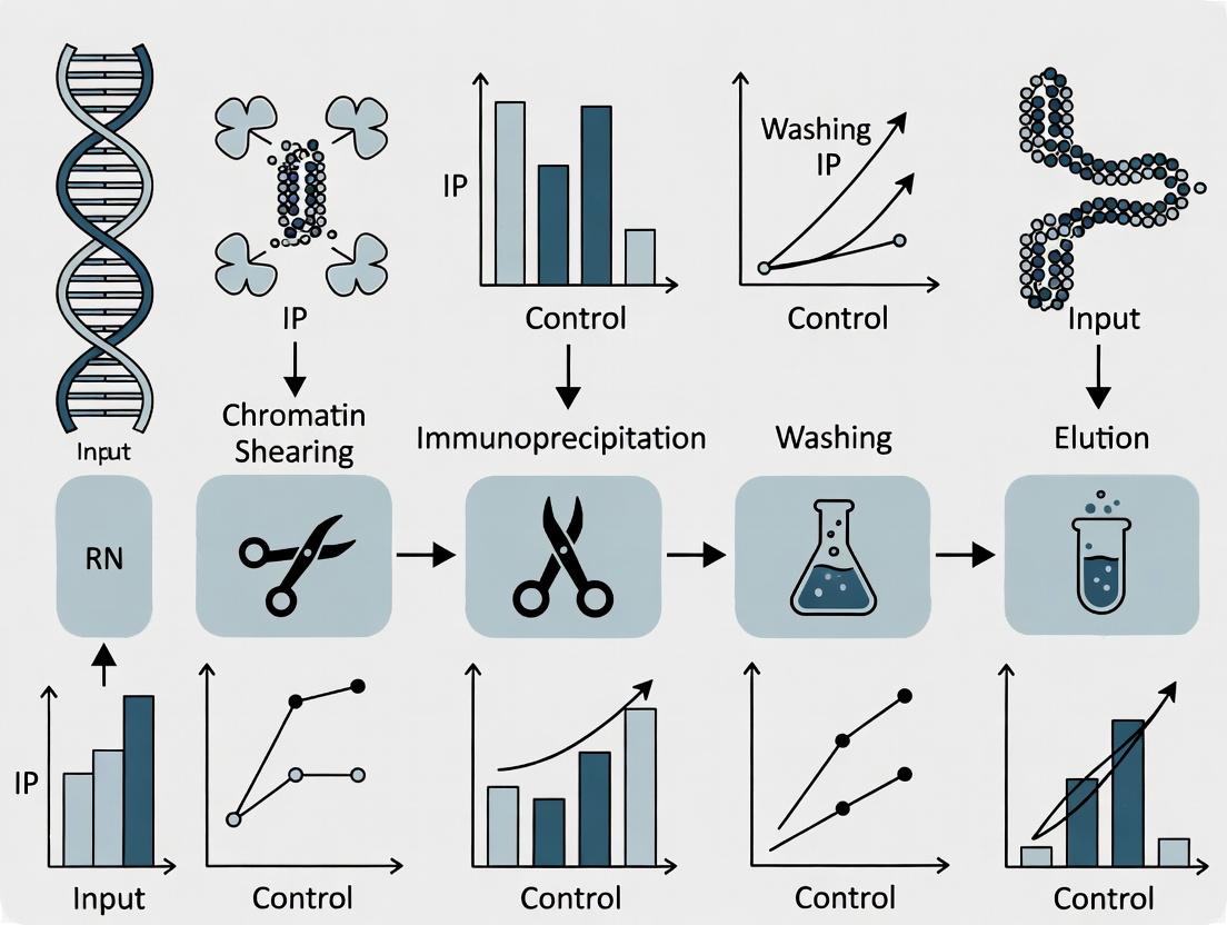
Within the broader thesis on Chromatin Immunoprecipitation (ChIP) assay methodology, the immunoprecipitation (IP) step is the critical purification phase that determines the specificity and yield of the entire experiment. This phase isolates the protein-DNA complexes of interest from the vast background of cellular lysate. The selection of the antibody and the solid-phase support (beads) directly dictates the success of subsequent steps, including washing, elution, and final analysis. This guide provides an in-depth technical analysis of the core considerations for optimizing this pivotal stage.
The Scientist's Toolkit: Research Reagent Solutions
| Item | Primary Function | Key Considerations for ChIP |
|---|---|---|
| Primary Antibody | Specifically binds to the target protein (or epitope-tag) in the crosslinked complex. | Must be validated for ChIP ("ChIP-grade"); recognizes target in fixed, denatured chromatin. Polyclonal often offers higher signal; monoclonal offers higher specificity. |
| Species-Matched Control IgG | Provides a negative control for non-specific binding. | Should be from the same host species as the primary antibody, lacking specific antigen reactivity. |
| Protein A/G Magnetic Beads | Solid-phase support that binds the Fc region of antibodies to capture immune complexes. | Magnetic beads allow for rapid, tube-free separations. Protein A/G mixtures offer broad species/isotype coverage. |
| Blocking Reagents | Reduce non-specific binding of chromatin to beads or tubes. | Commonly used: BSA, salmon sperm DNA, tRNA. Critical for low-background ChIP. |
| ChIP-Compatible Lysis & Wash Buffers | Maintain integrity of protein-DNA complexes while removing non-specifically bound material. | Contain detergents (e.g., SDS, DOC, NP-40) and salts; stringency increases with subsequent washes. |
| Elution Buffer | Releases immunoprecipitated complexes from the beads. | Typically contains SDS and NaHCO₃; designed to reverse crosslinks in the subsequent ChIP step. |
Choosing the Right Antibody
The antibody is the cornerstone of IP specificity. For ChIP, the antibody must recognize its target epitope even after formaldehyde crosslinking, which can mask or alter conformational epitopes.
Antibody Validation Metrics
Table 1 summarizes key validation data that should be sourced from supplier datasheets or literature.
Table 1: Quantitative Metrics for ChIP Antibody Evaluation
| Metric | Ideal/Recommended Value | Impact on Experiment |
|---|---|---|
| ChIP Validation | Datasheet shows successful ChIP-seq/ChIP-qPCR data. | Confirms epitope accessibility post-crosslinking. |
| Signal-to-Noise Ratio | ≥ 5-fold enrichment over IgG control in qPCR. | Indicates specific vs. non-specific DNA pull-down. |
| Target Specificity | Verified by knockout/knockdown cell lines (loss of signal). | Confirms absence of off-target binding. |
| Titer/Amount per IP | 1-10 µg per reaction is typical. | Optimize to balance yield with cost and background. |
| Species & Isotype | IgG; host species compatible with Protein A/G. | Determines bead choice (see Section 4). |
Experimental Protocol: Antibody Titration for ChIP
Objective: To determine the optimal amount of antibody that maximizes specific enrichment while minimizing non-specific background.
Materials:
- Sheared, crosslinked chromatin (e.g., from 1x10⁶ cells per IP point).
- ChIP-validated antibody.
- Species-matched control IgG.
- Protein A/G magnetic beads.
- Lysis Buffer, Wash Buffers, Elution Buffer.
- Rotating mixer at 4°C.
Method:
- Prepare Beads: For each IP point, wash 25 µL of bead slurry twice with lysis buffer. Block with 0.5 mg/mL BSA for 1 hour at 4°C.
- Set Up IP Reactions: Aliquot equal volumes of chromatin into separate tubes. Add the primary antibody in a dilution series (e.g., 0.5 µg, 1 µg, 2 µg, 5 µg). Include a tube with control IgG.
- Incubate: Incubate overnight at 4°C with rotation.
- Capture Complexes: Add pre-blocked beads to each tube. Incubate for 2 hours at 4°C with rotation.
- Wash & Elute: Wash beads sequentially with low-salt, high-salt, LiCl, and TE buffers. Elute complexes in Elution Buffer.
- Reverse Crosslinks & Analyze: Treat all samples (IP and Input) with NaCl and Proteinase K at 65°C. Purify DNA. Analyze enrichment at a known positive genomic locus and a negative control region via qPCR.
- Calculate: Determine % Input and fold-enrichment over IgG control for each antibody amount. The optimal amount yields the highest fold-enrichment with minimal increase in background signal at the negative locus.
Choosing the Right Beads
Beads provide the solid matrix for isolating antibody-bound complexes. Magnetic beads have largely replaced agarose for ChIP due to ease of handling.
Bead Type Comparison
Table 2: Comparison of Common Bead Types for ChIP
| Bead Type | Binding Principle | Advantages | Disadvantages |
|---|---|---|---|
| Protein A Magnetic | Binds Fc region of most mammalian IgGs, especially human, rabbit, mouse (IgG2a, IgG2b). | Strong binding, low non-specific DNA binding. | Poor binding to mouse IgG1, rat, goat IgG. |
| Protein G Magnetic | Broad affinity for IgG from many species, including mouse IgG1. | Excellent for mouse and rat antibodies. | Slightly higher non-specific binding than Protein A. |
| Protein A/G Magnetic | Recombinant fusion of A and G domains. | Broadest species/isotype coverage in one bead. | Can be more expensive. |
| Antibody-Conjugated | Primary antibody is covalently pre-coupled. | Reduces antibody co-elution, improves reproducibility. | Less flexible; dedicated to one target. |
Experimental Protocol: Bead Blocking and Preparation
Objective: To minimize non-specific binding of chromatin to beads, a major source of background.
Materials:
- Protein A/G magnetic beads.
- PBS/0.1% BSA.
- Sheared salmon sperm DNA (10 mg/mL).
- BSA (10 mg/mL).
Method:
- Wash: Resuspend bead slurry and transfer required volume. Place tube on a magnetic rack. Discard supernatant once clear. Resuspend in 1 mL PBS/0.1% BSA. Repeat wash twice.
- Block: After final wash, resuspend beads in 1 volume of PBS/0.1% BSA. Add sheared salmon sperm DNA to 0.5 mg/mL and BSA to 1 mg/mL.
- Incubate: Rotate bead suspension at 4°C for a minimum of 2 hours (overnight is optimal).
- Store: Beads can be stored in blocking buffer at 4°C for up to a week. Wash once with lysis buffer immediately before adding to the IP reaction.
Integrated Workflow and Decision Pathway
The following diagram illustrates the logical decision process for selecting the optimal antibody-bead combination within the ChIP workflow.
Title: ChIP IP Antibody and Bead Selection Workflow
The immunoprecipitation phase is a deterministic gatekeeper in ChIP assays. A rigorous, evidence-based selection of a ChIP-validated antibody, paired with the appropriate, thoroughly blocked beads, establishes the foundation for high-specificity, low-background results. Systematic titration and control experiments are non-negotiable for rigorous research. This optimization directly feeds into the reliability and interpretability of the final genomic data, a core tenet of any thesis on ChIP methodology.
Chromatin Immunoprecipitation (ChIP) is a cornerstone technique for mapping protein-DNA interactions in vivo. Following the immunoprecipitation of protein-DNA complexes, Phase 5 represents the critical final experimental steps: reversing the formaldehyde-induced crosslinks and purifying the target DNA. The efficacy of this phase directly dictates the quality, specificity, and quantifiability of downstream analyses, such as qPCR or next-generation sequencing (ChIP-seq). Incomplete reversal or impure DNA can lead to high background noise, false negatives, and unreliable data, undermining the entire assay.
Technical Guide to Reversal and Purification
Reversal of Crosslinks
Core Principle: The covalent bonds formed between proteins and DNA by formaldehyde are heat-labile. Incubation at elevated temperature in the presence of salt (NaCl) catalyzes the reversal of these crosslinks, freeing the immunoprecipitated DNA.
Detailed Protocol:
- Post-IP Wash: After the final wash of the Protein A/G beads, carefully remove all residual wash buffer.
- Elution Buffer Preparation: Prepare a fresh elution buffer (e.g., 1% SDS, 0.1M NaHCO₃). For standard ChIP, 100-200 µL is typically used per sample.
- Elution: Add the elution buffer to the beads. Vortex briefly and incubate at room temperature for 15 minutes with rotation. Pellet the beads and transfer the supernatant (containing the eluted complexes) to a new tube.
- Reversal Incubation: To the eluate, add NaCl to a final concentration of 200 mM (e.g., add 10 µL of 5M NaCl to 240 µL of eluate for a ~200 mM final concentration). Vortex to mix.
- Heat Denaturation: Incubate the samples at 65°C for a minimum of 4-6 hours, preferably overnight (~12-16 hours). This extended, high-temperature incubation ensures complete reversal of crosslinks and denaturation of proteins.
Note: For ChIP-seq, inclusion of Proteinase K (see below) is standard.
DNA Purification
Following reversal, the sample contains target DNA, residual proteins, RNA, salts, and SDS. Purification isolates DNA cleanly.
Detailed Protocol (Phenol-Chloroform Extraction & Ethanol Precipitation):
- Digestion: After the reversal incubation, cool samples to room temperature. Add 2 µL of 10 mg/mL RNase A and incubate at 37°C for 30 minutes to digest RNA.
- Proteinase K Treatment: Add 4 µL of 20 mg/mL Proteinase K. Incubate at 55°C for 1-2 hours to digest proteins. This step is critical for high-purity DNA, especially for sequencing.
- Phenol-Chloroform Extraction:
- Add an equal volume of phenol:chloroform:isoamyl alcohol (25:24:1).
- Vortex vigorously for 30 seconds.
- Centrifuge at >13,000 x g for 5 minutes at room temperature.
- Carefully transfer the upper aqueous phase (containing DNA) to a new tube.
- Ethanol Precipitation:
- Add 2.5 volumes of ice-cold 100% ethanol and 0.1 volume of 3M sodium acetate (pH 5.2). Include 1 µL of glycogen (20 mg/mL) as a carrier if DNA yield is expected to be low.
- Mix well and incubate at -80°C for at least 1 hour or overnight to precipitate DNA.
- Wash & Resuspension:
- Centrifuge at >13,000 x g for 30 minutes at 4°C. Carefully decant the supernatant.
- Wash the pellet with 500 µL of ice-cold 75% ethanol. Centrifuge again at >13,000 x g for 10 minutes at 4°C.
- Air-dry the pellet for 5-10 minutes (do not over-dry).
- Resuspend the purified DNA in 20-50 µL of TE buffer (10 mM Tris-HCl, pH 8.0, 1 mM EDTA) or nuclease-free water.
Alternative Method: Silica-membrane column-based purification kits (often designed for ChIP) offer faster processing and avoid hazardous organic solvents. Follow manufacturer protocols, often incorporating the RNase and Proteinase K steps prior to column binding.
Table 1: Key Parameters for Crosslink Reversal Efficiency
| Parameter | Optimal Condition | Effect of Deviation |
|---|---|---|
| Incubation Temperature | 65°C | <60°C: Incomplete reversal. >70°C: Increased DNA degradation. |
| Incubation Time | 6-16 hours | <4 hours: Substantially incomplete reversal. |
| [NaCl] in Reversal Mix | 200 mM | Lower conc.: Slower reversal kinetics. Higher conc.: Minimal additional benefit. |
| Proteinase K Digestion | 55°C for 1-2 hrs | Omission: Contaminating proteins carry over, inhibiting downstream assays. |
Table 2: Comparison of DNA Purification Methods
| Method | Average Recovery Yield | A260/A280 Purity | Time Required | Best For |
|---|---|---|---|---|
| Phenol-Chloroform + EtOH Precipitation | 60-80% | 1.7-1.9 | 3-4 hours (plus overnight precipitation) | High-yield inputs, routine qPCR. |
| Silica-Column Kit | 70-90% | 1.8-2.0 | 1-1.5 hours | High-throughput, ChIP-seq, avoiding organics. |
| SPRI Bead-Based Cleanup | 85-95% | 1.8-2.0 | 30-45 minutes | ChIP-seq library preparation, automation. |
The Scientist's Toolkit: Key Reagents & Materials
Table 3: Essential Reagents for Phase 5
| Item | Function | Critical Notes |
|---|---|---|
| SDS Elution Buffer | Disrupts antibody-antigen binding, releases complexes from beads. | Must be fresh; SDS can precipitate if cold. |
| 5M Sodium Chloride (NaCl) | Catalyzes the heat-driven reversal of formaldehyde crosslinks. | Critical component of reversal buffer. |
| RNase A | Degrades RNA contaminating the sample. | Prevents RNA from interfering with DNA quantification and assays. |
| Proteinase K | Broad-spectrum serine protease digests proteins, including nucleases. | Essential for high-purity DNA; inactivates by heating to 95°C. |
| Phenol:Chloroform:IAA | Organic extraction removes proteins and lipids from aqueous DNA solution. | Hazardous; requires proper disposal. IAA prevents foaming. |
| Glycogen (molecular grade) | Inert carrier to visualize pellet and improve recovery of low-nanogram DNA. | Do not use if downstream enzymatic steps are sensitive to contaminants. |
| TE Buffer (pH 8.0) | Resuspension buffer stabilizes DNA; EDTA chelates Mg²⁺ to inhibit DNases. | Preferable over water for long-term storage of DNA. |
| Silica-Membrane Spin Columns | Bind DNA under high-salt conditions; impurities are washed away. | Kit-specific binding/wash buffers must be used. |
Experimental Workflow Visualization
Title: Phase 5: Reversal & Purification Workflow
Key Signaling/Mechanistic Pathway
Title: Molecular Events in Crosslink Reversal & Cleanup
Within the broader thesis on Chromatin Immunoprecipitation (ChIP) assay methodologies, the selection and execution of downstream analysis represent a critical bifurcation. This phase determines the resolution, throughput, and biological insights gleaned from the enriched DNA. Two principal workflows dominate: the targeted, quantitative approach of ChIP-qPCR and the genome-wide, discovery-oriented approach of ChIP-seq. This guide provides an in-depth technical comparison, detailing protocols, data interpretation, and strategic application for researchers and drug development professionals.
Core Workflow Comparison
The fundamental steps following chromatin immunoprecipitation diverge significantly between the two methods.
Diagram Title: Decision Flow: ChIP-qPCR vs. ChIP-seq Downstream Paths
Detailed Experimental Protocols
ChIP-qPCR Protocol
Objective: To quantitatively measure protein-DNA enrichment at specific genomic loci.
Materials:
- ChIP-enriched DNA (eluted in TE buffer or water).
- Control DNA Samples: Input DNA (pre-IP), Negative Control IgG IP DNA.
- Sequence-Specific Primers (forward and reverse) for target and negative control regions.
- SYBR Green or TaqMan qPCR Master Mix.
- Real-Time PCR Instrument.
Procedure:
- DNA Dilution: Dilute ChIP and control DNA samples appropriately (typically 1:10 to 1:100) to fit the linear range of qPCR detection.
- Reaction Setup: In a 96-well plate, assemble reactions in triplicate:
- 5-10 µL diluted DNA template.
- 10 µL 2X qPCR Master Mix.
- 0.5-1.0 µM each primer.
- Nuclease-free water to 20 µL total.
- qPCR Cycling: Run on a real-time PCR instrument using standard cycling conditions (e.g., 95°C for 10 min, then 40 cycles of 95°C for 15 sec and 60°C for 1 min, followed by a melt curve analysis).
- Data Analysis: Calculate Cycle Threshold (Ct) values. Determine percent input or fold enrichment using the ΔΔCt method.
ChIP-seq Library Preparation Protocol
Objective: To prepare the enriched DNA for high-throughput sequencing.
Materials:
- ChIP-enriched DNA (1-50 ng).
- Library Prep Kit (e.g., Illumina TruSeq ChIP).
- DNA Cleanup Beads (SPRI beads).
- Thermocycler.
- Qubit Fluorometer and Bioanalyzer/TapeStation.
Procedure:
- End Repair: Convert overhangs into phosphorylated blunt ends using a mix of T4 DNA Polymerase, Klenow Fragment, and T4 Polynucleotide Kinase. Incubate at 20-30°C for 30 min.
- A-tailing: Add a single 'A' nucleotide to the 3' ends of the blunt fragments using Klenow exo- (3' to 5' exo minus) and dATP. Incubate at 37°C for 30 min. This prevents self-ligation and prepares for adapter ligation.
- Adapter Ligation: Ligate indexed sequencing adapters with a complementary 'T' overhang to the 'A'-tailed fragments using T4 DNA Ligase. Incubate at 20°C for 15-30 min.
- Size Selection: Purify the ligation product and select fragments of 200-500 bp using SPRI bead double-size selection to optimize for cluster generation.
- PCR Enrichment: Amplify the adapter-ligated DNA using 8-15 cycles of PCR with primers complementary to the adapter sequences. This step enriches for fragments that have adapters on both ends.
- Library QC: Quantify the final library using Qubit and assess size distribution and quality via Bioanalyzer. Pool equimolar amounts of indexed libraries for multiplexed sequencing.
Data Output and Analysis Pathways
The analysis of raw data from each method follows distinct computational or statistical pathways.
Diagram Title: ChIP-seq vs. ChIP-qPCR Data Analysis Pathways
Quantitative Comparison Table
Table 1: Strategic and Technical Comparison of Downstream Workflows
| Parameter | ChIP-qPCR | ChIP-seq |
|---|---|---|
| Primary Goal | Targeted validation & quantification | Genome-wide discovery & mapping |
| Throughput | Low (tens of loci) | High (entire genome) |
| Resolution | Locus-specific (primer-defined) | Base-pair (limited by fragment size) |
| Required DNA | Very low (0.1-1 ng per reaction) | Moderate to high (1-50 ng total) |
| Typical Cost | Low per sample, scales with loci | High per sample (sequencing costs) |
| Turnaround Time | Fast (hours to 1 day post-IP) | Slow (days to weeks for sequencing & bioinformatics) |
| Data Output | Ct values, % Input, Fold Enrichment | FASTQ files, aligned reads (BAM), peak calls (BED) |
| Bioinformatics Burden | Minimal (basic statistics) | Extensive (specialized pipelines required) |
| Ideal Application | Confirming known binding sites, time-course/dose-response studies, many samples | Identifying novel binding sites, characterizing global binding profiles, chromatin state |
Table 2: Typical Data Metrics from Published Studies (Representative Values)
| Metric | Typical ChIP-qPCR Result | Typical ChIP-seq Result |
|---|---|---|
| Positive Control Loci | 10- to 100-fold enrichment over IgG | Thousands to tens of thousands of significant peaks (p < 1e-5) |
| Negative Control Region | ~1-fold enrichment (no enrichment) | < 0.001% of reads in non-specific regions |
| Replicate Correlation | R² > 0.98 for technical replicates | Pearson correlation between biological replicates R > 0.9 |
| Key Validation Criterion | Significant difference (p < 0.05) from control IgG/region | Irreproducible Discovery Rate (IDR) < 0.05 for peaks |
The Scientist's Toolkit: Key Research Reagent Solutions
Table 3: Essential Materials for Downstream ChIP Analysis
| Item | Function | Example/Catalog |
|---|---|---|
| qPCR Master Mix (SYBR Green) | Contains DNA polymerase, dNTPs, buffer, and fluorescent dye for real-time quantification during PCR. | Applied Biosystems Power SYBR Green, Bio-Rad iTaq Universal SYBR Green. |
| Validated ChIP-qPCR Primers | Pre-designed, sequence-specific primers for positive and negative control genomic regions (e.g., GAPDH promoter, gene desert). | Qiagen EpiTect ChIP qPCR Assays, custom-designed from primer databases. |
| ChIP-seq Library Prep Kit | Integrated reagent suite for end repair, A-tailing, adapter ligation, and PCR enrichment of low-input DNA. | Illumina TruSeq ChIP Library Prep Kit, NEBNext Ultra II DNA Library Prep Kit. |
| Indexing Adapters (Multiplexing) | Unique oligonucleotide barcodes ligated to each library, enabling pooling and parallel sequencing of multiple samples. | Illumina TruSeq CD Indexes, IDT for Illumina UD Indexes. |
| SPRI Size Selection Beads | Magnetic beads for clean-up, size selection, and buffer exchange during library prep, critical for insert size range. | Beckman Coulter AMPure XP, KAPA Pure Beads. |
| High-Sensitivity DNA Assay Kit | Fluorometric or electrophoretic analysis for accurate quantification and quality control of libraries pre-sequencing. | Agilent High Sensitivity DNA Kit (Bioanalyzer), Qubit dsDNA HS Assay Kit. |
| Peak Calling Software | Bioinformatics tool to identify genomic regions with significant enrichment of sequencing reads compared to background. | MACS2 (Model-based Analysis of ChIP-Seq), HOMER (findPeaks). |
| Genome Browser | Visualization platform to view and interrogate aligned read (BAM) and peak (BED) files in a genomic context. | UCSC Genome Browser, Integrative Genomics Viewer (IGV). |
This technical whitepaper explores three advanced applications of the Chromatin Immunoprecipitation (ChIP) assay, framed within the broader thesis that ChIP is a foundational and versatile tool for elucidating gene regulatory mechanisms in health and disease. While standard ChIP identifies protein-DNA interactions at a single point in time, these advanced methodologies unlock dynamic, combinatorial, and clinically relevant insights crucial for modern research and therapeutic development.
Re-ChIP (Sequential ChIP)
Re-ChIP is a powerful technique used to investigate the simultaneous co-localization of two or more distinct proteins on the same genomic DNA fragment. This is critical for studying complex formation, such as transcription factor cooperativity or the coexistence of specific histone modifications.
Experimental Protocol
- First Immunoprecipitation: Perform a standard ChIP protocol using the first antibody (Ab1) and protein G/A magnetic beads. Elute the immunoprecipitated chromatin complexes using a gentle elution buffer (e.g., 25 mM DTT, 1% SDS) instead of reversing cross-links.
- Immunoprecipitation Elution Dilution: Dilute the eluate 1:50 with Re-ChIP buffer (1% Triton X-100, 2 mM EDTA, 150 mM NaCl, 20 mM Tris-HCl, pH 8.1).
- Second Immunoprecipitation: Use the diluted eluate as input for a second round of IP with an antibody against a second target (Ab2). Fresh beads are typically added.
- Wash, Elution, and Cross-link Reversal: Wash beads stringently, elute complexes, and reverse cross-links simultaneously for both the first IP, second IP, and Re-ChIP samples.
- DNA Purification & Analysis: Purify DNA and analyze by qPCR or sequencing (Re-ChIP-seq).
Key Considerations & Quantitative Data
Success depends on antibody specificity and stringent washing. Controls (IgG for each IP and sequential IP with non-related antibodies) are essential. Typical yields are lower than standard ChIP.
Table 1: Representative Re-ChIP-qPCR Data Analysis
| Sample | Target Locus (% Input) | Control Locus (% Input) | Enrichment (Fold over IgG) |
|---|---|---|---|
| Ab1 IP | 5.2 | 0.1 | 52.0 |
| Ab2 IP | 4.8 | 0.1 | 48.0 |
| Re-ChIP (Ab1+Ab2) | 0.5 | 0.05 | 10.0 |
| Sequential IgG | 0.05 | 0.06 | 0.8 |
Diagram Title: Re-ChIP Sequential Immunoprecipitation Workflow
Time-Course ChIP
Time-course ChIP involves performing ChIP assays on samples collected at sequential time points following a stimulus (e.g., drug addition, differentiation signal, infection). It maps the temporal dynamics of transcription factor binding, histone modification turnover, or polymerase recruitment.
Experimental Protocol
- Stimulus Application & Sampling: Apply a synchronized stimulus to cells or tissue. Harvest aliquots of cells or freeze tissue samples at defined time points (e.g., 0, 5, 15, 30, 60, 120 minutes). Include an unstimulated (t=0) control.
- Cross-linking: Immediately cross-link each sample at the moment of harvest using formaldehyde.
- Parallel Processing: Process all time-point samples in parallel using identical ChIP protocols (sonication, IP conditions, wash stringency) to enable direct comparison.
- Normalization: Use spike-in controls (e.g., exogenous chromatin from Drosophila or yeast) to normalize for technical variation between IPs across time points, especially crucial for global histone modification studies.
- High-Throughput Analysis: Analyze by qPCR for specific loci or, more commonly, by ChIP-seq for genome-wide profiling.
Key Considerations & Quantitative Data
Experimental design must account for the biological response kinetics. Robust normalization is critical. Data is often presented as fold-change over time zero or as normalized read density.
Table 2: Time-Course ChIP-qPCR for Transcription Factor Recruitment
| Time Post-Stimulation | Locus A (% Input) | Locus B (% Input) | Normalized Fold Change (vs t=0) |
|---|---|---|---|
| 0 min | 0.10 | 0.05 | 1.0 |
| 15 min | 0.85 | 0.07 | 8.5 |
| 30 min | 1.50 | 0.45 | 15.0 |
| 60 min | 0.60 | 0.90 | 6.0 |
| 120 min | 0.15 | 0.30 | 1.5 |
Diagram Title: Time-Course ChIP Experimental Design
ChIP from Clinical Samples
Adapting ChIP for clinical specimens (e.g., formalin-fixed paraffin-embedded [FFPE] tissue, primary patient cells, tumor biopsies) bridges basic research and translational medicine, enabling the study of epigenetic drivers in disease.
Experimental Protocol
For Frozen Tissue/Cells:
- Nuclei Isolation: Mechanically dissociate and homogenize tissue in lysis buffer to isolate nuclei.
- Cross-linking: Cross-link with formaldehyde. Quench with glycine.
- Chromatin Shearing: Sonicate nuclei to fragment chromatin. Optimization is critical due to sample variability.
For FFPE Tissue:
- Deparaffinization & Rehydration: Slice sections, treat with xylene and graded ethanol.
- Antigen Retrieval & Reversal: Use heat-induced epitope retrieval (HIER) in citrate/EDTA buffer to partially reverse cross-links and expose epitopes.
- Micrococcal Nuclease (MNase) Digestion: Preferred over sonication for FFPE. Digest tissue to release mononucleosomes.
- Standard IP: Proceed with immunoprecipitation using validated antibodies compatible with FFPE-derived chromatin.
Key Considerations & Quantitative Data
Sample quality and pre-analytical variables are major challenges. Input requirements are higher (10-20 sections of 10μm FFPE). Antibody validation on similar material is non-negotiable. Data is often correlative with patient outcomes.
Table 3: Comparison of ChIP from Different Clinical Sample Types
| Sample Type | Starting Material | Key Processing Step | Major Challenge | Typical DNA Yield per IP |
|---|---|---|---|---|
| Frozen Tissue | 20-50 mg tissue | Homogenization & Sonication | Cellular heterogeneity, RNase activity | 5-20 ng |
| FFPE Tissue | 10-20 x 10μm sections | HIER & MNase Digestion | Over-fixation, DNA fragmentation | 2-10 ng |
| Primary Cells | 0.5-1 x 10^6 cells | Standard cross-linking | Limited cell number, activation state | 1-5 ng |
Diagram Title: ChIP from Clinical Samples Processing Paths
The Scientist's Toolkit: Research Reagent Solutions
Table 4: Essential Reagents for Advanced ChIP Applications
| Item | Function & Application | Key Consideration |
|---|---|---|
| Validated ChIP-Grade Antibodies | Specific immunoprecipitation of target protein or histone modification. Critical for all applications, especially Re-ChIP. | Must be validated for ChIP (check databases like Cistrome DB). Re-ChIP requires antibodies from different host species or subtypes. |
| Protein A/G Magnetic Beads | Efficient capture of antibody-antigen complexes. Preferred for Re-ChIP for easier elution. | Magnetic separation minimizes background and eases sequential IP steps. |
| Cross-linking Reagents | Formaldehyde: Standard reversible cross-linker. DSG (Disuccinimidyl glutarate): Optional for distant cross-linking before formaldehyde. | FFPE samples use extensive formalin fixation; reversal is incomplete. |
| Chromatin Shearing Reagents | Covaris sonication shearing tubes: For frozen/cell samples. Micrococcal Nuclease (MNase): For FFPE or nucleosome positioning studies. | Sonication must be optimized per cell/tissue type. MNase digestion requires titration. |
| Spike-in Chromatin | Exogenous chromatin (e.g., Drosophila, S. pombe). Added prior to IP for normalization in time-course or clinical ChIP. | Allows correction for technical variation between samples, essential for quantitative comparisons. |
| Antigen Retrieval Buffer | Citrate or EDTA-based buffer (pH 6.0 or 9.0). Used in HIER to partially reverse FFPE cross-links and expose epitopes. | Optimal pH and time must be determined for each antibody-target pair in FFPE. |
| DNA Clean-up/Purification Kits | Silica-membrane or SPRI bead-based kits. For purifying low-abundance ChIP DNA after elution. | High recovery efficiency is critical for low-input samples from clinical or Re-ChIP experiments. |
| High-Sensitivity DNA Assay Kits | Fluorometric assays (e.g., Qubit). Accurately quantifies low-concentration ChIP DNA prior to library prep or qPCR. | More accurate than UV absorbance for dilute, fragmented ChIP DNA. |
ChIP Assay Troubleshooting: Solving Common Problems of Low Signal and High Background
Within the framework of chromatin immunoprecipitation (ChIP) assay research, antibody performance is the critical determinant of experimental success. Poor antibody performance manifests as high background, non-specific signals, or a lack of target enrichment, directly compromising data integrity and the validity of conclusions regarding protein-DNA interactions and epigenetic states. This technical guide dissects the core triumvirate of antibody diagnostics—specificity, titer, and validation—providing a systematic approach for researchers and drug development professionals to troubleshoot and optimize this fundamental reagent.
Specificity: The Cornerstone of Reliability
Specificity refers to an antibody's ability to bind exclusively to its intended target epitope. In ChIP, non-specific binding can lead to false-positive identification of genomic loci.
Diagnostic Experiments for Specificity
- Knockout/Knockdown Validation: The gold standard. Compare ChIP signal in wild-type vs. genetically modified (KO/KD) cell lines for the target protein. A specific antibody will show abrogated signal in the modified line.
- Peptide Blocking Competition: Pre-incubate the antibody with a molar excess of the immunizing peptide. Specific binding should be competitively inhibited.
- Western Blot Analysis: Subject the ChIP input lysate to immunoblotting. A specific antibody should produce a single band at the expected molecular weight, indicating minimal cross-reactivity.
Table 1: Specificity Validation Methods & Interpretations
| Method | Experimental Design | Interpretation of Positive Specificity |
|---|---|---|
| Genetic KO/KD | ChIP-qPCR in isogenic WT vs. target KO cell lines. | >70-80% reduction in signal at positive control loci in KO cells. |
| Peptide Blocking | ChIP with antibody pre-incubated ± immunizing peptide. | >80% inhibition of enrichment with peptide present. |
| Western Blot | SDS-PAGE of input chromatin supernatant, probed with ChIP antibody. | A single, dominant band at correct molecular weight. |
| Orthogonal Validation | Compare ChIP-seq profile with published data or an independent, validated antibody. | High correlation of peak calling and genomic distribution (e.g., Pearson's r > 0.7). |
Detailed Protocol: Knockout/Knockdown Validation ChIP-qPCR
- Cell Preparation: Culture isogenic wild-type and target gene knockout cell lines (e.g., via CRISPR-Cas9). Confirm knockout via western blot.
- Cross-linking & Harvest: Treat ~1x10^7 cells per line with 1% formaldehyde for 10 min at room temperature. Quench with 125 mM glycine.
- Chromatin Prep: Lyse cells, isolate nuclei, and shear chromatin via sonication to 200-500 bp fragments. Confirm fragment size by agarose gel.
- Immunoprecipitation: Aliquot sheared chromatin. Pre-clear with protein A/G beads. Incubate supernatant with 1-5 µg of test antibody overnight at 4°C. Include an IgG isotype control.
- Bead Capture & Washing: Add beads, incubate, then wash sequentially with low salt, high salt, LiCl, and TE buffers.
- Elution & De-crosslinking: Elute complexes, reverse crosslinks at 65°C overnight with 200 mM NaCl.
- DNA Purification: Treat with RNase A and Proteinase K, then purify DNA via column or phenol-chloroform.
- qPCR Analysis: Perform qPCR on purified DNA using primers for a known positive binding locus and a negative control locus. Calculate % input for each sample. Specificity is confirmed by signal loss in the KO line at the positive locus.
Titer and Affinity: Optimizing Signal-to-Noise
Antibody titer (optimal dilution) and inherent affinity significantly impact the signal-to-noise ratio. Using an antibody at too high a concentration increases non-specific binding.
Titer Determination Experiment
A chromatin immunoprecipitation titration is essential.
- Protocol: Perform identical ChIP reactions in parallel using a serial dilution of the antibody (e.g., 1 µg, 2 µg, 5 µg per reaction). Keep all other parameters constant.
- Analysis: Quantify enrichment via qPCR at positive and negative control regions. Plot enrichment (% Input) versus antibody amount. The optimal titer is the point just before the enrichment curve plateaus, while negative control signal remains minimal.
Table 2: Titer Optimization Outcomes
| Antibody Amount | Positive Locus Signal | Negative Locus Signal | Interpretation |
|---|---|---|---|
| Too Low (e.g., 0.5 µg) | Low/Undetectable | Low | Insufficient for detection. |
| Optimal (e.g., 2 µg) | High & Specific | Low | Ideal signal-to-noise. |
| Too High (e.g., 5 µg) | High (Plateaued) | Elevated | Increased non-specific binding, poor signal-to-noise. |
A Framework for Comprehensive Antibody Validation
Effective validation for ChIP requires application-specific testing.
Figure 1: Antibody validation workflow for ChIP assays.
The Scientist's Toolkit: Research Reagent Solutions
Table 3: Essential Reagents for Antibody Validation in ChIP
| Reagent/Material | Function in Diagnosis | Key Consideration |
|---|---|---|
| Validated Positive Control Cells | Provide known source of target antigen for specificity (KO) and titer tests. | Use isogenic pairs (WT/KO) from reputable sources (e.g., ATCC, Horizon). |
| Immunizing Peptide | Serves as competitor in blocking experiments to confirm epitope specificity. | Must match the antibody's immunogen sequence precisely. |
| Protein A/G Magnetic Beads | Capture antibody-antigen complexes; magnetic format improves wash efficiency. | Choose bead type (A, G, or A/G) matched to antibody species/isotype. |
| Cross-linking Reagent (Formaldehyde) | Preserves protein-DNA interactions in vivo prior to ChIP. | Fresh preparation (<6 months) is critical for consistent efficiency. |
| Chromatin Shearing System | Fragments DNA to 200-500 bp for resolution. | Sonicators (probe or bath) must be calibrated for each cell type/chromatin prep. |
| ChIP-Grade IgG Control | Species/isotype-matched non-immune antibody for background determination. | Essential for distinguishing true enrichment from non-specific bead binding. |
| qPCR Primers | Amplify known positive and negative genomic regions to quantify enrichment. | Positive locus must be established via literature or prior experiments. |
Integrated Validation Pathway for ChIP Antibodies
The following workflow integrates all diagnostic parameters into a single, logical sequence for implementation.
Figure 2: Integrated ChIP antibody testing and QC workflow.
Diagnosing antibody performance in ChIP research is a non-negotiable, multi-parameter process. Systematic assessment of specificity through genetic controls, determination of optimal titer via dilution series, and rigorous application-specific validation form an interdependent framework. This disciplined approach transforms antibodies from potential sources of error into reliable tools, thereby underpinning the generation of robust, reproducible data essential for advancing drug discovery and fundamental mechanistic research in epigenetics and gene regulation.
Within the broader context of a ChIP-seq assay, chromatin shearing represents a critical, rate-limiting step. The overarching thesis of successful Chromatin Immunoprecipitation (ChIP) research hinges on the efficient generation of chromatin fragments that are both appropriately sized for high-resolution mapping and of sufficient yield for robust downstream sequencing. Inadequate shearing leads to poor resolution and false-positive peaks, while overly aggressive shearing diminishes yield and compromises signal-to-noise ratios. This technical guide provides a comprehensive analysis of current methodologies to optimize this balance.
The Shearing Imperative in ChIP Workflow
Effective shearing solubilizes cross-linked chromatin, generating fragments typically between 150-500 base pairs (bp). The ideal target is 200-300 bp, which includes the nucleosome core (~147 bp) plus linker DNA. This size range is optimal for single-nucleosome resolution in sequencing.
Diagram Title: ChIP-seq Workflow with Shearing Core
Quantitative Comparison of Shearing Modalities
Current research identifies three primary shearing methods, each with distinct trade-offs between fragment size control, yield, and practicality.
Table 1: Quantitative Comparison of Chromatin Shearing Methods
| Method | Optimal Fragment Size Range | Typical Yield (μg DNA/10^6 Cells) | Hands-on Time | Equipment Cost | Key Advantage | Major Limitation |
|---|---|---|---|---|---|---|
| Ultrasonication (Covaris) | 150-500 bp | 2-5 μg | Low (Automated) | Very High | Precise, reproducible size tuning | High capital cost, sample heating risk |
| Bath Sonication (Bioruptor) | 200-1000 bp | 1-4 μg | Medium | Medium | Parallel processing, consistent cooling | Less precise, optimization intensive |
| Enzymatic Digestion (MNase/Tn5) | 100-300 bp | 3-8 μg | Low | Low | High yield, minimal equipment | Sequence bias, over-digestion risk |
Detailed Experimental Protocols
Protocol A: Optimized Ultrasonication (Covaris) for Cultured Mammalian Cells
Objective: Generate 200-300 bp fragments from cross-linked chromatin. Reagents: PBS, 1% formaldehyde, 2.5M glycine, Lysis Buffer (10 mM Tris-HCl pH 8.0, 10 mM NaCl, 0.2% NP-40), Shearing Buffer (0.1% SDS, 10 mM EDTA, 50 mM Tris-HCl pH 8.1).
- Cross-linking: Fix 10^7 cells in 1% formaldehyde for 10 min at RT. Quench with 125 mM glycine.
- Nuclei Preparation: Pellet cells, wash with cold PBS. Resuspend in 1 mL Lysis Buffer, incubate 15 min on ice. Pellet nuclei.
- Shearing Setup: Resuspend pellet in 1 mL Shearing Buffer. Transfer to a Covaris microTUBE.
- Covaris Settings (for ~250 bp): Peak Incident Power: 140W; Duty Factor: 5%; Cycles per Burst: 200; Time: 4-8 minutes (optimize). Temperature maintained at 4-6°C.
- Post-Shear: Centrifuge at 16,000 x g for 10 min at 4°C. Transfer supernatant (sheared chromatin) to a new tube. Analyze 50 μL on a 1.5% agarose gel or Bioanalyzer.
Protocol B: Enzymatic Shearing with Micrococcal Nuclease (MNase)
Objective: Generate nucleosome-sized fragments with high yield. Reagents: MNase (Worthington), MNase Digestion Buffer (50 mM Tris-HCl pH 7.9, 5 mM CaCl₂, 0.1% NP-40), 0.5 M EGTA (pH 8.0).
- Nuclei Preparation: Prepare nuclei from cross-linked cells as in Protocol A, Step 2. Resuspend in 1 mL MNase Digestion Buffer.
- Titration: Aliquot nuclei suspension (e.g., 100 μL per test). Add a dilution series of MNase (e.g., 0.5, 2, 5, 10 units). Incubate 10 min at 37°C with gentle mixing.
- Reaction Stop: Add EGTA to a final concentration of 10 mM to chelate Ca²⁺.
- Analysis: Centrifuge, run supernatant on gel. Select condition yielding majority of fragments at 1-3 nucleosomes (~150-450 bp).
Optimization Pathway & Decision Logic
Diagram Title: Shearing Method Selection Logic
The Scientist's Toolkit: Key Research Reagent Solutions
Table 2: Essential Reagents for Chromatin Shearing Optimization
| Item | Function & Role in Shearing | Example Product/Brand |
|---|---|---|
| Focused-Ultrasonicator | Applies controlled acoustic energy to physically fracture chromatin. Gold standard for precision. | Covaris S2, M220 |
| Water Bath Sonicator | Provides cavitation energy through a water bath for parallel shearing of multiple samples. | Diagenode Bioruptor Pico |
| Micrococcal Nuclease (MNase) | Endo-exonuclease that cleaves linker DNA between nucleosomes. Used for enzymatic shearing. | Worthington LS004798 |
| Magnetic Crosslinker | Rapid, consistent fixation of protein-DNA interactions prior to shearing. | Inventram ATCL-2 |
| Chip-seq Grade Antibodies | For IP after shearing; specificity is critical for meaningful results. | Cell Signaling Technologies, Abcam |
| DNA High Sensitivity Assay Kits | Accurate quantification of low-concentration sheared DNA post-purification. | Agilent Bioanalyzer HS DNA, Qubit dsDNA HS |
| Size Selection Beads | Post-shearing clean-up and selection of ideal fragment size range (e.g., 200-300 bp). | SPRIselect (Beckman), AMPure XP |
| Thermal Cycler | For reverse crosslinking and other enzymatic steps in enzymatic or hybrid protocols. | Applied Biosystems Veriti |
| Dynabeads Protein A/G | Magnetic beads for efficient immunoprecipitation of antibody-bound chromatin complexes. | Thermo Fisher Scientific |
Achieving the optimal equilibrium between chromatin fragment size and yield is not a one-size-fits-all endeavor but a deliberate, sample-aware optimization process. The choice between sophisticated ultrasonication, cost-effective bath sonication, or high-yield enzymatic digestion must align with the specific goals, sample constraints, and analytical requirements of the ChIP-seq experiment. By systematically applying the quantitative data, protocols, and decision frameworks outlined herein, researchers can robustly standardize this pivotal step, thereby ensuring the integrity and resolution of the subsequent genome-wide epigenetic data that underpins modern drug discovery and mechanistic biology.
Within the broader thesis of Chromatin Immunoprecipitation (ChIP) assay research, the specificity and signal-to-noise ratio of the final data are paramount. The core challenge lies in differentiating true, biologically relevant protein-DNA interactions from non-specific background. This technical guide delves into three critical, inter-related pillars for background reduction: the optimization of wash stringency, the effective blocking of solid-phase supports (beads), and the systematic formulation of immunoprecipitation and wash buffers. Mastery of these elements is fundamental for any researcher, scientist, or drug development professional aiming to derive robust, publication-quality data from ChIP assays, which underpin epigenetic research and target validation.
I. Wash Stringency: Principles and Optimization
Post-immunoprecipitation washes are the primary mechanism for removing loosely bound and non-specifically adsorbed contaminants. Stringency is controlled by ionic strength, detergent concentration, and pH.
Key Wash Buffer Components and Their Roles:
- Salt (NaCl/LiCl): Disrupts ionic interactions. Higher concentrations (>500 mM) increase stringency.
- Detergents (SDS, Triton X-100, Deoxycholate): Solubilize membranes and disrupt hydrophobic interactions. SDS is highly stringent.
- Buffer (Tris, HEPES): Maintains stable pH.
- EDTA/EGTA: Chelates divalent cations, inhibiting nucleases.
Optimization Strategy: A stepwise increase in stringency is standard. Early washes often use moderate-salt buffers with non-ionic detergents to remove contaminants without eluting the target complex. A final high-salt or low-detergent wash can be applied immediately before elution.
Table 1: Common ChIP Wash Buffer Formulations and Stringency
| Buffer Name | Common Formulation (Typical) | Primary Function | Stringency Level |
|---|---|---|---|
| Low Salt Wash | 20 mM Tris-HCl (pH 8.0), 150 mM NaCl, 2 mM EDTA, 1% Triton X-100 | Removes non-specific protein aggregates & contaminants. | Low |
| High Salt Wash | 20 mM Tris-HCl (pH 8.0), 500 mM NaCl, 2 mM EDTA, 1% Triton X-100 | Disrupts weak ionic protein-DNA/protein-protein interactions. | Medium-High |
| LiCl Wash | 10 mM Tris-HCl (pH 8.0), 250 mM LiCl, 1 mM EDTA, 1% NP-40, 1% Deoxycholate | Removes non-specific nucleic acid binding; effective for chromatin. | High |
| TE Buffer (Final) | 10 mM Tris-HCl (pH 8.0), 1 mM EDTA | Removes residual salts/detergents before elution; low nuclease activity. | Very Low |
II. Bead Blocking: Preventing Non-Specific Adsorption
Protein A/G or magnetic beads present surfaces that can passively adsorb chromatin fragments and proteins, generating high background. Pre-blocking is essential.
Detailed Protocol for Bead Blocking:
- Pre-wash: Pellet the required volume of beads. Remove storage solution and wash twice in 1x PBS + 0.1% BSA.
- Blocking Solution Preparation: Prepare a blocking buffer. A robust formulation is 0.5% BSA (w/v) and 0.2 μg/μL sheared salmon sperm DNA (or another inert carrier DNA) in Tris-EDTA buffer. For challenging targets, 0.1% Triton X-100 can be added.
- Incubation: Resuspend the washed beads in 5-10x their volume of blocking buffer.
- Incubation Time & Temperature: Rotate for a minimum of 1 hour at 4°C. Overnight blocking often yields lower background.
- Equilibration: Pellet beads, remove blocking buffer, and wash once with your chosen ChIP incubation/wash buffer before adding the chromatin-antibody mixture.
Critical Note: The blocking agent must be compatible with downstream detection. BSA is universal, but for downstream mass spectrometry, proprietary polymer-based blocking reagents may be preferable.
III. Buffer Optimization: A Systematic Approach
Optimal buffer composition is target-specific and must be determined empirically. A systematic optimization experiment is recommended.
Experimental Protocol for Buffer Optimization:
A. Design: Test a matrix of buffer conditions during the immunoprecipitation step. Key variables:
- Salt (NaCl): 100 mM, 150 mM, 300 mM.
- Detergent Type/Ratio: Triton X-100 (0.1% vs 1%), SDS (0.01% vs 0.1%).
- Blocking Additives: Include 0.1 mg/mL BSA or 0.2 μg/μL carrier DNA directly in the IP buffer.
B. Method:
- Aliquot a fixed amount of pre-cleared chromatin into separate tubes.
- Adjust each aliquot to the desired test buffer composition.
- Add a fixed, validated amount of specific antibody and isotype control to each condition.
- Proceed with standard IP, washes (using a fixed, stringent regime), and elution.
- Analyze all samples by qPCR for a positive control locus and a known negative control locus.
C. Analysis: Calculate the Signal-to-Noise (S/N) ratio for each condition: (Signal at Positive Locus from Specific Ab) / (Signal at Positive Locus from Isotype Control Ab). The condition yielding the highest S/N for the positive locus, while maintaining minimal signal at the negative locus, is optimal.
Table 2: Example Buffer Optimization Results (qPCR Cq Values)
| Test Condition | Specific Ab (Pos Locus) | Isotype Ctrl (Pos Locus) | Specific Ab (Neg Locus) | S/N Ratio |
|---|---|---|---|---|
| 150mM NaCl, 1% Triton | 24.5 | 32.1 | 33.8 | 282 |
| 300mM NaCl, 1% Triton | 25.1 | 31.5 | 34.2 | 181 |
| 150mM NaCl, 0.1% SDS | 26.8 | 32.0 | 35.0 | 82 |
| 300mM NaCl, 0.5% Deoxycholate | 28.3 | 30.9 | 32.5 | 23 |
S/N calculated as 2^(ΔCq), where ΔCq = Cq(Isotype) - Cq(Specific).
The Scientist's Toolkit: Research Reagent Solutions
| Item | Function & Importance |
|---|---|
| Protein A/G Magnetic Beads | Solid-phase support for antibody capture. Magnetic beads allow for rapid, tube-free washes. |
| Sheared Salmon Sperm DNA | Inert carrier DNA used in bead blocking and buffers to saturate non-specific DNA binding sites. |
| BSA (Fraction V, Protease-free) | Standard blocking protein to passivate bead and tube surfaces against non-specific protein adsorption. |
| Protease/Phosphatase Inhibitors | Cocktails added to all buffers pre-IP to maintain chromatin integrity and protein modifications. |
| High-Purity Triton X-100/SDS | Key detergents for modulating stringency. Consistency in grade is critical for reproducibility. |
| Glycogen (Molecular Biology Grade) | Carrier to precipitate DNA during the final ethanol precipitation step, improving recovery. |
| RNAse A (DNA-free) | Used post-IP to remove contaminating RNA, which can interfere with qPCR or library prep. |
| Dynabeads MyOne Streptavidin | For ChIP-seq protocols utilizing biotinylated antibodies or tagged proteins (e.g., CUT&RUN). |
Visualization: The Background Reduction Workflow
Title: Background Reduction Strategy in ChIP
Reducing non-specific background in ChIP assays is not a single step but an integrated strategy. Effective bead blocking creates a passive surface. Optimized immunoprecipitation and wash buffers create a thermodynamic environment that favors the retention of the target complex over spurious interactions. When executed within the framework of a rigorous experimental design, including appropriate controls, these techniques form the foundation for reproducible, high-fidelity ChIP data, ultimately strengthening the conclusions drawn in epigenetic research and drug discovery.
Within the broader context of ChIP assay research, a central challenge that consistently undermines data reliability and reproducibility is the recovery of low DNA yields following the immunoprecipitation (IP) step. This technical whitepaper deconstructs three critical, interdependent variables that govern post-IP DNA yield: crosslinking efficiency, elution efficacy, and downstream PCR amplification bias. Addressing these factors is paramount for generating high-quality, quantitative data essential for both basic research and drug target validation.
Crosslinking: The Foundational Step
Crosslinking stabilizes protein-DNA interactions but is a primary source of yield loss if not optimized. Under-crosslinking leads to complex dissociation, while over-crosslinking creates chromatin that is resistant to shearing and elution, directly reducing DNA recovery.
Quantitative Impact of Crosslinking on Yield:
| Crosslinking Agent & Condition | Typical Formaldehyde Concentration | Incubation Time | Impact on Post-IP DNA Yield | Recommended for |
|---|---|---|---|---|
| Formaldehyde (Reversible) | 1% | 8-10 min @ RT | Optimal balance | Most histone marks |
| Formaldehyde (Reversible) | 1% | >15 min @ RT | Yield decrease (over-crosslink) | N/A |
| DSG + Formaldehyde (Double) | DSG: 2mM; FA: 1% | DSG: 45 min; FA: 10 min | Increased yield for weak/transient interactions | Transcription factors, co-factors |
| EGS (Long-arm) | 1-2 mM | 45-60 min | Can improve yield for distal proteins | Specific architectural proteins |
Protocol: Optimization of Crosslinking for Maximum Yield
- Cell Preparation: Harvest ~1x10^6 cells per IP condition.
- Formaldehyde Fixation: Resuspend cell pellet in 1 mL of growth medium. Add 27 µL of 37% formaldehyde (final ~1%). Incubate at room temperature with gentle rotation for 10 minutes.
- Quenching: Add 100 µL of 1.25 M glycine (final 125 mM). Rotate for 5 minutes.
- Washing: Pellet cells, wash twice with ice-cold PBS containing protease inhibitors.
- Shearing Check: Lyse and sonicate a test sample. Analyze DNA fragment size (goal: 200-500 bp) and concentration via agarose gel electrophoresis and Qubit fluorometry. Low DNA concentration after shearing indicates over-crosslinking.
Title: Crosslinking Optimization Impact on DNA Yield
Elution: Recovering the Complex
Inefficient elution of the antibody-protein-DNA complex from beads is a major, often overlooked, contributor to low yield. Standard elution buffers may not fully reverse crosslinks or dissociate complexes.
Comparative Analysis of Elution Buffers:
| Elution Buffer Composition | Incubation | Avg. DNA Yield Improvement | Key Consideration |
|---|---|---|---|
| 1% SDS, 0.1M NaHCO3 (Standard) | 65°C, 15 min + 30 min | Baseline (1x) | May leave >20% complex on beads |
| 1% SDS, 0.1M NaHCO3 + 10mM DTT | 65°C, 15 min + 30 min | ~1.3x | Reduces disulfide bonds, improves protein elution |
| 0.5% N-Lauroylsarcosine, 0.1M NaHCO3 | 65°C, 15 min + 30 min | ~1.5x | Strong ionic detergent, enhances dissociation |
| 50 mM Tris-HCl pH 8.0, 1% SDS, 10 mM EDTA (qElu) | 65°C, 15 min; 95°C, 10 min | ~1.8x | Dual-temperature, most complete elution |
Protocol: Enhanced Dual-Temperature Elution for Maximum Recovery
- After final bead wash, remove all residual wash buffer.
- Add 100 µL of qElu Buffer (50 mM Tris-HCl pH 8.0, 1% SDS, 10 mM EDTA) to the beads.
- Incubate at 65°C for 15 minutes with vigorous shaking (1200 rpm).
- Briefly spin, then transfer the supernatant (Eluate 1) to a new tube.
- Add another 50 µL of qElu Buffer to the beads.
- Incubate at 95°C for 10 minutes with shaking.
- Combine the second eluate (Eluate 2) with Eluate 1. This combined eluate is ready for reverse crosslinking.
PCR Bias in Low-Yield Samples
Low-concentration DNA templates post-IP are exceptionally susceptible to amplification bias during qPCR or library amplification, distorting enrichment ratios. This is governed by stochastic sampling and amplification efficiency differences.
Factors Contributing to PCR Bias in ChIP-qPCR:
| Factor | Effect on Low-Template PCR | Mitigation Strategy |
|---|---|---|
| Stochastic Sampling | Allelic dropout; high Ct variance between replicates | Increase technical replicates (n≥4) |
| Amplification Efficiency | Small ΔΔCt magnified into large fold-change errors | Use TaqMan probes over SYBR Green |
| Inhibitor Carryover | SDS, salts from IP reduce polymerase efficiency | Dilute template or purify with silica columns |
| Primer Efficiency | Locus-specific primer efficiency varies greatly | Validate primer efficiency (90-105%) |
Protocol: Bias-Minimized qPCR Setup for Low-Yield ChIP DNA
- DNA Purification: Purify the reverse-crosslinked and proteinase K-treated DNA using a silica-column-based kit designed for low-input DNA (elute in 20-30 µL low-EDTA TE).
- Replicate Strategy: For each IP sample and control (Input, IgG), prepare at least four technical replicate qPCR reactions.
- Master Mix: Use a probe-based master mix (e.g., TaqMan Universal) with uracil-DNA glycosylase (UDG) to prevent amplicon carryover.
- Loading: Use up to 10 µL of purified ChIP DNA per 20 µL reaction. For standard curves, use a 5-log dilution series of input DNA.
- Cycle Conditions: Extend the number of cycles (e.g., 50 cycles) to detect low-abundance targets.
Title: PCR Bias Pathways and Mitigation in Low-Yield ChIP
The Scientist's Toolkit: Research Reagent Solutions
| Reagent / Material | Function & Role in Addressing Low Yield |
|---|---|
| Ultrapure Formaldehyde (1%, Methanol-free) | Ensures consistent, reversible crosslinking; methanol can inhibit shearing. |
| Dual Crosslinker (DSG) | Stabilizes weak protein-protein interactions prior to FA, improving complex recovery for low-abundance targets. |
| Magnetic Beads (Protein A/G) | Consistent binding capacity and low non-specific DNA retention is critical. |
| qElu Buffer (SDS/EDTA/Tris) | High-efficiency dual-temperature elution buffer to maximize complex release from beads. |
| Protease Inhibitor Cocktail (EDTA-free) | Prevents protein degradation during cell lysis without interfering with later steps. |
| RNase A | Removes RNA that can co-precipitate and interfere with DNA quantification. |
| Silica-Column DNA Cleanup Kit | Removes PCR inhibitors (SDS, salts) post-elution, crucial for low-template PCR. |
| TaqMan Probe Assays | Provides superior specificity and amplification efficiency over intercalating dyes for low-copy targets. |
| High-Sensitivity DNA Assay Kit (e.g., Qubit) | Accurately quantifies sub-nanogram amounts of DNA post-IP to assess yield. |
| Glycogen (Molecular Biology Grade) | Carrier for ethanol precipitation of very low concentration DNA samples. |
Integrated Workflow for Maximizing DNA Yield
Title: High-Yield ChIP Workflow from Fixation to qPCR
Low DNA yield post-IP is a multifactorial problem requiring a systematic approach. As framed within the broader thesis of ChIP assay optimization, the interplay between precisely controlled crosslinking, aggressive elution strategies, and bias-aware amplification is non-negotiable for quantitative data integrity. Implementing the protocols and solutions detailed herein provides a robust framework to overcome yield limitations, ensuring that results accurately reflect in vivo protein-DNA interactions, a cornerstone of modern genomic research and drug discovery.
Chromatin Immunoprecipitation followed by sequencing (ChIP-seq) is a cornerstone technique for mapping protein-DNA interactions in vivo. Within the framework of a comprehensive ChIP assay research thesis, robust and multi-stage quality control (QC) is paramount to generating biologically valid and reproducible data. Systematic failures at any step—from chromatin shearing to library quantification—can render costly sequencing runs uninterpretable. This technical guide focuses on two critical, post-library preparation QC checkpoints: the assessment of library fragment size distribution using capillary electrophoresis (Bioanalyzer/TapeStation) and the accurate quantification of amplifiable library molecules using quantitative PCR (qPCR). These checkpoints guard against common pitfalls, ensuring that only libraries meeting stringent criteria proceed to sequencing, thereby safeguarding research integrity and resource allocation.
Capillary Electrophoresis for Library Profiling
Following library preparation, it is essential to verify the success of adapter ligation and PCR amplification, and to determine the final library's average fragment size and size distribution. This is typically achieved using microfluidic capillary electrophoresis systems like the Agilent Bioanalyzer or TapeStation.
Experimental Protocol: Library Analysis on Agilent 4200 TapeStation
Principle: Samples are electrophoresed through a gel matrix within screen-taped wells. Intercalating dye fluorescence is measured, generating an electrophoretogram and gel-like image.
Detailed Methodology:
- Prepare the Working Dye: Thaw the TapeStation DNA ScreenTape dye (Agilent, part #5067-5582) and buffer (part #5067-5583). Mix 1 µL of dye with 500 µL of buffer in a 1.5 mL tube. Vortex and centrifuge briefly.
- Prepare Samples: Dilute the ChIP-seq library 1:10 in nuclease-free water. For the ladder, use the provided DNA ScreenTape ladder (part #5067-5580).
- Load the Tape: Dispense 15 µL of the prepared dye mix into each required well of a DNA ScreenTape. Load 5 µL of ladder into well 1A. Load 5 µL of each diluted sample into subsequent wells.
- Run and Analyze: Insert the tape into the Agilent 4200 TapeStation. Initiate the run using the D1000 assay protocol (for libraries ~100-1000 bp). The software automatically analyzes data, reporting concentration (ng/µL), molarity (nM), and size distribution.
Key QC Parameters and Data Interpretation
A successful ChIP-seq library should show a clean, monomodal peak corresponding to the adaptor-ligated fragments, with minimal adapter dimer contamination (~125-130 bp). The table below summarizes ideal QC metrics and common issues.
Table 1: Bioanalyzer/TapeStation QC Metrics for ChIP-seq Libraries
| QC Parameter | Ideal Outcome (Standard ChIP-seq) | Suboptimal Result | Potential Cause & Action |
|---|---|---|---|
| Peak Profile | Single, sharp monomodal peak. | Broad peak or multiple peaks. | Inconsistent chromatin shearing or over-amplification. Re-optimize shearing or reduce PCR cycles. |
| Average Fragment Size | 200-500 bp (depends on experimental goal). | Shifted outside expected range. | Incorrect size selection or calculation error. Verify size selection beads ratio. |
| Adapter Dimer Peak | Absent or minimal (<5% of main peak area). | Prominent peak at ~125 bp. | Inefficient cleanup post-ligation or overcycling. Perform double-sided size selection or re-clean. |
| Library Concentration | Typically > 1 ng/µL for reliable qPCR. | Very low concentration. | Low IP efficiency, poor ligation/amplification. Re-evaluate IP or library prep steps. |
| Molarity (nM) | Used for dilution planning. Varies. | N/A | Calculate from concentration and average size. |
qPCR Quantification for Sequencing Load Accuracy
While capillary electrophoresis provides a physical size distribution, it cannot distinguish between amplifiable library molecules with both adapters and non-amplifiable molecules or adapter dimers. qPCR quantification using adaptor-specific primers is the industry standard for determining the concentration of amplifiable library molecules, which is critical for accurate cluster density on flow cells.
Experimental Protocol: Library Quantification via qPCR
Principle: A dilution series of the library is amplified with primers specific to the universal adaptor sequences. The cycle threshold (Ct) values are compared to a standard curve of known concentration (e.g., KAPA Library Quantification Kit).
Detailed Methodology:
- Prepare Standards and Dilutions: Reconstitute the provided DNA standard (e.g., from KAPA Biosystems) and prepare a 6-point serial dilution (typically from 20 pM to 0.002 pM). Dilute the unknown ChIP-seq library 1:10,000 to 1:100,000 in nuclease-free water.
- Prepare Master Mix: For each reaction, combine: 12.5 µL of 2X SYBR Green qPCR Master Mix, 2.5 µL of Primer Premix (containing forward and reverse adaptor-specific primers), and 5 µL of nuclease-free water.
- Plate Setup: Aliquot 20 µL of master mix into each qPCR well. Add 5 µL of each standard or diluted library sample to respective wells. Include no-template controls (NTC). Perform replicates.
- Run qPCR Program: Use a standard SYBR Green protocol: 95°C for 5 min (enzyme activation), followed by 35 cycles of 95°C for 30 sec and 60°C for 45 sec, with a melting curve analysis step.
- Data Analysis: The software generates a standard curve (Ct vs. log concentration). Use the curve to interpolate the concentration of the unknown library dilutions, then calculate back to the original library concentration in nM.
Integrating qPCR Data into the ChIP-seq QC Workflow
The qPCR-derived concentration is used for precise pooling of multiplexed libraries and for calculating the volume to load onto the sequencer. A significant discrepancy (e.g., >2-fold) between TapeStation molarity and qPCR molarity often indicates a high proportion of non-amplifiable molecules (e.g., adapter dimers, primer dimers, or inefficiently ligated fragments).
Table 2: Comparison of Library Quantification Methods
| Method | Measures | Primary Use in QC | Advantages | Limitations |
|---|---|---|---|---|
| Bioanalyzer/TapeStation | Physical size and distribution of all nucleic acids. | Visual check of library profile, size selection success, and adapter dimer contamination. | Fast, visual, provides size data. | Cannot differentiate amplifiable molecules; less accurate for molarity. |
| qPCR | Concentration of amplifiable, adapter-ligated fragments. | Accurate quantification for sequencing cluster generation and library pooling. | Sequence-specific, highly accurate for functional concentration. | Does not provide size information; requires a standard curve. |
ChIP-seq Library QC Decision Workflow
Integrating Capillary and qPCR Quantification Data
The Scientist's Toolkit: Essential Reagents for ChIP-seq QC
Table 3: Key Research Reagent Solutions for Library QC
| Item | Function in QC | Example Product (Vendor) |
|---|---|---|
| High Sensitivity DNA Assay | Analyzes low-concentration libraries (pg/µL range) on Bioanalyzer. | Agilent High Sensitivity DNA Kit (5067-4626) |
| D1000/High Sensitivity ScreenTapes | Pre-manufactured gels and capillaries for TapeStation analysis. | Agilent D1000 ScreenTape (5067-5582) |
| Library Quantification Kit | Provides ready-to-use standards and primers for adaptor-specific qPCR. | KAPA Library Quantification Kit (Roche) |
| SYBR Green qPCR Master Mix | Sensitive, intercalating dye-based mix for quantification reactions. | Power SYBR Green Master Mix (Thermo Fisher) |
| Nuclease-free Water | Critical for all dilutions to prevent nucleic acid degradation. | Various molecular biology grade suppliers |
| Size Selection Beads | For post-ligation cleanup to remove adapter dimers prior to QC. | SPRIselect / AMPure XP Beads (Beckman Coulter) |
| DNA Reference Ladder | Provides accurate size calibration for capillary electrophoresis. | Agilent DNA Ladder (e.g., 5067-5580 for D1000) |
Within the framework of a broader thesis on chromatin biology, the Chromatin Immunoprecipitation (ChIP) assay stands as a cornerstone technique for investigating protein-DNA interactions. However, its multi-step, technically demanding nature makes it notoriously susceptible to variability. This guide details rigorous best practices in replicates, standardization, and documentation to ensure robust, reproducible ChIP outcomes, forming a reliable foundation for scientific discovery and drug target validation.
The Pillar of Replicates
Replicates are non-negotiable for distinguishing biological signal from technical noise. In ChIP, three types are critical.
Table 1: Types and Specifications for ChIP Replicates
| Replicate Type | Primary Purpose | Minimum Recommended Number | Key Implementation Note |
|---|---|---|---|
| Technical | Assess procedural variability. | 2-3 | Use the same biological sample, sheared chromatin aliquot, and reagent batch. Process in parallel. |
| Biological | Capture biological variation within a condition. | 3 (in vitro), 5+ (in vivo) | Use independently derived cell cultures or animal subjects treated identically. Process separately. |
| Experimental (Independent) | Confirm the entire finding. | 2+ | Complete repetition of the experiment from cell culture/animal treatment through analysis, on a different day. |
Detailed Protocol for Processing Biological Replicates:
- Cell Culture: Seed cells for each biological replicate from independent vials of frozen stock. Culture separately for the same number of passages before treatment and cross-linking.
- Chromatin Preparation: Lyse and sonicate each replicate sample individually. Do not pool samples before sonication.
- Immunoprecipitation: Perform IP for each chromatin sample using a master mix of antibodies and beads to minimize reagent variation.
- Analysis: Analyze qPCR or sequence libraries for each replicate independently. Statistical analysis (e.g., t-test, ANOVA) must be applied across biological replicates.
The Framework of Standardization
Standardization minimizes intra- and inter-lab variability, enabling data comparison across studies.
Table 2: Key Controls for ChIP Standardization & Interpretation
| Control Type | Function | Acceptable Result / Benchmark |
|---|---|---|
| Input DNA (% Input) | Normalizes for chromatin shearing efficiency and DNA concentration. | Typically 1-10% of total chromatin. Used as reference for IP enrichment. |
| Negative Control IgG | Assesses non-specific antibody/bead background. | Enrichment at target loci should be significantly lower than specific antibody. |
| Positive Control Locus | Validates antibody efficacy and overall protocol success. | A known binding site for the target protein should show high, consistent enrichment. |
| Negative Control Locus | A genomic region devoid of the target protein. | Demonstrates specificity. Enrichment should be near IgG control levels. |
| Spike-in Control (e.g., Drosophila chromatin) | Enables cross-sample normalization, especially for global histone modification comparisons. | Allows quantitative comparison between different treatments or cell lines. |
Standardized Sonication Protocol:
- Equipment: Use a focused ultrasonicator with microtip probes. Calibrate power output annually.
- Condition Optimization: For a new cell type, perform a sonication time-course (e.g., 5, 10, 15 min total duration). Aim for a fragment size distribution of 200-500 bp, verified by agarose gel electrophoresis or Bioanalyzer.
- Standardized Setting: Once optimized, document and fix: Cell count (e.g., 1x10^6 per sonication), volume (e.g., 1 mL), buffer, tube type, power (e.g., 20% amplitude), cycle (e.g., 30 sec ON, 30 sec OFF for 10 min total ON time), and water bath temperature (maintained at 4°C).
The Imperative of Documentation
Comprehensive metadata is the lifeline of reproducibility, allowing exact experimental reconstruction.
The Scientist's Toolkit: Essential ChIP Research Reagent Solutions
| Item | Function & Critical Specification |
|---|---|
| Crosslinking Agent (Formaldehyde) | Fixes protein-DNA interactions. Use high-purity, freshly opened or aliquoted stocks. Concentration and time (e.g., 1% for 10 min) must be standardized. |
| Protease Inhibitor Cocktail | Prevents protein degradation during lysis. Use a broad-spectrum, EDTA-free cocktail compatible with subsequent steps. |
| Magnetic Protein A/G Beads | For antibody-chromatin complex pulldown. Select beads based on antibody species/isotype. Pre-clear beads with sheared chromatin to reduce non-specific binding. |
| ChIP-Qualified Antibody | The most critical reagent. Must be validated for ChIP. Cite lot number. Polyclonals can show batch variability. |
| Chromatin Shearing Enzyme (Optional) | Enzymatic (e.g., MNase) alternative to sonication. Provides highly uniform fragment sizes but may have sequence bias. |
| DNA Cleanup Beads/Columns | For purifying immunoprecipitated DNA post-reversal. High recovery efficiency (>80%) is crucial for low-input samples. |
| qPCR Assay Primers | For specific locus validation. Design amplicons 60-120 bp within expected binding sites and negative control regions. Test primer efficiency (90-110%). |
| Spike-in Chromatin & Antibody | For normalization across conditions. Use a phylogenetically distant source (e.g., Drosophila S2 chromatin with its antibody). |
Visualizing Workflows and Logic
Title: ChIP Experimental Workflow & Critical Control Points
Title: Three Pillars Supporting Reproducible ChIP Data
In ChIP assay research, reproducibility is not an afterthought but an integral component of the experimental design. By systematically implementing adequate replicates, rigorous standardization with essential controls, and meticulous documentation of every reagent and step, researchers can produce data that withstands scrutiny, validates hypotheses within their thesis, and forms a credible basis for translational drug development.
Validating ChIP Data and Comparing Modern Epigenomic Profiling Techniques
Within the rigorous framework of chromatin immunoprecipitation (ChIP) research, a singular assay is never sufficient. ChIP identifies protein-DNA interactions under specific conditions but cannot, in isolation, confirm direct binding, functional consequence, or causal necessity. This necessitates orthogonal validation—the use of independent, methodologically distinct techniques to converge on a definitive conclusion. This guide details three core orthogonal methods: Electrophoretic Mobility Shift Assay (EMSA), Luciferase Reporter Assay, and CRISPR-based genome editing.
The Orthogonal Validation Triad
Electrophoretic Mobility Shift Assay (EMSA)
Purpose: To confirm direct, sequence-specific binding of a purified protein or nuclear extract to a target DNA sequence in vitro.
Detailed Protocol:
- Probe Preparation: A biotin- or radioactively-labeled double-stranded oligonucleotide (20-50 bp) containing the putative binding motif is synthesized.
- Protein Purification: The transcription factor of interest is purified via recombinant expression (e.g., with a His-tag) or a nuclear extract is prepared.
- Binding Reaction: The labeled probe is incubated with the protein/extract in a binding buffer (containing MgCl₂, DTT, EDTA, poly(dI:dC) as non-specific competitor) for 20-30 minutes at room temperature.
- Electrophoresis: The reaction mixture is loaded onto a pre-run, non-denaturing polyacrylamide gel (usually 4-10%). Protein-DNA complexes migrate slower than free probe.
- Detection: The gel is transferred to a nylon membrane and the labeled probe is detected via chemiluminescence (biotin) or autoradiography (³²P).
Key Controls:
- Cold competition: Excess unlabeled wild-type probe should abolish the shift; mutant probe should not.
- Supershift: Inclusion of a specific antibody to the protein should further retard the complex.
- Mutant probe: A labeled probe with a mutated binding site should show no shift.
Luciferase Reporter Assay
Purpose: To assess the functional transcriptional activity of a DNA regulatory element (e.g., an enhancer identified by ChIP) in a living cell.
Detailed Protocol:
- Reporter Construct Cloning: The genomic region of interest (wild-type or mutant) is cloned upstream of a minimal promoter driving the firefly luciferase gene in a plasmid vector.
- Cell Transfection: Cultured cells are co-transfected with:
- The experimental reporter construct.
- A Renilla luciferase control plasmid (for normalization of transfection efficiency).
- Optionally, an expression plasmid for the transcription factor of interest.
- Incubation & Lysis: Cells are incubated for 24-48 hours to allow gene expression, then lysed.
- Dual-Luciferase Measurement: Using a luminometer, firefly luciferase activity (experimental) and Renilla luciferase activity (control) are measured sequentially from the same sample. The ratio of Firefly/Renilla luminescence represents normalized transcriptional activity.
CRISPR/Cas9 Genome Editing
Purpose: To establish causal necessity of a specific cis-regulatory element or trans-acting factor for gene expression and cellular phenotype.
Detailed Protocol for Regulatory Element Deletion:
- Guide RNA (gRNA) Design: Two gRNAs are designed to flank the genomic region (e.g., an enhancer) identified by ChIP.
- Delivery: Cas9 nuclease and the gRNAs are delivered to cells via transfection or viral transduction.
- Screening & Cloning: Cells are screened for edits via PCR and sequencing. Clonal cell lines with homozygous deletions are isolated.
- Phenotypic Analysis: The knockout clonal line is compared to wild-type/isogenic control for:
- Expression of the target gene (qRT-PCR, RNA-seq).
- Chromatin state (H3K27ac ChIP, ATAC-seq).
- Relevant cellular phenotypes (proliferation, differentiation).
Table 1: Comparison of Orthogonal Validation Methods
| Method | Core Principle | Readout | Throughput | Key Strength | Primary Limitation |
|---|---|---|---|---|---|
| EMSA | In vitro protein-DNA binding | Gel shift (retardation) | Low | Proves direct, physical binding. Quantitative for affinity. | Lacks cellular context. Requires purified protein. |
| Luciferase | Transcriptional activity in cells | Luminescence (Relative Light Units) | Medium-High | Measures functional output. Tunable with expression vectors. | Can be influenced by episomal chromatin state. |
| CRISPR | Genomic perturbation in situ | Genotype, expression, phenotype | Low (clonal) to Medium (pooled) | Establishes causal, endogenous necessity. | Time-consuming to generate clonal lines. Off-target effects. |
Table 2: Typical Experimental Outcomes for a Validated Enhancer
| Assay | Experimental Condition | Expected Result vs. Control | Interpretation |
|---|---|---|---|
| EMSA | WT Probe + TF Protein | Shifted band | Direct binding occurs. |
| Mutant Probe + TF Protein | No shift | Binding is sequence-specific. | |
| Luciferase | WT Reporter in TF+ cells | 5-50x increase in activity | Element is sufficient for TF-driven transcription. |
| Mutant Reporter in TF+ cells | <2x change | Activity depends on intact TF binding site. | |
| CRISPR | Enhancer Deletion Clone | 70-95% reduction in target gene mRNA | Element is necessary for full gene expression. |
Visualizing the Orthogonal Validation Workflow
(Diagram 1: Orthogonal validation workflow from ChIP discovery.)
(Diagram 2: Dual-luciferase reporter assay construct design and readout.)
The Scientist's Toolkit: Key Research Reagent Solutions
| Reagent / Material | Function in Orthogonal Validation | Key Considerations |
|---|---|---|
| Biotin-labeled DNA Oligonucleotides (EMSA) | High-sensitivity, non-radioactive probe for gel shifts. | Requires streptavidin-HRP and chemiluminescent detection. Longer probes may need PCR labeling. |
| Recombinant Tagged Transcription Factors (EMSA) | Provides pure protein for definitive binding assays. | His-, GST-, or MBP-tags common. Ensure tag does not interfere with DNA-binding domain. |
| Poly(dI:dC) (EMSA) | Non-specific competitor DNA to reduce background binding. | Critical when using nuclear extracts. Titration is required for optimal signal-to-noise. |
| Dual-Luciferase Reporter Vectors (e.g., pGL4 series) | Backbone for cloning enhancers; includes firefly and Renilla genes. | Choose vectors with minimal background promoters. pGL4 vectors have improved codon optimization. |
| Normalization Control Plasmids (e.g., pRL-SV40) | Controls for variation in transfection efficiency and cell viability. | Renilla luciferase under constitutive promoter. Co-transfection ratio (experimental:control) is critical. |
| Lipid-Based Transfection Reagents | Delivers DNA plasmids into mammalian cells for luciferase assays. | Optimize for cell type. High efficiency is crucial for robust luminescence signal. |
| Dual-Glo or Dual-Luciferase Assay Kits | Provides optimized buffers/substrates for sequential firefly/Renilla measurement. | Essential for reliable, linear detection. Stop-and-Glo technology quenches firefly signal. |
| CRISPR/Cas9 Ribonucleoprotein (RNP) Complex | For efficient, transient knockout without DNA integration. | Complex of purified Cas9 protein and synthetic gRNA. Reduces off-target effects and speeds editing. |
| Nucleofection System | High-efficiency delivery of RNP or plasmids into hard-to-transfect cells (e.g., primary cells). | Electroporation-based. Cell-type-specific kits are essential for viability and editing efficiency. |
| Isogenic Control Cell Line | The gold-standard control for CRISPR experiments, differing only in the edited locus. | Generated from the same parental clone, often via a "rescued" allele or sibling wild-type clone. |
Within a comprehensive thesis on chromatin immunoprecipitation (ChIP) assay research, bioinformatic validation is not merely a supplementary step; it is the critical bridge from raw sequencing data to biologically meaningful conclusions. This guide details the core computational workflows—peak calling, motif discovery, and multi-omics integration—that transform aligned reads into validated insights about transcription factor binding or histone modifications, solidifying the thesis's mechanistic claims.
2.1. Foundational ChIP-Seq Protocol (Cited)
- Cross-linking & Sonication: Cells are fixed with 1% formaldehyde. Chromatin is sheared via sonication to 200-500 bp fragments.
- Immunoprecipitation: Protein-specific antibody (e.g., anti-H3K27ac, anti-CTCF) binds target, followed by pull-down with Protein A/G beads.
- Library Prep & Sequencing: DNA is reverse-crosslinked, purified, and prepared for high-throughput sequencing (typically Illumina platforms), generating paired-end 50-150 bp reads.
- Control Experiment: A matched input DNA or IgG control sample is processed in parallel, essential for downstream peak calling.
2.2. Complementary RNA-Seq Protocol
- RNA Extraction & Enrichment: Total RNA is extracted. For mRNA-seq, poly-A selection is performed.
- Library Construction: RNA is fragmented, reverse-transcribed to cDNA, and adaptor-ligated.
- Sequencing: Libraries are sequenced on Illumina platforms to a depth of 20-40 million reads per sample.
Core Bioinformatic Workflows
3.1. Peak Calling: Identifying Enriched Genomic Regions
Peak calling algorithms statistically compare the ChIP sample to the control to identify significant enrichment sites.
Table 1: Common Peak Calling Algorithms & Quantitative Outputs
| Algorithm | Primary Use Case | Key Statistical Metric | Typical Output (Example) |
|---|---|---|---|
| MACS2 (Model-based Analysis) | Sharp peaks (TFs) & Broad peaks (histones) | q-value (FDR) | ~15,000 peaks at q-value < 0.01 |
| SEACR (Sparse Enrichment) | Histone marks (e.g., H3K4me3) with controls | AUC threshold (e.g., 0.99) | Top 1% of peaks by AUC |
HOMER (findPeaks) |
Both sharp/broad, with de novo motif option | Fold-change vs. control, p-value | Peaks with fold-enrichment > 4, p < 1e-5 |
| SICER2 (Spatial Clustering) | Broad, diffuse histone marks | FDR, Window size | Clusters of reads, FDR < 0.05 |
3.2. Motif Analysis: Discovering Binding Signatures
- De novo Motif Discovery: Tools like MEME-ChIP or HOMER scan peak sequences for overrepresented DNA patterns without prior assumptions.
- Known Motif Enrichment: Tools (HOMER, RSAT) match peaks against databases (JASPAR, CIS-BP) to identify which known TF binding motifs are significantly enriched.
Table 2: Motif Analysis Tools & Databases
| Tool/Database | Function | Key Output |
|---|---|---|
HOMER findMotifsGenome.pl |
De novo & known motif discovery | Motif logo, p-value, target % vs. background % |
| MEME-ChIP Suite | De novo discovery, motif centering | E-value, matched site locations |
| JASPAR 2024 | Curated, non-redundant TF binding profiles | Position Frequency Matrices (PFMs) |
| STREME (MEME suite) | De novo discovery on large genomic sets | Significantly enriched motifs (p-value) |
3.3. Integration with RNA-seq: Linking Binding to Function
Integration contextualizes binding events by correlating them with transcriptional changes in matched RNA-seq data.
- Proximity Assignment: Peaks are associated with genes based on genomic proximity (e.g., within promoter ± 3 kb, or to the nearest TSS).
- Differential Expression Correlation: Bound genes are overlapped with differentially expressed genes (DEGs) from RNA-seq (e.g., |log2FC| > 1, FDR < 0.05). Functional enrichment analysis (GO, KEGG) is performed on this overlapping gene set.
Table 3: Exemplar Integration Data from a Hypothetical TNFα ChIP/RNA-seq Study
| Gene Set | Number of Genes | Example Enriched Pathway (FDR) | Interpretation |
|---|---|---|---|
| TNFα-bound genes (ChIP-seq) | 1,250 | NF-kB signaling (1e-8) | Direct targets of the factor. |
| Upregulated DEGs (RNA-seq) | 980 | Inflammatory response (1e-12) | Global transcriptional outcome. |
| Overlap: Bound & Upregulated | 420 | Apoptosis signaling (1e-9) | High-confidence direct, functional targets. |
The Scientist's Toolkit: Research Reagent Solutions
Table 4: Essential Materials for ChIP-Seq & RNA-seq Validation
| Item | Function | Example/Note |
|---|---|---|
| High-Quality Antibody | Target-specific immunoprecipitation. | Validated for ChIP-seq (e.g., Cell Signaling Tech "ChIP Certified"). |
| Protein A/G Magnetic Beads | Efficient antibody-antigen complex retrieval. | Enable low-background, scalable pulldowns. |
| Library Prep Kit | Prepares sequencing libraries from low-input DNA/RNA. | Illumina TruSeq ChIP & NEBNext Ultra II RNA. |
| Size Selection Beads | (e.g., SPRIselect) Clean and size-fragment libraries. | Critical for consistent insert size distribution. |
| qPCR Reagents & Primers | Validate peak regions pre- and post-sequencing. | SYBR Green assays with positive/negative control primers. |
| Cell Line or Tissue | Biologically relevant model system. | Includes appropriate experimental controls (e.g., knockout, stimulus). |
Visualized Workflows & Pathways
ChIP-seq Bioinformatics Validation Workflow
TF Binding Drives Expression via Chromatin State
Within the broader thesis of chromatin immunoprecipitation (ChIP) assay-explained research, selecting the appropriate downstream analysis method is critical. ChIP-qPCR and ChIP-seq are complementary but fundamentally different approaches. This technical guide provides a detailed comparison to inform researchers, scientists, and drug development professionals.
Core Principles and Quantitative Comparison
ChIP-qPCR quantitatively measures the enrichment of specific, pre-defined genomic regions using quantitative polymerase chain reaction. It is a targeted, high-sensitivity method.
ChIP-seq (Chromatin Immunoprecipitation followed by sequencing) provides a genome-wide, unbiased profile of protein-DNA interactions or histone modifications.
The following table summarizes the key quantitative and operational differences:
| Parameter | ChIP-qPCR | ChIP-seq |
|---|---|---|
| Throughput | Low (typically 1-10 targets per assay) | High (genome-wide) |
| Hypothesis | Targeted (confirmatory) | Discovery (unbiased) |
| Required Input DNA | 1-10 ng | 1-50 ng (library-dependent) |
| Typical Cost per Sample | $50 - $200 | $500 - $2,000+ |
| Time to Data (post-ChIP) | 4-24 hours | 3-10 days |
| Dynamic Range | ~7-8 orders of magnitude | ~3-4 orders of magnitude |
| Resolution | Amplicon-defined (~50-200 bp) | Library fragment-defined (~50-300 bp) |
| Primary Output | Cycle threshold (Ct), % Input | Sequence reads (FASTQ), aligned peaks (BAM/BED) |
| Key Metric | Fold enrichment over control | Peak count, read depth, FDR |
Detailed Experimental Protocols
Protocol 1: ChIP-qPCR Analysis
- ChIP Eluate Preparation: Reverse cross-links of the immunoprecipitated DNA and control Input DNA samples at 65°C for 4-6 hours. Purify DNA using phenol-chloroform extraction or spin columns.
- qPCR Assay Design: Design primers (18-25 bp) flanking the genomic region of interest, generating an amplicon of 70-200 bp. Verify specificity using in silico PCR and melt curve analysis.
- qPCR Reaction Setup: Use SYBR Green or TaqMan chemistry. Prepare reactions in triplicate for each ChIP sample, Input DNA control (typically diluted 1:10 to 1:100), and a no-template control. A standard curve from serial dilutions of Input DNA is recommended for absolute quantification.
- Data Analysis: Calculate percent input for each sample: % Input = 2^(Ct[Input] - Ct[ChIP]) x Dilution Factor x 100. Fold enrichment is derived by comparing % Input of the specific antibody to that of an irrelevant IgG control.
Protocol 2: ChIP-seq Library Preparation (Illumina TruSeq)
- End Repair & A-tailing: Purified ChIP DNA is blunt-ended using T4 DNA Polymerase and Klenow Fragment. A single 'A' nucleotide is added to the 3' ends using Klenow Exo-.
- Adapter Ligation: Illumina-indexed adapters with a complementary 'T' overhang are ligated to the A-tailed fragments.
- Size Selection: Ligated fragments of desired size (typically 200-600 bp) are selected using AMPure XP beads.
- PCR Enrichment: Library fragments are amplified with 10-15 cycles of PCR using primers complementary to the adapter sequences.
- Quality Control & Quantification: Library fragment size distribution is verified using a Bioanalyzer or TapeStation. Quantification is performed via qPCR (KAPA Library Quant Kit) for accurate cluster generation.
- Sequencing: Pooled libraries are sequenced on an Illumina platform (e.g., NovaSeq) to a depth of 10-50 million reads per sample for transcription factors, or 20-80 million for histone marks.
Signaling and Workflow Visualizations
ChIP Assay Core Workflow
Method Selection Decision Tree
The Scientist's Toolkit: Key Research Reagent Solutions
| Item | Function | Key Considerations |
|---|---|---|
| Specific Antibody | Binds the target protein or histone modification for immunoprecipitation. | Validation for ChIP is essential (ChIP-grade). Check species reactivity. |
| Protein A/G Magnetic Beads | Facilitate antibody-antigen complex capture and washing. | Higher binding capacity and ease of use over agarose beads. |
| Cell Lysis & Sonication Buffers | Lyse cells and nuclei, then shear chromatin to optimal fragment size (200-600 bp). | Include protease inhibitors. Sonication efficiency must be empirically determined. |
| qPCR Master Mix (SYBR Green) | Enables quantitative PCR amplification of target DNA sequences. | Requires primer optimization and melt curve analysis for specificity. |
| ChIP-seq Library Prep Kit | Converts purified ChIP DNA into a sequencing-ready library. | Select kits optimized for low-input DNA. Indexing allows sample multiplexing. |
| DNA Cleanup & Size Selection Beads | Purify DNA after enzymatic steps and select library fragments by size. | AMPure XP beads are standard. Ratio of beads:sample determines size cutoff. |
| DNA Quantitation Kit (qPCR-based) | Accurately quantifies sequencing library concentration. | Critical for optimal cluster density on sequencer. Fluorometric assays overestimate. |
1. Introduction within the Thesis Context The Chromatin Immunoprecipitation (ChIP) assay has been the cornerstone of in vivo protein-DNA interaction analysis for decades, forming a critical methodology in the broader thesis of epigenetics and gene regulation research. This thesis on "ChIP assay explained" must now evolve to encompass revolutionary alternatives: CUT&RUN (Cleavage Under Targets and Release Using Nuclease) and CUT&Tag (Cleavage Under Targets and Tagmentation). This guide provides a technical comparative analysis, positioning these cleavage-based methods not as mere alternatives but as transformative advancements that address key limitations of traditional ChIP.
2. Core Methodologies and Workflows
2.1. Chromatin Immunoprecipitation (ChIP) Protocol:
- Crosslinking: Treat cells with formaldehyde (1% final concentration, 10 min at room temp) to fix protein-DNA interactions.
- Lysis & Sonication: Lyse cells and shear chromatin via sonication to ~200-500 bp fragments.
- Immunoprecipitation (IP): Incubate sheared chromatin with a target-specific antibody (e.g., anti-H3K4me3). Capture antibody-bound complexes using Protein A/G beads.
- Wash & Reverse Crosslinking: Wash beads stringently, then elute and reverse crosslinks at 65°C with high salt.
- DNA Purification: Treat with Proteinase K and RNase A, followed by phenol-chloroform extraction or column-based purification.
- Analysis: Quantify target DNA via qPCR or next-generation sequencing (ChIP-seq).
2.2. CUT&RUN (Cleavage Under Targets and Release Using Nuclease) Protocol:
- Permeabilization: Bind cells or nuclei to Concanavalin A-coated magnetic beads. Permeabilize with digitonin.
- Antibody Binding: Incubate with a primary antibody against the target protein in permeabilization buffer.
- pA-Micrococcal Nuclease (pA-MN) Binding: Add Protein A-MN fusion protein to bind the antibody.
- Cleavage Activation: Add Ca²⁺ to activate MN, which cleaves DNA flanking the antibody-bound target (~50-900 bp fragments).
- Release & Stop: Release cleaved fragments into supernatant by incubating at 0°C with EDTA. Stop reaction with EGTA.
- DNA Extraction & Analysis: Purify released DNA (simple spin-column protocol) for qPCR or sequencing. High-molecular-weight DNA remains bead-bound.
2.3. CUT&Tag (Cleavage Under Targets and Tagmentation) Protocol:
- Permeabilization & Binding: Similar to CUT&RUN—bind nuclei to Concanavalin A beads and permeabilize with digitonin.
- Sequential Antibody Incubation: Incubate with primary antibody, then a secondary antibody (e.g., guinea pig anti-rabbit).
- pA-Tn5 Transposase Binding: Add a Protein A-Tn5 transposase fusion protein preloaded with sequencing adapters ("tagmentation" adapters).
- Tagmentation Activation: Add Mg²⁺ to activate Tn5, which simultaneously cleaves and ligates adapters to DNA adjacent to the target.
- DNA Extraction & Amplification: Extract DNA via SDS-proteinase K treatment. Perform a PCR amplification directly using the ligated adapters to generate sequencing-ready libraries.
Diagram Title: Comparative Workflows of ChIP, CUT&RUN, and CUT&Tag
3. Quantitative Comparison Table
Table 1: Technical and Performance Comparison
| Feature | Chromatin Immunoprecipitation (ChIP-seq) | CUT&RUN | CUT&Tag |
|---|---|---|---|
| Starting Material | 0.5-10 million cells | 10,000 - 500,000 cells | 1,000 - 100,000 cells |
| Assay Time | 3-5 days | ~1 day | ~1 day |
| Key Step | Crosslinking & Sonication | In situ Cleavage by pA-MN | In situ Tagmentation by pA-Tn5 |
| Signal-to-Noise Ratio | Low-Medium (High background) | Very High | Highest |
| Sequencing Depth Required | High (~20-40M reads) | Low (~1-5M reads) | Very Low (~0.5-3M reads) |
| Background DNA | High (from sonication) | Very Low (controlled cleavage) | Extremely Low (targeted tagmentation) |
| Resolution | 100-300 bp (limited by sonication) | Single-nucleotide (MNase cut sites) | Single-nucleotide (Tn5 insertion sites) |
| Compatibility | Fixed chromatin, any protein | Native chromatin, some TFs challenging | Native chromatin, some TFs challenging |
| Multiplexing Potential | Low | Medium (using barcoded pA-MN) | High (using barcoded pA-Tn5) |
| Cost per Sample | High (reagents, sequencing) | Medium | Low (reagents, sequencing) |
Table 2: Typical Experimental Output Metrics
| Metric | ChIP-seq | CUT&RUN | CUT&Tag |
|---|---|---|---|
| Fraction of Reads in Peaks (FRIP) | 1-20% | 30-80% | 50-90% |
| Peak Concordance (vs. gold standard) | 100% (baseline) | 70-90% | 80-95% |
| DNA Yield per 100k Cells | 1-50 ng (variable) | 0.1-5 ng (target-specific) | 0.01-1 ng (amplifiable) |
4. The Scientist's Toolkit: Key Research Reagent Solutions
Table 3: Essential Materials and Their Functions
| Item | Primary Function | Key Consideration |
|---|---|---|
| Formaldehyde (37%) | Reversible crosslinker for ChIP; fixes protein-DNA/RNA interactions. | Quenching with glycine is critical. Over-crosslinking reduces ChIP efficiency. |
| Digitonin | Mild detergent for cell/nuclear permeabilization in CUT&RUN/Tag. | Concentration optimization is vital for antibody/protein access while retaining nuclear integrity. |
| Protein A/G Magnetic Beads | Solid-phase support for antibody-antigen complex capture in ChIP. | Pre-blocking with BSA/sheared salmon sperm DNA reduces non-specific binding. |
| Concanavalin A Magnetic Beads | Binds glycosylated cell/nuclear surfaces to immobilize samples for CUT&RUN/Tag. | Enables efficient buffer exchange and localized reaction in situ. |
| pA-MNase Fusion Protein | Key enzyme for CUT&RUN; antibody-directed, calcium-activated DNA cleavage. | Commercial recombinant proteins ensure consistent, high-specificity activity. |
| pA-Tn5 Transposase | Key enzyme for CUT&Tag; antibody-directed, simultaneous cleavage and adapter ligation. | Must be pre-loaded with sequencing adapters. Barcoded versions enable multiplexing. |
| Adaptamer-Loaded Tn5 | Pre-complexed Tn5 transposase with mosaic end adapters for CUT&Tag library generation. | Enables direct PCR amplification after tagmentation, streamlining workflow. |
| SPRI Beads | Solid-phase reversible immobilization beads for post-reaction DNA size selection and cleanup. | Replaces traditional phenol-chloroform extraction; essential for low-input NGS library prep. |
5. Pathway and Strategic Decision Logic
Diagram Title: Technique Selection Decision Pathway
6. Conclusion The evolution from ChIP to CUT&RUN and CUT&Tag represents a paradigm shift in epigenomic mapping. While ChIP remains a robust, versatile tool—particularly for challenging transcription factors or when crosslinking is essential—the cleavage-based techniques offer superior resolution, efficiency, and signal-to-noise for most histone mark and chromatin regulator studies. Integrating this comparative analysis into the broader thesis on "ChIP assay explained" demonstrates the dynamic nature of genomic technology, where understanding core principles allows researchers to adopt faster, cheaper, and more precise methods that accelerate discovery in basic research and drug development.
Chromatin Immunoprecipitation (ChIP) assays have long been the cornerstone for investigating in vivo protein-DNA interactions, particularly those of transcription factors and histone modifications. A comprehensive thesis on "ChIP assay explained" must, however, contextualize this technique within the modern epigenomic toolkit. This comparative analysis positions ChIP against core chromatin accessibility assays—ATAC-seq and DNase-seq—elucidating their complementary roles. While ChIP reveals the occupancy of specific proteins, accessibility assays map the regulatory landscape that governs their binding. Understanding their synergies is critical for deciphering gene regulatory networks in development, disease, and drug discovery.
ChIP-seq isolates DNA fragments bound by a protein of interest using a specific antibody, followed by sequencing. It answers "Where does this specific protein bind?".
DNase-seq exploits the enzyme DNase I to cleave nucleosome-depleted, accessible DNA regions. ATAC-seq (Assay for Transposase-Accessible Chromatin) uses a hyperactive Tn5 transposase to simultaneously fragment and tag accessible DNA with sequencing adapters. Both answer "Which genomic regions are accessible?".
Table 1: High-Level Comparison of Techniques
| Feature | ChIP-seq | ATAC-seq | DNase-seq |
|---|---|---|---|
| Primary Output | Protein-specific binding sites | Genome-wide chromatin accessibility | Genome-wide chromatin accessibility |
| Core Principle | Antibody-based immunoprecipitation | Transposase insertion into open chromatin | DNase I cleavage of open chromatin |
| Key Requirement | High-quality, specific antibody | Permeabilized nuclei / live cells | Isolated nuclei |
| Typical Input | Crosslinked or native chromatin | 50,000 - 500,000 nuclei | 1-50 million nuclei |
| Resolution | ~50-200 bp (based on fragment size) | Single-nucleotide (insertion sites) | ~10-50 bp (cleavage sites) |
| Experimental Time | 3-5 days | 1 day | 2-3 days |
| Multiomics Potential | Can be combined with RNA-seq (ChIP-RNA) | Can infer transcription factor footprints, nucleosome position | Can infer transcription factor footprints |
Detailed Experimental Protocols
Protocol A: Standard Crosslinked ChIP-seq
- Crosslinking: Treat cells with 1% formaldehyde for 8-12 minutes at room temperature to fix protein-DNA interactions.
- Cell Lysis & Chromatin Shearing: Lyse cells and isolate nuclei. Sonicate chromatin to 200-500 bp fragments using focused ultrasonication.
- Immunoprecipitation: Incubate sheared chromatin with a target-specific antibody (e.g., anti-H3K27ac) bound to magnetic Protein A/G beads overnight at 4°C.
- Washing & Elution: Wash beads stringently to remove non-specific binding. Elute protein-DNA complexes and reverse crosslinks at 65°C overnight.
- DNA Purification & Library Prep: Purify DNA using SPRI beads. Prepare sequencing library with end-repair, A-tailing, adapter ligation, and PCR amplification.
- Sequencing & Analysis: Sequence on an NGS platform (e.g., Illumina). Align reads and call peaks against an input control sample.
Protocol B: Omni-ATAC-seq (Current Standard)
- Nuclei Isolation & Permeabilization: Harvest and lyse cells in ice-cold lysis buffer (10mM Tris-HCl pH 7.4, 10mM NaCl, 3mM MgCl2, 0.1% IGEPAL CA-630). Pellet and wash nuclei.
- Tagmentation Reaction: Resuspend 50,000 nuclei in Tagmentation Buffer (33mM Tris-acetate, 66mM K-acetate, 11mM Mg-acetate, 16% DMF). Add Tn5 transposase (Illumina) and incubate at 37°C for 30 minutes.
- DNA Purification: Immediately purify tagmented DNA using a Qiagen MinElute PCR Purification Kit.
- Library Amplification: Amplify the purified DNA with 10-12 cycles of PCR using indexed primers. Include a qPCR side reaction to determine optimal cycle number.
- Library Clean-up & Sequencing: Purify the final library using SPRI beads. Validate quality (e.g., Bioanalyzer) and sequence. Paired-end sequencing is standard.
Protocol C: DNase-seq
- Nuclei Preparation: Isolate nuclei as in ATAC-seq, but typically in larger quantities.
- DNase I Titration & Digestion: Perform a pilot titration of DNase I concentration to achieve optimal partial digestion. For the main reaction, incubate nuclei with optimal DNase I at 37°C for a brief period (e.g., 3-5 min).
- Reaction Stop & DNA Extraction: Stop digestion with EDTA/SDS and purify total genomic DNA via phenol-chloroform extraction.
- Size Selection: Gel-purify fragments smaller than 500 bp to enrich for accessible regions.
- Library Preparation & Sequencing: Construct sequencing libraries from the size-selected DNA using standard NGS library prep kits.
Signaling Pathways and Workflow Visualizations
Title: ChIP-seq Experimental Workflow
Title: ATAC-seq vs DNase-seq Core Workflow
Title: Relationship Between Accessibility and Protein Binding
The Scientist's Toolkit: Essential Research Reagents & Materials
Table 2: Key Reagent Solutions for Chromatin Profiling
| Reagent/Material | Primary Function | Typical Application |
|---|---|---|
| Formaldehyde (37%) | Crosslinks proteins to DNA, preserving in vivo interactions. | ChIP-seq (crosslinked). |
| Protein A/G Magnetic Beads | Bind the Fc region of antibodies, enabling isolation of immune complexes. | ChIP-seq. |
| Target-Specific Antibody (e.g., anti-H3K4me3) | Specifically recognizes and binds the epigenetic mark or protein of interest. | ChIP-seq (critical for success). |
| Hyperactive Tn5 Transposase | Simultaneously fragments and tags accessible chromatin with sequencing adapters. | ATAC-seq (core enzyme). |
| DNase I (RNase-free) | Enzyme that cleaves DNA in accessible, nucleosome-free regions. | DNase-seq. |
| IGEPAL CA-630 (NP-40) | Non-ionic detergent for cell membrane permeabilization and nuclear isolation. | ATAC-seq, DNase-seq, ChIP. |
| SPRI (Solid Phase Reversible Immobilization) Beads | Magnetic beads for size-selective purification and cleanup of DNA fragments. | All protocols (post-tagmentation, post-IP, library cleanup). |
| PCR Indexing Primers (Unique Dual Indexes) | Amplify libraries and add unique barcodes for multiplexed sequencing. | All library preparations. |
| High-Sensitivity DNA Assay Kit (e.g., Bioanalyzer/Qubit) | Precisely quantify and assess the size distribution of DNA libraries. | Quality control for all final libraries. |
Data Interpretation and Synergistic Applications
Accessibility assays provide a global map of potential regulatory elements (promoters, enhancers). ChIP-seq defines the actual occupancy of proteins (TFs, co-activators, histone marks) at those elements. Integrating both reveals mechanistic insights:
- Enhancer Validation: An accessible region (ATAC-seq peak) with H3K27ac and TF binding (ChIP-seq peaks) is a strong candidate for an active enhancer.
- Footprinting Analysis: High-resolution ATAC-seq/DNase-seq data can show protected "footprints" within accessible regions, indicating where a TF is bound, even without ChIP-grade antibody.
- Drug Development: In disease models, identifying key differentially accessible regions and the TFs driving expression via ChIP can reveal novel therapeutic targets.
Table 3: Quantitative Data Output Comparison
| Metric | Typical ChIP-seq | Typical ATAC-seq | Typical DNase-seq |
|---|---|---|---|
| Recommended Sequencing Depth | 20-50 million reads (histones); 50-100M (TFs) | 50-100 million reads (human/mouse) | 50-200 million reads |
| Fraction of Reads in Peaks (FRiP) | 1-5% (TFs) to >30% (histones) | 20-60% | 10-40% |
| Primary Analysis Tools | MACS2, SEACR, HOMER | MACS2, Genrich, HOMER | MACS2, F-seq, HOMER |
| Key Complementary Analysis | Motif discovery, pathway enrichment. | Nucleosome positioning, TF footprinting. | TF footprinting, hypersensitivity score. |
Within the comprehensive framework of Chromatin Immunoprecipitation (ChIP) assay research, selecting the appropriate downstream analysis platform is a critical decision point that directly impacts data interpretation and biological conclusions. This guide provides an in-depth technical comparison of the dominant platforms, focusing on throughput, genomic resolution, and sample requirements to inform robust experimental design.
The choice of platform involves trade-offs between scale, detail, and practical constraints. The following table summarizes the core specifications of current major platforms.
Table 1: Comparison of ChIP-Seq Analysis Platforms
| Platform | Throughput (Samples per Run) | Effective Genomic Resolution | Typical Input Requirement (After ChIP) | Primary Application |
|---|---|---|---|---|
| Microarray (ChIP-on-Chip) | Moderate (4-24) | Limited by probe spacing (50-100 bp) | 10-100 ng | Focused studies on known genomic regions (e.g., promoter arrays). |
| Next-Gen Sequencing (ChIP-Seq) | High (Multiplexed, 10s-100s) | Single base-pair (peak calling dependent) | 1-10 ng | Genome-wide discovery of binding sites & histone modifications. |
| Automated Liquid Handling Systems | Very High (96-384 well plate scale) | Dependent on downstream detection (qPCR or Seq) | Can enable lower inputs via miniaturization | High-throughput screening or validation across many conditions/targets. |
| Quantitative PCR (qPCR) | Low to Moderate (1-96 targets) | Single amplicon (80-150 bp) | 0.1-1 ng | Validation of specific candidate regions from genome-wide studies. |
Detailed Methodologies for Key Experiments
Protocol 1: Standard ChIP-Seq Library Preparation for Illumina Platforms
- Materials: Immunoprecipitated DNA, End Repair Mix, Klenow Fragment, T4 Polynucleotide Kinase, dNTPs, dATP, A-Tailing Mix, T4 DNA Ligase, Platform-specific Adapters, PCR Amplification Mix, Size Selection Beads (e.g., SPRI beads).
- Procedure:
- End Repair: Convert staggered DNA ends to blunt ends using End Repair Mix. Incubate at 20°C for 30 minutes. Purify using bead-based cleanup.
- A-Tailing: Add a single 'A' nucleotide to the 3' ends of blunted DNA using dATP and A-Tailing enzyme. Incubate at 37°C for 30 minutes. Purify.
- Adapter Ligation: Ligate platform-specific adapters with a 3' 'T' overhang to the A-tailed DNA using T4 DNA Ligase. Incubate at 20°C for 15 minutes. Purify.
- Size Selection: Perform double-sided bead-based size selection (e.g., 150-300 bp fragments) to isolate adapter-ligated DNA.
- PCR Enrichment: Amplify the library using primers complementary to the adapter sequences for 8-15 cycles. Perform final bead-based cleanup.
- Quality Control: Assess library concentration (qPCR) and size distribution (Bioanalyzer/TapeStation).
Protocol 2: High-Throughput ChIP-qPCR Validation Using Automated Systems
- Materials: 96-well or 384-well PCR plates, Automated Liquid Handler, ChIP DNA, SYBR Green qPCR Master Mix, Primers for Target and Control Regions.
- Procedure:
- Plate Setup: Program liquid handler to aliquot 1-2 µL of each ChIP sample into designated wells of a PCR plate, in triplicate.
- Master Mix Dispensing: Prepare a qPCR master mix containing SYBR Green, primers, and water. The liquid handler dispenses a uniform volume (e.g., 18 µL) into each sample well.
- Sealing & Centrifugation: Automatically seal the plate, then centrifuge briefly to collect contents.
- qPCR Run: Place plate in real-time PCR instrument. Use cycling conditions: 95°C for 2 min; 40 cycles of 95°C for 15 sec, 60°C for 1 min; followed by melt curve analysis.
- Data Analysis: Calculate % input or fold enrichment for each target region relative to a negative control region.
Mandatory Visualizations
Decision Workflow for ChIP Analysis Platform
Integrative Analysis of TF Function via Multi-Platform Data
The Scientist's Toolkit: Research Reagent Solutions
Table 2: Essential Materials for ChIP Workflows
| Item | Function in ChIP Assay |
|---|---|
| Crosslinking Agent (e.g., Formaldehyde) | Reversibly links DNA-binding proteins to DNA, preserving in vivo interactions. |
| Chromatin Shearing Enzymes (Micrococcal Nuclease) | Enzymatically cuts chromatin at linker regions, yielding mononucleosomes for histone mark ChIP. |
| Chromatin Shearing Hardware (Ultrasonicator) | Physically fragments crosslinked chromatin via acoustic shearing for transcription factor ChIP. |
| Protein A/G Magnetic Beads | Solid-phase support for efficient antibody-antigen complex capture and washing. |
| High-Specificity Antibodies | Key reagent that determines target specificity; must be validated for ChIP application. |
| DNA Cleanup & Size Selection Beads (SPRI) | Magnetic beads for consistent purification and size selection of DNA during library prep. |
| Platform-Specific Adapters & Indexes | Oligonucleotides that enable sequencing cluster generation and sample multiplexing. |
| qPCR Primers for Control Regions | Essential for assessing ChIP efficiency (positive locus) and background (negative locus). |
Conclusion
The ChIP assay remains an indispensable, though technically demanding, tool for decoding the genomic regulatory landscape. Mastering its foundational principles, meticulous protocol execution, rigorous troubleshooting, and robust validation is paramount for generating reliable data. As the field evolves, integrating ChIP findings with newer, lower-input techniques like CUT&Tag and multi-omics datasets will provide unprecedented systems-level views of gene regulation. For drug development, this translates to better identification of dysregulated transcription factors and epigenetic drivers in disease, paving the way for more targeted epigenetic and gene-targeted therapies. Future advancements in antibody specificity, single-cell ChIP methodologies, and computational integration will further solidify its role in mechanistic research and translational medicine.